DOCT OR AL DISSERT A TION IN ODONT OL OG Y ANN A KLIN GE MALMÖ UNIVERSIT CR ANIOF A CIAL HEIGHT IN REL A TION T O AL VEOL AR BONE MORPHOL OG Y
ANNA KLINGE
CRANIOFACIAL HEIGHT
IN RELATION TO ALVEOLAR
BONE MORPHOLOGY
C R A N I O F A C I A L H E I G H T I N R E L A T I O N T O A L V E O L A R B O N E M O R P H O L O G Y
Malmö University
Faculty of Odontology Doctoral Dissertation 2020
© Copyright Anna Klinge 2020 Foto/illustration: Carina Chi ISBN 978-91-7877-149-3 (print) ISBN 978-91-7877-150-9 (pdf) DOI 10.24834/isbn.9789178771509 Holmbergs, Malmö 2020
ANNA KLINGE
CRANIOFACIAL HEIGHT IN
RELATION TO ALVEOLAR
BONE MORPHOLOGY
Malmö University, 2020
Faculty of Odontology
Department of Oral and
Maxillofacial Surgery and
Oral Medicine
Publication also available online, www.mau.se/muep or muep.mau.se
This work is dedicated to the light and vision: To the light—my family: Ellie, my daughter, and Dennis, my love, Björn, my father and inspiration in many aspects of life, my mother Kerstin and brother Magnus whose love, encouragement, and patience made this work a goal to be reached. To the vision—Dr. Jonas Becktor and Dr. Karin Binner Becktor whose clinical
CONTENTS
THESIS AT A GLANCE ... 9
ABSTRACT ... 10
POPULÄRVETENSKAPLIG SAMMANFATTNING ... 13
LIST OF PAPERS ... 16
Contribution by the respondent ...17
ABBREVIATIONS AND SYMBOL ... 18
DEFINITIONS ... 19
INTRODUCTION ... 20
General introduction ...20
Craniofacial growth ...22
Radiography ...39
AIMS AND HYPOTHESES ... 43
SUBJECTS AND METHODS ... 45
Radiography ...45
Ethical considerations ...55
Statistical analysis ...56
SUMMARY OF RESULTS ... 58
Alveolar bone morphology ...59
Interrater agreement ...67
Intrarater agreement ...67
CONCLUSIONS AND FUTURE RESEARCH ...100
ACKNOWLEDGEMENTS ...102
REFERENCES ...105
THESIS A
T A GL
AN
CE
St udy A im Il lu str ati on K ey fi ndi ng s I Cra ni of ac ia l he ight in r el at ion to c ros s-se ct iona l m axi lla ry a nd m andi bul ar m or phol ogy. To in ve stig ate if th er e is a n as so ci at io n b et w een th e cr os s-se ct iona l m or phol ogy of the m axi lla ry a nd m andi bul ar al ve ol ar bone a nd t he cr ani of ac ia l he ight , us ing im ag es fr om C on e B eam C om put ed T om ogr aphy (CBCT ). Lo w , n orm al & h igh a ngl e A n ove ra ll pa tte rn w ith di ff er enc es be tw ee n the c ra ni of ac ia l gr oups w er e fo un d, es peci al ly in t he a nt er ior re gi on, e.g . pa tie nt s w ith a la rge c ra nio fa cia l h eig ht (hi gh a ngl e) ha d s igni fic ant ly hi ghe r a lve ol ar bone , bot h i n t he m axi lla a nd i n t he m andi bl e. II A ra te r a gr ee m en t s tu dy o n m eas ur em en ts in cr os s-se ct iona l C B C T i m age s expl or ing t he a ss oc ia tion be tw ee n a lve ol ar bone m or phol ogy a nd cr an io faci al he ight . To in ves tig at e r at er ag reem en t re ga rdi ng m ea sur em ent s of he ight a nd w idt h of the m axi lla and m andi bl e on c ros s-se ct iona l i m age s f rom C B C T exa m ina tions . O ve ra ll int er ra te r a gr ee m ent IC C w as m os tly exc el le nt or good for he ight m eas ur em en ts . III Cra ni of ac ia l he ight in r el at ion to c ros s-se ct iona l m or phol ogy of the a nt er ior m axi lla -an ana tom ic al c ons ide ra tion i n im pl ant the ra py. To in ve stig ate if c ra nio fa cia l he ight a nd s ex a re a ss oc ia te d w ith th e al ve ol ar b on e di m ens ions in t he an ter io r m ax illa . Si gn ifi ca nt d iff er en ce s i n bo th h ei gh t a nd w id th m ea su re m en ts o f t he a lv eo la r b on e m or phol ogy w ere pr es ent be tw ee n m al es a nd fem al es i n al l si tes . IV The ri sk f or inf ra pos iti on of de nt al im pl ant s a nd a nky -los ed te eth in th e a nte rio r m ax illa rel at ed to cr an io faci al g ro w th , a s ys tem at ic r ev iew . To in ve stig ate th e p ote ntia l as so ci at io n b et w een ag e, sex , and c ra ni of ac ia l he ight a nd inf ra pos iti on due to c on tinue d al ve ol ar de ve lopm ent in t he an te rio r m ax illa , in a sys te m at ic re vi ew . Evi de nc e f or c ont inue d gr ow th i n t he a lve ol ar bone of the a nt er ior m axi lla a ss oc ia te d t o a ge , se x, an d cr an io faci al h ei gh t i s of ve ry l ow qua lit y.10
ABSTRACT
There is a constant development towards a more aesthetically oriented dentistry. New techniques and new materials are introduced to improve and optimise treatment results and shorten treatment time. This development can be favourable for both the dentist and the patient. If missing one or more teeth due to congenital reason or an incident (dental trauma), it is often challenging to already at an early stage choose the best and most optimal treatment for the particular clinical situation and individual. This thesis presents an overview of the basic biological factor of importance for a succesful treatment outcome: the relation between craniofacial height and alveolar bone height and width (morphology). The idea is to not just focus on the area of the missing tooth, but to incorporate other important factors, such as craniofacial height, sex, and age, in a broader context.
The overall aim of this thesis is to investigate the relation between craniofacial height and alveolar bone morphology (height and width of the maxilla and mandible) as a support and tool in therapy planning of individuals in need of tooth replacement through orthodontic treatment and/or dental implant treatment.
Paper I reports results of an investigation regarding the relation between the craniofacial height and height and width of the maxilla and mandible. The craniofacial height of the patients is divided into three groups: low, normal, and high angle. A radiographic profile image was used to establish the craniofacial height. Two separate lines are traced between anatomical structures on the radiographic
image. The angle formed between these two lines is used for the classification of craniofacial height (low < 27°, normal 27-37°, high >37°).
The alveolar bone is measured in millimeter with a digital caliper on radiographs obtained in three planes. The measurements of the jaws are performed both in the maxilla and in the mandible, between the teeth in the front (midline), between the premolars, and between the molars, on the right and left side respectively. An association between the craniofacial height and the height and width of the alveolar bone (morphology) was found. The greatest differences between the three craniofacial groups were seen in the anterior region, both in the maxilla and in the mandible. This means that an individual from the high-angle group displays a higher alveolar bone which also is more narrow in the anterior region, especially in the mandible. The opposite pattern is displayed in the low-angle group where the subjects present a low and wide alveolar bone.
In paper II, the measurement technique for the maxilla and mandible was evaluated. Five raters with different experiences within the field of dentistry repeats the same measurements, independent of each other. The results show strong reliability of the measurement technique.
In paper III, new measurements of the alveolar bone were presented, however this time focus was on the anterior part of the maxilla, which is a more aesthetically challenging area. The results from this study strengthen the association previously reported between the craniofacial height and height and width of the maxilla measured on cross-sectional images.
Paper IV is a systematic review of the scientific literature, evaluating risk factors for a maxillary anterior tooth to end up in infraposition over time and to not follow the growth pattern of the adjacent teeth. Infraposition occurs when a tooth or dental implant does not continue to erupt and follow the anticipated growth pattern of the individual. This results in a growth cessation of the topical area and a dental implant still remain in the same place as immediately following treatment, whereas the surrounding teeth continue to erupt
12
as the jaw grows. Dental infraposition can be a disadvantage from both an aesthetic and a functional perspective. An individual with a high angle (high craniofacial height), who presents with a thinner and longer alveolar bone, especially in the anterior part of the jaw, seems to be at a greater risk of developing infraposition of a traumatised tooth or a dental implant since growth seems to continue for longer (both as measured in mm and in years) compared to individuals with a low craniofacial height. The different risk factors investigated were age, sex, and craniofacial height. According to the findings in our systematic review, there is a lack of new scientific papers of high quality and the topic needs to be explored further in order for any conclusions to be drawn.
In conclusion, the results of the present thesis show that craniofacial height should be considered in therapy planning in order to achieve the best long-term treatment results for the patients. This seems to be especially relevant in young individuals where continued growth is expected.
POPULÄRVETENSKAPLIG
SAMMANFATTNING
Det pågår en ständig utveckling mot en mer estetisk tandvård. Nya tekniker och nya material introduceras för att optimera behandlingsresultatet och vinna tid, vilket kan vara en fördel för både behandlare och patient. Om en eller flera tänder saknas på grund av medfödd orsak eller olycksfall är det ofta en utmaning att i ett tidigt stadie välja bästa behandlingsalternativ för just den individen. Med denna avhandling ges en överblick över grundläggande biologiska faktorer som är av betydelse för framgångsrik behandling: relationen mellan ansiktetshöjden (kraniofacial höjd) och käkbenets höjd och bredd (alveolarutskottets morfologi). Avsikten är att inte bara se luckan där tanden saknas och tekniska möjligheter, utan lyfta blicken och se ett större sammanhang.
Övergripande mål med denna avhandling är att undersöka relationen mellan individens ansiktshöjd och käkbenets höjd och bredd som underlag för behandlingsplanering av individer i behov av exempelvis tandreglering och/eller tandimplantat (som består av en titan eller keramisk skruv inopererad och fastläkt i käkbenet för att ersätta en tandrot och fästa en konstgjord tandkrona på implantatskruven).
I delarbete I undersöks och kartläggs sambandet mellan ansiktshöjd och käkbenets höjd och bredd. Ansiktshöjden delas in i tre olika grupper: låg, normal och hög. En profilröntgenbild används för att fastställa ansiktshöjden. Två separata linjer dras mellan olika
14
anatomiska strukturer på röntgenbilden. Det gradtal på vinkeln som formas mellan dessa två linjer ligger till grund för klassificeringen (låg < 27°, normal 27-37°, hög >37°).
Käkbenet mäts i millimeter med digital linjal på röntgenbilder i tre mot varandra vinkelräta plan. Mätningen av käkbenet görs separat i över- och underkäken, mellan tänderna längst fram i mitten, mellan tänderna längst bak i munnen och vid ett ställe mellan dessa två mätområden (på höger och vänster sida). Vi ser ett samband mellan ansiktshöjden och käkbenets höjd och bredd. De största skillnaderna mellan de tre grupperna uppmäts mellan framtänderna i både över- och underkäken. Där ser man att en individ med hög ansiktshöjd också har ett långt/högt, men smalt käkben jämfört med någon med låg ansiktshöjd. Där ser man istället ett lågt/kort och även bredare käkben.
I delarbete II utvärderas mätmetoden av käkbenet. Fem tandläkare med olika lång erfarenhet inom yrket gör om alla mätningarna, helt oberoende av varandra. Hög överrensstämmelse uppmäts inom och mellan observatörerna vid upprepning av mätningarna. Detta visar att det är god tillförlitlighet i mätmetoden.
I delarbete III presenteras nya mätningar av käkbenet, med fokus längst fram i överkäken, vilket är ett område som anses vara extra krävande estetiskt vid behandling av tandförluster. Resultaten från denna studie stärker sambandet mellan ansiktshöjd och käkbenets höjd och bredd.
Delarbete IV är en systematisk litteraturöversikt med syfte att inventera och undersöka dagens vetenskapliga kunskapsläge gällande olika riskfaktorer för att en framtand inte ska följa med i käkbenets utveckling över tid. Det kallas på fackspråk att tanden hamnar i ”infraposition”. Infraposition är alltså när en tand eller implantat inte följer med i individens tillväxt utan blir kvar på samma ställe som den placerades vid behandlingen, medan övriga tänder fortsätter att följa med i tillväxten av käkbenet. Infraposition kan vara besvärande både estetiskt, men också funktionellt.
En individ med hög ansiktshöjd, verkar ha högre och smalare käkben särskilt i framtandsområdet, och troligtvis ha en ökad risk för infraposition då käkbenet fortsätter att växa, både i millimeter, men troligen även högre upp i åldern. De olika riskfaktorerna vi undersökte var ålder, kön och ansiktshöjd. Vår genomgång visar att det behövs nya studier med högre kvalité inom ämnet för att det ska vara möjligt att kunna dra några säkra slutsatser.
Sammanfattningsvis visar resultaten i avhandlingen att ansiktshöjden bör beaktas vid behandlingsplanering för att uppnå bästa resultat för patienten. Detta gäller i synnerhet för unga individer med återstående tillväxt.
16
LIST OF PAPERS
This thesis is based on the following articles. The articles are referred to by their Roman numbers and appended at the end of the thesis.
I. Klinge A, Becktor K, Lindh C, Becktor JP. Craniofacial
height in relation to cross-sectional maxillary and mandibular morphology. Prog Orthod. 2017 Oct 23;18(1):32. doi: 10.1186/s40510-017-0187-8. PMID: 29058096
II. Klinge A, Al-Okshi A, Becktor JP, Lindh C. A rater
agreement study on measurements in cross-sectional CBCT images exploring the association between alveolar bone morphology and craniofacial height. Submitted.
III. Klinge A, Ahmad M, Eldh P, Ulvan R, Andersson L,
Becktor JP. Craniofacial height in relation to cross-sectional morphology of the anterior maxilla - an anatomical consideration in implant therapy. Int J Oral Maxillofac Implants. 2020 Mar/Apr;35(2):386-394. doi:10.11607/jomi.7776. PMID: 32142576
IV. Klinge A, Tranæus S, Becktor JP, Winitsky N,
Naimi-Akbar A. The risk for infraposition of dental implants and ankylosed teeth in the anterior maxilla related to craniofacial growth, a systematic review. Acta Odont Scand. 2020 Aug 24;1-10. doi: 10.1080/00016357.2020.1807046. PMID: 32835562.
Contribution by the respondent
The respondent performed most of the work, from going through the CBCT image database, excluding a vast majority to prepare the final included sample, to being responsible for planning and arranging the measurement sessions, produce spreadsheets for data/measurement management. Assisting during all measurement sessions, compilation of all the data. Main author of the ethical application. The respondent was also the main contributor to writing the manuscripts.
18
ABBREVIATIONS AND SYMBOL
2 D Two dimensional
3 D Three dimensional
ANS Anterior Nasal Spine
BMD Bone Mineral Density
BMU Basic Multicellular Unit of bone remodelling
CBCT Cone Beam Computed Tomography
DACM Dentoalveolar Compensatory Mechanism
FOV Field Of View
GBD Global Burden of Disease
GBR Guided Bone Regeneration
Go Gonion
Me Menton
ML Mandibular Line
N Nasion
NL Nasal Line
NSL Nasion Sella Line
PDL Periodontal Ligament
PNS Posterior Nasal Spine
S Sella
SOS Spheno-Occipital Synchondrosis
SR Systematic Review
TADs Temporary Anchorage Devices
TDI Traumatic Dental Injury
Mean
DEFINITIONS
Adolescent A person between 13 and 18 years of age
(MeSH)
Alveolar bone Refers to the part of the jaw which houses
the teeth, (in this thesis) also including the basal bone. The extent in the maxilla is from the top of the alveolar crest to the nasal floor (NL)
Alveolar bone Form and structure of the alveolar bone in
morphology the maxilla and mandible (height and width)
Basal bone The osseous foundation tissue of the maxilla
and mandible (except the alveolar bone)
Craniofacial height Defined by the angle formed between the
nasion sella line and the mandibular line
Mandibular line A reference line for the lower boarder of the
mandible
Nasal line A reference line from the anterior- to the
posterior nasal spine visualising/highlighting the nasal floor in the sagittal direction
Nasion-Sella line A reference line from nasion to the middle
part of sella
Normal The craniofacial height “normal” 27-37° can
20
INTRODUCTION
General introduction
The craniofacial complex can be divided into four different components: The cranial base, the maxilla, the mandible and the alveolar bone (Figure 1).
As with all tissues and organs in the body, when it comes to formation of the tissues of the cranium and face, embryotic development and formation takes place around the three primary germinal layers; ectoderm, mesoderm, and endoderm. The cells in the mesodermal layer will develop into skeleton, muscles, and blood cells. Bone and cartilage develop from the mesoderm. Dentin, pulp, cementum, periodontal ligaments (PDL) and alveolar bone have ectomesenchymal origins, whereas the tooth enamel originates from the ectoderm (1). The events required to organise the tissue of the human face occur from the fourth to the seventh prenatal week. The face starts to develop around day 24-28 of pregnancy. Tooth formation in the region of the future upper and lower jaws also begins at this early stage. The cells of the neural crest are responsible for the initiation of the process by which different genes regulate the migration of ecto-mesenchymal cells (2). Membranous bone, with no cartilage precursor, develops directly from mesenchyme, and forms the facial bones and cranial vault, while the cranial base undergoes an endochondral ossification (3).
The face of an infant is relatively round and wide because the lateral craniofacial growth occurs earlier than the vertical growth (3).
During the postnatal growth period, growth in the Spheno-Occipital Synchondrosis (SOS) cartilage (Figure 1) and the cartilage of the condyle occur. Both of these growth processes have a considerable influence on craniofacial development. Also, adaptive growth of the maxillary sutures and dentoalveolar bone development will contribute to an increase in facial dimensions (4).
ence on craniofacial development. Also, adaptive growth of the maxil-lary sutures and dentoalveolar bone development will contribute to an increase in facial dimensions (4).
Figure 1. Schematical drawing of the craniofacial complex Grey: the cranial base, blue: the maxilla,
pink: the mandible, green: the dento-alveolar bone and à: Spheno-occipital synchondrosis
Most synchondroses ossifies already in the late fetus stage or during the first year of life. However, the growth in the SOS cartilage continues well into adult life (5).
Biologic factors which could have an impact on craniofacial morpholo-gy are nutrition (6), growth hormones (7), height, (8, 9) and skeletal maturation (10-12). Even so, the epigenetic and environmental compo-nents of craniofacial development are to a large extent still unknown and are the object of intense discussion.
During childhood a significant part of the development of the craniofa-cial complex takes place, including development of the maxilla and mandible as well as tooth eruption (5).
à
Figure 1. Schematic drawing of the craniofacial complex Blue: the cranial base, grey: the maxilla,
light blue: the mandible, green: the dentoalveolar bone and
à: Spheno-occipital synchondrosis
Most synchondroses ossifies already in the late fetus stage or during the first year of life. However, the growth in the SOS cartilage continues well into adult life (5).
Biologic factors which could have an impact on craniofacial morphology are nutrition (6), growth hormones (7), height, (8, 9) and skeletal maturation (10-12). Even so, the epigenetic and environmental components of craniofacial development are to a large extent still unknown and are the object of intense discussion. During childhood a significant part of the development of the craniofacial complex takes place, including development of the maxilla and mandible as well as tooth eruption (5).
Figure 4. Schematic drawing of the processes of the mandible
Grey: coronoid, light blue: condylar, blue: gonial angle and green: alveolar bone (5). Figure 1. Schematic drawing of the craniofacial complex
Blue: the cranial base, grey: the maxilla,
light blue: the mandible, green: the dento-alveolar bone and : Spheno-occipital synchondrosis
22
Craniofacial growth
Cranial base
The flexure of the cranial base is determined by connecting the following anatomical landmarks by a line: Nasion, sella turcica, and basion (Figure 2).
Craniofacial growth
Cranial baseThe flexure of the cranial base is determined by connecting the follow-ing anatomical landmarks by a line: Nasion, sella turcica, and basion (Figure 2).
Figure 2. Cranial base: Nasion, Sella, Basion
Development of the neurocranium involves growth of the cranial vault and cranial base which is related to the growth of the brain. The cranial vault grows intensively during the first decade of life, reaching 95% of its adult size during that period. Predominantly, the growth occurs in the sutural area, which to a large extent decreases and ossifies towards the end of the second decade of life. Growth of the cranial base occur by expansion by sutural growth, and by surface modelling and endochon-dral growth of the synchondrosis. The SOS is a temporary growth centre that enables a separating mechanism and a displacement of the facial
Figure 2. Cranial base: Nasion, Sella, Basion
Development of the neurocranium involves growth of the cranial vault and cranial base which is related to the growth of the brain. The cranial vault grows intensively during the first decade of life, reaching 95% of its adult size during that period. Predominantly, the growth occurs in the sutural area, which to a large extent decreases and ossifies towards the end of the second decade of life. Growth of the cranial base occur by expansion by sutural growth, and by surface modelling and endochondral growth of the synchondrosis. The SOS is a temporary growth centre that enables a separating
mechanism and a displacement of the facial bones as it grows. The greatest change in the cranial base occurs during the first two postnatal years, although the SOS shows continued activity until the age of approximately 13, but not as extensive and in a more stable manner (13). The growth process of the craniofacial complex occurs in a three-dimensional direction including a vertical (height), sagittal (length), and transversal (width) aspect (13). The postnatal growth of the craniofacial skeleton is a complex physiological process including basic growth mechanisms, such as increase in size, bone remodelling, and displacement. Each bone has its own individual growth pattern, which can differ in onset, offset, timing, and velocity.
Bone surface modelling is a mechanism of deposition and resorption which resulting in the change of shape and size of the bones. Displacement is the movement of bones away from each other resulting from enlargement in size and can be due to forces of a primary or secondary nature, also referred to as active and passive displacement. Primary displacement is caused by enlargement and growth of the bone itself, while secondary displacement is due to enlargement and growth of the other bones in the area, which then leads to changes in the location of the neighbouring bones (14, 15). During embryo and fetal development, bone formation can occur in three different ways. The different mechanisms are endochondral growth, where the cartilage is replaced by bone, intra-membranous growth, where bone formation occur in the mesenchyme, and, finally, sutural growth. The base of the skull and the articular region of the mandible develop from endochondral bone formation. In intramembranous bone formation, the bone forms directly in the soft connective tissue such as in the maxilla and mandibular body during fetal development. Sutural bone growth play an important role in the growth and development of the skull and face. The sutures are the fibrous joints between the bones and the growth mechanism is based on the knowledge of the periosteum of the bone as a two-layer structure (3).
24
Bone growth and development during childhood and onward can according to Björk (5) be divided into four mechanisms and named depending on location, e.g. periosteal, endosteal, sutural, and periodontal. Periosteal bone growth cessation occur at around 20-25 years. The endosteal growth is located in the trabecles of the bone, resulting in a gain in bone density. The sutural bone growth, as previously mentioned. The sutures of the face are named according to the adjacent bones. The periodontal growth, described as a form of appositional growth, takes place at the PDL. The new formation of bone in the site of the alveolus wall are one of the factors contributing to tooth eruption. These are all growth sites contributing to development of the facial form (5).
Maxilla
Arne Björk (16) inserted mini implants into the maxilla in growing individuals, and by subsequently making lateral radiographs at several occasions it was possible to superimpose the images and study maxillary growth and development.
The maxilla and mandible are both formed from tissues of the first branchial arch. The pre-maxilla, maxilla, and palatine bones form the upper jaw. The first two fuse at an early stage. Growth of the upper jaw occurs mainly through two mechanisms: the surface modelling and the sutural growth. Surface modelling play a major role in the overall increase in size. The midface accommodates the circummaxillary- and intermaxillary sutures. These sutures are important sites of growth and grow mainly as a consequence of the mid-facial displacement. Growth of the maxilla occurs as a result of both enlargement and passive displacement (5, 17). Maxillary enlargement occurs in a three-dimensional manner; in vertical, sagittal, and transversal dimensions. Passive displacement occurs as the maxilla is displaced downward and forward by growth and flexure of the cranial base and a complex system of sutures in the midface (5, 17).
The maxilla contains both vertical and horizontal sutures which contribute to the forward and downward growth. There is also a mid-line suture contributing to the transverse growth (Figure 3).
are one of the factors contributing to tooth eruption. These are all growth sites contributing to development of the facial form (5).
Maxilla
Arne Björk (16) inserted mini implants into the maxilla in growing indi-viduals, and by subsequently making lateral radiographs at several occa-sions it was possible to superimpose the images and study maxillary growth and development.
The maxilla and mandible are both formed from tissues of the first bran-chial arch. The pre-maxilla, maxilla, and palatine bones form the upper jaw. The first two fuse at an early stage. Growth of the upper jaw occurs mainly through two mechanisms: the surface modelling and the sutural growth. Surface modelling play a major role in the overall increase in size. The midface accommodates the circummaxillary- and intermaxil-lary sutures. These sutures are important sites of growth and grow main-ly as a consequence of the mid-facial displacement. Growth of the max-illa occurs as a result of both enlargement and passive displacement (5, 17). Maxillary enlargement occurs in a three-dimensional manner; in vertical, sagittal, and transversal dimensions. Passive displacement oc-curs as the maxilla is displaced downward and forward by growth and flexure of the cranial base and a complex system of sutures in the mid-face (5, 17).
The maxilla contains both vertical and horizontal sutures which contrib-ute to the forward and downward growth. There is also a mid-line suture contributing to the transverse growth (Figure 3).
Figure 3. Sutures of the maxilla
Figure 3. Sutures of the maxilla
Vertical enlargement: The maxillary height increase occurs by sutural growth and growth of the alveolar bone associated with tooth eruption. In connection with the vertical development a re-modulation of the hard palate takes place, including apposition on the lower aspect of the hard palate and alveolar bone, and the upper surface of the hard palate is resorbed. An enlargement of the nasal cavity is thus enabled, which would not be possible by a lowering of the maxilla alone. Studies using mini implants have indicated that this re-location increased when sutural lowering was reduced (17). Sagittal enlargement: From early infancy, growth in the sagittal direction unfolds from the posterior part of the maxilla. Growth in length is known to occur in the sutures towards the frontal bone of the maxilla; the zygomatic- and the palatine bone. There is an increase of about 1mm per year, in sagittal dimension due to sutural growth, during the juvenile period and about 2mm during the peak of puberty (5). There is also an sagittal enlargement by periosteal apposition at the maxillary tuberosity. In a study by Björk et al. no cases showed growth in length by periosteal apposition on the anterior surface of the maxilla, except from the alveolar bone (5, 16).
26
Transversal enlargement: The most important factor for enlargement in the transversal dimension of the maxilla is growth in the midpalatal suture. The posterior part enlarges three and a half times more than the anterior part of the palate. Growth of the maxilla will take place until sutural fusion occur (17). The transversal enlargement by growth in the median palatine suture proceed until about 17 years of age in boys and about 15-16 years of age in girls.
During the infantile period (0-2 years), the growth rate increases steeply to a point where there is a sudden change to a much slower phase; this phase continues during the juvenile period towards a minimum at the pre-pubertal stage. During the adolescence period the growth rate increases towards a pubertal maximum and thereafter it decreases until adulthood when there is a growth cessation.
The timing of the pubertal growth spurt in general height/stature of adolescents differed between girls and boys. For girls, average age at the beginning of the spurt were 9.0-10.4 years, 11.7-12.2 years at peak height velocity and 12.9-14.8 at end of the spurt. The corresponding age in boys were 10.7-12.8 at beginning of the spurt, 13.9-14.1 years at peak height velocity and 15.4-17.1 years at end of the spurt. To summarise, all the events occurred later in boys, with a difference of 1.6-2.2 years between the sexes (18).
Björk compared growth intensity variation between growth in stature, mandibular condyles, and sutures. The age at pubertal growth peak coincided between sutural and condylar growth and occurred at around 14 years of age. Pubertal growth peak in stature occurred a couple of months earlier. Age at growth cessation coincided between height in stature and mandibular condyles, at just before 19 years of age. The sutural growth cessation occurred earlier, at about 17 years of age (for boys).
Mandible
During the first year of life the mandible consists of two bones, they then fuse in the midline and are thereafter regarded as one bone (1).
Arne Björk also inserted mini implants into the mandible, and due to the subsequent lateral radiographs it was possible to superimpose the images and study mandibular growth and development (19). The major growth site of the mandible is the condyles, as confirmed by the tantalum mini implant method in humans (16). Surface modelling of the mandible maintains and modifies the shape until adulthood. From a growth and developmental perspective, the mandible consists of basal bone, (corpus, ramus, and symphysis) and the alveolar bone. There are also several processes present: coronoid, condylar and gonial angle (5) (Figure 4).
Björk compared growth intensity variation between growth in stature, mandibular condyles, and sutures. The age at pubertal growth peak co-incided between sutural and condylar growth and occurred at around 14 years of age. Pubertal growth peak in stature occurred a couple of months earlier. Age at growth cessation coincided between height in stature and mandibular condyles, at just before 19 years of age. The su-tural growth cessation occurred earlier, at about 17 years of age (for boys).
Mandible
During the first year of life the mandible consists of two bones, they then fuse in the midline and are thereafter regarded as one bone (1). Arne Björk also inserted mini implants into the mandible, and due to the subsequent lateral radiographs it was possible to superimpose the imag-es and study mandibular growth and development (19). The major growth site of the mandible is the condyles, as confirmed by the tanta-lum mini implant method in humans (16). Surface modelling of the mandible maintains and modifies the shape until adulthood. From a growth and developmental perspective, the mandible consists of basal bone, (corpus, ramus, and symphysis) and the alveolar bone. There are also several processes present: coronoid, condylar and gonial angle (5) (Figure 4).
Figure 4. Schematic drawing of the processes of the mandible Grey: coronoid, pink: condylar, green: gonial angle and yellow: alve-olar bone (5).
Figure 4. Schematic drawing of the processes of the mandible Grey: coronoid, light blue: condylar, blue: gonial angle and green: alveolar bone (5).
Condylar growth will be accompanied by a displacement of the mandible relative to the articular fossae, in the opposite direction, but of the same magnitude (5). As described by Björk, the yearly condylar growth increase was on average 3mm during the juvenile period, and on average 5mm per year at maximum pubertal growth in boys. However, it was emphasised that development of the mandible encompasses the largest individual variation of the entire facial skeleton (5). In a study by Björk and Skieller it was shown that the mandible rotated, in average, six degrees in a forward direction
Figure 4. Schematic drawing of the processes of the mandible
Grey: coronoid, light blue: condylar, blue: gonial angle and green: alveolar bone (5).
Figure 1. Schematic drawing of the craniofacial complex
Blue: the cranial base, grey: the maxilla,
light blue: the mandible, green: the dento-alveolar bone and
: Spheno-occipital synchondrosis
28
during the six years of puberty, in orthodontically untreated subjects (Range:16° forward to five degrees backward (20).
The rotations give rise to different craniofacial types and post-eruptive tooth movement patterns (Figure 5) (20, 21).
Condylar growth will be accompanied by a displacement of the mandi-ble relative to the articular fossae, in the opposite direction, but of the same magnitude (5). As described by Björk, the yearly condylar growth increase was on average 3mm during the juvenile period, and on aver-age 5mm per year at maximum pubertal growth in boys. However, it was emphasised that development of the mandible encompasses the largest individual variation of the entire facial skeleton (5). In a study by Björk and Skieller it was shown that the mandible rotated, in average, six degrees in a forward direction during the six years of puberty, in or-thodontically untreated subjects (Range:16° forward to five degrees backward (20).
The rotations give rise to different craniofacial types and post-eruptive tooth movement patterns (Figure 5) (20, 21).
Figure 5a and b. Anterior (a) vs. posterior (b) rotation of the mandible.
Solid outer line of the mandible: before rotation Broken lines: after rotation
Figure 5a and b. Anterior (a) vs. posterior (b) rotation of the mandible. Solid outer line of the mandible: before rotation
Broken lines: after rotation
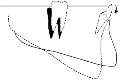
Post-eruptive compensation of the incisors in the mandible at backward rotation (22) (Figure 6).
Post-eruptive compensation of the incisors in the mandible at backward rotation (22) (Figure 6).
Figure 6. Posterior rotation of the mandible. Solid outer line of the mandible: before rotation Black shadow at molar region: before rotation Broken lines: after rotation
The alveolar bone
The formation of the alveolar bone begins early in fetal life, around the tooth germs. The growth of the dentoalveolar bone is associated with tooth eruption, and is therefore dependent on a normal eruption process (23). The more the teeth are erupting, the larger the alveolar bone will be (24). The existence of alveolar bone is entirely dependent on the presence of teeth (3). Thus, there seems to be a somewhat mutual rela-tion between the alveolar bone and the teeth, in which both are depend-ent on one another. In the vertical plane, a positive relationship between craniofacial height and height of the alveolar bone in both the maxilla and mandible was demonstrated in an adult sample by Beni Solow (25). This means that in subjects with a high craniofacial dimension, the alve-olar bone will be longer in the incisor region, and comparatively shorter in the molar region, which generates a downward inclined occlusal plane. On the contrary, subjects with a low craniofacial height display a shorter alveolar bone in the incisor region, with a similar height towards the molar region, generating a more parallel occlusal plane (20).
Similar to other skeletal bones, the alveolar bone consists of an outer layer of cortical (compact) bone and an inner layer of trabecular bone.
Figure 6. Posterior rotation of the mandible. Solid outer line of the mandible: before rotation Black shadow at molar region: before rotation Broken lines: after rotation
The alveolar bone
The formation of the alveolar bone begins early in fetal life, around the tooth germs. The growth of the dentoalveolar bone is associated with tooth eruption, and is therefore dependent on a normal eruption process (23). The more the teeth are erupting, the larger the alveolar bone will be (24). The existence of alveolar bone is entirely dependent on the presence of teeth (3). Thus, there seems to be a somewhat mutual relation between the alveolar bone and the teeth, in which both are dependent on one another. In the vertical plane, a positive relationship between craniofacial height and height of the alveolar bone in both the maxilla and mandible was demonstrated in an adult sample by Beni Solow (25). This means that in subjects with a high craniofacial dimension, the alveolar bone will be longer in the incisor region, and comparatively shorter in the molar region, which generates a downward inclined occlusal plane. On the contrary, subjects with a low craniofacial height display a shorter alveolar bone in the incisor region, with a similar height towards the molar region, generating a more parallel occlusal plane (20).
Similar to other skeletal bones, the alveolar bone consists of an outer layer of cortical (compact) bone and an inner layer of trabecular bone. This architectural construction offer a combination of rigidity
30
and low weight. The size and morphology are in part genetically determined and in part the result of functional loading. The outer part of the bone surface is covered by the periosteum (24).
There is also compact bone at the outer layer of the tooth socket. It looks like two types viewed microscopically; a compact bone and also a part perforated by periodontal ligament fibres (PDL). The perforating bundles of collagen fibres are knows as Sharpey´s fibres. Due to the bundles of collagen fibres in this area this can also be called bundle bone, also synonymous with lamina dura which is observed as a thin radiopaque layer of dense bone around sound teeth (3).
The development of the alveolar bone takes place in three dimensions in the maxilla and mandible. Vertical growth of the alveolar bone is depended on the height development of the face and in accordance with the mandibular rotation (Figure 6). Normally the teeth adapt to the overall changes induced by growth and rotation by compensation in eruption and inclination (26).
This architectural construction offer a combination of rigidity and low weight. The size and morphology are in part genetically determined and in part the result of functional loading. The outer part of the bone sur-face is covered by the periosteum (24).
There is also compact bone at the outer layer of the tooth socket. It looks like two types viewed microscopically; a compact bone and also a part perforated by periodontal ligament fibres (PDL). The perforating bun-dles of collagen fibres are knows as Sharpey´s fibres. Due to the bunbun-dles of collagen fibres in this area this can also be called bundle bone, also synonymous with lamina dura which is observed as a thin radiopaque layer of dense bone around sound teeth (3).
The development of the alveolar bone takes place in three dimensions in the maxilla and mandible. Vertical growth of the alveolar bone is de-pended on the height development of the face and in accordance with the mandibular rotation (Figure 6). Normally the teeth adapt to the over-all changes induced by growth and rotation by compensation in eruption and inclination (26).
Figure 7 DACM
Low angle, short anterior alveolar bone height High angle, long anterior alveolar bone height
Growth in sagittal dimension will take place through forward directed tooth eruption and by maturation of the molar tooth germs. The sagittal growth also evolves in connection with apposition at the maxillary tu-berosity and resorptions on the anterior surface of the mandibular ramus (25). Transverse growth takes place through buccal periosteal apposition and surface resorption on the lingual side (25).
Figure 7. DACM
Low angle, short anterior alveolar bone height High angle, long anterior alveolar bone height
Growth in sagittal dimension will take place through forward directed tooth eruption and by maturation of the molar tooth germs. The sagittal growth also evolves in connection with apposition at
the maxillary tuberosity and resorptions on the anterior surface of the mandibular ramus (25). Transverse growth takes place through buccal periosteal apposition and surface resorption on the lingual side (25).
The alveolar bone of the jaw are intriguing since, multiple interventions can be applied e.g. teeth can be moved, extracted, transplanted and replaced by implants with favourable results. The continued turnover of the alveolar bone allows a response to manipulation (3).
Tooth eruption and root development
All human teeth, primary and permanent dentition, develops through successive stages known as the bud, cap, and bell stage. During the bud stage differentiation of the cells forming the hard tissue of the teeth takes place and sets the stage for enamel and dentin formation. During the crown formation and development of teeth a pre-eruptive movement begins (27). After crown formation and mineralisation, the root development starts, with subsequent development of the supporting tissue of the teeth, namely the cementum, PDL, and alveolar bone. Thereafter the crown starts to erupt into the oral cavity (3). The teeth are surrounded by mesenchymal cells known as the dental follicle.
Dental follicle
Before emersion of the teeth into the oral cavity, the intraosseous stage of eruption occurs. The dental follicle overlying the teeth changes during the last stage of eruption and forms a pathway for the tooth emerging. Macrophages (large white specialised blood cells) appear in the tissue of the eruption pathway leading to a release of enzymes aiding in the destruction of cells and fibres that hinder tooth eruption. Osteoclasts (cells that break down bone tissue) responsible for bone resorption, are found in the bone area overlying the teeth. Osteoblasts (bone forming cells) work constantly in orchestra with osteoclasts to enable the new space needed at movement and tooth development (28). In a dog experiment that involved removal of the dental follicle, eruption of the tooth was prevented and there was a lack of signs of enlargement of the eruptive pathway, or root elongation (radiographically or histologically). This was evaluated
32
at a follow-up time when the contralateral, (un-operated) tooth erupted. According to these data, the intraosseous tooth eruption does not take place in the absence of the dental follicle (29).
Periodontal ligament (PDL)
The PDL, the alveolar bone and the cementum develop from cells from the dental follicle surrounding the tooth (1). The PDL comprises collagen fibres attaching the cementum and the alveolar bone. The minimal periodontal space is between 0.06-0.025mm, with a average measure of 0.12mm (30). Interstitial spaces contain nerve bundles and blood vessels. The PDLs function is to support the teeth and also transmit neural input at/during masticatory function. Another very important role of the PDL is to function as a suture, forming intramembranous bone during continued tooth eruption (5). In addition epithelial cells in the PDL have an important role in keeping the alveolar bone at a distance from the tooth (31).
Dentoalveolar compensatory mechanism (DACM)
As a result of the mid-facial and mandibular growth and rotation, there is a post-eruptive movement of the teeth which will occur to compensate for the rotations and to attain and preserve an occlusal stability (20) (Figure 6 and 7). This phenomenon is referred to as the dentoalveolar compensatory mechanism (DACM) (25). Inter-jaw and inter-dental space, created by craniofacial growth and displacement is the main reason for this mechanism and depends on the dental balance and the strive to achieve equilibrium in relation to the neighbouring teeth (13). Forces caused by lip, tongue, and cheek muscles also influence the movements of the teeth (32).
The DACM acts as the link between the growth of the cranial base, the maxilla and the mandible and the alveolar bone (25). The mechanism is meant to adapt and maintain a sufficient occlusion of the teeth.
Growth of the alveolar bone takes place in connection with tooth eruption, and is therefore dependent on a normal eruption process (23). Significant adaptation in the magnitude and direction of tooth eruption can occur especially in the vertical, but also the transverse and sagittal development of the face (5). There is a noticeable
variation in the rate of tooth eruption, with an increase in eruption velocity at the time of the puberty peak in body height growth (33). Presuming that continued eruption is a compensatory mechanism for craniofacial growth, the tooth eruption ought to follow the general pattern of craniofacial growth (23). A structure that most likely play an important role for the continued eruption is the PDL which functions as a suture forming intramembranous bone as the teeth erupt (5). The PDL has also been described as a suture with only one periosteal layer (5).
Growth of the alveolar bone can be influenced by soft tissue, function (e.g sucking and swallowing habits) and orthodontic treatment (23). For example, orthodontic treatment can be used in an attempt to prevent DACM by maintaining the teeth in a more optimal position in relation to the jaw base in young individuals with extreme growth where a need for orthognatic surgery appears early on. Early transverse expansion in the midpalatal suture of the maxilla can also be used in order to prevent the DACM and thus buccal inclination of the teeth (34). The amount of eruption, especially in the incisor region, seems to differ depending on craniofacial height. Therefore, knowledge of the expected growth pattern of the alveolar bone is important in relation to orthodontic treatment of growing individuals and dental implant treatment after growth cessation. The main movement of the teeth occur during eruption. However, the post-eruptive compensatory movements will continue even though occlusion is reached (13), e.g. as a response to mandibular rotation and toothwear. Change is the only constant throughout aging, development is present through all stages of life although it differs in the relative amount and activity (35).
Bone remodelling
Bone is a living tissue, which responds continuously to mechanical stress, nutritional status, hormones, and concentrations of circulating calcium.
Bone modelling dominates in growing individuals and bone remodelling is dominant in adults. Bone modelling is the process by
34
which bones are shaped or reshaped. In growing individuals, bone formation exceeds bone resorption. In bone modelling, osteoclasts and osteoblasts work independently, whereas they are coupled in bone remodelling (e.g. resorption is followed by bone formation). In remodelling, the shape of the bone is not changed, but damaged or old bone is detected by osteocytes, removed by osteoclasts, and thereafter an equal amount of new bone is formed by osteoblasts at the resorption site. In adults, the resorption and formation of bone is balanced, whereas bone resorption exceeds bone formation after menopause and in old age. The active bone resorption period in adults is approximately 29 days, and the bone formation period 134 days (36). In the first year of life, almost 100% of the skeleton is replaced, whereas in adults, the skeleton is renewed by remodelling every 10 years.
In a study by Pietrokovski (37) it was shown that the amount of resorption of the alveolar bone width after single tooth loss was greater along the buccal surface than along the lingual/palatal surface in all subjects. However, the individual variation was extensive and the absolute amount lost varied widely. The measurement range in the maxilla was between 2.58- 5.25mm on the buccal surface and 0.86-3.12mm in the palatal surface (depending on tooth position), mean value 3.58 and 1.99mm. The corresponding measurements in the mandible were between 2.08-4.69mm on the buccal surface and 0.91-3.0mm in the palatal surface (depending on tooth position), mean value 3.51 and 1.69mm. The post-extraction alteration caused a shift in the centre of the edentulous alveolar bone toward the lingual/palatal side.
Araújo and Lindhe (2005) also investigated the dimensional ridge alterations after tooth extraction. Alterations of the height of the alveolar bone during healing showed that the buccal bone wall after two to eight weeks post extraction was consistently located more apically compared to its lingual counterpart. Regarding the alterations of the width of the alveolar bone during healing, the buccal bone wall was thinner than the lingual bone wall. Further, the buccal and the lingual bone walls were thicker apically than coronally (38).
In a study by Schropp et al. (2003) an investigation of bone healing and soft tissue contour was performed. The alveolar bone width immediately after tooth extraction was on average 12.0mm (range 8.6-16.5mm). The width of the ridge was reduced, approximately 50%, from 12.0-5.9mm (6.1mm; range 2.7-12.2mm). There was alterations in both height and width, where the largest changes and reduction occurred within the first three month after extraction (39).
Other factors influencing the alveolar bone morphology
The role of the alveolar bone in orthodontic treatment, as well as in implant treatment, is well-known. In terms of morphology, inadequacy of alveolar bone dimensions limits the amount of tooth movement and may lead to treatment complications. Besides individual growth patterns, other factors influence the shape and size of the alveolar bone.
Tooth loss
In connection with tooth loss due to, for instance, tooth extraction, the alveolus is filled with a blood coagulum which will be replaced by fibrous tissue. A couple of weeks after the loss of the tooth, bone remodelling begins and initial bone formation occurs (24). After approximately two months the alveolus appears healed but with a reduction of hard tissue, most apparent in the buccal marginal area (40). A maturation of the trabecular bone morphology occurs to match the surrounding bone and the gingiva will also regain its original appearance (24).
After tooth extraction, the alveolar bone is resorbed to varying degrees (41). The decrease in size can be comprehensive (37) (Figure 8).
36
Figure 8. Example of horizontal bone resorption after tooth extraction.
There are some factors that seem to play an evident role regarding the amount of resorption that occurs, such as the length of time that the pa-tient has been edentulous, denture wear, and consumption of tobacco (41). However, the basal parts of the jaws are more resilient and e.g. not resorbed by denture wear (42).
Additional factors involved in determining the degree of alveolar bone resorption after tooth loss are e.g. muscular activity, prosthetic rehabili-tation, age, sex, and metabolic disease (43). The amount of bone that is orginially present (in mm) is individual and as is the amount of resorp-tion. This should be kept in mind when evaluating data in the literature presenting bone loss in mm and/or %.
The functional loading of the edentulous area may be reduced after tooth extraction, leading to bone loss. There is a great variation between viduals regarding remodelling of the edentulous areas, where some indi-viduals lose less bone and others undergo a more extensive resorption (42, 44).
Figure 8. Example of horizontal bone resorption after tooth extraction.
There are some factors that seem to play an evident role regarding the amount of resorption that occurs, such as the length of time that the patient has been edentulous, denture wear, and consumption of tobacco (41). However, the basal parts of the jaws are more resilient and e.g. not resorbed by denture wear (42).
Additional factors involved in determining the degree of alveolar bone resorption after tooth loss are e.g. muscular activity, prosthetic rehabilitation, age, sex, and metabolic disease (43). The amount of bone that is originally present (in mm) is individual and as is the amount of resorption. This should be kept in mind when evaluating data in the literature presenting bone loss in mm and/or %. The functional loading of the edentulous area may be reduced after tooth extraction, leading to bone loss. There is a great variation between individuals regarding remodelling of the edentulous areas, where some individuals lose less bone and others undergo a more extensive resorption (42, 44).
Figure 8. Example of horizontal bone resorption after tooth extraction.
There are some factors that seem to play an evident role regarding the amount of resorption that occurs, such as the length of time that the pa-tient has been edentulous, denture wear, and consumption of tobacco (41). However, the basal parts of the jaws are more resilient and e.g. not resorbed by denture wear (42).
Additional factors involved in determining the degree of alveolar bone resorption after tooth loss are e.g. muscular activity, prosthetic rehabili-tation, age, sex, and metabolic disease (43). The amount of bone that is orginially present (in mm) is individual and as is the amount of resorp-tion. This should be kept in mind when evaluating data in the literature presenting bone loss in mm and/or %.
The functional loading of the edentulous area may be reduced after tooth extraction, leading to bone loss. There is a great variation between viduals regarding remodelling of the edentulous areas, where some indi-viduals lose less bone and others undergo a more extensive resorption (42, 44).
Figure 9. Bone resorption after tooth extraction (vertical and horizontal). Ill. Fredrik Persson.
Bones adapt their strength to the physiological load placed on them (Frost, 2003). Frost theorises about two loading thresholds. Modelling takes place above a threshold (weightlifter’s hip and spine) and a remodelling occurs below a threshold because of disuse (e.g. after leg or hip fracture).
This theory can also be applied to alveolar bone. Teeth are loaded intermittently during biting/chewing and the reaction to changes in loading has been shown in a study of the alveolar bone in growing rats. Depending on whether or not the rats were given a soft or hard diet, the bone was more or less dense (Kiliaridis, Bresin, Holm, & Strid, 1996).
Agenesis
Tooth agenesis is a condition in which teeth are missing. Hypodontia is of genetic origin and usually involves one to five absent teeth. The prevalence of a single congenitally missing permanent tooth (28 tooth dentition) was between 2-10% summarised from both Nordic countries as well as other countries (45). The mandibular second premolar was found to be the most affected tooth, followed by the maxillary lateral incisor (45). In patients with ectodermal dysplasia (ED) severe oligodontia (six or more missing teeth) may be displayed, which subsequently requires implant therapy. Mandibular bone hypoplasia, both in bone quality and quantity was observed in association with tooth agenesis, and both are determining factors in the success of implant therapy (46, 47). The effect on craniofacial development in subjects with multiple missing teeth have been investigated by Johnson et al. Their findings suggested that the craniofacial morphology could be adversely affected by the lack of function due to severe hypodontia (48). A positive relation between
38
the severity in numbers of missing permanent teeth and severity of craniofacial dysmorphology in ED children was presented (48). In the ED patients, vertical growth of the mandible by condylar growth and increase in ramus height occur, however the alveolar bone remains atrophic resulting in a low-angle vertical growth pattern (49). Another condition where subjects are often affected by tooth agensis is osteogenesis imperfecta (OI). Investigations of the craniofacial development of subjects with OI show an association to smaller vertical craniofacial dimensions (50, 51).
Traumatic dental injuries (TDI)
TDI occurs very frequently and affects younger patients to a greater extent (52-54). Approximately one in five children have experienced a TDI to their permanent anterior teeth before leaving school (52). The prevalence is about 20% and the incidence about 1-2% overall. The numbers presented are based on global data. Thus, there are variations both between and within countries (54, 55).
The majority of the TDIs involve the anterior teeth, especially the maxillary central and lateral incisors (55). Overall, an estimation of 71–92% of all TDIs sustained during a lifetime occur before the age of 19 (55). This means that a treatment plan needs to be initiated in still growing individuals.
Ankylosis after TDI
In situations where the PDL has been severely damaged, especially in tooth avulsion and intrusion, there is a high risk for irreversible injury to the PDL, which will result in ankylosis of the tooth (56-58). Ankylosis is a bony ingrowth in the PDL, fusing the tooth to the alveolar bone (57-59). Ankylosis will result in the tooth being osseointegrated and unable to contribute to the growth of the alveolar bone (60). The region of such a tooth will be inhibited, so called infraposition (56, 60, 61).
Infraposition of a tooth can lead to tilting of the adjacent teeth as there is a local arrest of dentoalveolar development at the traumatic tooth area (56, 60, 61).
39
Osseointegrated implants and infraposition
Ossointegration of dental implants is a condition similar to tooth ankylosis. The implant is fused to the bone and inhibits growth of the alveolar bone in young, growing patients in the same way that an ankylosed tooth inhibits growth (Figure 10). Infraposition is observed in implants in a manner similar to what is observed with ankylosed teeth (61-66).
Figure 10. Infraposition of the upper left central and lateral incisor. Photo: Courtesy of Prof. Lars Andersson.
Radiography
Radiography has long been the method of choice to investigate the rela-tionship between craniofacial height and the morphology of the alveolar bone. Until the early 2000s the available methods were panoramic and lateral cephalometric imaging.
Panoramic imaging
Panoramic radiography produces a single image including both the max-illary and mandibular dental arches as well as the teeth and other facial structures (67).There are a number of advantages associated with pano-ramic imaging e.g. the image provides an overview of the facial bones and teeth, and the examination is convenient for the patient (68). How-ever, panoramic radiography is sensitive to patient positioning because of a narrow sharp layer and shape of the focal trough. Interpretation of the radiographs is uncertain especially in the anterior regions of the jaws. Furthermore, the cervical vertebrae generates artefacts in the ante-rior region of the mandible and maxilla complicating assessments in these regions (69).
Cephalometric imaging
The standard radiographs obtained with a cephalostat are lateral and postero-anterior cephalograms. They are mainly used to evaluate dental and skeletal relationships in the sagittal and vertical plane as well as asymmetries (70).
Figure 10. Infraposition of the upper left central and lateral incisor. Photo: Courtesy of Prof. Lars Andersson.
Radiography
Radiography has long been the method of choice to investigate the relationship between craniofacial height and the morphology of the alveolar bone. Until the early 2000s the available methods were panoramic and lateral cephalometric imaging.
Panoramic imaging
Panoramic radiography produces a single image including both the maxillary and mandibular dental arches as well as the teeth and other facial structures (67). There are a number of advantages associated with panoramic imaging e.g. the image provides an overview of the facial bones and teeth, and the examination is convenient for the patient (68). However, panoramic radiography is sensitive to patient positioning because of a narrow sharp layer and shape of the focal trough. Interpretation of the radiographs is uncertain especially in the anterior regions of the jaws. Furthermore, the cervical vertebrae
40
generates artefacts in the anterior region of the mandible and maxilla complicating assessments in these regions (69).
Cephalometric imaging
The standard radiographs obtained with a cephalostat are lateral and postero-anterior cephalograms. They are mainly used to evaluate dental and skeletal relationships in the sagittal and vertical plane as well as asymmetries (70).
By combining the information provided by the lateral cephalometric image and the panoramic radiograph, an evaluation of the patient’s craniofacial height and alveolar bone height in the maxilla and mandible can be used to investigate an association.
Neither with panoramic radiography nor with lateral or posterior-anterior cephalometric imaging is it possible to obtain cross-sectional site-specific images of the maxilla and mandible. Technological advancements in radiological imaging have moved the technique from film radiography towards digital 3D (68, 70, 71). This has been achieved through the use of conventional tomography (72), computed tomography (70) and, more recently, CBCT (73).
CBCT
CBCT was introduced in the early 1980s, primarily for angiography (74), but has since become the most significant advance in dental- and maxillofacial imaging. Development of specialised CBCT scanners for use in dentistry begun in the late 1990s (75). The first commercial CBCT (NewTom 9000) was introduced on the European market in 1999 (76). CBCT utilises a cone-shaped ionizing radiation source, in shape similar to the beam of conventional 2D-imaging. CBCT in dental practice is an emerging technique for diagnostic imaging, and provides images with a high level of detail, in three planes. The additional information regarding the width dimension that CBCT supplies is the missing piece previously not provided by the two-dimensional images.
Rater agreement
For imaging methods, both reliability and agreement are important concepts intended to provide information about the amount of error inherent in any diagnosis, score, or measurement (77) which in turn determins the validity of the study results (78). Agreement “is the degree to which scores or ratings are identical”. Interrater agreement refer to different raters, using the same scale or classification when assessing the same subjects/objects. Intrarater agreement refers to the same rater, using the same scale or classification when assessing the same subjects/object at different times (77). Consequently, for studies on imaging methods and validity of study results, several raters should be included to provide information about the generalisability of results (77).
With regards to measurements of alveolar bone height and width and the association to craniofacial height, presentation of intrarater and interrater agreement is infrequent. Furthermore, studies presenting intrarater agreement have either used dry skulls with only three raters (79) or a patient sample but with only two raters (80). The limited number of raters result in questionable study results.
Consequently, there is a need for studies on rater agreement regarding measurements in CBCT images to establish the relationship between alveolar bone morphology and craniofacial height in adherence with the guidelines for Reporting Reliability and Agreement Studies (GRRAS) that have been proposed (Kottner 2011) in order to validate a conceivable relationship.
Systematic reviews
The continuous growth of research publications generates an increased need to systematically summarise the available scientific evidence. To give a brief history of systematic reviews, the first known example of a systematic review was conducted by James Lind in 1753. He published a paper with the aim of providing an unbiased and concise summary of evidence on scurvy (81, 82).
Systematic reviews (SR) have a number of benefits. Primarily, they deliver a clear and comprehensive overview of existing available
42
evidence on a given topic. In addition, SRs can be used to identify where evidence and clear answers already exists in the available literature and where no further research is necessary at present. This helps avoid duplicate research, waste of production and reporting research (83). Finally, SRs also help to identify research gaps within the field of interest. Methodological concerns in research studies can be highlighted and thereafter used to improve future research in the topic area (84).
AIMS AND HYPOTHESES
The difference in the duration and amount of DACM seems associated with vertical craniofacial height. Knowledge in this area could benefit in treatment planning of young individuals. Therefore this thesis will investigate further the alveolar bone morphology in relation to craniofacial height.
The overriding aim of this thesis was to investigate the morphology of the alveolar bone in relation to the vertical craniofacial development using measurements in CBCT images. In addition, the thesis sought to investigate the existing scientific knowledge related to dental infraposition, in order to clarify if the degree of infraposition was associated to craniofacial heigh.
Specific aims and hypotheses:
Paper I
• To investigate if there is an association between the cross-sectional morphology of the maxillary and mandibular alveolar bone and the craniofacial height, using images from Cone Beam Computed Tomography (CBCT).
• To investigate differences between male and female patients regarding the association between the cross-sectional morphology of the maxillary and mandibular alveolar bone and the craniofacial height.
44
Hypothesis: There is an association between the morphology of the alveolar bone and the craniofacial height.
Null hypothesis: There is no association between the morphology of the alveolar bone and the craniofacial height.
Paper II
• To investigate rater agreement regarding measurements of height and width of the maxilla and mandible on cross-sectional images from CBCT examinations.
Hypothesis: There is an agreement between raters when measuring the morphology of the alveolar bone.
Null hypothesis: There is no agreement between raters when measuring the morphology of the alveolar bone.
Paper III
• To investigate if craniofacial height and sex are associated with the alveolar bone dimensions in the anterior maxilla.
Hypothesis: There is an association between the craniofacial height, sex, and morphology of the alveolar bone in the anterior maxilla. Null hypothesis: There is no association between the craniofacial height, sex, and morphology of the alveolar bone in the anterior maxilla.
Paper IV
• To investigate the potential association between age, sex, and craniofacial height and infraposition due to continued alveolar development in the anterior maxilla, in a systematic review.