R E S E A R C H A R T I C L E
Open Access
Intracellular Ser/Thr/Tyr phosphoproteome
of the oral commensal Streptococcus
gordonii DL1
Carolina Robertsson
1*, Gunnel Svensäter
1, Zoltan Blum
2and Claes Wickström
1Abstract
Background: To respond and adapt to environmental challenges, prokaryotes regulate cellular processes rapidly and reversibly through protein phosphorylation and dephosphorylation. This study investigates the intracellular proteome and Ser/Thr/Tyr phosphoproteome of the oral commensal Streptococcus gordonii. Intracellular proteins from planktonic cells of S. gordonii DL1 were extracted and subjected to 2D-gel electrophoresis. Proteins in general were visualized using Coomassie Brilliant Blue and T-Rex staining. Phosphorylated proteins were visualized with Pro-Q Diamond Phosphoprotein Gel Stain. Proteins were identified by LC-MS/MS and sequence analysis.
Results: In total, sixty-one intracellular proteins were identified in S. gordonii DL1, many of which occurred at multiple isoelectric points. Nineteen of these proteins were present as one or more Ser/Thr/Tyr phosphorylated form. The identified phosphoproteins turned out to be involved in a variety of cellular processes.
Conclusion: Nineteen phosphoproteins involved in various cellular functions were identified in S. gordonii. This is the first time the global intracellular Ser/Thr/Tyr phosphorylation profile has been analysed in an oral streptococcus. Comparison with phosphoproteomes of other species from previous studies showed many similarities. Proteins that are consistently found in a phosphorylated state across several species and growth conditions may represent a core phosphoproteome profile shared by many bacteria.
Keywords: 2DE, Oral bacteria, Phosphoproteome, Pro-Q diamond, Streptococci, Streptococcus gordonii
Background
Streptococcus gordonii is a pioneer oral colonizer,
in-volved in the establishment of oral biofilms [1]. Due to the numerous different adhesins expressed on its sur-face, S. gordonii cells readily attach to and colonize
den-tal surfaces [2]. Moreover, S. gordonii can passively
migrate from small oral lesions through the blood stream and cause infective endocarditis by opportunistic infection of the heart valves [3]. This mainly saccharoly-tic species is considered a commensal with ubiquitous habitation in humans, and given the acid production and
acid tolerance of S. gordonii, it prevails at the acidic con-ditions that periodically occur in oral biofilms [4–6]. When carbohydrate concentrations are low, S. gordonii like other oral streptococci utilizes a carbohydrate phos-photransferase transport system (PTS) with high glucose affinity [7]. Upon spikes in carbohydrate concentration, oral streptococci are at risk for“sugar killing” from dam-aging effects caused by accumulated glycolytic interme-diates [8]. To evade such inflictions, oral streptococci switch to carbohydrate transport systems with lower af-finity for glucose, e.g. the permease system. The glyco-lytic rate can also be regulated by switching to alternative pathways, e.g. through activation of lactate dehydrogenase (ldh) in pyruvate conversion for faster re-generation of NAD+[9]. In this way, cells reduce glucose © The Author(s). 2020 Open Access This article is licensed under a Creative Commons Attribution 4.0 International License, which permits use, sharing, adaptation, distribution and reproduction in any medium or format, as long as you give appropriate credit to the original author(s) and the source, provide a link to the Creative Commons licence, and indicate if changes were made. The images or other third party material in this article are included in the article's Creative Commons licence, unless indicated otherwise in a credit line to the material. If material is not included in the article's Creative Commons licence and your intended use is not permitted by statutory regulation or exceeds the permitted use, you will need to obtain permission directly from the copyright holder. To view a copy of this licence, visithttp://creativecommons.org/licenses/by/4.0/. The Creative Commons Public Domain Dedication waiver (http://creativecommons.org/publicdomain/zero/1.0/) applies to the data made available in this article, unless otherwise stated in a credit line to the data.
* Correspondence:carolina.robertsson@mau.se
1Department of Oral Biology and Pathology, Faculty of Odontology, Malmö University, 20506 Malmö, Sweden
addition, oral streptococci employ strategies colloquially
termed as the “acid tolerance response” (ATR) that
en-hance cell survival in acidic environments [4, 10, 11]. The ATR is dependent on molecular chaperone activity [4,8] that sustains correct protein folding during biosyn-thesis even at low pH.
To keep up with environmental fluctuations, prokary-otes have been suggested to regulate the activity of pro-teins involved in the central carbon metabolism rapidly and reversibly through phosphorylation and
dephosphory-lation mechanisms [12, 13]. The reversible regulatory
phosphorylation events operate on a much faster time scale than changes in protein expression [14]. The earliest studies on protein phosphorylation and dephosphorylation as regulatory events in bacteria focused on phosphoryl-ation of histidine and aspartate residues in relphosphoryl-ation to
two-component systems [15]. However, phosphorylation
events on serine, threonine, and tyrosine (Ser/Thr/Tyr) have also been found to play important roles in prokary-otic intracellular signalling. Although the most common group of Ser/Thr kinases (i.e. Hanks-type kinases) are often referred to as“eukaryotic kinases”, many prokaryotic Ser/Thr kinases also belong to this group [16]. There are many examples of phosphorylation events regulating cel-lular activities in bacteria, relating to house-keeping func-tions as well as stress responses and virulence [12,17,18]. In Lactobacillus rhamnosus, phosphorylation of glycolytic enzymes is upregulated as a response to acidic stress [19]. In that study, the phosphorylation state of threonine and serine residues on glyceraldehyde 3-phosphate dehydro-genase (gapdh) differed between protein species depend-ing on the growth conditions, while the overall abundance of the different proteins remained unchanged. This sup-ports the idea that phosphorylation can regulate enzyme activity separately from regulation of gene expression. Dif-ferent forms of the same protein may display increased or decreased catalytic activity, altered subcellular localization, or modified interaction with non-substrates [20,21]. Mul-tisite phosphorylation can coordinate several such effects, determine the duration of a response, or mediate signal
integration [21]. Protein phosphorylation may also
completely alter the biological functions of proteins in the cell, a phenomenon referred to as protein moon-lighting [22]. The regulatory effects of phosphorylation events on specific enzymes have been studied in some oral bacteria, mainly the oral streptococci [23–25], but to the best of our knowledge, global Ser/Thr/Tyr phos-phorylation profiles of S. gordonii have not yet been detailed. The aim of the current study was to identify the intracellular protein expression profile, with special attention to Ser/Thr/Tyr phosphorylated proteins, in
S. gordoniiDL1.
Intracellular proteomes from planktonic cells of S.
gor-donii DL1 were extracted and separated by 2DE. The
total intracellular proteome was visualized with Coomas-sie Brilliant Blue stain (Sigma). In total, 222 protein spots were detected. Discrete spots were manually ex-cised for identification with LC-MS/MS. Molecular weights (MWs) and isoelectric points (pIs) for identified proteins were estimated from the gels as well as gathered from the mass spectrometry data. MWs, pIs, MASCOT scores, number of matched peptides and % coverages are
listed in Table 1. The sequenced peptides identified by
LC-MS/MS can be found in supplemental material (Additional file 1). In total, 61 proteins were identified, many of which were present at multiple isoelectric points (Fig.1, Table1).
Intracellular Ser/Thr/Tyr phosphorylation profile
Spots containing Ser/Thr/Tyr phosphorylated proteins were visualized with Pro-Q Diamond stain and phos-phoimaging. The total intracellular proteome was visual-ized with T-Rex protein labelling for orientation of the phosphorylated spots. In total, 49 phosphorylated spots were detected. These were generally found on the acidic side of the 2DE gels (Fig.2).
Phosphorylated protein spots where identified with
LC-MS/MS (Table1). Six of the 49 phosphorylated spots
remained unidentified, due to a lack of significant hits from the Mascot search (four spots) or absence from Coomassie gels, preventing excision for identification (two spots). In total, 19 putative Ser/Thr/Tyr phosphory-lated proteins were identified, see Table1.
Cellular processes associated with Ser/Thr/Tyr phosphorylated proteins
The proteins detected as Ser/Thr/Tyr phosphorylated are involved in various cellular processes (Fig. 3). All phosphoproteins involved in the carbon metabolism, ex-cept for HPr and tpi, were present as more than one phosphorylated species, occurring at different pIs on the gels. HPr and tpi were also present as one additional,
non-phosphorylated species respectively (Figs. 1 and 2,
Table 1). Pfk and pgm were both present as two species
each, all phosphorylated. Enolase was identified in both unphosphorylated and phosphorylated forms. Four of the phosphorylated enolase species were found at the ex-pected MW. Pyk was present as four adjacent species, two of which were phosphorylated. Eight spots were identified as gapdh, three of which were phosphorylated. Nine species of pgk were identified, of which three were phosphorylated. Three species of ldh were identified, two phosphorylated. Six phosphorylated proteins in-volved in biosynthesis were identified. Deob, pgam and
Table 1 S. gordonii intracellular proteins and phosphoproteins, separated by 2DE and identified with LC-MS/MS Abbr Protei n Gene b) Acc. No. c) pI estimat ed from gel/ theo retical d) MW , Da es timate d from ge l/ the oretical d) LC-MS /MS Ser/Thr/Tyr phosp hory-lated +/− MASC OT score No. of peptides e) Cove rage Trans mem brane tra nsport ABC a) ABC trans porter ATP -binding prot ein SP171 5 SGO_ 1342 A8AXW 3 6.7/6.25 2 3 ,000/26 325 1124 18 72% – Sugar trans port HPr a) Phosp hocarrier protei n H Pr ptsH (SG O_1556 ) A8AYH5 5.1/4.74 1 1 ,000/89 35 124 2 55% + Glyco lysis Tpi-1 a) Trio sephosp hate isome rase tpiA (SGO _0762 ) A8AWA 1 4.5/4.75 2 3 ,000/26 524 652 9 52% + Tpi-2 a) 4.3/4.75 2 3 ,000/26 524 540 9 52% -pfk-1 a) Phosp hofructokinas e pfkA (SGO_ 134 0) A8AXW 1 6.6/5.62 3 5 ,000/35 346 1043 17 49% + pfk-2 6.8/5.62 3 5 ,000/35 346 1476 22 56% + fba-1 f) Fruc tose-1,6 -bisphosphate ald olase, class II Fba (SGO_ 1745) A8AZ 06 5.1/5.00 2 7 ,000/31 394 -fba-2 a) 5.2/5.00 2 7 ,000/31 394 771 12 51% -fba-3 a) 5.3/5.00 2 7 ,000/31 394 800 14 47% -fba-4 f) 5.4/5.00 2 7 ,000/31 394 -fba-5 a) 5.4/5.00 1 7 ,000/31 394 164 3 11% -gap dh-1 a) Glyce raldehy de-3-ph osphate deh ydroge nase gap (SGO _0207 ) A8AUR7 5.5/5.37 3 9 ,000/35 918 1153 20 51% + gap dh-2 a) 5.7/5.37 3 9 ,000/35 918 1299 21 79% + gap dh-3 a) 5.9/5.37 3 9 ,000/35 918 1304 22 58% + gap dh-4 5.5/5.37 4 0 ,000/35 918 89 2 8 % -gap dh-5 5.7/5.37 4 0 ,000/35 918 276 4 14% -gap dh-6 6.0/5.37 3 9 ,000/35 918 84 2 6 % -gap dh-7 5.9/5.37 4 0 ,000/35 918 325 5 16% -gap dh-8 5.2/5.37 1 8 ,000/35 918 141 2 8 % -pgk-1 Phosp hoglyc erate kinase pgk (SGO_ 0209) A8AUR9 4.9/4.88 4 5 ,000/42 089 397 6 18% + pgk -2 a) 5.0/4.88 4 5 ,000/42 089 1454 23 63% + pgk -3 a) 4.3/4.88 2 1 ,000/42 089 340 5 17% + pgk -4 f) 5.1/4.88 4 5 ,000/42 089 -pgk -5 f) 5.2/4.88 4 5 ,000/42 089 -pgk − 6 4.3/4.88 4 5 ,000/42 089 159 3 9 % -pgk -7 a) 5.9/4.88 2 4 ,000/42 089 431 7 18%
-Table 1 S. gordonii intracellular proteins and phosphoproteins, separated by 2DE and identified with LC-MS/MS (Continued) Abbr Protei n Gene b) Acc. No. c) pI estimat ed from gel/ theo retical d) MW , Da es timate d from ge l/ the oretical d) LC-MS /MS
Ser/Thr/Tyr phosp lated +/−
MASC OT score No. of peptides e) Cove rage pgk -8 a) 4.2/4.88 2 0 ,000/42 089 830 11 41% -pgk -9 a) 5.9/4.88 2 0 ,000/42 089 561 9 28% -pgm-1 a) Phosp hoglyc erate mu tase gpma (SGO_ 070 4) A8AW4 6 5.9/5.41 2 2 ,000/26 044 904 18 84% + pgm-2 a) 6.1/5.41 2 2 ,000/26 044 933 18 87% + eno-1 Enol ase eno (SGO_ 1426) A8AY4 6 4.4/4.71 4 8 ,000/47 062 1368 18 61% + eno − 2 4.5/4.71 4 8 ,000/47 062 1260 13 52% + eno − 3 4.6/4.71 4 8 ,000/47 062 1721 18 63% + eno -4 a) 4.7/4.71 4 8 ,000/47 062 2245 31 77% + eno − 5 4.6/4.71 2 6 ,000/47 062 1316 17 58% + eno -6 a) 4.5/4.71 2 4 ,000/47 062 460 8 26% + eno -7 a) 4.1/4.71 2 2 ,000/47 062 809 9 30% + eno -8 a) 4.2/4.71 2 1 ,000/47 062 673 9 28% + eno -9 a) 4.3/4.71 2 1 ,000/47 062 595 10 32% + eno -10 a) 4.4/4.71 2 1 ,000/47 062 989 11 34% + eno -11 a) 4.4/4.71 2 8 ,000/47 062 1485 15 55% -eno -12 a) 4.8/4.71 2 5 ,000/47 062 950 14 47% -eno -13 a) 4.4/4.71 2 3 ,000/47 062 460 8 26% -eno -14 a) 4.2/4.71 2 3 ,000/47 062 746 11 36% -eno -15 a) 4.8/4.71 2 3 ,000/47 062 627 9 32% -eno -16 a) 4.8/4.71 2 2 ,000/47 062 631 9 31% -eno -17 a) 4.9/4.71 2 2 ,000/47 062 636 9 31% -eno -18 a) 4.4/4.71 1 7 ,000/47 062 230 4 11% -eno − 19 4.5/4.71 1 4 ,500/47 062 931 9 41% -eno − 20 4.6/4.71 1 4 ,000/47 062 533 7 23% -eno -21 a) 5.0/4.71 1 3 ,000/47 062 437 7 23% -eno -22 a) 4.7/4.71 1 2 ,500/47 062 722 9 33% -eno -23 a) 4.3/4.71 1 1 ,000/47 062 418 6 20% -eno -24 a) 5.4/4.71 1 8 ,000/47 062 684 9 27% - pyk-1 a) Pyruv ate kinase pyk (SGO _1339) A8AXW 0 5.15/4 .94 58 ,000/54 799 150 3 6% + pyk-2 a) 5.2/4.94 5 8 ,000/54 799 1474 29 62% + pyk-3 f) 5.1/4.94 5 8 ,000/54 799
-Table 1 S. gordonii intracellular proteins and phosphoproteins, separated by 2DE and identified with LC-MS/MS (Continued) Abbr Protei n Gene b) Acc. No. c) pI estimat ed from gel/ theo retical d) MW , Da es timate d from ge l/ the oretical d) LC-MS /MS Ser/Thr/Tyr phosp hory-lated +/− MASC OT score No. of peptides e) Cove rage pyk-4 f) 5.3/4.94 5 8 ,000/54 799 -Cofactor biosynt hesis nade a) NH(3 )-depende nt NAD (+) syn thetase nadE (SG O_0583 ) A8AVT9 5.4/5.11 3 5 ,000/30 248 268 3 16% – Acetoin catab olism aced-1 Acet oin de hydro genase butA (SGO_ 109 6) A8AX7 5 5.4/5.20 2 6 ,000/26 547 1008 15 77% - aced-2 a) 5.7/5.20 2 6 ,000/26 547 1247 17 75% - aced-3 a) 5.7/5.20 2 4 ,000/26 547 809 13 62% -Carb ohydr ate catab olism dera a) Deoxyri bose -phosph ate ald olase deoC (SG O_1080 ) A8AX5 9 4.9/4.84 2 0 ,000/23 380 691 8 49% – Pyruv ate conver sion ldh-1 a) L-lact ate de hydro genase ldh (SGO _1232 ) A8AXK9 5.4/5.24 3 6 ,000/35 267 327 6 28% + ldh-2 5.6/5.24 3 6 ,000/35 267 436 6 28% + ldh-3 5.8/5.24 3 6 ,000/35 267 641 8 32% -adh a) Alcoh ol de hydro genase adhA (SG O_0565 ) A8AVS1 5.2/4.94 3 9 ,000/35 958 911 13 69% – pox Pyruv ate oxidase spxB (SGO_0 292) A8AV0 1 5.3/5.06 6 5 ,000/65 283 161 3 5 % – Pento se pho spha te pathw ay deo b Phosp hopent omutase deoB (SGO_1 264) A8AXN 8 5.3/4.96 4 7 ,000/44 508 172 3 9 % + Ami no aci d metabol ism alad Alani ne de hydroge nase ald (SGO_0 708) A8AW5 0 6.1/5.29 4 5 ,000/38 960 1378 19 63% – dap h a) 2,3,4, 5-tetrahydropy ridine-2, 6-dic arboxylate N-a cetyltransferase dapH (SGO_0 158) A8AUL9 5.2/5.06 2 2 ,000/24 255 291 5 25% – glya Serine hyd roxyme thyltransferas e glyA, (SGO _1151 ) A8AXC 8 5.4/5.07 4 8 ,000/45 566 583 8 28% – sed Homos erin e deh ydroge nase hom (SGO _0801 ) A8AWE0 5.0/4.86 4 5 ,000/46 154 245 3 11% – Nucleo tide metabol ism adk a) Aden ylate kinase adk (SGO_1964) A8AZ K4 5.2/4.95 2 2 ,000/ 23 ,857 318 6 38% – aprt a) Aden ine phosp horibos yltra nsferase apt (SGO_1 001) A8AWY 0 5.0/4.90 1 7 ,000/18 840 875 12 74% –
Table 1 S. gordonii intracellular proteins and phosphoproteins, separated by 2DE and identified with LC-MS/MS (Continued) Abbr Protei n Gene b) Acc. No. c) pI estimat ed from gel/ theo retical d) MW , Da es timate d from ge l/ the oretical d) LC-MS /MS
Ser/Thr/Tyr phosp lated +/−
MASC OT score No. of peptides e) Cove rage imdh-1 Inosi ne-5 ′-mono phosp hate deh ydroge nase guaB (SGO_ 000 8) A8AU7 0 5.4/5.19 5 8 ,000/52 717 116 2 5 % + imdh-2 5.5/5.19 5 6 ,000/52 717 252 4 12% -pyrb Asp artate carbam oyltransferase pyrB (SG O_1109 ) A8AX8 8 5.6/5.04 3 5 ,000/34 741 54 1 g) 4% – xprt Xant hine phosp horibos yltra nsferase xpt (SG O_1158 ) A8AXD5 6.2/5.79 1 7 ,000/20 849 684 12 77% – Molecular cha peron e activ ity and/o r prot ein fold ing Dnak -1 Mol ecular chaperone DnaK dnaK (SGO_ 040 2) A8AVA 8 4.4/4.69 6 8 ,000/64 724 266 4 9 % + DnaK-2 a) 4.5/4.69 6 8 ,000/64 724 1557 26 50% + DnaK-3 a) 5.2/4.69 2 5 ,000/64 724 134 2 3 % + DnaK-4 f) 4.6/4.69 6 8 ,000/64 724 -DnaK-5 4.1/4.69 4 4 ,000/64 724 254 4 10% -DnaK-6 a) 5.3/4.69 3 6 ,000/64 724 345 6 14% -GroEL-1 Mol ecular chaperone GroEL groEL (SGO _1885 ) A8AZ E1 4.2/4.66 6 4 ,000/56 754 776 13 34% + GroEL-2 4.3/4.66 6 4 ,000/56 754 1500 22 47% + GroEL-3 4.4/4.66 6 4 ,000/56 754 794 13 32% -GroES Co-c hapero ne GroES groES (SGO _1886 ) A8AZ E2 5.4/5.11 6 4 ,000/96 81 65 1 g) 2% – grpE a) Mol ecular chaperone GrpE grpE (SGO_0 401) A8AVA 7 4.8/4.83 2 3 ,000/20 265 186 3 18% – hyd a) Hydro lase, haloac id deh alogenase family/ pept idyl-prolyl cis-tran s isome rase, cyclophilin type SGO_ 0604 A8AVW 0 4.2/4.74 2 2 ,000/51 936 516 10 30% – tig a) Trigg er facto r (chaperone ) tig (SG O_0412 ) A8AVB 8 4.2/4.53 5 5 ,000/47 242 531 10 31% + Trans cription GreA Tra nscription elong ation facto r GreA greA (SG O_0519 ) A8AVM5 4.9/4.85 1 5 ,500/17 515 581 9 68% – rpoA a) DNA -directed RNA polym erase subuni t alpha rpoA (SG O_195 9) A8AZ J9 4.5/4.70 4 1 ,000/34 502 578 7 29% – rpoZ DNA -directed RNA polym erase subuni t omega rpoZ (SGO_0 595) A8AVV 1 6.7/5.82 1 2 ,000/11 851 231 5 44% – y454 Prob able transcriptiona l regul atory protei n A8AVG0 4.6/4.45 3 0 ,000/25 722 227 4 26% –
Table 1 S. gordonii intracellular proteins and phosphoproteins, separated by 2DE and identified with LC-MS/MS (Continued) Abbr Protei n Gene b) Acc. No. c) pI estimat ed from gel/ theo retical d) MW , Da es timate d from ge l/ the oretical d) LC-MS /MS Ser/Thr/Tyr phosp hory-lated +/− MASC OT score No. of peptides e) Cove rage Trans lation ef-G Elo ngation facto r G fusA (SGO_0 206) A8AUR6 4.7/4.88 5 0 ,000/76 785 53 1 g) 2% – ef-Ts Elo ngation facto r Ts tsf (SGO_2 000) A8AZ P0 4.9/4.84 4 1 ,000/37 284 326 5 20% – ef-tu-1 a) Elo ngation facto r tu tuf (SGO_ 0761) A8AWA 0 4.1/4.86 5 5 ,000/44 011 519 8 31% + ef-tu-2 5– 6/4. 86 55 ,000/44 011 1253 22 63% + ef-tu-3 a) 4.2/4.86 3 4 ,000/44 011 532 6 23% + ef-tu-4 a) 4.3/4.86 2 5 ,000/44 011 167 3 8 % + ef-tu-5 a) 4.3/4.86 2 9 ,000/44 011 553 6 25% -ef-tu-6 a) 4.7/4.86 2 3 ,000/44 011 420 6 18% -ef-tu-7 a) 5.3/4.86 2 0 ,000/44 011 433 6 16% -rL1 a) 50S ribos omal prot ein L1 rplA (SGO _1455 ) A8AY7 4 5.5/9.22 2 3 ,000/24 399 202 3 16% – rL4-1 a) 50S ribos omal prot ein L4 rplD (SGO_ 1984) A8AZ M4 5.2/10 .03 19 ,000/22 279 285 4 26% -rL4-2 a) 5.5/10 .03 19 ,000/22 279 327 4 24% -rL10 a) 50S ribos omal prot ein L10 rplJ (SGO_1 192) A8AXG 9 5.2/5.06 1 5 ,000/17 462 1002 15 86% – rL17 50S ribos omal prot ein L17 rplQ (SG O_1958 ) A8AZ J8 5.3/9.86 1 3 ,000/14 511 141 2 17% – rrf a) Ribosom e recy cling factor frr (SGO _1451 ) A8AY7 0 6.8/5.90 1 8 ,000/20 640 421 8 46% – rS1 30S ribos omal prot ein S1 rpsA (SG O_1234 ) A8AXL0 4.5/4.91 5 2 ,000/44 149 130 2 5 % + rS2 30S ribos omal prot ein S2 rpsB (SGO_2 001) A8AZ P1 4.6/5.07 3 3 ,000/29 125 515 8 35% – rS5-1 a) 30S ribos omal prot ein S5 rpsE (SGO_ 1968) A8AZ K8 5.1/9.69 1 4 ,000/17 079 335 4 33% -rS5 –2 5.3/9.69 1 4 ,000/17 079 283 4 33% -rS8 a) 30S ribos omal prot ein S8 rpsH (SGO_1 971) A8AZ L1 5.3/9.48 1 2 ,500/14 753 120 2 25% – rS17 30S ribos omal prot ein S17 rpsQ (SGO_1 976) A8AZ L6 5.0/9.92 1 1 ,000/10 010 58 1 g) 10% – sys-1 Serine tR NA-ligase serS (SGO _1683) A8AYV 0 5.4/5.11 4 8 ,000/48 110 342 7 21% -sys-2 a) 5.7/5.11 2 1 ,000/48 110 95 1 g) 2% -Protei n catabolism clpp ATP-d epend ent Clp prot ease prot eolytic subuni t clpP (SG O_1632 ) A8AYP 9 4.3/4.72 1 8 ,000/21 363 200 3 12% – pept Pept idase T pepT (SGO_1312) A8AXT 3 4.2/4.59 4 9 ,000/45 200 332 4 13% – Vita min B biosy nthe sis pxk a) Pyrid oxine kinase SGO_ 0409 A8AVB 5 5.3/5.16 2 3 ,000/27 598 583 10 52% –
Table 1 S. gordonii intracellular proteins and phosphoproteins, separated by 2DE and identified with LC-MS/MS (Continued) Abbr Protei n Gene b) Acc. No. c) pI estimat ed from gel/ theo retical d) MW , Da es timate d from ge l/ the oretical d) LC-MS /MS
Ser/Thr/Tyr phosp lated +/−
MASC OT score No. of peptides e) Cove rage Cell e nvelope biosy nthesis murd a) UDP-N-a cetylmu ramoylalanine D-gl utama te lig ase murdD (SGO _0671 ) A8AW1 4 5.4/5.07 4 6 ,000/48 257 253 4 12% – pga m a) Phosp hoglu cosamine mutas e glmM (SG O_0889 ) A8AWM 5 4.6/4.71 5 2 ,000/48 396 592 7 26% + rmla-1 Gluc ose-1-ph osphate thymi dylyltran sferase rfbA-1 (SGO_ 100 9) A8AWY 8 5.1/4.92 2 8 ,000/32 213 42 1 g) 3% + rmla-2 a) 5.3/4.92 2 8 ,000/32 213 136 2 7 % + rmlb dTD P-glucose 4,6-d ehydratase rfbB-1 (SG O_101 1) A8AWZ0 6.3/5.55 4 0 ,000/39 314 168 3 12% – Cell di vision SepF Cell division prot ein SepF sepF (SGO _0677 ) A8AW2 0 6.6/5.61 1 9 ,000/21 654 367 6 41% – Antio xidan t activ ity sod a) Supe roxide dismutase sodA (SGO_1 599) A8AYL 7 4.9/4.78 1 9 ,000/22 446 474 8 46% – tox Thi ol pe roxidase Tpx (SGO_1 803) A8AZ 62 4.2/4.52 1 6 ,000/18 015 349 6 59% – Protei n dephosphory lation ppp a) Phosp hoprot ein phosp hatase SGO_ 0599 SGO_ 0599 A8AVV 5 4.3/4.62 2 3 ,000/26 870 696 11 61% + a) Two or more proteins were identified from this spot b) Gene name in the UniProt database as entered for S. gordonii DL1 proteins c) Accession number in the UniProt database for S. gordonii DL1 d) Theoretical values for S. gordonii DL1 proteins e) The sequenced peptides identified by LC-MS/MS can be found in supplemental material (Additional file 1 ) f) Protein identity deduced from adjacent identified spot g) Proteins identified by only one peptide are given when only a single match was yielded from the database search
rS1 were all present as single phosphorylated species, and one of the two species of imdh was phosphorylated. Like enolase, ef-tu was found at several pIs, of which four were phosphorylated. Rmla was detected as two phosphorylated species. Three phosphorylated chaper-ones were identified. Six species of DnaK were identified, three of which were phosphorylated. GroEL was present as three species, two of which were phosphorylated. Tig was present as a single phosphorylated species. The pro-tein phosphatase SGO_0599 (ppp) was detected as one phosphorylated species.
Comparison of Ser/Thr/Tyr phosphoproteomes to other bacteria as detected with Pro-Q Diamond Phosphoprotein gel stain on 2DE-gels
To investigate whether the identified phosphoproteome of
S. gordonii DL1 coincided with phosphoproteomes of
other bacteria, literature searches were performed in the PubMed, Web of Science and Cochrane databases. Six studies were identified that examined the global Ser/Thr/ Tyr phosphoproteomes in human commensal or patho-genic bacteria using 2DE gels and Pro-Q Diamond stain.
Two studies on the model organism Bacillus subtilis were also found and included (Table 2). From planktonic cells grown under varying conditions, the previous studies revealed in total 27 and 29 putative Ser/Thr/Tyr phos-phorylated proteins in B. subtilis, 15 in L. rhamnosus, 51 in Neisseria meningitidis, 73 in Staphylococcus aur-eus, and between 10 and 26 in different species of mycoplasma. All phosphoproteins identified in the current study except for three (pfk, rmla, ppp), were also detected as Ser/Thr/Tyr-phosphorylated in those studies, with varying occurrences. The number of phos-phorylated proteins in S. gordonii DL1 detected in this study make up approximately 1% of the total number of annotated proteins in the UniProt database [45] for this species. This corresponds well to the other species tested with similar methodology, whose Ser/Thr/Tyr-phosphoproteomes as detected with Pro-Q Diamond stain varied between 0.7 to 2.6% of the total proteomes. Confirmed or putative Ser/Thr/Tyr phosphorylation sites in all proteins identified as phosphorylated in this study or homologues, except for rmla and ppp, were found in the literature (Table2).
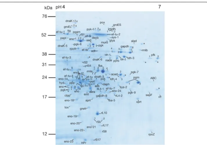
Fig. 1 Representative 2DE protein expression profile extracted from S. gordonii DL1, visualized with Coomassie Brilliant Blue stain. Proteins identified by mass spectrometry are indicated (abbreviations are listed in Table1). Spots with no significant hits in the Mascot database search are labelled with an asterisk (*). Gels were produced in triplicates from three different cultures of S. gordonii DL1
Of the 19 identified putative phosphoproteins in S. gordonii DL1, 13 were replicated in B. subtilis, 10 in S. aureus, 8 in N. meningitidis, 7 in L. rhamnosus, 8 in M. penetrans, 5 in M. pneumoniae, 4 in M. genitalium and 2 in M. gallisepticum. As in S. gordonii DL1, several of the phosphorylated proteins were also present at two or more pIs in these bacteria (Table2).
Discussion
Ser/Thr/Tyr phosphoproteome of S. gordonii DL1
Putative Ser/Thr/Tyr phosphorylated intracellular pro-teins in S. gordonii DL1 were identified from 2DE gels using Pro-Q Diamond stain and LC-MS/MS. From the 49 phosphorylated spots detected, 19 Ser/Thr/Tyr phospho-proteins were identified in S. gordonii. Determined or pu-tative phosphorylation sites on Ser, Thr and/or Tyr in all phosphorylated proteins or homologues except for two, rmla and ppp, were found in the literature (Table2).
A variety of cellular processes were associated with the
phosphoproteins identified in S. gordonii DL1 (Fig. 3).
This supports the notion that Ser/Thr/Tyr phosphoryl-ation events are integrated in the pathways that regulate different cellular responses in this species. Many of the phosphorylated proteins were present at two or more pIs in S. gordonii. As is evident from Table 2, this is a common occurrence in other bacteria as well. Variably phosphorylated species of proteins are of interest
be-cause they may differ in function [21] and hence be of
biological relevance. With 2DE, although laborious, the occurrence of physiologically distinct forms of one pro-tein can be readily visualized in a more“hands on”, com-prehensible overview of the present proteomic profile, were differently modified protein species often occur at adjacent pIs. Studies on deletion mutants can be employed to investigate the role of specific
kinases/phos-phatases on phenotypic presentation or different
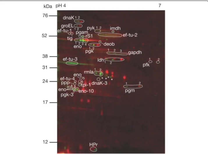
Fig. 2 Representative 2DE phosphoprotein profile extracted from S. gordonii DL1. The red stain indicates intracellular proteins visualized with T-Rex Protein Labelling kit detecting lysine residues. The green stain indicates phosphorylated proteins visualized with Pro-Q Diamond Phosphoprotein Gel Stain detecting phosphate groups attached to serine, threonine or tyrosine residues by O-phosphorylation. Phosphorylated proteins identified by mass spectrometry are indicated (abbreviations are listed in Table1). Phosphorylated spots with no significant hits in the Mascot database search are labelled with an asterisk (*). Gels were produced in triplicates from three different cultures of S. gordonii DL1
bacterial functions [46]. The current material constitutes a first step towards future studies that may utilize such targeted approaches. Studies of this character can be car-ried out from any perspective of microbial physiology and biological interaction of interest. Future studies using clinical isolates as well as biofilm growth flow cell models with saliva as a nutritious substrate are also of
interest to increase the resemblance to in vivo
conditions.
Core phosphoproteome
The total number of identified Ser/Thr/Tyr phosphory-lated proteins as detected with the Pro-Q Diamond stain in other bacteria grown in various growth conditions was between 10 and 73 [19, 27, 28, 32, 33, 37, 40, 42], making up between 0.7 to 2.6% of the total proteomes as
annotated in the UniProt database [45]. This is in
ac-cordance with the 19 phosphoproteins identified in S.
gordoniiDL1 (approximately 1% of the total proteome).
All phosphoproteins identified in the current study ex-cept for three (pfk, rmla, ppp) were also previously de-tected as Ser/Thr/Tyr-phosphorylated with the Pro-Q Diamond stain in other bacteria (Table2). Proteins that are consistently found in their phosphorylated state across several species and growth conditions may repre-sent a core phosphoproteome profile shared by many bacteria. The mapping of such a core phosphoproteome may facilitate the identification of phenotypic character-istics that deviate from this core pattern. Identification of distinct phenotype phosphorylation patterns that re-flect microbial activities may be crucial for pursuing a
further understanding of biofilm formation and
colonization of the commensal microbiota. Based on our findings, pfk, rmla and ppp represent possible proteins
of interest for further investigation. Two phosphorylated protein species of pfk were detected in S. gordonii. In Streptococcus pneumoniae, pfk was found to be phos-phorylated on a tyrosine residue, and phosphorylation of this enzyme was suggested to be involved in regulation
of the metabolic flux in the cell [31]. One
phosphory-lated protein species of ppp was detected in S. gordonii. The phosphatase stp1 in Streptococcus agalactiae was identified as a homologue to S. gordonii ppp (100% simi-larity) in theUniProt.org database [45]. Stp1 is involved in group B streptococcal virulence [47]. Two phosphory-lated protein species of rmla was identified in S. gordonii. Activation and deactivation of rmla is an important regulating mechanism of peptidoglycan biosynthesis in gram positive bacteria, making it an interesting candi-date in the search for new antibiotics [48]. Hardly any-thing can be found in the literature about potential regulation of catalytic activity by phosphorylation of rmla, but the current findings suggest that Ser/Thr/Tyr phosphorylation events are involved. These three pro-teins represent possible candidates for uniquely phos-phorylated proteins in S. gordonii, however, because phosphorylation patterns are dependent on growth conditions, further experiments comparing the phospho-proteomes of different species grown in the same envi-ronments are needed.
Phosphoproteins in S. gordonii DL1 involved in carbohydrate transport and metabolism
The carbohydrate transporting protein HPr was detected as Ser/Thr/Tyr phosphorylated in S. gordonii. HPr is a known phosphoprotein with roles in the phosphotrans-ferase system (PTS), for uptake and phosphorylation of certain carbohydrates in the central carbon metabolism.
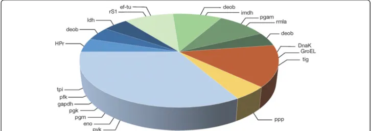
Fig. 3 Ser/Thr/Tyr O-phosphorylated proteins involved in various cellular functions in S. gordonii DL1. Blue slices; carbon metabolism. Green slices; biosynthesis. Orange slice; chaperone function. Yellow slice; protein dephosphorylation. glycolysis, carbohydrate transport, pentose phosphate pathway, pyruvate conversion, protein translation, nucleotide biosynthesis, cell wall biosynthesis, amino acid biosynthesis, molecular chaperone function, protein dephosphorylation
Protein Protein also detected as phosphorylated with Pro-Q Diamond in
Number of phosphorylated protein species Carbohydrate transport HPrb[7,25,26] B. subtilis 168 trpC2 [27] M. penetrans GTU-54 [28] 1 1 Glycolysis
tpib[29,30] L. rhamnosus GG (ATCC 53103) [19] M. penetrans GTU-54 [28] 1 1 pfkb[31] – – gapdhb[29,30] B. subtilis 168 trpC2 [27] L. rhamnosus GG (ATCC 53103) [19] S. aureus COL [32] N. meningitidis Z4970 (serogroup A) [33] M. penetrans GTU-54 [28] 1 2 3 2 1 pgkb[29,34,35] B. subtilis 168 trpC2 [27] L. rhamnosus GG (ATCC 53103) [19] N. meningitidis Z4970 (serogroup A) [33] S. aureus COL [32] 1 1 2 2 pgmb[29,34] B. subtilis 168 trpC2 [27] S. aureus COL [32] M. penetrans GTU-54 [28] 2 3 1 enob[20,29,30,34,36] B. subtilis 168 trpC2 [27] B. subtilis 168 trpC2 [37] S. aureus COL [32] N. meningitidis Z4970 (serogroup A) [33] 3 3 3 2 pykb[29,30,34,38,39] B. subtilis 168 trpC2 [27] B. subtilis 168 trpC2 [37] L. rhamnosus GG (ATCC 53103) [19] S. aureus COL [32] M. penetrans GTU-54 [28] 4 1 1 5 1 Pyruvate conversion ldha[29,35] M. genitalium G37 (ATCC 33530) [40] M. pneumoniae M129-B170 (ATCC 29343) [40] 2 1 Pentose phosphate pathway, amino acid and nucleotide biosynthesis
deobb[34,35] S. aureus COL [32]
B. subtilis 168 trpC2 [27]
2 1 Nucleotide biosynthesis
imdhb[41] B. subtilis 168 trpC2 [27] 2
Molecular chaperone activity
DnaKa[30,34] B. subtilis 168 trpC2 [27] N. meningitidis Z4970 (serogroup A) [33] M. penetrans GTU-54 [28] M. gallisepticum S6 [42] M. genitalium G37 (ATCC 33530) [40] M. pneumoniae M129-B170 (ATCC 29343) [40] 1 2 1 1 1 1 GroELb[30,35,39] B. subtilis 168 trpC2 [27] L. rhamnosus GG (ATCC 53103) [19] N. meningitidis Z4970 (serogroup A) [33] M. penetrans GTU-54 [28] M. gallisepticum S6 [42] M. genitalium G37 (ATCC 33530) [40] M. pneumoniae M129-B170 (ATCC 29343) [40] 1 1 4 1 1 1 1
The PTS has a high affinity for glucose, and a preference for glucose over alternative carbohydrates. HPr carries known phosphorylation sites on histidine, per se in-volved in the phosphotransfer catalytic activity, and serine, involved in the regulation of catalytic phospho-transfer activity [49]. Phosphorylation on HPr serine re-duces the phosphotransfer activity, thereby reducing the uptake of PTS-carbohydrates, while simultaneously en-hancing the uptake of alternative carbohydrates [7, 26,
49]. In this way, phosphorylation events on HPr can
regulate both the quantity and types of carbohydrates that are processed by the cell. Seven glycolytic proteins (tpi, pfk, gapdh, pgk, pgm, eno, pyk), as well as ldh, in-volved in pyruvate conversion, were Ser/Thr/Tyr phos-phorylated in S. gordonii.
Prokaryotes have been suggested to regulate glycolytic activity rapidly and reversibly through protein phosphor-ylation and dephosphorphosphor-ylation in response to environ-mental changes [12,13]. Our findings indicate that these mechanisms are present in S. gordonii as well. Aside from controlling the metabolic rate and character of the metabolic end products of glycolysis, e.g. by activation of ldh, regulation of some glycolytic enzymes, e.g. tpi, con-trols the switch between different metabolic pathways [50,51]. Regulation of enzymes with reversible catalytic function, e.g. pgk, eno, may enable switching between ca-tabolism and anabolism [45]. Rapid and reversible regu-lation of carbohydrate transport, control of rate of
glycolysis, and alternative carbohydrate metabolizing pathways by phosphorylation, increase cell fitness by en-abling adaptation of the cell metabolism in response to variations in carbohydrate concentration. Thereby, dam-age from processes such as sugar killing can be mitigated [8, 9, 12]. Our findings indicate that Ser/Thr/Tyr phos-phorylation is involved in regulating the activities of these proteins. Control of glycolysis not only by the levels of metabolic substrates and products, but by trans-duction of a variety of other signals through enzyme phosphorylation events, suggests a high complexity of metabolic control in S. gordonii. This supports the idea that integration of metabolic activity and environmental factors in bacterial cells is more convoluted than previ-ously believed.
Phosphoproteins in S. gordonii DL1 involved in acid tolerance response
To cope with the intermittent fluctuations in pH that nat-urally occur in oral biofilms, partly as a consequence of accumulation of lactic acid and other acids from the cen-tral carbon metabolism, oral streptococci employ the so-called acid tolerance response (ATR) [4, 10, 11]. Aside from responsive regulation of carbohydrate transport and metabolism, increased molecular chaperone activity com-prises another important aspect of the ATR in oral streptococci [4,8]. The chaperones represent the group of
proteins most frequently detected as Ser/Thr/Tyr
Table 2 Comparison to Ser/Thr/Tyr phosphoproteomes of other bacteria as detected with Pro-Q Diamond Phosphoprotein Gel Stain on 2DE gels (Continued)
Protein Protein also detected as phosphorylated
with Pro-Q Diamond in
Number of phosphorylated protein species
Tigb[19] L. rhamnosus GG (ATCC 53103) [19]
B. subtilis 168 trpC2 [27] B. subtilis 168 trpC2 [37] S. aureus COL [32] N. meningitidis Z4970 (serogroup A) [33] M. pneumoniae M129-B170 (ATCC 29343) [40] 1 1 1 1 1 1 Protein translation ef-tub[30,43] B. subtilis 168 trpC2 [27] B. subtilis 168 trpC2 [37] S. aureus COL [32] N. meningitidis Z4970 (serogroup A) [33] M. penetrans GTU-54 [28] M. genitalium G37 (ATCC 33530) [40] M. pneumoniae M129-B170 (ATCC 29343) [40] 1 1 1 2 1 1 1 rS1b[19] L. rhamnosus GG (ATCC 53103) [19] N. meningitidis Z4970 (serogroup A) [33] 1 2 Cell envelope biosynthesis
pgama[44] S. aureus COL [32] 2
rmla – –
Protein dephosphorylation
ppp – –
a
Phosphorylation site on Ser/Thr/Tyr identified in S. gordonii DL1 according to theUniProt.orgdatabase [45]
b
lar chaperones (DnaK, GroEL, tig). Ser/Thr/Tyr phos-phorylation has been suggested to activate all these three chaperones in bacteria [27,37,39,52]. By modulating the character and acidity of metabolic end products that accu-mulate locally, while retaining effective metabolism and other cellular functions at lower environmental pH with the help of ATR responses, cells increase their competi-tiveness towards less aciduric species [53]. From an evolu-tionary point of view, these mechanisms may have contributed to the long-term survival of these species in the strenuous oral environment.
Phosphoproteins in S. gordonii DL1 involved in biosynthesis
Six phosphorylated proteins involved in biosynthesis were identified in S. gordonii DL1 (deob, imdh, ef-tu, rS1, pgam, rmla). These proteins are involved in amino acid, nucleotide and cell wall biosynthesis, as well as protein translation (Table 1). Except for the chaperones, ef-tu was the protein most commonly detected with Pro-Q Diamond stain on 2DE gels in the previously
studied bacteria (Table 2). Phosphorylation on a
threo-nine residue in Escherichia coli ef-tu seems to inactivate the protein by decoupling the tRNA from the ribosome A-site [54, 55], triggering a rapid inhibition of protein translation. In contrast, phosphorylation on a threonine residue in the ribosomal protein rS1 in E. coli was found to activate protein translation [56]. Rapid and reversible regulation of biosynthesis through protein phosphoryl-ation events enables the cell to be frugal regarding en-ergy expenditure, and streamline cellular functions by modification of biomolecule production.
Moonlighting and multisite regulation of identified phosphoproteins
Several of the proteins identified as phosphorylated in S.
gordoniiDL1 have known moonlighting functions, often
in adhesion, e.g. tpi [57], gapdh [58], eno [59], and GroEL [60], alternative metabolic pathways, e.g. gapdh in iron metabolism [61], or other functions. In prokaryotes, cell surface associated enolase is involved in adhesion to host components in connective tissue or saliva [59], and retention of exported enolase on the bacterial cell sur-face was found to increase in acidic environments [58]. Maintaining cell adhesion during acidic shifts that may inactivate other adhesins is a tentative strategy for in-creased competitiveness in oral biofilms. In E. coli eno-lase, apart from reducing its glycolytic activity, lysine phosphorylation also prevented the enzyme export re-lated to its moonlighting functions [62]. These findings suggest that phosphorylation of enolase may affect cell adhesion during acidic shifts. Regulation of several of
multisite phosphorylation events [30, 34, 39, 63]. This further supports the idea that regulation of cellular activ-ities in prokaryotes is complex and involves integrated patterns of signal transduction.
Conclusion
This study clearly shows that Ser/Thr/Tyr phosphoryl-ation is present in an array of cytoplasmic proteins from the oral commensal S. gordonii. In total, 61 intracellular proteins were identified in S. gordonii DL1, and 19 of these turned out to be present as phosphorylated. Most of the phosphorylated proteins are involved in the car-bon metabolism, specifically related to glycolysis, which is no surprise for this saccharolytic oral streptococcus. Ser/Thr/Tyr phosphorylation presents a possible mech-anism for regulation of multiple cellular processes in S. gordonii, and phosphorylated proteins species were often present at several pIs with potential variation in bio-logical function. Many similarities were found between the identified phosphoproteome of S. gordonii DL1 and that of previously studied species, despite differences in basic cell physiology and the growth conditions applied. Identification of core regulatory pathways involved in the interaction between bacteria and their environment should provide useful insights regarding new strategies to manage biofilm-induced diseases. Studies in micro-biology are often focused on investigating the role of bacteria in disease. However, the physiology of commen-sals that prevent shifts towards dysbiosis are just as rele-vant. To investigate mechanisms of biofilm formation and homeostasis in the oral cavity, it is essential to examine bacterial responses and regulation of responses in relation to specific environmental challenges.
Methods
Bacterial strain and culture conditions
S. gordonii DL1 was routinely grown overnight in 25%
Todd-Hewitt Yeast Extract (¼ THYE, Becton Dickin-son), at 37 °C in 5% CO2. Cell cultures were diluted 1:10
in 25% THYE + 20 mM glucose (¼ THYE+G) and grown as described above until the mid-exponential phase (OD600nm = 0.5–0.6) was reached. Planktonic cells were retrieved by centrifugation (3000 rpm, 10 min, 5 °C), washed, and resuspended in a 10 mM Tris HCl-buffer
pH 6.8 containing 1 mM EDTA and 5 mM MgSO4, and
stored at− 20 °C until protein extraction.
Protein extraction
Harvested cells were subjected to three freeze-thaw
cy-cles before washing and then resuspended in 700μl lysis
buffer containing 8 M urea, 2% (v/v) CHAPS, 64.8 mM DTT, 2% IPG buffer pH 4–7 (Pharmacia Amersham
Biotech, Sweden). Ultrasonication of the samples was then performed with homogenizing 0.2 mm glass beads for 4 × 5 min (5 s pulses, amplitude 40, Vibra-Cell™ Ultrasonic Processor, SONICS), with alternate periods of cooling. Intact cells were sedimented by centrifugation at 17000×g for 10 min at 4 °C and the supernatants
(pro-tein extracts) were stored at − 20 °C. The protein
con-centration was determined using the 2-D Quant kit (GE Healthcare Life Sciences).
2D gel electrophoresis
Intracellular proteins were extracted and separated by two-dimensional sodium dodecyl sulfate-polyacrylamide gel electrophoresis (2D SDS-PAGE; 2DE) as described previously [64]. Isoelectric focusing was carried out on 18 cm Immobiline Dry Strips pH 4–7 (Amersham Phar-macia Biotech, Sweden) followed by gel electrophoresis on 14% polyacrylamide gels (185 × 200 × 1.0 mm). Gels
for Coomassie staining were loaded with 150μg protein.
Gels for fluorescent staining were loaded with 50μg pro-tein, and proteins were pre-labelled with the T-Rex La-belling kit (NH DyeAGNOSTICS) before rehydration
(see “Staining procedures” below). The gels were then
fixed in the appropriate fix solution as recommended by the respective stain manufacturer (Coomassie stain 2% acetic acid and 40% ethanol and fluorescent stain 10% acetic acid and 50% ethanol). Gels for Coomassie stain-ing were fixed for at least 1 h and gels for fluorescent staining were fixed overnight, protected from light. All gels were produced in triplicates from separate cell cultures.
Staining procedures
Coomassie gels were stained overnight in 300 ml Coo-massie brilliant blue staining solution containing 17% Coomassie Brilliant Blue G - Colloidal Concentrate (Sigma) and 21% ethanol. After staining, Coomassie gels were destained with 25% ethanol for approximately 1 h and stored in Ultra-High Quality water (UHQ) at 4 °C until scanning and excision of spots. For fluorescent staining, lysine residues of the general proteome were pre-labelled with T-Rex Labelling kit (NH DyeAGNOS-TICS) before rehydration, to facilitate orientation of the phosphorylation profiles. Pre-labelling was performed according to the manufacturer’s protocol. Briefly, 2 μl
T-Rex solvent and 50μg of the extracted proteins were
transferred into a T-Rex labelling vial and incubated on ice for 30 min. Thereafter the gels were prepared as de-scribed above. After fixation, each T-rex stained gel was washed with UHQ for 3 × 15 min and pre-scanned once at 647 nm with a photomultiplier tube (PMT) setting of
800, pixel size 100μm, before staining with Pro-Q
Dia-mond Phosphoprotein Gel Stain (Invitrogen™,
Thermo-Fisher Scientific) according to the manufacturer’s
instruction. Phosphoprotein staining was performed pro-tected from light, and with minor modifications based
on results from Agrawal and Thelen [65]. Each gel was
stained with Pro-Q Diamond Phosphoprotein Gel Stain diluted 1:2 in UHQ to a final volume of 250 ml for 2 h. Gels were destained in 250 ml Pro-Q Diamond Phospho-protein Gel Destaining Solution (Invitrogen™, Thermo-Fisher Scientific) per gel for 3 × 30 min on a shaker and then washed in UHQ for 3 × 5 min.
Phosphoimage analysis
Phosphoimaging was performed with a Fujifilm FLA-9000 (Science Imaging Scandinavia AB). T-Rex labelled
proteins were visualized at 647 nm, PMT 800, 100μm
(displayed as red). Pro-Q Diamond stained proteins were
visualized at 532 nm, PMT 600, 100μm (displayed as
green). Phosphorylation profiles were analysed by three operators and then compared and discussed for calibra-tion, with good concordance. Quantification of Pro-Q Diamond Phosphoprotein Gel Stain signals from phos-phorylated spots was not possible due to variations be-tween triplicates.
Identification of streptococcal proteins by LC-MS/MS
Protein spots were manually excised from Coomassie brilliant blue-stained gels and subjected to in-gel
diges-tion with trypsin as previously described [64]. The
resulting protein fragments were separated with liquid chromatography (LC) and characterized using tandem mass spectrometry (MS/MS) (Aberdeen Proteomics, University of Aberdeen). In short, proteins were treated with DTT for reduction (60 °C, 20 min), iodacetamide for S-alkylation (25 °C, 10 min) and trypsin for digestion (37 °C, 8 h). Following drying by rotary evaporation (SC110 Speedvac, Savant Instruments), the peptide ex-tract was dissolved in 0.1% formic acid. Analysis of pep-tide solutions was carried out using an HTCultraPTM Discovery system (Bruker Daltonics) coupled to an Ul-tiMate 3000 LC system (Dionex). Separation of tryptic peptides was performed on a monolithic capillary
col-umn (200μm internal diameter × 5 cm, Dionex). The
gradient consisted of 5% acetonitrile in UHQ containing 0.5% formic acid, to 37% acetonitrile in UHQ containing
0.45% formic acid over 12 min at a flow rate of 2.5μl
min− 1. Data-dependent mode was employed to acquire
peptide fragment mass spectra, AutoMS (2). The scan range was 300–1500 m/z, with three averages, and up to three precursor ions that were selected from the MS scan (100–2200 m/z). Active exclusion of precursors was performed within a 1.0 min window, as well as exclusion of all singly charged ions. Detection and deconvolution of peptide peaks were carried out automatically using Data Analysis software (Bruker). Mass lists in the form of Mascot Generic files were created automatically and
using the Matrix Science web server [66]. Parameters were set to 0.5 Da peptide mass tolerance, methionine oxidation and carboxyamidomethylation of cysteine. The maximum number of missed cleavages was set to 1. Pro-teins were identified by at least two peptides with a Mas-cot score of 33 or higher, the latter as suggested by Koenig et al., 2008 [67].
Literature search
The PubMed, Web of Science and Cochrane databases were searched for studies that examined global Ser/Thr/ Tyr phosphoproteomes in human commensal and pathogenic bacteria using Pro-Q Diamond Phosphopro-tein Gel Stain on 2DE gels. Studies on the model organ-ism Bacillus subtilis were also included.
Supplementary information
Supplementary information accompanies this paper athttps://doi.org/10.
1186/s12866-020-01944-y.
Additional file 1. The sequenced peptides identified by LC-MS/MS can be found in supplemental material (Additional file1). Sequenced peptides identified by LC-MS/MS; Ser/Thr/Tyr phosphorylated protein species (Table A), and non-phosphorylated protein species (Table B).
Abbreviations
2DE:Two-dimensional gel electrophoresis; ¼ THYE: 25% Todd-Hewitt Yeast Extract; ¼ THYE+G: 25% THYE + 20 mM glucose; ATR: Acid tolerance response; MW: Molecular weight; PTS: Phosphotransferase transport system; Ser/Thr/Tyr: Serine, threonine, tyrosine; UHQ: Ultra-High Quality water Acknowledgements
We would like to thank Agnethe Henriksson and Madeleine Blomqvist (Malmö) for their excellent technical assistance.
Authors’ contributions
CR, GS and CW participated in the design and coordination of the study. CR performed the experiments and drafted the manuscript. CR, GS, and CW analyzed and interpreted the data. GS, CW and ZB provided extensive suggestions on the research as it progressed and were major contributors in revising and editing the final manuscript. All authors read and approved the final manuscript.
Authors’ information Not applicable. Funding
This study was supported by grants from Malmö University, Sweden, supporting the interdisciplinary research programme FORESIGHT, and the Swedish Research Council (201601994). Open access funding provided by Malmö University.
Availability of data and materials
All datasets generated or analysed during this study are included in this published article and its supplementary information files, see (Additional file
1). The gel triplicates are available from the corresponding author on reasonable request.
Ethics approval and consent to participate Not applicable.
Competing interests
The authors declare that they have no competing interests. Author details
1Department of Oral Biology and Pathology, Faculty of Odontology, Malmö University, 20506 Malmö, Sweden.2Department of Biomedical Science, Malmö University, 20506 Malmö, Sweden.
Received: 26 June 2020 Accepted: 11 August 2020
References
1. Li J, Helmerhorst EJ, Leone CW, Troxler RF, Yaskell T, Haffajee AD, Socransky SS, Oppenheim FG. Identification of early microbial colonizers in human dental biofilm. J Appl Microbiol. 2004;97:1311–8.
2. Schachtele CF, Nobbs A, Zhang Y, Costalonga M, Herzberg MC. Oral streptococci: commensals and opportunistic pathogens. In: Hakenbeck R, Chhatwal S, editors. Molecular biology of streptococci. 1st ed. Norfolk: Horizon Bioscience; 2007. p. 411–62.
3. Knox KW, Hunter N. The role of oral bacteria in the pathogenesis of infective endocarditis. Aust Dent J. 1991;36:286–92.
4. Takahashi N, Yamada T. Acid-induced acid tolerance and acidogenicity of non-mutans streptococci. Oral Microbiol Immunol. 1999;14:43–8. 5. Stephan RM. Changes in hydrogen-ion concentrations on tooth surfaces
and in carious lesions. J Am Dent Assoc. 1940;27:718–23.
6. Stephan RM. Intra-oral hydrogen-ion concentrations associated with dental caries activity. J Dent Res. 1944;23:257–66.
7. Vadeboncoeur C, Pelletier M. The phosphoenolpyruvate:sugar
phosphotransferase system of oral streptococci and its role in the control of sugar metabolism. FEMS Microbiol Rev. 1997;19:187–207.
8. Lemos JAC, Abranches J, Burne RA. Responses of cariogenic streptococci to environmental stresses. Current Issues in Mol Biol. 2005;7:95–108. 9. Yamada T, Carlsson J. Regulation of lactate dehydrogenase and change of
fermentation products in streptococci. J Bacteriol. 1975;124:55–61. 10. Hamilton IR, Buckley ND. Adaptation by Streptococcus mutans to acid
tolerance. Oral Microbiol Immunol. 1991;6:65–71.
11. Svensäter G, Larsson UB, Greif ECG, Cvitkovitch DG, Hamilton IR. Acid tolerance response and survival by oral bacteria. Oral Microbiol Immunol. 1997;12:266–73.
12. Cozzone AJ. Protein phosphorylation in prokaryotes. Annu Rev Microbiol. 1988;42:97–125.
13. Pereira SFF, Goss L, Dworkin J. Eukaryote-like serine/threonine kinases and phosphatases in Bacteria. Microbiol Mol Biol Rev. 2011;75:192–212. 14. Hammes GG, Wu CW. Kinetics of allosteric enzymes. Annu Rev Biophys
Bioeng. 1974;3:1–33.
15. Kennelly PJ, Potts M. Fancy meeting you here! A fresh look at‘prokaryotic’ protein phosphorylation. J Bacteriol. 1996;178:4759–64.
16. Deutscher J, Saier MH. Ser/Thr/Tyr protein phosphorylation in bacteria - for long time neglected, now well established. J Mol Microbiol Biotechnol. 2006;9:125–31.
17. Cozzone AJ. Role of protein phosphorylation on serine/threonine and tyrosine in the virulence of bacterial pathogens. J Mol Microbiol Biotechnol. 2005;9:198–213.
18. Ge R, Shan W. Bacterial Phosphoproteomic analysis reveals the correlation between protein phosphorylation and bacterial pathogenicity. Genomics Proteomics Bioinformatics. 2011;9:119–27.
19. Koponen J, Laakso K, Koskenniemi K, Kankainen M, Savijoki K, Nyman TA, de Vos WM, Tynkkynen S, Kalkkinen N, Varmanen P. Effect of acid stress on protein expression and phosphorylation in Lactobacillus rhamnosus GG. J Proteome. 2012;75:1357–74.
20. Jers C, Pedersen MM, Paspaliari DK, Schütz W, Johnsson C, Soufi B, Macek B, Jensen PR, Mijakovic I. Bacillus subtilis BY-kinase PtkA controls enzyme activity and localization of its protein substrates. Mol Microbiol. 2010;77:287–99. 21. Cohen P. The regulation of protein function by multisite phosphorylation
-a 25 ye-ar upd-ate. Trends Biochem Sci. 2000;25:596–601.
22. Jeffery CJ. Protein species and moonlighting proteins: very small changes in a protein’s covalent structure can change its biochemical function. J Proteome. 2016;134:19–24.
23. Mimura CS, Poy F, Jacobson GR. ATP-dependent protein kinase activities in the oral pathogen Streptococcus mutans. J Cell Biochem. 1987;33:161–71. 24. Banu LD, Conrads G, Rehrauer H, Hussain H, Allan E, van der Ploeg JR. The
Streptococcus mutans serine/threonine kinase, PknB, regulates competence development, bacteriocin production, and cell wall metabolism. Infect Immun. 2010;78:2209–20.
25. Frey N, Nessler S, Fieulaine S, Vaillancourt K, Frenette M, Vadeboncoeur C. The HPr(Ser) kinase of Streptococcus salivarius: a hexameric bifunctional enzyme controlled by glycolytic intermediates and inorganic phosphate. FEMS Microbiol Lett. 2003;224:67–72.
26. Reizer J, Romano AH, Deutscher J. The role of phosphorylation of HPr, a phosphocarrier protein of the phosphotransferase system, in the regulation of carbon metabolism in gram-positive bacteria. J Cell Biochem. 1993;51:19–24.
27. Eymann C, Becher D, Bernhardt J, Gronau K, Klutzny A, Hecker M. Dynamics of protein phosphorylation on Ser/Thr/Tyr in Bacillus subtilis. Proteomics. 2007;7:3509–26.
28. Ferrer-Navarro M, Gómez A, Yanes O, Planell R, Avilés FX, Piñol J, Pons JAP, Querol E. Proteome of the bacterium Mycoplasma penetrans. J Proteome Res. 2006;5:688–94.
29. Macek B, Mijakovic I, Olsen JV, Gnad F, Kumar C, Jensen PR, Mann M. The serine/threonine/tyrosine Phosphoproteome of the model bacterium Bacillus subtilis. Mol Cell Proteomics. 2007;6:697–707.
30. Misra SK, Milohanic E, Aké F, Mijakovic I, Deutscher J, Monnet V, Henry C. Analysis of the serine/threonine/tyrosine phosphoproteome of the pathogenic bacterium Listeria monocytogenes reveals phosphorylated proteins related to virulence. Proteomics. 2011;11:4155–65.
31. Ahmad Z, Morona R, Standish AJ. In vitro characterization and identification of potential substrates of a low molecular weight protein tyrosine phosphatase in Streptococcus pneumoniae. Microbiol. 2018;164:697–703. 32. Bäsell K, Otto A, Junker S, Zühlke D, Rappen G-M, Schmidt S, Hentschker C, Macek
B, Ohlsen K, Hecker M, Becher D. The phosphoproteome and its physiological dynamics in Staphylococcus aureus. Int J Med Microbiol. 2014;304:121–32. 33. Bernardini G, Laschi M, Serchi T, Arena S, D’Ambrosio C, Braconi D, Scaloni
A, Santucci A. Mapping phosphoproteins in Neisseria meningitidis serogroup a. Proteomics. 2011;11:1351–8.
34. Macek B, Gnad F, Soufi B, Kumar C, Olsen JV, Mijakovic I, Mann M. Phosphoproteome analysis of E. coli reveals evolutionary conservation of bacterial Ser/Thr/Tyr phosphorylation. Mol Cell Proteomics. 2008;7:299–307. 35. Soufi B, Gnad F, Jensen PR, Petranovic D, Mann M, Mijakovic I, Macek B. The
Ser/Thr/Tyr phosphoproteome of Lactococcus lactis IL1403 reveals multiply phosphorylated proteins. Proteomics. 2008;8:3486–93.
36. Virmani R, Sajid A, Singhal A, Gaur M, Joshi J, Bothra A, Garg R, Misra R, Singh VP, Molle V, Goel AK, Singh A, Kalia VC, Lee JK, Hasija Y, Arora G, Singh Y. The Ser/Thr protein kinase PrkC imprints phenotypic memory in Bacillus anthracis spores by phosphorylating the glycolytic enzyme enolase. J Biol Chem. 2019;294:8930–41.
37. Lévine A, Vannier F, Absalon C, Kuhn L, Jackson P, Scrivener E, Labas V, Vinh J, Courtney P, Garin J, Séror SJ. Analysis of the dynamic Bacillus subtilis Ser/ Thr/Tyr phosphoproteome implicated in a wide variety of cellular processes. Proteomics. 2006;6:2157–73.
38. Singh DK, Singh PK, Tiwari S, Singh SK, Kumari R, Tripathi DK, Srivastava KK. Phosphorylation of pyruvate kinase a by protein kinase J leads to the altered growth and differential rate of intracellular survival of mycobacteria. Appl Microbiol Biotechnol. 2014;98:10065–76.
39. Arora G, Sajid A, Virmani R, Singhal A, Kumar CMS, Dhasmana N, Khanna T, Maji A, Misra R, Molle V, Becher D, Gerth U, Mande SC, Singh Y. Ser/Thr protein kinase PrkC-mediated regulation of GroEL is critical for biofilm formation in Bacillus anthracis. NPJ Biofilms Microbiomes. 2017;3:7. 40. Su HC, Hutchison CA, Giddings MC. Mapping phosphoproteins in
Mycoplasma genitalium and Mycoplasma pneumoniae. BMC Microbiol. 2007;7:63.
41. Rajagopal L, Vo A, Silvestroni A, Rubens CE. Regulation of purine biosynthesis by a eukaryotic-type kinase in Streptococcus agalactiae. Mol Microbiol. 2005;56:1329–46.
42. Demina IA, Serebryakova MV, Ladygina VG, Rogova MA, Zgoda VG, Korzhenevskyi DA, Govorun VM. Proteome of the bacterium Mycoplasma gallisepticum. Biochem. 2009;74:165–74.
43. Lippmann C, Lindschau C, Vijgenboom E, Schroder W, Bosch L, Erdmann VA. Prokaryotic elongation factor Tu is phosphorylated in vivo. J Biol Chem. 1993;268:601–7.
44. Jolly L, Ferrari P, Blanot D, van Heijenoort J, Fassy F, Mengin-Lecreulx D. Reaction mechanism of phosphoglucosamine mutase from Escherichia coli. Eur J Biochem. 1999;262:202–10.
45. UniProt database.www.uniprot.org. Accessed 18 Aug 2020. 46. Bender MH, Cartee RT, Yother J. Positive correlation between tyrosine
phosphorylation of CpsD and capsular polysaccharide production in Streptococcus pneumoniae. J Bacteriol. 2003;185:6057–66.
47. Burnside K, Lembo A, Harrell MI, Gurney M, Xue L, Binh Tran NT, Connelly JE, Jewell KA, Schmidt BZ, de los Reyes M, Tao WA, Doran KS, Rajagopal L. Serine/threonine phosphatase Stp1 mediates post-transcriptional regulation of hemolysin, autolysis, and virulence of group B Streptococcus. J Biol Chem. 2011;286:44197–210.
48. Blankenfeldt W, Asuncion M, Lam JS, Naismith JH. The structural basis of the catalytic mechanism and regulation of glucose-1-phosphate
thymidylyltransferase (RmlA). EMBO J. 2000;19:6652–63. 49. Deutscher J, Kessler U, Alpert CA, Hengstenberg W. Bacterial
Phosphoenolpyruvate-dependent Phosphotransferase system: P-Ser-HPr and its possible regulatory function. Biochemistry. 1984;23:4455–60.
50. Kang TS, Korber DR, Tanaka T. Regulation of dual glycolytic pathways for fructose metabolism in heterofermentative Lactobacillus panis PM1. Appl Environ Microbiol. 2013;79:7818–26.
51. Solem C, Koebmann B, Jensen PR. Control analysis of the role of triosephosphate isomerase in glucose metabolism in Lactococcus lactis. IET Syst Biol. 2008;2:64–72. 52. Sherman MY, Goldberg AL. Heat shock of Escherichia coli increases binding
of dnaK (the hsp70 homolog) to polypeptides by promoting its phosphorylation. Proc Natl Acad Sci U S A. 1993;90:8648–52. 53. Abbe K, Carlsson J, Takahashi-Abbe S, Yamada T. Oxygen and the sugar
metabolism in oral streptococci. Proc Finn Dent Soc. 1991;87:477–87. 54. Pereira SFF, Gonzalez RL, Dworkin J. Protein synthesis during cellular
quiescence is inhibited by phosphorylation of a translational elongation factor. Proc Natl Acad Sci U S A. 2015;112:3274–81.
55. Talavera A, Hendrix J, Versées W, Jurėnas D, Van Nerom K, Vandenberk N, Singh RK, Konijnenberg A, De Gieter S, Castro-Roa D, Barth A, De Greve H, Sobott F, Hofkens J, Zenkin N, Loris R, Garcia-Pino A. Phosphorylation decelerates conformational dynamics in bacterial translation elongation factors. Sci Adv. 2018;4:9714–28.
56. Robertson ES, Nicholson AW. Phosphorylation of Escherichia coli translation initiation factors by the bacteriophage T7 protein Kinaset. Biochemistry. 1992;31:4822–7.
57. Furuya H, Ikeda R. Interaction of triosephosphate isomerase from Staphylococcus aureus with plasminogen. Microbiol Immunol. 2011;55:855–62.
58. Wang G, Xia Y, Cui J, Gu Z, Song Y, Chen YQ, Zhang H, Chen W. The roles of moonlighting proteins in Bacteria. Curr Issues Mol Biol. 2014;16:15–22. 59. Huberts DHEW, van der Klei IJ. Moonlighting proteins: an intriguing mode
of multitasking. Biochim Biophys Acta. 2010;1803:520–5.
60. Bergonzelli GE, Granato D, Pridmore RD, Marvin-Guy LF, Donnicola D, Corthésy-Theulaz IE. GroEL of Lactobacillus johnsonii La1 (NCC 533) is cell surface associated: potential role in interactions with the host and the gastric pathogen Helicobacter pylori. Infect Immun. 2006;74:425–34. 61. Boradia VM, Raje M, Raje CI. Protein moonlighting in iron metabolism:
Glyceraldehyde-3-phosphate dehydrogenase (GAPDH). Biochem Soc Trans. 2014;42:1796–801.
62. Boël G, Pichereau V, Mijakovic I, Mazé A, Poncet S, Gillet S, Giard JC, Hartke A, Auffray Y, Deutscher J. Is 2-phosphoglycerate-dependent automodification of bacterial enolases implicated in their export? J Mol Biol. 2004;337:485–96. 63. Shi L, Ravikumar V, Derouiche A, Macek B, Mijakovic I. Tyrosine 601 of
Bacillus subtilis DnaK undergoes phosphorylation and is crucial for chaperone activity and heat shock survival. Front Microbiol. 2016;7:533–46. 64. Davies JR, Svensäter G, Herzberg MC. Identification of novel LPXTG-linked
surface proteins from Streptococcus gordonii. Microbiology. 2009;155:1977–88. 65. Agrawal GK, Thelen JJ. Development of a simplified, economical polyacrylamide
gel staining protocol for phosphoproteins. Proteomics. 2005;5:4684–8. 66. Matrix Science web server.www.matrixscience.com. Accessed 18 Aug 2020. 67. Koenig T, Menze BH, Kirchner M, Monigatti F, Parker KC, Patterson T, Steen
JJ, Hamprecht FA, Steen H. Robust prediction of the MASCOT score for an improved quality assessment in mass spectrometric proteomics. J Proteome Res. 2008;7:3708–17.
Publisher’s Note
Springer Nature remains neutral with regard to jurisdictional claims in published maps and institutional affiliations.