Citation for the published paper:J. Neilands, D. Sutherland, A. Resin, P.L. Wejse, L.E. Chávez de Paz Chitosan Nanoparticles Affect the Acid Tolerance Response in Adhered Cells of Streptococcus mutans” 2011, Caries Research, vol 45, issue 6: pp 501-505.
URL: <http://dx.doi.org/10.1159/000331206>. Access to the published version require subscription.
Chitosan Nanoparticles Affect the Acid Tolerance Response in Adhered
Cells of Streptococcus mutans
J. Neilands a D. Sutherland b A. Resin b P.L. Wejse c L.E. Chávez de Paz a a
Department of Oral Biology, Faculty of Odontology, Malmö University, Malmö , Sweden;
b Interdisciplinary Nanoscience Center (iNANO), Faculty of Science, Aarhus University, Aarhus , and c Arla Foods Ingredients R&D, Nørre Vium , Denmark
Keywords
Acid tolerance, Biofilms, Chitosan, Nanoparticles, Streptococcus mutans
Abstract
In this study we evaluated the effect of chitosan nanoparticles on the acid tolerance response (ATR) of adhered Streptococcus mutans. An ATR was induced by exposing S. mutans to pH 5.5 for 2 h and confirmed by exposing the acid-adapted cells to pH 3.5 for 30 min, with the majority of cells appearing viable according to the LIVE/DEAD® technique. However, when chitosan nanoparticles were present during the exposure to pH 5.5, no ATR occurred as most cells appeared dead after the pH 3.5 shock. We conclude that the chitosan nanoparticles tested had the ability to hinder ATR induction in adhered S. mutans.
Copyright © 2011 S. Karger AG, Basel
Frequent consumption of easily fermentable carbohydrates leads to progressively lower pH values in human dental plaque. These low pH fluctuations in dental plaque are caused by microorganisms such as Streptococcus mutans, which can rapidly metabolize dietary carbohydrates into acid end products [Loesche, 1986; Bowden, 1991; Carlsson and Hamilton, 1994]. This acidification of dental plaque contributes to the demineralization of tooth enamel during caries development [Stephan, 1944]. Organisms associated with caries not only contribute to the acidification of the plaque biofilm, but they must also be capable of growth and survival during the prolonged periods of low pH typical of caries development. This acidtolerant microflora promotes further demineralization of the enamel and the development of many forms of dental caries [Marsh, 1994, 2003]. In response to repeated periods of low pH, some microorganisms are able to initiate an acid tolerance response (ATR), which involves changes in their physiology. Mainly, this response allows these microorganisms to survive and to continue to ferment carbohydrates and produce acids even though the pH in the environment decreases [Hamilton and Buckley, 1991; Svensäter et al., 1997; Lemos et al., 2005]. In S. mutans, in vitro experiments have shown that only a small percentage of cells survive a pH change from 7.5 to 3.5; however, if they are first incubated at pH 5.5 for 2 h, the cells induce an ATR, which leads to enhanced survival at lethal pH values (pH 3.5) [Welin-Neilands and Svensäter, 2007].
Citation for the published paper:J. Neilands, D. Sutherland, A. Resin, P.L. Wejse, L.E. Chávez de Paz Chitosan Nanoparticles Affect the Acid Tolerance Response in Adhered Cells of Streptococcus mutans” 2011, Caries Research, vol 45, issue 6: pp 501-505.
URL: <http://dx.doi.org/10.1159/000331206>. Access to the published version require subscription.
In recent years there has been an increase in dental caries, and there is a need for new preventive strategies [Bagramian et al., 2009]. Being able to prevent bacteria from inducing an ATR could be an effective measure in the prevention of dental caries. The recent application of biodegradable polymeric nanoparticles has shown immense potential for oral delivery of different types of therapeutic agents including drugs, vaccines, genes, proteins and peptides [des Rieux et al., 2006]. To date, most of the advanced nanoparticulate drug carriers have been developed by utilizing either synthetic or natural polymers, or by their combination [Sarmento et al., 2007]. Among the various natural polymers available, chitosan is one of the most widely used biopolymers for the preparation of nanoparticles [Allemann et al., 1998]. As shown by Decker et al. [2005], some chitosans had no activity against attached cells of Streptococcus sanguinis, while others affected their cell viability. These differences in antimicrobial activity of chitosan depend on their molecular weight and the degree of deacetylation [Sudarshan et al., 1992; Rabea et al., 2003; Chávez de Paz et al., 2011]. In the present study, we investigated the effect of a chitosan nanoparticle complex on acid adaptation in adhered cells of S. mutans. We hypothesized that due to the high solubility of chitosan nanoparticles, these could have an effect in preventing an ATR in S. mutans cells. Material and Methods
Strain, Growth Conditions and Media
The S. mutans strain UA159 (ATCC 700610) used in this study was a kind gift from Dr. Dennis Cvitkovitch, University of Toronto, Canada. The inoculum for the biofilm experiments was prepared by growing S. mutans in MM4 minimal medium [Hamilton and Svensäter, 1998] (pH 7.5) at 37 ° C in air with 5% CO 2 until it had reached the mid-exponential phase (optical density at 600 nm: approx. 0.7). S. mutans cells were harvested by centrifugation (5,000 g for 5 min) and resuspended at 2 times the concentration
in MM4 pH 7.5. The pH of the MM4 medium for the ATR experiments was adjusted to 5.5 and 3.5 with HCl.
Chitosan Nanoparticle System
A chitosan of 550 kDa molecular weight and a deacetylation degree of 85% (NovaMatrix, Sandvika, Norway) was used to prepare nanoparticles. This chitosan preparation had previously been shown to possess low antimicrobial activity [Chávez de Paz et al., 2011]. All aqueous solutions were prepared with deionized water and filtered through 200-nm pore filters. Nanoparticle assemblies were formulated by drop-wise adding a chitosan solution (100 μ l at 1 mg/ml in an acetic acid buffer) to a premixed solution composed of 50 μ l of
sodium tripolyphosphate solution (0.1%) in water and 500 μ l PBS buffer (pH 7.4). The mass
ratio of chitosan to sodium tripolyphosphate was kept constant at 2: 1. The particles were adjusted to pH 7.5 by the addition of 2 M NaOH and equilibrated at room temperature for 1 h under mixing. The nanoparticle solution was further characterized in a Malvern Zetasizer Nano ZS instrument (particle size: approx. 40 nm). The chitosan nanoparticle solution was
Citation for the published paper:J. Neilands, D. Sutherland, A. Resin, P.L. Wejse, L.E. Chávez de Paz Chitosan Nanoparticles Affect the Acid Tolerance Response in Adhered Cells of Streptococcus mutans” 2011, Caries Research, vol 45, issue 6: pp 501-505.
URL: <http://dx.doi.org/10.1159/000331206>. Access to the published version require subscription.
and adjusted to the appropriate pH using either HCl or NaOH.
Miniflow Cell System
The flow cell system μ-Slide VI for live cell analysis (Ibidi ® ; Ibidi GmbH, Martinsried, Germany) was used to grow S. mutans biofilms, and it consisted of 6 channels each with a
total working volume of 150 μ l and total growth areas of 0.6 cm 2 .
Adhesion of S. mutans
S. mutans inoculums prepared as described above were allowed to adhere for 2 h at 37 ° C at a
flow rate of 3.6 ml/h (shear rate: 10.1 s –1 ). After the 2-hour incubation period, non-adhered cells were washed away using MM4 pH 7.5 during a 1-hour rinse period, also at 3.6 ml/h.
Acid Tolerance Response
To test whether the chitosan nanoparticles could affect the induction of an ATR in biofilm cells of S. mutans, a previously used method for testing the effect of fluoride on acid adaptation was employed [Welin-Neilands and Svensäter, 2007]. Three-hour biofilms were exposed to MM4 pH 7.5 containing chitosan nanoparticles for 15 min, after which the cells were exposed to MM4 pH 5.5 with chitosan nanoparticles for 2 h. After the 2-hour adaptation period, the flow cells were rinsed with MM4 pH 5.5 devoid of chitosan nanoparticles for 15 min to remove any free chitosan nanoparticles and then exposed to MM4 pH 3.5 without chitosan nanoparticles for 30 min. Control cells were treated in the same way, but with MM4 only.
Cell Viability
The LIVE/DEAD ® BacLight ™ Bacterial Viability kit for microscopy (L0707; Molecular Probes) was used to assess the membrane integrity of biofilm cells. The LIVE/DEAD mixture was prepared by mixing components A + B in the kit, consisting of SYTO9 and propidium iodide, at a ratio of 1: 100 with the corresponding medium. After removing the culture fluid
from the flow chamber and washing with PBS, 30 μ l of the LIVE/DEAD mixture was added.
The chambers were incubated at room temperature for 10 min. The fluorescence from stained cells was viewed using confocal laser scanning microscopy (CLSM).
CLSM and Image Analysis
The fluorescence from stained cells was viewed using an Eclipse TE2000 inverted confocal laser scanning microscope (Nikon). A total of 20 CLSM images were acquired by means of the software EZ-C1 version 3.40 build 691 (Nikon) at a resolution of 512 x 512 pixels and
with a zoom factor of 1.0, yielding a final pixel resolution of 0.42 μ m/pixel. The CLSM
mages were analysed with the software bioImage_L [Chávez de Paz, 2009], which produced information on the total biofilm population as well as the independent subpopulations represented by red and green fluorescent colors. All experiments were run in duplicate on three separate occasions.
Citation for the published paper:J. Neilands, D. Sutherland, A. Resin, P.L. Wejse, L.E. Chávez de Paz Chitosan Nanoparticles Affect the Acid Tolerance Response in Adhered Cells of Streptococcus mutans” 2011, Caries Research, vol 45, issue 6: pp 501-505.
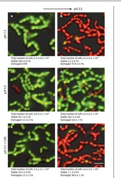
URL: <http://dx.doi.org/10.1159/000331206>. Access to the published version require subscription. Figure 1. High magnification of representative LIVE/DEAD CLS micrographs showing ATR and the effect of chitosan nanoparticles in adhered S. mutans UA159 cells. a , b At pH 7.5, most cells are viable (fluorescent green), and after lethal acid shock, most cells are viable (fluorescent green), and after lethal acid shock, most cells are damaged (fluorescent red). c , d Cells are allowed to generate an ATR at pH 5.5, and after lethal acid shock, most cells remain viable. e , f Cells are exposed to pH 5.5 + chitosan nanoparticles (CH3), and after acid shock, most cells are damaged. Scale bar = 2 μ m.
Citation for the published paper:J. Neilands, D. Sutherland, A. Resin, P.L. Wejse, L.E. Chávez de Paz Chitosan Nanoparticles Affect the Acid Tolerance Response in Adhered Cells of Streptococcus mutans” 2011, Caries Research, vol 45, issue 6: pp 501-505.
URL: <http://dx.doi.org/10.1159/000331206>. Access to the published version require subscription.
S. mutans UA159 formed single-layered biofilms with few clusters at 3 h of incubation. The
approximate number of adhered cells was of 6.7 ± 0.6 x 10 4 , which corresponds to 36.2 ± 4.8% of the surface. The proportion of viable cells was of 99.6 ± 0.1%. Our results corresponded well with data from previous studies using a larger flow cell system [Welin-Neilands and Svensäter, 2007]. In summary, we were able to run biofilm experiments under flow in a system compatible with CLSM and where conditions such as nutrient availability and pH could be controlled by the continuous addition of new medium.
The Presence of Chitosan Nanoparticles Hinders ATR Induction
We have previously shown that exposure of S. mutans to a sublethal pH value (pH 5.5) will induce an ATR in the cells that enhances survival at lower pH values (pH 3.5–3.0) [Svensäter et al., 1997; Welin-Neilands and Svensäter, 2007]. In this study, the control cells exposed to pH 5.5 (fig. 1 c) showed a viability of 95.7 ± 2.1%, which is similar to the viability of biofilm cells kept at pH 7.5 (99.6%) (fig. 1 a). After exposure to pH 3.5, the pH 5.5-adapted cells showed high viability (90.1 ± 8 3.2%), indicating that they had induced an ATR (fig. 1 d). As expected, the control cells kept at pH 7.5 showed low viability with only 2.2 8 0.1% viable cells (fig. 1 b). No significant reduction in the number of adhered cells was observed. The involvement of chitosan nanoparticles in the development of an ATR was tested by treating adhered S. mutans cells with the nanoparticles during exposure to the sublethal pH of 5.5. First, we confirmed that the exposure to chitosan did not affect cell viability at pH 5.5, with 94.5 ± 4.3% viable cells (fig. 1 e). Although the addition of nanoparticles did not affect the cell viability of S. mutans at pH 5.5, these cells showed very low viability after exposure to pH 3.5, with only 1.1 ± 0.5% viable cells (fig. 1 f).
Discussion
In S. mutans, the ATR involves changes in protein expression, changes in membrane fatty-acid synthesis, increased expression of proteins involved in DNA and protein repair and increased ATPase activity leading to increased proton efflux [Bowden and Hamilton, 1998; Quivey et al., 2000; Fozo et al., 2004; Fozo and Quivey, 2004]. S. mutans has been shown to undergo changes in membrane composition after exposure to pH 5.0, with the membrane profile changing from short-chained fatty acids to long-chained fatty acids [Quivey et al., 2000; Fozo and Quivey, 2004]. Cells treated with cerulenin to inhibit fatty acid biosynthesis were not able to undergo these changes in membrane composition and were more sensitive to a low pH [Quivey et al., 2000]. Chitosan has been shown to interact with the cell membrane and alter membrane permeability [Kong et al., 2008]. Chitosan has also been shown to inhibit the synthesis of both mRNA and proteins [Hadwiger et al., 1985]. It is possible that the chitosan nanocomplex has interfered with fatty acid biosynthesis or with the synthesis of stress response proteins, two factors important in ATR induction since chitosan-treated cells did not induce an ATR as seen in the control cells. Previous studies have shown that chitosan
Citation for the published paper:J. Neilands, D. Sutherland, A. Resin, P.L. Wejse, L.E. Chávez de Paz Chitosan Nanoparticles Affect the Acid Tolerance Response in Adhered Cells of Streptococcus mutans” 2011, Caries Research, vol 45, issue 6: pp 501-505.
URL: <http://dx.doi.org/10.1159/000331206>. Access to the published version require subscription.
interacts with the cell membrane, leading to leakage of intracellular constituents [Pedro et al., 2009]. The state of the cellular membranes visualized by means of the LIVE/DEAD BacLight technique showed that the chitosan nanocomplex did not seem to have a direct effect on the cell membrane since almost all cells at pH 5.5 were viable with intact membranes (fig. 1, fluorescent green). Still, further investigation is needed to understand the mechanisms of action of chitosan on ATR induction.
In this study we investigated the effect of chitosan nanoparticles on ATR in a strain known to induce a strong ATR. Previous work [Welin-Neilands and Svensäter, 2007] has shown that fresh clinical isolates of S. mutans had a lower capacity to induce ATR than UA159, which makes it suitable as a model organism to test the effect of this novel chitosan nanoparticle solution. However, using a single-strain model will not fully represent conditions in the oral cavity, and future experiments have to be directed towards testing the effect of chitosan nanoparticles on other acid-tolerating streptococci as well as on multispecies biofilms to get a more complete picture of the effect of chitosan on ATR induction in oral biofilms.
References
Allemann E, Leroux J, Gurny R: Polymeric nano- and microparticles for the oral delivery of peptides and peptidomimetics. Adv Drug Deliv Rev 1998; 34: 171–189.
Bagramian RA, Garcia-Godoy F, Volpe AR: The global increase in dental caries: a pending public health crisis. Am J Dent 2009; 22: 3–8.
Bowden G: Which bacteria are cariogenic in humans? In Johnson NM (ed): Dental Caries. Markers of High and Low Risk Groups and Individuals. Cambridge, Cambridge
University Press, 1991, vol 1, pp 266–286.
Bowden GH, Hamilton IR: Survival of oral bacteria. Crit Rev Oral Biol Med 1998; 9: 54– 85.
Carlsson J, Hamilton I: Metabolic activities of oral bacteria; in Thylstrup A, Fejerskov O (eds): Textbook of Clinical Cariology, ed 2. Copenhagen, Munksgaard, 1994, pp 71–88. Chávez de Paz LE: Image analysis software based on color segmentation for
characterization of viability and physiological activity of biofilms. Appl Environ Microbiol 2009; 75: 1734–1739.
Chávez de Paz LE, Resin A, Howard KA, Sutherland DS, Wejse PL: Antimicrobial effect of chitosan nanoparticles on Streptococcus mutans biofilms. Appl Environ Microbiol 2011; 77: 3892–3895.
Citation for the published paper:J. Neilands, D. Sutherland, A. Resin, P.L. Wejse, L.E. Chávez de Paz Chitosan Nanoparticles Affect the Acid Tolerance Response in Adhered Cells of Streptococcus mutans” 2011, Caries Research, vol 45, issue 6: pp 501-505.
URL: <http://dx.doi.org/10.1159/000331206>. Access to the published version require subscription.
Decker EM, von Ohle C, Weiger R, Wiech I, Brecx M: A synergistic chlorhexidine/ chitosan combination for improved antiplaque strategies. J Periodontal Res 2005; 40: 373–377.
des Rieux A, Fievez V, Garinot M, Schneider YJ, Préat V: Nanoparticles as potential oral delivery systems of proteins and vaccines: a mechanistic approach. J Control Release 2006; 116: 1–27.
Fozo EM, Kajfasz JK, Quivey RG Jr: Low pH-induced membrane fatty acid alterations in oral bacteria. FEMS Microbiol Lett 2004; 238: 291–295.
Fozo EM, Quivey RG Jr: Shifts in the membrane fatty acid profile of Streptococcus
mutans enhance survival in acidic environments. Appl Environ Microbiol 2004; 70: 929–
936.
Hadwiger LA, Kendra DF, Fristensky BW, Wagoner W: Chitosan both activates genes in plants and inhibits RNA synthesis in fungi; in Muzzarelli RAA, Jeuniaux C, Gooday GW (eds): Chitin in Nature and Technology. New York, Plenum, 1985, pp 209–222.
Hamilton IR, Buckley ND: Adaptation by Streptococcus mutans to acid tolerance. Oral Microbiol Immunol 1991; 6: 65–71.
Hamilton IR, Svensäter G: Acid-regulated proteins induced by Streptococcus mutans and other oral bacteria during acid shock. Oral Microbiol Immunol 1998; 13: 292–300. Kong M, Chen XG, Liu CS, Liu CG, Meng XH, Yu LJ: Antibacterial mechanism of chitosan microspheres in a solid dispersing system against E coli. Colloids Surf B Biointerfaces 2008; 65: 197–202.
Lemos JA, Abranches J, Burne RA: Responses of cariogenic streptococci to environmental stresses. Curr Issues Mol Biol 2005; 7: 95–107.
Loesche WJ: Role of Streptococcus mutans in human dental decay. Microbiol Rev 1986; 50: 353–380.
Marsh PD: Microbial ecology of dental plaque and its significance in health and disease. Adv Dent Res 1994; 8: 263–271.
Marsh PD: Are dental diseases examples of ecological catastrophes? Microbiology 2003; 149: 279–294.
Citation for the published paper:J. Neilands, D. Sutherland, A. Resin, P.L. Wejse, L.E. Chávez de Paz Chitosan Nanoparticles Affect the Acid Tolerance Response in Adhered Cells of Streptococcus mutans” 2011, Caries Research, vol 45, issue 6: pp 501-505.
URL: <http://dx.doi.org/10.1159/000331206>. Access to the published version require subscription.
Pedro AS, Cabral-Alburqueque E, Ferreira D, Sarmento B: Chitosan: an option for development of essential oil delivery systems for oral cavity care? Carbohydr Polym 2009; 76: 501–508.
Quivey RG Jr, Faustoferri R, Monahan K, Marquis R: Shifts in membrane fatty acid profiles associated with acid adaptation of Streptococcus mutans. FEMS Microbiol Lett 2000; 189: 89–92.
Rabea EI, Badawy ME, Stevens CV, Smagghe G, Steurbaut W: Chitosan as antimicrobial agent: applications and mode of action. Biomacromolecules 2003; 4: 1457–1465.
Sarmento B, Ribeiro A, Veiga F, Sampaio P, Neufeld R, Ferreira D: Alginate/chitosan nanoparticles are effective for oral insulin delivery. Pharm Res 2007; 24: 2198–2206. Stephan RM: Intra-oral hydrogen-ion concentration associated with dental caries. J Dent Res 1944; 23: 257–266.
Sudarshan NR, Hoover DG, Knorr D: Antibacterial action of chitosan. Food Biotechnol 1992; 6: 257–272.
Svensäter G, Larsson UB, Greif EC, Cvitkovitch DG, Hamilton IR: Acid tolerance response and survival by oral bacteria. Oral Microbiol Immunol 1997; 12: 266–273. Welin-Neilands J, Svensäter G: Acid tolerance of biofilm cells of Streptococcus mutans. Appl Environ Microbiol 2007; 73: 5633–5638.