CELINA WIERZBICKA
NEW FRACTIONATION
TOOLS TARGETING ELUSIVE
POST- TRANSLATIONAL
MODIFICATIONS
MALMÖ UNIVERSIT Y HEAL TH AND SOCIET Y DOCT OR AL DISSERT A TION 20 1 7 :3 CELIN A WIERZBIC KA MALMÖ UNIVERSIT Y 20 NEW FR A CTION A TION T OOL S TAR GETIN G ELUSIVE POS T -TR ANSL A TION AL MODIFIC A TIONSN E W F R A C T I O N A T I O N T O O L S T A R G E T I N G E L U S I V E
P O S T - T R A N S L A T I O N A L M O D I F I C A T I O N S
Malmö University
Health and Society, Doctoral Dissertation 2017:3
© Copyright Celina Wierzbicka 2017
Front illustration: Fishing the phosphotyrosine peptides out of a peptide mixture
by Katarzyna Czyżyńska-Gołos
ISBN 978-91-7104-728-1 (print)
ISBN 978-91-7104-729-8 (pdf)
ISSN 1653-5383
Malmö University, 2017
Faculty of Health and Society
Department of Biomedical Science
CELINA WIERZBICKA
NEW FRACTIONATION
TOOLS TARGETING ELUSIVE
POST-TRANSLATIONAL
CONTENTS
ABBREVIATIONS ... 7
ABSTRACT ... 9
POPULÄRVETENSKAPLIG SAMMANFATTNING ... 11
LIST OF PUBLICATIONS ... 13
INTRODUCTION ... 15
Protein post-translational modifications ... 15
Protein phosphorylation ... 16
Protein histidine phosphorylation --- an elusive PTM ... 17
The role of phosphorylation in disease pathogenesis ... 19
Limitations in phosphoproteomic studies ... 20
Enrichment strategies for phosphoproteomics ... 21
Enrichment techniques ... 21
Fractionation techniques ... 23
Combined methods ... 24
Molecularly imprinted polymers ... 25
MIPs for phosphoproteomics ... 26
Urea-based functional monomers ... 28
Imidazolium-based functional monomers ... 30
METHODS ... 32
Batch binding test ... 32
Molecularly imprinted solid phase extraction ... 35
High-performance liquid chromatography ... 36
Mass spectrometry ... 39
Nuclear magnetic resonance ... 40
RESULTS AND DISCUSSION ... 44
Paper I ... 44
Paper II ... 45
Paper III ... 47
Paper IV ... 49
Paper V ... 50
CONCLUDING REMARKS AND FUTURE OUTLOOK... 52
ACKNOWLEDGEMENTS ... 54
REFERENCES ... 56
7
ABBREVIATIONS
ACN
Acetonitrile
BSA
Bovine serum albumin
Da
Dalton
ESI
Electrospray ionization
EDMA
Ethylene glycol dimethacrylate
HPLC
High-performance liquid chromatography
His
Histidine
IMAC
Immobilized metal affinity chromatography
LC-MS
Liquid chromatography-mass spectrometry
MeOH
Methanol
MS
Mass spectrometry
MALDI
Matrix assisted laser desorption/ionization
MOAC
Metal oxide affinity chromatography
MIP
Molecularly imprinted polymer
Fmoc
N-(9-fluorenylmethoxy)carbonyl
NIP
Non-imprinted polymer
PAC
Phosphoramidate chemistry
PETA
Pentaerythritol triacrylate
pHis
Phosphohistidine
pSer
Phosphoserine
pThr
Phosphothreonine
pTza
Phosphonotriazolylalanine
pTyr
Phosphotyrosine
PTM
Post-translational modification
SAX
Strong anion exchange
SCX
Strong cation exchange
Ser
Serine
Thr
Threonine
TEA
Triethylamine
TFA
Trifluoroacetic acid
9
ABSTRACT
Protein phosphorylation is a reversible post-translational modification (PTM)
playing a central role in numerous biological events including disease
patho-genesis. Thus, the analysis of phosphoproteome is crucial for understanding
cellular regulation processes and can facilitate the development of new
diag-nostic and therapeutic tools.
Phosphoproteins are typically analyzed using liquid chromatography
cou-pled with mass spectrometry (LC-MS) after proteolytic processing. However,
phosphopeptides are notoriously difficult to analyze by LC-MS due their low
abundance and transient nature. This creates a need for effective enrichment
tools for phosphorylated proteins and peptides prior to mass spectrometry
analysis.
The work presented in this thesis is focused on development and validation
of methods and tools for enrichment of phosphopeptides with the use of
mo-lecular imprinting technology. In particular, the targeted PTMs include
phos-phorylation on tyrosine (pTyr) and histidine (pHis).
The key recognition element employed in developed synthetic receptors was
1,3-diaryl urea functional monomer FM1. This monomer is a potent hydrogen
bond donor forming strong cyclic hydrogen bonds with oxyanions such as
phosphates. The bias of the imprinted urea-based receptor towards different
phosphorylated residues can be programmed by selection of the template.
Thus, the N, C-protected phosphotyrosine and phosphonotriazolylalanine
were used as templates to generate phosphotyrosine (pTyr MIP) and
phospho-histidine (pHis MIP) selective molecularly imprinted polymers, respectively.
The application of previously reported pTyr MIP for phosphoproteomic
studies was validated on complex biological samples of the mouse brain lysate
digest spiked with standard peptides and HeLa cells digested proteins.
Fur-thermore, the pTyr MIP was developed in the format of microspherical porous
beads characterized by uniformly sized and shaped particles with increased
surface area and pore size as well as improved binding affinity and selectivity
for larger pTyr peptides (2-3 kDa). This opens the way to generation of
cap-ture materials suitable for middle-down phosphoproteomics.
In response to the lack of adequate tools and methods for enrichment of
ac-id-labile phosphohistidine peptides a pHis MIP-based approach is proposed as
a solution. The method involving selective dephosphorylation of
phosphoserine (pSer) peptide by alkali treatment of the sample, followed by
extraction of base-stable pHis peptides with MIP was demonstrated on the
sample of bovine serum albumin digest spiked with standard pSer and pHis
peptides.
The last part of this thesis is focused on improving the recognition of
phosphopeptides in aqueous media – the natural environment of biological
samples. Guided by the principles of supramolecular chemistry, novel cationic
host monomers were introduced for binding phosphates by ionic hydrogen
bonds. These were used to synthesize MIPs showing enhanced binding of
phosphopeptides in aqueous media.
11
POPULÄRVETENSKAPLIG
SAMMANFATTNING
Människokroppen är en komplex maskin baserad på tusentals biokemiska
re-aktioner varje sekund. En avgörande faktor för att säkerställa integriteten hos
hela organismen är kommunikationen mellan enskilda celler. En av de
meto-der som används av levande organismer för detta ändamål är reversibel
fosfo-rylering av proteiner. Studien av kommunikationsvägar mellan celler är
myck-et viktigt för att förstå biologiska processer och sjukdomsförlopp.
Syftet med denna avhandling var att utveckla verktyg och metoder som
un-derlättar studier av fosforylerade proteiner och peptider. Dagens analytiska
metoder bygger på att proteinerna i ett biologiskt prov först bryts ned och att
de resulterande peptiderna därefter anrikas följt av kvantitativ eller kvalitativ
analys oftast med hjälp av mass spektrometri. Denna avhandling fokuserar på
utveckling av syntetiska receptorer som kan diskriminera mellan fosforylerade
och icke fosforylerade peptider. Dessa material framställdes med hjälp av
mo-lekylär imprinting teknik som gör det möjligt att skapa polymera material med
en fördefinierad selektivitet för en speciell mål-molekyl eller molekyl-klass.
Avtrycken görs genom polymerisering av syntetiska monomerer I närvaro av
en mall (exempelvis en fosforylerad peptid eller dess fragment) vilken
avlägs-nas då polymeren har bildats. Detta ger upphov till hålrum med minne för
mallen med avseende på dess form, storlek och funktionalitet. Likt en
biolo-gisk receptor kan polymeren därför känna igen molekyler som innehåller
fragment med mallens struktur.
Detta enkla koncept användes för att generera syntetiska receptorer för
mo-lekylär igenkänning av peptider innenhållande fosforylerade tyrosin och
histi-din-rester vilka idag är svåra att analysera. Metoder för anrikning av
fosfopep-tider under användning av molekylärt präglade polymerer validerades med
av-seende på existerande standardmetoder och enkla modelsystem samt på
kom-plexa cell prov.
Fördelarna med materialen och metoderna som rapporteras i denna
avhand-ling är först och främst att de i ett fall erbjuder en unik lösning
(phospho-histidin) men generellt är fördelarna deras höga selektivitet kombinerat med
hög stabilitet, reproducerbarhet, och enkla protokoll. Dessa erbjuder alltså
både nya lösningar samt attraktiva alternativ till dagens etablerade
anriknings-tekniker.
13
LIST OF PUBLICATIONS
I.
Chen, J.; Shinde, S.; Subedi, P.; Wierzbicka, C.; Sellergren, B.; Helling,
S.; Marcus, K. Validation of molecularly imprinted polymers for side
chain selective phosphopeptide enrichment. Journal of Chromatography
A, 1471 (2016) 45–50.
II.
Bllaci, L.; Torsetnes, S. B.; Wierzbicka, C.; Shinde, S.; Sellergren, B.;
Rogowska-Wrzesinska, A.; Jensen, O. N. Phosphotyrosine biased
en-richment of tryptic peptides from cancer cells exploiting combined pY
MIP and TiO
2affinity. Manuscript.
III. Wierzbicka, C.; Torsetnes, S. B.; Jensen, O. N.; Shinde, S.; Sellergren, B.
Hierarchically templated beads with tailored pore structure for
phosphopeptide capture and phosphoproteomics. RSC Advances,
Accepted (2017).
IV. Wierzbicka, C.; Gajoch, K.; Jensen, O. N.; Sellergren, B. Selective
en-richment of histidine phosphorylated peptides combining
β-elimination
and MIP-based pHis capture. Submitted for publication.
V.
Wierzbicka, C.; Liu, M.; Irgum, K.; Sellergren, B. Cationic pTyr/pSer
imprinted polymers based on a bis-imidazolium host monomer:
Phosphopeptide recognition in aqueous buffers demonstrated by
μ-liquid chromatography and monolithic columns. Journal of Materials
Chemistry B, 5 (2017) 953-960.
Contribution:
Paper I. Synthesis of templates, functional monomers and polymers.
Paper II. Synthesis of templates, functional monomers and polymers.
Partici-pation in manuscript preparation.
Paper III. Synthesis of all materials. Performance of experiments and
charac-terizations (except SEM imaging, elemental analysis and nitrogen sorption),
evaluation of data and writing the manuscript.
Paper IV. Synthesis of all materials. Performance of all experiments,
evalua-tion of data and writing the manuscript.
Paper V. Synthesis of templates, functional monomers and polymers (except
capillary monoliths). Performance of experiments (except characterization of
capillary monoliths and micro-liquid chromatography), evaluation of data and
writing the draft of the manuscript.
15
INTRODUCTION
Protein post-translational modifications
Biosynthesis of proteins is a complex multistep process involving transcription
of information encoded in DNA to mRNA followed by translation step, where
amino acids are assembled to form proteins. Post-translational modifications
(PTMs) occur on proteins after the translation is complete and are responsible
for change of structure and functions of proteins. These covalent
modifica-tions, including phosphorylation, acetylation, methylation, glycosylation and
ubiquitination (Table 1) regulate nearly all biological processes in living
or-ganisms.
1-3Table 1. Common post-translational modifications.
PTM
Function (examples)
Phosphorylation
Cell proliferation and differentiation,
signal transduction
Acetylation
Protein stability
Methylation
Regulates protein–protein
and protein–nucleic acid interactions
Glycosylation
Protein–ligand interactions, protein folding
and conformation
Ubiquitination
Protein degradation
For example, reversible phosphorylation of proteins acts as a function on/off
switch. Acetylation of lysine residues in histone is linked to transcription
regu-lation.
4There is also a link between acetylation and cellular metabolism.
5, 616
protein aging and repair.
8Glycosylation is an essential modification involved
in fundamental processes such as inflammation, immune response, cell growth
or protein folding and stability, just to name a few.
9, 10Proteolytic destruction
of proteins, on the other hand, is initiated by protein modification with
ubiq-uitin (ubiqubiq-uitination).
11The analysis of PTMs is therefore essential for
under-standing fundamental biological functions.
Protein phosphorylation
Phosphorylation is one of the most widely studied PTMs affecting basic
bio-logical processes such as cell growth, proliferation, differentiation, and
apop-tosis as well as cellular signal transduction.
12It can occur on nine amino acids,
i.e. serine, threonine, tyrosine, histidine, lysine, arginine, aspartic acid,
glutam-ic acid, and cysteine. However, the phosphorylation of the former three amino
acids capture most attention in phosphoproteomics research (Figure 1).
Figure 1. Structures of phosphoserine, phosphothreonine and phosphotyrosine.
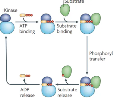
This reversible modification is tightly regulated by the action of protein
kinas-es and phosphataskinas-es being rkinas-esponsible for phosphorylation and
dephosphorylation, respectively. Protein kinases catalyze the transfer of the
γ-phosphate from adenosine triγ-phosphate (ATP) to the specific residues in
pro-teins (Figure 2).
12Protein phosphatases, on the other hand, remove the
phos-phate group by enzymatic hydrolysis.
It is estimated that approximately 30% of proteins in eukaryotic organisms
are phosphorylated at any time.
13In human, there are about 500 kinases and
100 phosphatases coded by ca. 2% of human genome.
14In eukaryotic
organ-isms phosphorylation occurs predominantly on Ser, Thr and Tyr residues with
16
16
protein aging and repair.
8Glycosylation is an essential modification involved
in fundamental processes such as inflammation, immune response, cell growth
or protein folding and stability, just to name a few.
9, 10Proteolytic destruction
of proteins, on the other hand, is initiated by protein modification with
ubiq-uitin (ubiqubiq-uitination).
11The analysis of PTMs is therefore essential for
under-standing fundamental biological functions.
Protein phosphorylation
Phosphorylation is one of the most widely studied PTMs affecting basic
bio-logical processes such as cell growth, proliferation, differentiation, and
apop-tosis as well as cellular signal transduction.
12It can occur on nine amino acids,
i.e. serine, threonine, tyrosine, histidine, lysine, arginine, aspartic acid,
glutam-ic acid, and cysteine. However, the phosphorylation of the former three amino
acids capture most attention in phosphoproteomics research (Figure 1).
Figure 1. Structures of phosphoserine, phosphothreonine and phosphotyrosine.
This reversible modification is tightly regulated by the action of protein
kinas-es and phosphataskinas-es being rkinas-esponsible for phosphorylation and
dephosphorylation, respectively. Protein kinases catalyze the transfer of the
γ-phosphate from adenosine triγ-phosphate (ATP) to the specific residues in
pro-teins (Figure 2).
12Protein phosphatases, on the other hand, remove the
phos-phate group by enzymatic hydrolysis.
It is estimated that approximately 30% of proteins in eukaryotic organisms
are phosphorylated at any time.
13In human, there are about 500 kinases and
100 phosphatases coded by ca. 2% of human genome.
14In eukaryotic
organ-isms phosphorylation occurs predominantly on Ser, Thr and Tyr residues with
16
17
the ratio of 1800:200:1 (pSer:pThr:pTyr).
15Nevertheless, phosphorylation on
histidine can also occur although it often remains undetected due to its
acid-labile nature.
Figure 2. Protein phosphorylation cycle catalyzed by a kinase. Reproduced from
Ubersax and Ferrell
12with permission from Nature Publishing Group.
In recent years, however, an increasing interest in analysis of this elusive
phos-phorylation has been observed resulting in development of techniques for
en-richment, detection and analysis of pHis peptides.
16-20Protein histidine phosphorylation – an elusive PTM
Phosphorylation of histidine is well known to be involved in two component
signaling pathways in prokaryotes and lower eukaryotes
21-23and it is now
be-coming recognized in mammals and implicated in certain human disease
states.
24-27It has been estimated that histidine phosphorylation in eukaryotes
accounts for 6% of total protein phosphorylation,
28thus it is significantly
more abundant than pTyr modification. Nonetheless, little is known about the
biological role of pHis as compared to phosphoester modifications. This is due
to significant technical challenges in enrichment and detection of this PTM.
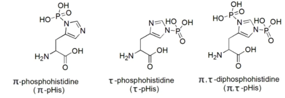
There are three possible isomers of phosphohistidine (pHis), i.e. π-pHis,
τ-pHis and π, τ-pHis (Figure 3) of which the former two have been found in
18
vivo.
16Unlike the phosphoesters (pSer, pThr, pTyr), the phosphoryl group of
pHis is attached to nitrogen atom making it a phosphoramidate. This high
en-ergy phosphoramidate bond is susceptible to hydrolysis in acidic environment
and it defines unique stability profile of phosphohistidine.
29, 30Figure 3. Structures of
π-phosphohistidine, phosphohistidine and π,
τ-diphosphohistidine.
While O-phosphorylated amino acids, pSer and pThr, are stable in acidic
me-dia and labile in alkali environment, the phosphoramidates display reversed
stability profile, i.e. they are base-stable and acid-labile. Most of the methods
used in the phosphopeptide enrichment and detection involve acidic treatment,
and thus fail to preserve pHis modification. Therefore, dedicated methods and
tools are needed to study this PTM.
To date, several methods for detection and analysis of pHis have been
re-ported.
17, 29, 30Generation of synthetic pHis proteins and peptides has clearly
facilitated the development of such methods. Medzihradszky et al.
31used
po-tassium phosphoramidate to synthesize histidine phosphorylated peptides.
This method is highly selective for histidine and does not phosphorylate any
other residues. The complex kinetics of the reaction prove that initially
π-pHis
is formed followed by
π, τ-pHis and τ-pHis. Long reaction times (>12h) result
in the disappearance of
π-pHis yielding only the latter two isomers.
32This
method was also successfully used for phosphorylation of proteins.
17, 18, 33Although chemical phosphorylation gives access to pHis protein and
pep-tide standards, it does not solve the problems related to high acid lability of
this modification. For example, attempts to use pHis-based immunogens to
produce antibodies failed due to facile dephosphorylation of pHis.
34, 35To
overcome these issues, several stable analogues of pHis have been developed
such as phosphonofurylalanine (1),
36phosphonopyrrolylalanine (2)
29and
phsosphonotriazolylalanine (3, 4)
34, 37, 38based analogues (Figure 4).
18
18
vivo.
16Unlike the phosphoesters (pSer, pThr, pTyr), the phosphoryl group of
pHis is attached to nitrogen atom making it a phosphoramidate. This high
en-ergy phosphoramidate bond is susceptible to hydrolysis in acidic environment
and it defines unique stability profile of phosphohistidine.
29, 30Figure 3. Structures of
π-phosphohistidine, phosphohistidine and π,
τ-diphosphohistidine.
While O-phosphorylated amino acids, pSer and pThr, are stable in acidic
me-dia and labile in alkali environment, the phosphoramidates display reverse
stability profile, i.e. they are base-stable and acid-labile. Most of the methods
used in the phosphopeptide enrichment and detection involve acidic treatment
and thus fail to preserve pHis modification. Therefore dedicated methods and
tools are needed to study this PTM.
To date, several methods for detection and analysis of pHis have been
re-ported.
17, 29, 30Generation of synthetic pHis proteins and peptides has clearly
facilitated the development of such methods. Medzihradszky et al.
31used
po-tassium phosphoramidate to synthesize histidine phosphorylated peptides.
This method is highly selective for histidine and does not phosphorylate any
other residues. The complex kinetics of the reaction prove that initially
π-pHis
is formed followed by
π, τ-pHis and τ-pHis. Long reaction times (>12h) result
in the disappearance of
π-pHis yielding only the latter two isomers.
32This
method was also successfully used for phosphorylation of proteins.
17, 18, 33Although chemical phosphorylation gives access to pHis protein and
pep-tide standards, it does not solve the problems related to high acid lability of
this modification. For example, attempts to use pHis-based immunogens to
produce antibodies failed due to facile dephosphorylation of pHis.
34, 35To
overcome these issues, several stable analogues of pHis have been developed
such as phosphonofurylalanine (1),
36phosphonopyrrolylalanine (2)
29and
phsosphonotriazolylalanine (3, 4)
34, 37, 38based analogues (Figure 4).
18
18
19
Figure 4. Examples of stable phosphohistidine analogues.
Kee et al.
34successfully incorporated analog 3 in human histone H4 peptide
sequence by solid-phase peptide synthesis (SPPS). This peptide was next used
as an antigen to raise sequence specific antibody that could recognize histidine
phosphorylated histone H4 but not the unmodified protein. After this first
re-port on successful generation of pHis antibodies, the
phosphoryltriazolyl-based analogues were used to produce pan-specific polyclonal pHis antibody
39and monoclonal 1- and 3-pHis antibodies.
40Despite a remarkable progress in analysis of pHis in recent years, this area
of phosphoproteomics research is still in its infancy, especially in higher
eu-karyotes. Without a doubt, development of new mild enrichment techniques
and advances in mass spectrometry analysis will have significant impact on
elucidation of the biological role of pHis.
The role of phosphorylation in disease pathogenesis
Phosphorylation is responsible for regulation of many important processes
such as cell proliferation, differentiation and apoptosis and plays a vital role in
signal transduction. Disruptions in signaling pathways, regulated by kinases
and phosphatases, are often recognized as a cause or a consequence of various
diseases, including cancer,
41-43neurodegenerative diseases,
44-46cardiovascular
diseases,
47, 48inflammatory diseases,
49, 50and diabetes.
51This has led to an
in-creasing effort to identify kinases and phosphorylated proteins that could
serve as disease biomarkers or drug targets, especially in cancer.
More than 1200 proteins have been reported to be differentially expressed
in human cancer,
52of which many belong to kinase family. These proteins are
potential candidates for cancer biomarkers, however, due to technical
limita-19
19
Figure 4. Examples of stable phosphohistidine analogues.
Kee et al.
34successfully incorporated analog 3 in human histone H4 peptide
sequence by solid-phase peptide synthesis (SPPS). This peptide was next used
as an antigen to raise sequence specific antibody that could recognize histidine
phosphorylated histone H4 but not the unmodified protein. After this first
re-port on successful generation of pHis antibodies, the
phosphoryltriazolyl-based analogues were used to produce pan-specific polyclonal pHis antibody
39and monoclonal 1- and 3-pHis antibodies.
40Despite a remarkable progress in analysis of pHis in recent years, this area
of phosphoproteomics research is still in its infancy, especially in higher
eu-karyotes. Without a doubt, development of new mild enrichment techniques
and advances in mass spectrometry analysis will have significant impact on
elucidation of the biological role of pHis.
The role of phosphorylation in disease pathogenesis
Phosphorylation is responsible for regulation of many important processes
such as cell proliferation, differentiation and apoptosis and plays a vital role in
signal transduction. Disruptions in signaling pathways, regulated by kinases
and phosphatases, are often recognized as a cause or a consequence of various
diseases, including cancer,
41-43neurodegenerative diseases,
44-46cardiovascular
diseases,
47, 48inflammatory diseases
49, 50and diabetes.
51This has led to an
in-creasing effort to identify kinases and phosphorylated proteins that could
serve as disease biomarkers or drug targets, especially in cancer.
More than 1200 proteins have been reported to be differentially expressed
in human cancer,
52of which many belong to kinase family. These proteins are
potential candidates for cancer biomarkers, however due to technical
20
tions in proteomic analysis of low abundant proteins as well as rigorous
re-quirements for marker validation, only a limited number of these proteins has
been approved by the US Food and Drug Administration (FDA) for clinical
use. In fact, only three kinases were approved so far as cancer biomarkers, i.e.
epidermal growth factor receptor for selection of therapy for colon cancer,
KIT for diagnosis and selection of therapy of gastrointestinal stromal tumor
and Her2 for prognosis, monitoring and selection of therapy for breast
can-cer.
41, 53Kinases involved in cancer development can also serve as drug targets. In
targeted cancer therapy two main approaches for kinase inhibition have
emerged, i.e. based on monoclonal antibodies and small-molecule agents.
54The prominent examples of trastuzumab (Herceptin®, Genentech) and
imatinib (Gleevec®, Novartis) being the first FDA approved monoclonal
anti-body (1998) and small-molecule (2001) kinase inhibitors, respectively, paved
the way for a new era of targeted cancer therapy. As many as 28
small-molecule kinase inhibitors have been approved by FDA by June 2015.
55, 56The
fact that 19 of these drugs were approved since 2011, indicates a huge
poten-tial of such therapeutic approach.
Limitations in phosphoproteomic studies
It is evident that advances in phosphoproteomics had an enormous impact on
the identification of novel disease biomarkers and drug targets. For a complete
understanding of signaling pathways and its role in disease pathogenesis a
comprehensive analysis of phosphoproteome is needed. This includes the
iden-tification of phosphoproteins and phosphopeptides, localization of the
phos-phorylation sites as well as quantitation of phosphos-phorylation.
13Despite a
tre-mendous advancement in MS-based techniques in recent years, the analysis of
phosphoproteins and phosphopeptides is not a trivial task. Main challenges
arise from low stoichiometry of phosphorylation, its transient nature and low
abundance of signaling molecules. These issues are particularly evident when it
comes to analysis of tyrosine phosphorylation. For example, in vertebrate
cells, the tyrosine phosphorylation accounts for 0.05% of total protein
phos-phorylation.
57Furthermore, the ionization of phosphopeptides in MS is often
suppressed in presence of nonmodified peptides.
13For these reasons, it is a
common practice to implement enrichment and/or fractionation steps prior to
MS analysis.
21
Enrichment strategies for phosphoproteomics
Over the past years, many techniques to concentrate phosphoproteins and
phosphopeptides from complex biological samples have been developed.
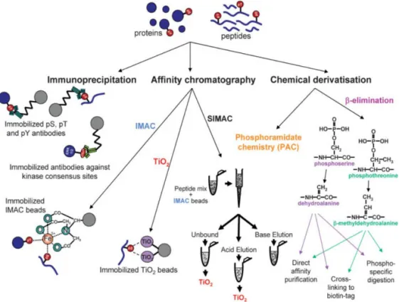
58-60Overall, these methods can be classified into two categories, i.e. more specific
enrichment techniques (e.g. immunoaffinity-based methods, chemical
derivatization and affinity chromatography, see Figure 5) and less specific
fractionation techniques (HILIC, SAX, SCX). In a comprehensive
phosphoproteomic studies enrichment and fractionation methods are often
combined for better coverage of phosphoproteome.
Enrichment techniques
Immunoaffinity-based methods rely on selective capture of phosphoproteins
by specific antibodies against phosphorylated serine, threonine and tyrosine
residues.
61-63In general, however, antibodies raised against pSer and pThr
suf-fer from limited selectivity and therefore are not routinely used in
phosphoproteomic studies.
64Anti-phosphotyrosine antibodies, on the other
hand, display high selectivity and are extensively used in the analysis of this
low abundant modification at the level of both phosphoproteins
65-67and
phosphopeptides.
68-72Nevertheless, the immunoaffinity-based methods suffer
from a number of drawbacks. For example, typically a large amount of
pro-tein sample (in mg scale) is required.
73Furthermore, the high price of
antibod-ies characterized by low stability and non-reusability along with laborious
protocols call for more cost- and time-efficient alternative methods.
Immobilized metal affinity chromatography (IMAC) is based on
interac-tions of negatively charged phosphopeptides with positively charged metal
ions (Fe
3+, Al
3+, Ga
3+, Co
2+, Zr
4+and Ti
4+) chelated to nitrilotriacetic acid or
iminodiacetic acid coated beads. This method is applicable for efficient
extrac-tion of pSer, pThr and pTyr proteins
74and peptides.
75-77However, this
tech-nique suffers from significant non-specific binding of nonphosphorylated
pep-tides bearing acidic residues. O-methyl esterification of carboxylic side chains
was reported to significantly reduce this effect,
78nevertheless, this
derivatization method is not quantitative, thus it results in an increase of
sam-ple comsam-plexity.
79Alternatively, loading the sample at low pH (50% ACN
0.1% TFA) on IMAC resin proved to reduce non-specific binding due to
pro-tonation of carboxyl groups of peptides.
80Recently Zhou et al.
81, 82reported
novel Ti
4+-IMAC resin showing superior selectivity and efficiency compared to
Fe
3+-IMAC, Zr
4+-IMAC, TiO
22
tryptic peptides as well as digest of mouse liver lysate. The Cu
2+-IMAC, on the
other hand, was applied for extraction of pHis peptide from histidine
phos-phorylated protein digest.
83Figure 5. Analytical strategies for phosphoprotein and phosphopeptide
enrich-ment. Reproduced from Thingholm et al.
58with permission from John Wiley and
Sons.
Metal oxide affinity chromatography (MOAC) is another technique for
ex-traction of phosphopeptides. Although a number of different metal oxides,
in-cluding zirconium, aluminum, iron, and tin were applied for phosphopeptide
enrichment
84the titanium dioxide (TiO
2
) is most widely used.
85, 86
The nature
of metal oxide and phosphate group interaction is characterized by Lewis
ac-id/base interaction with bidentate mode
84, 87meaning that phosphopeptides are
well retained in acidic conditions. Thus, typically loading of the sample is
per-formed at pH 2-3 while elution occurs in strongly alkaline conditions. In
gen-eral, TiO
2enrichment is considered to be more specific for binding of
phosphopeptides compared to IMAC,
60although it is not free from unspecific
23
binding of nonphosphorylated peptides. This issue was however largely
dimin-ished by optimization of the protocol, e.g. by using additives in the loading
step such as 2,5-dihydroxybenzoic acid,
86lactic acid,
88glycerol
89or glycolic
ac-id
90that compete with nonphosphorylated peptides for binding to low affinity
sites. The TiO
2-based phosphopeptide enrichment has been employed in
sever-al large scsever-ale phosphoproteomic studies.
91-94Chemical derivatization methods are based on exploiting unique properties
of phosphorylated residues that can be selectively modified prior to
enrich-ment step. For example, pSer and pThr undergo facile dephosphorylation via
elimination in alkali conditions resulting in dehydroalanine and
β-methyldehdroalanine residues, respectively. A subsequent Michael addition
reaction with ethanedithiol allows for modification with biotin tag and
conse-quent enrichment with immobilized avidin
95or direct affinity purification on
activated thiol resin.
96These methods are however specific only for enrichment
of pSer and pThr since pTyr does not undergo
β-elimination. Additionally, a
co-elution of O-glycosylated proteins/peptides, that are susceptible to
β-elimination, can occur. Another method, applicable for enrichment of pSer,
pThr and pTyr peptides is based on phosphoramidate chemistry (PAC).
97Phosphopeptides are immobilized on a solid support in a multistep process via
phosphoramidate bond and subsequently isolated by hydrolysis in acidic
me-dia. In general, chemical derivatization methods are not routinely used in
phosphoproteomic studies due to laborious protocols and non-quantitative
character of reactions that leads to increase of sample complexity.
Fractionation techniques
Fractionation techniques are usually employed in a form of column liquid
chromatography allowing for a gradual separation of a sample based on
prop-erties such as charge or hydrophilicity. These methods are not as efficient and
specific as enrichment techniques, nevertheless, they are often employed in
phosphoproteomics workflows, especially in combination with enrichment
techniques.
Ion exchange chromatography allows to separate phosphopeptides from
nonphosphorylated peptides due to their different charge states. In strong
cat-ion exchange (SCX) negatively charged phosphopeptides interact poorly with
anionic stationary phase and therefore elute early from the column. This
con-trasts with the strong anion exchange (SAX) where phosphopeptides are
strongly retained on the cationic stationary phase.
98However, the separation is
24
number of present acidic and basic residues in the sequence. Both SCX and
SAX have been used for prefractionation of phosphopeptides prior to
enrich-ment, e.g. with IMAC or TiO
2.
69, 75, 79, 99, 100
In hydrophilic interaction chromatography (HILIC) the neutral hydrophilic
stationary phase is used.
101Typically the elution is performed in the
acetoni-trile-water system starting with high organic mobile phase. Thus, peptides are
separated according to their hydrophilicity/hydrophobicity with more polar
peptides (for example phosphopeptides) eluting later. HILIC was shown to be
an efficient prefractionation method prior to specific phosphopeptide
enrich-ment.
102Combined methods
Due to partial complementarity of described methods, it is a common practice
to combine two or more techniques in a multidimensional enrichment
work-flow. Typically, a prefractionation method (e.g. SCX, SAX, HILIC) is applied
before specific enrichment step (e.g. IMAC or MOAC) although combination
in reverse order has also been reported.
103In another approach, the so-called
SIMAC (sequential elution from IMAC),
104the multi-phosphorylated peptides
are bound to IMAC resin while the unbound fraction containing
mono-phosphorylated peptides is loaded on the TiO
2. Thus, the elution fractions
from IMAC and TiO
2are enriched with multi- and mono-phosphorylated
pep-tides, respectively.
Overall, each of the enrichment and fractionation methods has advantages and
limitations and none of these techniques can provide full coverage of
phosphoproteome. Due to significant differences in reported protocols such as
sample type and amount, instrument setup, data processing, etc. it is very
dif-ficult to make a direct comparison of available enrichment methods.
Bodenmiller et al.
105made an attempt to compare and assess reproducibility
and specificity of commonly used methods, i.e. Fe
3+-IMAC, PAC ad TiO
2
using
tryptic digest of the cytosolic fraction of Drosophila melanogaster Kc167 cells.
It was shown that all the investigated methods are highly reproducible,
how-ever, IMAC and PAC gave the best result allowing for identification of >500
phosphopeptides and insignificant contamination with nonphosphorylated
peptides (9 and 34, respectively). Importantly, the overlap of identified
phosphopeptides was only 33-35% indicating differences in selectivity of
in-vestigated methods. This confirms that a combination of several methods can
increase the coverage of studied phosphoproteome.
25
Molecularly imprinted polymers
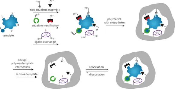
Molecular imprinting is an attractive approach to form synthetic materials with
molecular recognition properties.
106-108This technique is based on
copolymeriza-tion of funccopolymeriza-tional monomer(s) and crosslinker in presence of a template, which
is removed subsequently. Thus, a molecularly imprinted polymer (MIP) possess
cavities that are complementary in size, shape and functionality to the template
allowing for selective rebinding of the template and its close analogues (Figure
6). This simple concept is applicable to a variety of target molecules ranging
from ions and small molecules to macromolecules (e.g. proteins) and
microor-ganisms. Likewise, many different approaches to form strong interactions
be-tween template and functional monomer(s) are available, e.g. covalent and
non-covalent interactions as well as ligand exchange (Figure 6).
As a result, the molecular imprinting technology (MIT) has found a number
of applications including purification and separation,
109, 110sensing
111, 112and
drug delivery
113owing to unique recognition properties of MIPs characterized
by high affinity and specificity for target molecule.
114Furthermore, MIPs are
robust materials with high stability in a wide range of temperature and
sol-vents. The low cost of production and possibility of multiple reuse of the
ma-terial are additional attractive features of MIPs.
Figure 6. Schematic representation of the molecular imprinting process showing
possible interactions between template and functional monomer, including
cova-lent and non-covacova-lent interactions as well as ligand exchange. Reproduced from
Alexander et al.
11526
Nevertheless, the molecular imprinting technology is not free from limitations.
Most commonly used non-covalent imprinting approach
116suffers from a
number of technical challenges owing to the non-covalent nature of
interac-tions between template and functional monomer(s), e.g. hydrogen bonding,
van der Waals interactions or hydrophobic interactions. The dynamic nature
of prepolymerization self-assembled complex being in equilibrium with free
monomers and template leads to formation of heterogeneous binding sites
with different affinity for the target analyte. An excess of functional monomer
can be used to shift the equilibrium towards monomer-template complex,
however, the polymerization of remaining free functional monomer often
leads to high degree of non-specific binding. Instead, a stoichiometric
imprint-ing can be used.
117, 118In this approach the functional monomer interacts very
strongly with the template providing a high degree of template complexation,
thus it can be used in stoichiometric ratio resulting in more uniform binding
sites and low non-specific binding.
Another commonly postulated difficulty in MIT is the imprinting of water
soluble biological macromolecules, for example proteins.
119, 120This is due to
large size and complexity of such targets resulting in slow mass transfer to the
binding site. Two main approaches to overcome these difficulties were
devel-oped, namely epitope imprinting and surface imprinting. The epitope
imprint-ing approach is based on usimprint-ing a small fragment of protein or peptide as a
template.
121, 122The binding sites formed in this way are able to recognize not
only template but also larger molecules bearing the imprinted fragment. In the
surface imprinting approach, the immobilization of template on the
support-ing material (e.g. silica micro- and nanoparticles, magnetic nanoparticles or
nanowires/nanotubes) enables creation of imprinted sites on the surface of the
polymer.
123, 124As a result the highly accessible binding sites located on the
sur-face of the imprinted material are formed.
MIPs for phosphoproteomics
Several approaches to develop phosphopeptide selective molecularly imprinted
polymers have been reported to date.
125-133The first attempt to apply molecular
imprinting technology for phosphoproteomics was reported by
Sellergren et al.
125The neutral urea-based receptor was synthesized using
N, C-protected phosphotyrosine as a template and ethylene glycol
dimethacrylate (EDMA) as a crosslinker. This crushed monolith material
27
showed bias for pTyr peptides in presence of pSer and Tyr peptides. Further
optimization and validation of this system resulted in successful extraction of
pTyr peptide spiked at a femtomol level in bovine fetuin digest.
131Later on,
Chen et al.
132showed a complementary selectivity of pTyr and pSer imprinted
polymers in a mixture of twelve peptides and incorporation of pSer MIP
en-richment in combination with SCX in proteomics workflow. The MIP-based
method showed superior performance compared to TiO
2-based enrichment.
Recently, the pTyr MIP-based phosphopeptide enrichment was validated in a
comparative study with state of the art phosphopeptide enrichment
tech-niques, i.e. immunoaffinity- and TiO
2-based enrichments (Paper I). It was
shown that pTyr MIP is an attractive alternative for established
phosphoenrichment methods allowing for a controlled bias of material
to-wards specific phosphorylation site.
In another approach, the monodisperse 4-vinylpyridine-based MIPs
target-ing organophosphates were shown to non-selectively trap phosphorylated
pep-tides (pSer, pThr, pTyr) and phosphopeppep-tides from a tryptic digest of
α-casein.
126Ren et al.
127, 128have used charged metal ion-based functional
mono-mers (Zn
2+and Ti
4+) for imprinting of phenylphosphonic acid (PPA) as an
epitope template. Obtained materials displayed the affinity for phosphorylated
peptides in aqueous solution with a preference for tyrosine phosphorylated
peptides as contrasted with unbiased enrichment with TiO
2.
Surface imprinting approach has also been employed for development of
phosphopeptide selective MIPs.
130, 133For example, Li et al.
133reported on
sur-face imprinted fluorescent receptor based on CdTe quantum dots. The
recep-tor gave linear fluorescence enhancement response to pTyr peptide which
could also be detected in a spiked sample of tryptic digest of skim milk.
How-ever, the concentration of pTyr peptide tested in this work was in μM range
which is well above the range found in biological samples. Recently Chen et
al.
130reported on phosphate imprinted mesoporous silica nanoparticles using
dual-template docking oriented molecular imprinting approach. Obtained
im-printed nanoparticles displayed an excellent affinity for template analogue, i.e.
adenosine monophosphate (AMP) and were able to extract pSer peptides from
β-casein digest as well as from nonfat milk sample. However, the affinity for
peptides phosphorylated on Thr and Tyr residues was not studied.
28
Urea-based functional monomers
The core of this thesis relies on the use of urea-based functional monomer
FM1 (Figure 8A) as a recognition unit in MIPs targeting phosphorylated
pep-tides reported in Papers I-IV.
1,3-Disubsituted ureas have long been exploited as neutral receptors for
an-ions such as carboxylates, phosphates and sulfonates.
134-136This type of hosts
are potent hydrogen bond donors capable of forming two-fold cyclic hydrogen
bonds with anion guest (Figure 7). The strength of interaction increases with
the acidity of urea protons and basicity of complexed anion. Thiourea is more
acidic than urea (pK
a= 21.1 and 26.9, respectively in DMSO),
136
thus thioureas
should form stronger hydrogen bonds with guest compared to ureas.
137Figure 7. Schematic representation of the interaction between urea (X=O) or
thiourea (X=S) receptors and carboxylate anion.
Another factor influencing the strength of interaction is the type of solvent in
which the recognition occurs. Neutral receptors such as ureas and thioureas,
which rely solely on hydrogen bond interactions are highly susceptible to
competition with polar protic solvents for anion binding. Thus, this type of
receptors are most effective in aprotic organic solvents.
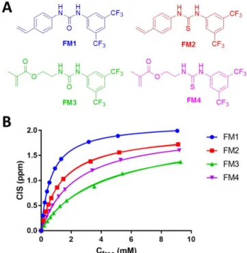
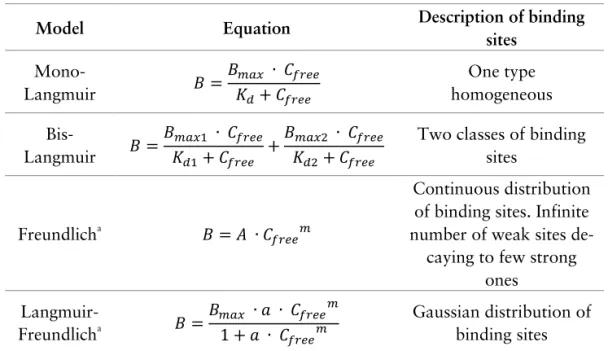
138Functional monomers FM1-FM4 (Figure 8A) were evaluated by NMR
titra-tion experiments with tetrabutylammonium hydrogen-1-naphtyl phosphate
(TBAHNP) guest in DMSO-d6 (Figure 8B). The data points obtained for each
monomer were fitted to 1:1 interaction model to determine values of
associa-tion constant (K
a) and maximum complexation induced shift (CIS
max) (Table
2).
The 1,3-diaryl monomers (FM1 and FM2) interacted more strongly with
guest compared to alkyl-aryl counterparts (FM3 and FM4). Furthermore,
thiourea monomer FM4 interacted more strongly with guest compared to urea
analogue FM3 as expected based on the difference in acidity of ureas and
thioureas (vide supra).
28
28
Urea-based functional monomers
The core of this thesis relies on the use of urea-based functional monomer
FM1 (Figure 8A) as a recognition unit in MIPs targeting phosphorylated
pep-tides reported in Papers I-IV.
1,3-Disubsituted ureas have long been exploited as neutral receptors for
an-ions such as carboxylates, phosphates and sulfonates.
134-136This type of hosts
are potent hydrogen bond donors capable of forming two-fold cyclic hydrogen
bonds with anion guest (Figure 7). The strength of interaction increases with
the acidity of urea protons and basicity of complexed anion. Thiourea is more
acidic than urea (pK
a= 21.1 and 26.9, respectively in DMSO)
136
thus thioureas
should form stronger hydrogen bonds with guest compared to ureas.
137Figure 7. Schematic representation of the interaction between urea (X=O) or
thiourea (X=S) receptors and carboxylate anion.
Another factor influencing the strength of interaction is the type of solvent in
which the recognition occurs. Neutral receptors such as ureas and thioureas,
which rely solely on hydrogen bond interactions are highly susceptible to
competition with polar protic solvents for anion binding. Thus this type of
re-ceptors are most effective in aprotic organic solvents.
138Functional monomers FM1-FM4 (Figure 8A) were evaluated by NMR
titra-tion experiments with tetrabutylammonium hydrogen-1-naphtyl phosphate
(TBAHNP) guest in DMSO-d6 (Figure 8B). The data points obtained for each
monomer were fitted to 1:1 interaction model to determine values of
associa-tion constant (K
a) and maximum complexation induced shift (CIS
max) (Table
2).
The 1,3-diaryl monomers (FM1 and FM2) interacted more strongly with
guest compared to alkyl-aryl counterparts (FM3 and FM4). Furthermore,
thiourea monomer FM4 interacted more strongly with guest compared to urea
analogue FM3 as expected based on the difference in acidity of ureas and
thioureas (vide supra).
28
28
29
Table 2. Values of association constants (K
a) and maximum complexation induced
shifts (CIS
max) for FM1-FM4 titrated with TBAHNP in DMSO-d6.
Monomer
K
a(M
-1)
CIS
max(ppm)
FM1
1489±7
2.14
FM2
743±10
1.96
FM3
266±14
1.91
FM4
439±5
2.00
However, this relationship did not hold true for FM1 and FM2 pair of
mon-omers. This unexpected finding can be ascribed to previously reported
prefer-ence of diaryl thioureas for (Z, E) conformation as opposed to preferential
(Z, Z) conformation of diaryl ureas
139and it is in agreement with the previous
study on FM1 and FM2.
125Figure 8. (A) Structures of functional monomers FM1-FM4 and (B) plot of
complexation induced shift (CIS) vs. free concentration of guest (C
free) fitted to 1:1
interaction model for
1H MNR titrations of FM1-FM4 with tetrabutylammonium
hydrogen-1-naphtyl phosphate (TBAHNP) in DMSO-d6 at 25 °C.
29
29
Table 2. Values of association constants (K
a) and maximum complexation induced
shifts (CIS
max) for FM1-FM4 titrated with TBAHNP in DMSO-d6.
Monomer
K
a(M
-1)
CIS
max(ppm)
FM1
1489±7
2.14
FM2
743±10
1.96
FM3
266±14
1.91
FM4
439±5
2.00
However, this relationship did not hold true for FM1 and FM2 pair of
mon-omers. This unexpected finding can be ascribed to previously reported
prefer-ence of diaryl thioureas for (Z, E) conformation as opposed to preferential
(Z, Z) conformation of diaryl ureas
139and it is in agreement with the previous
study on FM1 and FM2.
125Figure 8. (A) Structures of functional monomers FM1-FM4 and (B) plot of
complexation induced shift (CIS) vs. free concentration of guest (C
free) fitted to 1:1
interaction model for
1H MNR titrations of FM1-FM4 with tetrabutylammonium
hydrogen-1-naphtyl phosphate (TBAHNP) in DMSO-d6 at 25 °C.
30
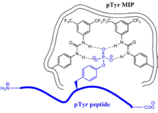
Based on the above results, the FM1 was selected as a functional monomer for
synthesis of molecularly imprinted polymers targeting pTyr and pHis peptides
(Papers I-IV). Schematic representation of interactions between tyrosine
phos-phorylated peptide and binding site in the imprinted polymer is shown in
Fig-ure 9.
Figure 9. Schematic representation of interactions between tyrosine
phosphory-lated peptide and phosphotyrosine imprinted polymer (pTyr MIP).
Imidazolium-based functional monomers
Recognition of anions in water is essential for biological and environmental
applications, however, it remains a great challenge in modern supramolecular
chemistry.
140Neutral receptors bearing urea, amide or pyrrole functionality
were extensively studied for recognition of anions,
141, 142nevertheless, their
compatibility with aqueous media is limited. Water is a polar solvent with the
ability of hydrogen bond donation and acceptance, thus it effectively competes
with neutral receptors for binding anions. Charged receptors, on the other
hand, can take advantage of the electrostatic effect, and thus compete more
effectively with polar protic solvent.
Commonly used charged receptors for anions based on guanidinium or
ammonium employ
+N−H∙∙∙X
−type of ionic hydrogen bond. In contrast,
imidazolium-based receptors can interact with anions through (C−H)
+∙∙∙X
−type of ionic hydrogen bond (Figure 10).
14330
30
Based on the above results the FM1 was selected as a functional monomer for
synthesis of molecularly imprinted polymers targeting pTyr and pHis peptides
(Papers I-IV). Schematic representation of interactions between tyrosine
phos-phorylated peptide and binding site in the imprinted polymer is shown in
Fig-ure 9.
Figure 9. Schematic representation of interactions between tyrosine
phosphory-lated peptide and phosphotyrosine imprinted polymer (pTyr MIP).
Imidazolium-based functional monomers
Recognition of anions in water is essential for biological and environmental
applications, however, it remains a great challenge in modern supramolecular
chemistry.
140Neutral receptors bearing urea, amide or pyrrole functionality
were extensively studied for recognition of anions
141, 142nevertheless, their
compatibility with aqueous media is limited. Water is a polar solvent with the
ability of hydrogen bond donation and acceptance thus it effectively competes
with neutral receptors for binding anions. Charged receptors, on the other
hand, can take advantage of electrostatic effect and thus compete more
effec-tively with polar protic solvent.
Commonly used charged receptors for anions based on guanidinium or
ammonium employ
+N−H∙∙∙X
−type of ionic hydrogen bond. In contrast,
imidazolium-based receptors can interact with anions through (C−H)
+∙∙∙X
−type of ionic hydrogen bond (Figure 10).
14330
30
31
Figure 10. Schematic representation of the ionic hydrogen bond interaction
be-tween imidazolium receptor and anion X
−.
A number of such receptors was reported before in the literature and reviewed
elsewhere.
144-147For example, Kwon et al.
148reported water-soluble
imidazolium anthracene derivative (Figure 11) which acts as a potential
fluo-rescent receptor for guanosine 5′-triphosphate (GTP) in 100% aqueous
solu-tion. The value of K
aderived from the fluorescence titration with GTP in 10
mM HEPES buffer (pH = 7) was 87000 M
-1.
Figure 11. Fluorescent receptor for GTP.
In Paper V a set of imidazolium-based functional monomers was used to
syn-thesize MIPs. Materials prepared in capillary monolith format were able to
strongly retain phosphopeptides in highly aqueous mobile phase as opposed to
nonphosphorylated ones.
31
31
Figure 10. Schematic representation of ionic hydrogen bond interaction between
imidazolium receptor and anion X
−.
A number of such receptors was reported before in the literature and reviewed
elsewhere.
144-147For example, Kwon et al.
148reported water-soluble
imidazolium anthracene derivative (Figure 11) which acts as a potential
fluo-rescent receptor for guanosine 5′-triphosphate (GTP) in 100% aqueous
solu-tion. The value of K
aderived from the fluorescence titration with GTP in 10
mM HEPES buffer (pH = 7) was 87000 M
-1.
Figure 11. Fluorescent receptor for GTP.
In Paper V a set of imidazolium-based functional monomers was used to
syn-thesize MIPs. Materials prepared in capillary monolith format were able to
strongly retain phosphopeptides in highly aqueous mobile phase as opposed to
nonphosphorylated ones.
32
METHODS
Analytical methods for identification, purification, separation and
quantifica-tion of chemical compounds as well as material characterizaquantifica-tion techniques
were extensively used in this thesis. This section explains the basic principles
of the most important methods used during the course of this work. Details
about instrumentation and conditions of all analyses are given in Experimental
Sections of Papers I-V.
Batch binding test
One of the most basic and simple tests for characterization of binding
proper-ties of MIPs is the batch binding test.
149It relies on equilibration of a known
amount of the polymer (m, in g) with the solution of a known amount of the
analyte (n
0, in moles) until the equilibrium is reached. The polymer is then
separated from the solution and the amount of unbound analyte (n
free, in
moles) in the solution is measured. The amount of bound analyte per unit
mass of the polymer (B, in mol g
-1) can be then calculated from equation (1):
(1)
𝐵 = (𝑛
0
− 𝑛
𝑓𝑟𝑒𝑒
)/𝑚
The experiment is typically done in parallel for both MIP and NIP under
iden-tical conditions (Figure 12). The difference between materials originating from
the imprinting phenomenon can be expressed as imprinting factor (IF) and it
can be calculated from equation (2):
(2)
𝐼𝐹 =
𝐷
𝑀𝐼𝑃𝐷
𝑁𝐼𝑃=
𝐵
𝑀𝐼𝑃/𝐶
𝑓𝑟𝑒𝑒 𝑀𝐼𝑃𝐵
𝑁𝐼𝑃/𝐶
𝑓𝑟𝑒𝑒 𝑁𝐼𝑃Where D (L g
-1) is the distribution ratio and C
free
(mol L
-1