1
The accuracy of apical palpation and percussion
when diagnosing apical periodontitis
Gunilla Karlson
Helena Tengvall
Supervisor: Eva Wolf, department of endodontics, Faculty of
Odontology, Malmö University
Running title: Diagnosing apical periodontitis
Mesh terms: diagnostic tests, routine; palpation; percussion; periapical
periodontitis; predictive value of tests; sensitivity and specificity.
Master’s thesis (30hp) Malmö University Dentistry Faculty of Odontology
2
Abstract
The aim of this study was to evaluate the accuracy of apical palpation and percussion when diagnosing symptomatic apical periodontitis and asymptomatic apical periodontitis in non-root filled teeth.
Fifteen patients participated in the study; eight patients with symptomatic apical periodontitis and seven with asymptomatic apical periodontitis. The patients were recruited from three different clinics in the south of Sweden.
Initially, the patients answered questions concerning recent intake of medication and graded their pain on a Visual Analogue Scale. The observer performed the two diagnostic tests on the test tooth, the control tooth and their adjacent teeth. As reference test, the pulp condition was examined by direct inspection at the access opening of the test tooth.
For apical palpation, when diagnosing symptomatic apical periodontitis/asymptomatic apical periodontitis, the sensitivity was 0.50/0.14, the specificity was 1/1, the positive predictive value was 1/1 and the negative predictive value was 0.67/0.54. For percussion, when diagnosing symptomatic apical periodontitis /asymptomatic apical periodontitis, the
sensitivity was 0.38/0.29, the specificity was 1/1, the positive predictive value was 1/1 and the negative predictive value was 0.62/0.58.
No general conclusions can be made from this study due to the small population size. However, the results indicate that the accuracy of apical palpation and percussion when diagnosing symptomatic apical periodontitis and asymptomatic apical periodontitis in non-root filled teeth is low. The two diagnostic tests should not be used alone to identify teeth with symptomatic apical periodontitis and asymptomatic apical periodontitis.
3
Sammanfattning
Syftet med den aktuella studien var att undersöka tillförlitligheten av apikal palpation och perkussion vid diagnostik av symtomatisk apikal parodontit och asymptomatisk apikal parodontit på icke rotfyllda tänder.
Femton patienter undersöktes; åtta patienter med symtomatisk apikal parodontit och sju patienter med asymptomatisk apikal parodontit. Patienterna rekryterades från tre kliniker i södra Sverige.
Först besvarade patienterna frågor om aktuellt läkemedelsintag och graderade sin smärta på en Visuell Analog Skala. Observatören utförde de två diagnostiska testerna på testtanden, kontrolltanden samt deras intilliggande tänder. Som referenstest kontrollerades pulpans tillstånd genom direkt inspektion vid kavumpreparation av testtanden.
För apikal palpation, vid diagnostik av symtomatisk apikal parodontit/asymptomatisk apikal parodontit, var sensitiviteten 0,50/0,14, specificiteten 1/1, det positiva prediktiva värdet 1/1 samt det negativa prediktiva värdet 0,67/0,54. För perkussion, vid diagnostik av symtomatisk apikal parodontit/asymptomatisk apikal parodontit, var sensitiviteten 0,38/0,29, specificiteten 1/1, det positiva prediktiva värdet 1/1 samt det negativa prediktiva värdet 0,62/0,58.
Inga generella slutsatser kan dras från studien på grund av den begränsade studiepopulationen. Resultatet indikerar dock att tillförlitligheten för apikal palpation och perkussion vid
diagnostik av symtomatisk apikal parodontit och asymptomatisk apikal parodontit på icke-rotfyllda tänder är låg. Dessa två diagnostiska tester bör inte användas enskilt för att
4
Introduction
The word diagnosis comes from the Greek word ”dia´gnōsis” which means examination or judgment of a certain illness (1). Diagnostics is built on information gathered from the patient’s experiences and the caretaker’s clinical observations, which are later interpreted (1, 2). Adequate diagnostics separate sickness from health (2). To be able to make a decision on whether treatment is necessary and which treatment would be the best, a correct diagnosis is necessary. Not only will this mean a possibility of the health care provider to make an
adequate prognostic evaluation but also imply a better treatment result. A further advantage of significance is that treatment costs will be lower, if thorough diagnostics are used (3).
When the dentist uses a diagnostic test, it is important that he or she has knowledge of what tissues and what function the chosen test is testing. Of importance is also to have knowledge of the test´s accuracy when analyzing and valuing the results of the tests in the clinical decision making (3).
Sensitivity describes the tests ability to correctly identify sick patients compared to a method of reference, in other words the amount of true positive tests amongst all sick patients in the studied group. Specificity is the tests ability to correctly identify healthy patients compared to a method of reference, in other words the amount of true negative tests amongst all healthy patients in the studied group (4). The sensitivity and the specificity are related to each other, thus when the sensitivity increases, the specificity decreases and opposite. The sensitivity and the specificity are comparisons with a method of reference and unrelated to the disease prevalence (5).
5 To calculate the probability that a positive test result is correct, the positive predictive value is calculated. It is the proportion of true positive results out of all positive results. To calculate the probability that a negative test result is correct, the negative predictive value is calculated. It is the proportion of true negative results out of all negative results (4). The predictive values are of special significance in the clinical practice during the diagnostic procedure since these values are dependent on the disease prevalence.
A bacterial infection in combination with a carious lesion is the most common reason for pulpal and periapical diseases. The bacterial infection of the pulp consists of a mixed microbial flora including anaerobic microorganisms whose toxins and degradation products initiate an inflammatory response in the pulpal tissue. When parts of the coronal pulpal tissue become necrotic, microorganisms can colonize, which leads to a spreading of the disease. The microorganisms in the infected root canal are unreachable for the body’s immune system due to the lack of blood circulation. An untreated infection in the root canal causes an
inflammation in the periodontal ligament around the tooth and can initiate a periapical bone destruction which usually is asymptomatic, but occasionally becomes symptomatic (6).
Apical periodontitis is an inflammation caused by bacteria in the necrotic tooth and develops in the periradicular tissue, often surrounding the tooth’s apex (7).The American board of endodontics defines symptomatic apical periodontitis (SAP) and asymptomatic apical periodontitis (AAP) as follows:
SAP: “Inflammation, usually of the apical periodontium, producing clinical symptoms including a painful response to biting and/or percussion or palpation. It might or might not be associated with an apical radiolucent area.”
6
AAP: “Inflammation and destruction of apical periodontium that is of pulpal origin, appears as an apical radiolucent area, and does not produce clinical symptoms.”(8)
Nociceptive fibers are activated by mediators synthesized by the inflammation which releases neuropeptides that increase the inflammation further. The neuropeptides also sensitize the nociceptors which reduces the pain threshold on stimulations such as apical palpation and percussion (9).
Apical palpation and percussion are two diagnostic tests which are often used when
diagnosing apical periodontitis (5), although the information concerning the accuracy of the tests still are scarce (2, 10). Since apical palpation and percussion are two diagnostic tests which are easy to perform and which do not cause any complications (11) it is important that studies of good quality are made to examine the accuracy of the tests when diagnosing apical periodontitis.
The aim of this study was to study the sensitivity, specificity, positive and negative predictive values of apical palpation and percussion when diagnosing symptomatic and asymptomatic apical periodontitis in non-root filled teeth.
The hypotheses were:
The apical palpation test is accurate when diagnosing SAP
The percussion test is accurate when diagnosing SAP
The apical palpation test is not accurate when diagnosing AAP
7
Material and Methods
Fifteen patients participated in this experimental clinical study, eight patients with SAP and seven patients with AAP. The patients were collected among patients at the Department of Endodontics and the Comprehensive Care Clinic at the Faculty of Odontology, Malmö University, Malmö, Sweden; at Folktandvården Löttorp, Öland, Sweden and at Christerssons Tandvårdsteam c/o Smile, Helsingborg, Sweden. The patients were examined from April 2008 to February 2009 and from February 2012 to October 2012.
The inclusion criteria were:
Seeking help for tooth ache or referred for endodontic treatment of an asymptomatic tooth with apical periodontitis (test tooth)
Having a contra lateral tooth or an adjacent tooth to the contra lateral tooth which was intact or had a shallow restoration (control tooth)
>19 years of age
No bleeding from at least one of the root canals of the test tooth
The exclusion criteria were:
Analgesic medication within 24 hours prior to the examination
Antibiotic medication within the past month prior to the examination
Root filled test tooth or root filled contra lateral tooth
8 An examination protocol (attachment 1) was developed and used in a pilot study at the
Comprehensive Care Clinic at the Faculty of Odontology, Malmö University, Malmö, Sweden. The pilot study was used as a part of this study.
The observers were two dental students (HT, GK) and two dentists (TE, MS) from the Faculty of Odontology, Malmö University, Malmö, Sweden, one dental student (GK) at
Folktandvården Löttorp, Öland, Sweden and an endodontic specialist (EG) at Christerssons Tandvårdsteam c/o Smile, Helsingborg, Sweden. The observers were calibrated concerning apical palpation and percussion with a specialist in endodontics (MP).
Before the examination, the patient filled in a form with questions concerning recent analgesic and antibiotic medication, as well as pain intensity graded on a Visual Analogue Scale (VAS). The VAS consisted of a straight line of 10-cm, where the left end of the line was defined as “no pain” and the right end of the line was defined as “worst possible pain” (12). The patients with a registration on VAS of 0 were considered to have an AAP. The patients with a
registration on VAS of more than 0 were considered to have a SAP.
The two index tests were performed as follows:
Tenderness to apical palpation. The alveolar ridge on both sides of the alveolar process at the apices of the teeth was palpated with the thumb and the index finger.
Tenderness to percussion. The cusps of each tooth were percussioned three times with the shaft of a straight probe.
The index tests were performed on five teeth in each half of the jaw including the test tooth and the control tooth respectively. The test tooth and its adjacent teeth were first examined followed by the control tooth and its adjacent teeth. The teeth were tested in a non-controlled randomized order. The order of which apical palpation and percussion were tested on the teeth
9 was also randomized in a non-controlled order. The reference test (direct inspection of the pulp) was performed immediately after the index test in direct conjunction with the adequate treatment chosen.
The patient’s responses to apical palpation and percussion of the test tooth and the control tooth were registered according to the following criteria:
0 = No reaction and a non-affirmative answer to the question whether pain was felt. 1 = Discomfort. An affirmative answer to the question whether pain was felt.
2 = Large discomfort. An affirmative answer to the question whether pain was felt. The patient flinched when examined.
Direct inspection of the pulp’s condition at the access opening, was used as the reference test. The diagnosis was set at the orifice of the root canals. In order to note the pulp’s condition as “necrotic”, at least one root canal of the examined tooth had to be necrotic.
The sensitivity, specificity, negative and positive predictive values were calculated according to the following formulas:
Sensitivity = True positive
(False negative + True positive)
Specificity = True negative
(False positive + True negative) (13)
NPV = (Specificity)(1 – Prevalence)
(Specificity)(1 – Prevalence) + (1 – Sensitivity)(Prevalence)
PPV = (Sensitivity)(Prevalence)
10 The study has been approved by the Regional Ethical Review Board at Lund University (Dnr 2008/3) and the Local Ethical Review Board at the Faculty of Odontology, Malmö University, according to the guidelines of the Helsinki declaration (15). The patients received verbal and written information of the study and signed an informed consent. No economical or other compensation was given for their participation.
11
Results
In total eight patients with SAP and seven patients with AAP were examined and included in the study. The population consisted of seven (47 %) women and eight (53 %) men. Of the examined test teeth ten (67 %) were molars, two (13 %) were premolars and three (20 %) were incisors. Of the examined control teeth three (20 %) were molars, seven (47 %) were premolars, four (27 %) were canines and one (6 %) was an incisor. The patients were 29 to 84 years of age and the mean age of the patients was 53 years.
The VAS value interval of the patients with SAP was between 0.5 and 9.7. The mean VAS value of the patients with SAP was 3.7.
Distribution of sex, age, pain intensity, tenderness to apical palpation and percussion of the test tooth and control tooth of seven patients with AAP and eight patients with SAP are presented in Table 1-2. In total four patients with SAP and one patient with AAP experienced tenderness to apical palpation. Three patients with SAP and two patients AAP experienced tenderness to percussion (Fig. 1). Since every patient had one test tooth and one healthy contra lateral control tooth examined, the prevalence of SAP and AAP in the studied population was 50 %.
The sensitivity, specificity, positive and negative predictive values for apical palpation and percussion when diagnosing SAP and AAP are presented in Figure 2-3. When combining apical palpation and percussion to diagnose SAP, the sensitivity is 0.75, the specificity is 1, the NPV is 0.80 and the PPV is 1. When combining apical palpation and percussion to diagnose AAP, the sensitivity is 0.29, the specificity is 1, the NPV is 0.58 and the PPV is 1.
12
Discussion
The accuracy of apical palpation and percussion was high when diagnosing healthy teeth but lower when diagnosing SAP and AAP. All results of this study are uncertain since the population size was limited. Whether a diagnostic test is considered to be accurate or not depends on in which circumstance it is used. It is not possible to verify or falsify the hypotheses formulated in this study concerning the sensitivity, the specificity, the negative predictive values and the positive predictive values or to classify the predictive values as “accurate” or “not accurate” because of the limited population size. However, the sampling will continue.
The exclusion criteria concerning analgesic and antibiotic medication limited the amount of participating patients. The criteria were chosen in order to decrease the number of false negative results, since these medications might have affected the patients´ sensation of pain. (16). To include patients with antibiotic and analgesic medication would have probably better represented the general population, but the aim was to study the accuracy of the two
diagnostic tests without the influence of external factors that might affect the outcome. As a further research project to study the relevance of the actual diagnostic tests in a true clinical context, it might be suggested to include patients with antibiotic and analgesic medication.
If at least one of the root canals of the test tooth was necrotic, the probability of a root canal infection was considered to be high. To exclude teeth with periodontal disease (gingival pockets ≥ 5 mm), further increased the probability of the pain from the symptomatic tooth to have an endodontic origin. The choice of using an intact tooth or a tooth with a shallow
13 restoration including a visibly healthy periapical area as the control tooth was considered relevant, since the probability of a healthy pulp tissue under these circumstances was high.
An exact observer calibration can be considered as almost impossible since the pressure and force used at apical palpation and percussion respectively, is dependent on many different factors such as the tooth’s mobility, the tooth position in the mouth and the instrument angle used during the test procedures. Including more observers performing the diagnostic tests might probably have meant a larger study population but also a risk of greater observer
variability. On the other hand, including more observers and more patients per observer might have meant a more relevant clinical applicability, since most clinical practitioners perform apical palpation and percussion with different forces.
VAS used as a pain grading instrument is one of the most common tools within healthcare to evaluate pain. It is reported to have a high reliability (17) and constructed to allow an
objective evaluation (18). Mantha et al. report that VAS values in the range of 0-30 mm are considered to be a “zone of analgesic success” (18) but since our aim was not to evaluate a decrease of pain, but instead register the patients´ actual pain symptoms, the decision was made to classify all VAS values > 0 as SAP.
Both of the studied diagnostic tests aim at detecting an inflamed tissue at the apices of teeth as a sign of pulpal disease. Before performing the study, the tests were believed to be accurate for apical palpation and percussion when diagnosing SAP since the nerves in the periodontal ligament at the tooth’s apex in this case would be sensitized with a lowered pain threshold (6, 9). The tests were not believed to be as accurate when diagnosing AAP since, in this case, the
14 patient was not experiencing pain and thus, nerve endings were considered not to be
sensitized.
In our study, the sensitivity for apical palpation was lower than the sensitivity in the study by Klausen et al. (10) where it was 0.71. On the other hand, the result of percussion, also with a low sensitivity in our study, correlates with the results of other studies (19, 20). Since there were no false positive results in our study, the specificity of both the diagnostic tests was 1.0 for both SAP and AAP, which is higher than in the study of Klausen et al. (10) who reported a specificity of 0.63 for apical palpation and 0.51 for percussion. It is difficult to conclude why the results differ from Klausen et al. (10), since they do not specify if the examined teeth had SAP or AAP. One explanation to the difference might be the population size; 15 patients in our study compared to 30 patients in the study of Klausen et al. (10). Our results of the specificity concerning percussion seem to correlate better to the results of other studies (20, 21, 22) where the specificity was reported to 0.83-0.89.
The low sensitivity and high specificity values lead to the risk of a high amount of false negative diagnoses which, in the clinical practice, means that if a tooth does not show any symptoms when performing apical palpation or percussion, there is still a risk that the tooth has apical periodontitis. This knowledge could mean that under diagnostics and thereby under treatment of teeth with SAP and AAP increases when using apical palpation and percussion. The dentist would thereby leave a number of the teeth with apical periodontitis untreated. However, when combining apical palpation and percussion to diagnose SAP, the diagnostic tests are accurate and therefore can be considered a valuable tool in clinical dentistry. On the other hand, the diagnostic tests are not accurate when used together to
15 diagnose AAP which still imply a need of further diagnostic tests to be used in the clinical situation in order to diagnose these inflammatory conditions.
Apical palpation and percussion must also be discussed in regard to different disease prevalence. The PPV’s for both the diagnostic tests in this study were 1.0 while the NPV’s range between 0.54 and 0.67 for apical palpation, and 0.58 and 0.62 for percussion. This means that both tests are sensitive in identifying healthy teeth but less precise in identifying teeth with apical periodontitis. Hyman and Cohen (5) calculated the NPV and the PPV of the results from two studies (20, 22) using the disease prevalence of 18 %. Using the same prevalence, our NPV’s for apical palpation as well as percussion are in accordance with the results of Hyman and Cohen (5); 0.90 compared to 0.85-0.87 and 0.88 compared to 0.74-0.88 respectively.
Swedish Council on Health Technology Assessment (SBU) (2) and Pretty and Maupome (3) . mean that the lower the prevalence of disease is, the lower the PPV will be, even if both the sensitivity and the specificity are high, with an increasing risk of false positive results. This means that the two diagnostic tests will be more accurate in a clinical environment where the prevalence of SAP and AAP is high, for example in an emergency clinic for SAP and in an endodontic specialist clinic for AAP.
Frisk (23) reports that the prevalence of apical periodontitis on a tooth level is 1-14 % in a general population which is significantly lower than the prevalence in our study and also the probable prevalence of disease in a specialist clinic. This low prevalence will make the PPV’s lower and the NPV’s higher. With low disease prevalence, the risk of under diagnostics diminishes with a higher risk of over diagnostics as a sequel (2). It is therefore important that
16 the clinical practitioner has knowledge of the disease prevalence in the actual population. The results from this study are not generalizable. The predictive values from one study cannot be compared with the predictive values from another, if the prevalence of disease is not
comparable (2).
To correctly diagnose SAP and especially AAP it seems preferable to combine the diagnostic tests of apical palpation and percussion with other diagnostic methods such as pulp sensitivity tests, clinical findings such as caries and deep fillings and also radiography (23). The task of the dentist in the clinical situation is to diminish the diagnostic insecurity in order to be able to estimate the prognosis and to perform the adequate treatment. Therefore, not only knowledge of the accuracy of different diagnostic tests is important but also, in the first place, the
understanding of pulp biology and disease development.
It would have been interesting to perform studies on what the optimal pressure for apical palpation and percussion would be, when diagnosing apical periodontitis. If studies like this were performed, calibration of future studies on apical palpation and percussion as diagnostic methods when diagnosing apical periodontitis would be easier and more reliable. It would also be interesting to make studies with fewer exclusion criteria to be able to compare how accurate the diagnostic tests of apical palpation and percussion are if they are affected by pain influencing factors such as analgesic and antibiotic medication. Future studies should include a better randomization of patient selection and performance, and a larger population size. It would also be beneficial if the study was performed at different types of clinics such as an emergency clinic, an endodontic specialist clinic and a general clinic in order to study the accuracy of apical palpation and percussion in different prevalence of disease.
17
Conclusions
The accuracy of apical palpation and percussion was high when diagnosing healthy teeth but lower diagnosing SAP and AAP. This means that both tests are sensitive in identifying healthy teeth but less precise in identifying teeth with apical periodontitis.
The results of this study are uncertain since the population size was limited. However, the results indicate that apical palpation and percussion, if used independently, are not accurate when diagnosing SAP and AAP. However, a combination of the tests indicates the tests to be accurate when diagnosing SAP. There is a considerable risk for under diagnostics and thereby under treatment of SAP and AAP if either of these two diagnostic tests is used as the only diagnostic method to diagnose SAP and AAP in non-root filled teeth.
18
References
1. Rössner S. Diagnos. Available at: http://www.ne.se/lang/diagnos. Accessed 28 October, 2011
2. SBU, Swedish Council on Health Technology Assessment. Methods of Diagnosis and Treatment in Endodontics. 2010; 203: 51-112.
3. Pretty IA, Maupome G. A closer look at diagnosis in clinical dental practice: part 1. Reliability, validity, specificity and sensitivity of diagnostic procedures. J Can Dent Assoc. 2004; 70: 251-255.
4. SBU, Swedish Council on Health Technology Assessment. Är diagnosmetoden bra? Available at:
http://www.sbu.se/sv/Vetenskap--Praxis/Vetenskap-och-praxis/Ar-diagnosmetoden-bra/. Accessed 12 July 2011. Medicinsk vetenskap & praxisinformation från SBU. 2006; nr 3-4.
5. Hyman JJ, Cohen ME. The predictive value of endodontic diagnostic tests. Oral Surg Oral Med Oral Pathol. 1984; 58: 343-346.
6. Yu C, Abbott PV. An overview of the dental pulp: its functions and responses to injury. Aust Dent J. 2007; 52: S4-16.
7. Rocas IN, Alves FR, Santos AL, Rosado AS, Siqueira JF,Jr. Apical root canal microbiota as determined by reverse-capture checkerboard analysis of cryogenically ground root samples from teeth with apical periodontitis. J Endod. 2010; 36: 1617-1621.
19 8. Eleazer PD, Glickman GN, McClanahan SB, Webb TD, Justman BC. Glossary of
Endodontic terms. Available at:
http://www.nxtbook.com/nxtbooks/aae/endodonticglossary/index.php#/2. Accessed 15 November 2012. American Board of Endodontics 2012.
9. Hargreaves KM, Swift JQ, Roszkowski MT, Bowles W, Garry MG, Jackson DL.
Pharmacology of peripheral neuropeptide and inflammatory mediator release. Oral Surg Oral Med Oral Pathol. 1994; 78: 503-510.
10. Klausen B, Helbo M, Dabelsteen E. A differential diagnostic approach to the
symptomatology of acute dental pain. Oral Surg Oral Med Oral Pathol. 1985; 59: 297-301.
11. Bergenholtz G, Høṟsted-Bindslev P, Reit C. Textbook of endodontology. Oxford: Wiley-Blackwell; 2010.
12. Gutmann JL, Baumgartner JC, Gluskin AH, Hartwell GR, Walton RE. Identify and define all diagnostic terms for periapical/periradicular health and disease states. J Endod. 2009; 35: 1658-1674.
13. Parikh R, Mathai A, Parikh S, Chandra Sekhar G, Thomas R. Understanding and using sensitivity, specificity and predictive values. Indian J Ophthalmol. 2008; 56: 45-50.
14. Altman DG, Bland JM. Diagnostic tests 2: Predictive values. BMJ. 1994; 309: 102.
15. World Medical Association Inc. Declaration of Helsinki. Ethical principles for medical research involving human subjects. J Indian Med Assoc. 2009; 107: 403-405.
16. FASS : förteckning över humanläkemedel 2010. LÄkemedelsindustriföreningen (LIF). 2009: 3404 s.
20 17. Bijur PE, Silver W, Gallagher EJ. Reliability of the visual analog scale for measurement of acute pain. Acad Emerg Med. 2001; 8: 1153-1157.
18. Lee S J. Pain measurement: understanding existing tools and their application in the emergency department. Emerg Med. 2001;13: 279-287.
19. Mantha S, Thisted R, Foss J, Ellis JE, Roizen MF. A proposal to use confidence intervals for visual analog scale data for pain measurement to determine clinical significance. Anesth Analg. 1993; 77: 1041-1047.
20. Seltzer S, Bender IB, Ziontz M. The dynamics of pulp inflammation: correlations between diagnostic data and actual histologic findings in the pulp. Oral Surg Oral Med Oral Pathol. 1963; 16: 846-71 contd.
21. Tyldesley WR, Mumford JM. Dental pain and the histological condition of the pulp. Dent Pract Dent Rec 1970; 20; 333-336
22. Dummer PM, Hicks R, Huws D. Clinical signs and symptoms in pulp disease. Int Endod J. 1980; 13: 27-35.
23. Frisk F. Epidemiological aspects on apical periodontitis. Studies based on the Prospective Population Study of Women in Goteborg and the Population Study on Oral Health in
Jonkoping, Sweden. Swed Dent J Suppl. 2007; (189): 11-78, 1 p preceding table of contents.
24. .Akerblom A, Rohlin M, Hasselgren G. Individualised restricted intraoral radiography versus full-mouth radiography in the detection of periradicular lesions. Swed Dent J. 1988;12(4):151-9.
21
Acknowledgements
The authors thank Thomas Engström (TE) and Mahjid Sorhabi (MS) from Malmö University, and Emma Gatmon (EG) from c/o Smile in Helsingborg for their help with collecting material for the study. They would also like to thank Per-Erik Isberg for helping them with their statistic calculations.
22
Tables
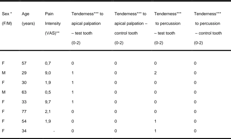
Table 1. Distribution of sex, age, pain intensity, tenderness to apical palpation and percussion of the test tooth and control
tooth of eight patients with SAP
Sex * (F/M) Age (years) Pain Intensity (VAS)** Tenderness*** to apical palpation – test tooth (0-2) Tenderness*** to apical palpation – control tooth (0-2) Tenderness*** to percussion – test tooth (0-2) Tenderness*** to percussion – control tooth (0-2) F 57 0,7 0 0 0 0 M 29 9,0 1 0 2 0 F 30 1,9 1 0 0 0 M 63 0,5 1 0 0 0 F 33 9,7 1 0 0 0 F 77 2,1 0 0 0 0 F 54 1,9 0 0 1 0 F 34 - 0 0 1 0 * F = female; M = male
** VAS = Visual Analogue Scale ranging from 0 to 10 where 0 represents “no pain” and 10 represents “worst possible pain”.
*** 0 = No reaction and a negative answer to the question whether pain is felt; 1 = Discomfort. An affirmative answer to the question whether pain is felt; 2 = Large discomfort. A positive answer to the question whether pain is felt. The patient flinches when examined.
23
Table 2. Distribution of sex, age, pain intensity, tenderness to apical palpation and percussion of the test tooth and control
tooth of seven patients with AAP
Sex * (F/M) Age (years) Pain Intensity (VAS)** Tenderness*** to apical palpation – test tooth (0-2) Tenderness*** to apical palpation – control tooth (0-2) Tenderness*** to percussion – test tooth (0-2) Tenderness*** to percussion – control tooth (0-2) M 72 0 0 0 1 0 M 74 0 0 0 0 0 M 51 0 0 0 0 0 M 67 0 0 0 0 0 M 35 0 0 0 0 0 F 53 0 0 0 0 0 M 56 0 1 0 1 0 * F = female; M = male
** VAS = Visual Analogue Scale ranging from 0 to 10 where 0 represents “no pain” and 10 represents “worst possible pain”.
*** 0 = No reaction and a negative answer to the question whether pain is felt; 1 = Discomfort. A positive answer to the question whether pain is felt; 2 = Large discomfort. An affirmative answer to the question whether pain is felt. The patient flinches when examined.
24
Figures
Figure 1
25 Figure 2
26 Figure 3
27
Figure legends
Figure 1. Number of test teeth (n) with and without tenderness to apical palpation and
percussion when diagnosing symptomatic apical periodontitis (SAP) and asymptomatic apical periodontitis (AAP).
Figure 2. Sensitivity, specificity, positive and negative predictive values for apical palpation when diagnosing symptomatic apical periodontitis (SAP) and asymptomatic apical
periodontitis (AAP).
Figure 3. Sensitivity, specificity, positive and negative predictive values for percussion when diagnosing symptomatic apical periodontitis (SAP) and asymptomatic apical periodontitis (AAP).
28
Attachment 1
Denna sida fylls i av patienten
Personnummer: _________________________________________
Hur länge har du haft tandvärk? ______________________________ dagar
Markera din smärta just nu:
Ingen smärta Värsta tänkbara smärta
Har du tagit någon värktablett de senaste 24 timmarna? Ja Nej
29
Denna sida fylls i av observatören
Datum för den kliniska undersökningen:_____________________
Undersökt tand:
________________________Apikalömhet: Ja Nej
Perkussionsömhet: Ja Nej
När tanden öppnas upp noteras pulpans tillstånd och graderas enligt nedan med ett kryss
Röntgenologiska fynd
Ingen apikal bendestruktion Apikal benförtätning Periapikal bendestruktion
Kontrolltand:
_______________________ Intakt: Ja Nej Fyllning: ____________ Material:_____________ Apikalömhet: Ja Nej Perkussionsömhet: Ja Nej Röntgenologiska fyndIngen apikal bendestruktion Apikal benförtätning Periapikal bendestruktion
Kommentarer
___________________________________________________________________________ ___________________________________________________________________________ ___________________________________________________________________________ ___________________________________________________________________________