Contamination Controls for Root Canal Sample Analysis
by Molecular Methods – A pilot study
Mauj Abdul Hussain Suzan Hawaz Ali Tutor: Malin Brundin
ABSTRACT
When exploring the root canal flora, before and after treatment, it is crucial to eliminate bacteria from the tooth surface before entering the pulp. If not, bacteria from the surface might contaminate the root canal sample resulting in false information. When using molecular techniques, not only bacteria are to be eliminated but also DNA from bacteria to avoid
contamination from the surface. The aim of the study was to examine if DNA from bacteria can be eliminated using a modified disinfection protocol. Samples from the tooth surface were taken from ten intact teeth stored in ethanol/ glycerol (50 % / 50 %) prior to the experiment. The teeth were sampled before and after cleaning the surface with H2O2 (30 %), NaOCl (3
%) and EDTA (0.5 M). Samples were taken from buccal, occlusal and lingual enamel and dentin surfaces. All samples were analyzed with polymerase chain reaction and culture. In a second experiment, ten teeth were placed in a bacterial solution containing Enterococcus
faecalis for three days and sampled as above. No growth could be detected using the
conventional culture technique from the post-wash samples. The results show that the teeth could not entirely become free from bacterial DNA using the performed cleaning routine as all samples were positive for bacterial DNA after cleaning. The average amount of detectable bacterial DNA was decreased with 95 % after the cleaning procedure. The results confirm reduction of bacterial DNA after cleaning, however, detectable bacterial DNA is still evident after disinfection.
INTRODUCTION
Apical periodontitis is an inflammatory condition caused by infection in the root canal and intra radicular infections are caused by poly microbial colonization (Sundqvist, 1976; Ricucci and Siqueira, 2010). These infections appear when there is a lesion or a fracture in the tooth admitting bacteria to enter the tooth pulp. The most common reason for endodontic treatments is caries (Ridell et al., 2006). Other known reasons are trauma, cracks and deep restorations (Kim et al., 2013). To ensure that the treatment has the best prognosis all bacteria must be eliminatedfrom the root canal (Sundqvist et al., 1998). To ensure a bacteria-free outcome a bacterial sample can be taken post treatment.
Another factor that can impact the prognosis of the endodontic treatment is the operator. The operator must work under sterile conditions to eliminate bacteria and reduce the risk of contamination. For these purposes, there are instruments and processing steps that needs to be used throughout the treatment; cleaning and disinfecting the tooth surface and the rubber dam, using sterile instruments (Siqueira et al.; 2000, Brito et al., 2009), using endodontic
medicaments that provides advantages like “bactericidal action, substantively, biocompatibility and low toxicity” (Nalawade et al., 2016).
Using these disinfection routines can improve the treatment and can reach a success rate up to 91 % (Sjögren et al., 1990).
In some cases, a sample from the root canal can be indicated to evaluate the antibacterial treatment or to ensure elimination of bacteria prior to filling the root canal system. Such a sample can be analyzed either by culture techniques or by modern molecular techniques, nowadays the most used technique is the latter. Studieshave shown that conventional culture techniques have limitations such as the difficulty of culture all species of bacteria due to their sensitivity to different environments (Josephson et al., 1993; Siqueira and Rocas, 2009). With PCR it is possible to detect bacteria that cannot be cultured due to sensitivity regarding
nutrition (Stolovitzky and Cecchi, 1996), gases and acids. The technique is also highly specific, timesaving (Siqueira and Rocas, 2003) and the samples can be frozen prior to analysis (Opel et al., 2010). Molecular techniques are based on detecting DNA from bacterial cells, which makes this technique unable to distinguish whether the bacteria are viable or dead (Josephson et al., 1993). Studies have shown a specific affinity between hydroxyapatite and DNA and also that DNA bound to hydroxyapatite is more resistant to degradation than
unbound DNA (Bernardi, 1965; Brundin et al., 2013; Burger et al., 2017). DNA from dead bacteria can persist and be detected years after cell death in root canals in vitro (Brundin et al., 2010). Dead bacteria are irrelevant to the success of the endodontic treatment but still can be detected by PCR technique. DNA from dead bacteria may be picked up during sampling resulting in a false positive result.
Before entering the pulp space the tooth surface should be clean to eliminate the risk of contamination of the canal by bacteria from the oral cavity. Cleaning protocols used today mainly works to eliminate viable bacteria samples are analyzed by conventional culture techniques (Sjögren et al., 1990; Siqueira and Rocas, 2009). However, today most root canal samples are analyzed with molecular techniques and a relevant question is if the surface cleaning methods used today can eliminates both viable bacteria and also bacterial DNA that otherwise might contaminate the clinical sample.
This in vitro pilot study aims to evaluate a modified method used to sterilize the tooth surface before taking samples from root canals and investigate if it is adequate to eliminate
contaminating DNA. The hypothesis of this study is that the PCR method will detect bacterial DNA from the teeth surfaces that the conventional culture technique will not be able to detect after cleaning the teeth with a modified protocol.
MATERIALS AND METHODS
In the study, biological material consisting of (anonymously donated) extracted human teeth will be used. It is impossible to identify people who donated teeth, so anonymity can be guaranteed. Since this is a biological material, it will be treated with good care; that is, if there is an opportunity to use the teeth, for example, in the endodontics department, they will be donated to the relevant department for further use. Data was selected using Pubmed search bar and the following keywords “Endodontics”, “Endodontic treatments”, “Endodontic
medicaments”, “PCR”, “Root canal sample”, “Contamination”, “Molecular methods” etc. Permission for this study was granted by the Ethical Committee.
First experiment
This first experiment aimed to determine whether the tooth surface could be free from bacteria after using a modified cleaning routine.
Tooth selection
Ten intact teeth (no caries, no fillings) without observable cracks were anonymously donated from the orthodontic department. The teeth had been stored in a container with ethanol/ glycerol (50%/50%).
Setup
Before the experiment started, all teeth were X-rayed to determine the thickness of the buccal and lingual enamel. The teeth were embedded in plaster to the cement-enamel junction to simplify the handling of the teeth during the experiment.
Initial sample
An initial pre-cleaning sample (IS) was taken with sterile mini-foams; one dry and one soaked in thiosulphate for culture technique-analysis, and one dry and one soaked in thiosulphate for PCR-analysis. The samples were taken from the following sample sites: buccal (IS-B1), occlusal (IS-O1), and from the rubber dam (IS-R1) 1 cm from the clamp on the buccal side (sample 1).
Cleaning of the tooth surface
The teeth were cleaned with pumice and rubber dam was applied. For each tooth, all the areas, the rubber dam, and the clamp was cleaned with peroxide (30%) for one minute. The same surfaces were cleaned with EDTA (0.5 M) for one minute and lastly with NaOCl (3%) for 2 minutes. The chlorine was deactivated with thiosulphate (5%) and the surfaces were left to dry.
Second sample
This sample was a post-cleaning sample (SS) taken from smooth surface enamel. It was taken as above for initial sample, marked as buccal surface (SS-B1), occlusal surface (SS-O1) and rubber dam (SS-R1).
Preparation of the cavities
After cleaning a cavity was prepared on the buccal surface on each tooth using high speed hand piece and a cylindrical bur with no water spray, to the enamel-dentine junction. Another cavity was also prepared on the lingual surface in the same way. This cavity was deeper; ending just before entering the pulp space.
Third sample
All the surfaces were again cleaned as above. The second post-cleaning sample (TS) was taken from cut open dentin. Samples were taken from the two cavities (as above for initial sample); buccal surface (TS-CB1) and lingual surface (TS-CL1).
Controls
The mini-foams and the paper points used for this experiment were tested for contaminating DNA before the experiment.
Second experiment
Setup
Ten intact teeth were embedded in plaster to the cemento-enamel junction and, using a highspeed hand piece, a cavity was drilled to the enamel-dentine junction. The teeth were placed in a bacterial solution containing Enterococcus faecalis for three days.
A known strain of Enterococcus faecalis (JH2-2) was used for this experiment. Bacteria were aerobically grown on Fastidious Anaerobe Agar (FAA; 37° C) overnight and then suspended in phosphate buffered saline to an optical density of 1 (corresponding approximately 109 colony forming units /mL).
Initial sample
The teeth were dried with a paper towel and an initial pre-cleaning sample was taken with two sterile mini-foams, both soaked in thiosulphate, for each tooth; again, one sample for PCR and
one for culture technique. This time the sample sites were: buccal (2IS-B), occlusal (2IS-O) and lingual (2IS-L) surfaces.
Cleaning
The same cleaning routine was used for this experiment as used in the first.
Second sample
Another sample was taken after cleaning from the same sites as mentioned in the initial sample, using two sterile mini-foams soaked in thiosulphate for each tooth; one for PCR analysis and one for culture technique-analysis.
DNA extraction and Quantitative PCR
DNA was extracted according to the manufacturer’s protocol (Bacterial Genomic DNA kit; Gene Elute, Sigma, Saint Louis, Missouri, USA) and eluted in 200 µL elution solution. For real-time PCR, universal primers were used producing a 170 bp amplicon. PCR amplifications were prepared in a 10 µL final reaction volume: 1 µL total DNA template, 0.5 mM of each primer; 5 µL PCR mix (KAPA SYBR FAST universal qPCR kit, KAPA Biosystems, Boston, MA, United States) Ultrapure water (Sigma-Aldrich,) was added to give the final reaction volume.
Real-time PCR was performed using a qPCR machine (Corbett RESEARCH, RG-6000, Sydney, Australia). An initial incubation at 95°C (10 min) was followed by 35 cycles of denaturing at 95°C (10 sec); annealing 68° C (5 sec); amplification 72°C (7 sec). At end of each cycle fluorescent products were registered. All samples were run in duplicates. Quantification of amplification products was performed using Rotor-Gene 6000 Series Software 1.7. Standard curves were created using DNA from pure cultures with known concentrations of bacteria (Enterococcus faecalis; JH2-2)) determined by cultivation. Providing standard curves tenfold serial dilutions of known amount of DNA (108 to 102 cells) were processed. Ultrapure water (Sigma) replaced the bacterial DNA template in the negative control.
RESULTS
CultureAll samples taken before wash from the first and second experiment and that were analyzed with conventional culture technique showed growth. However, there was no growth detected in the conventional culture technique from the post-washed samples taken in both first experiment (uninfected teeth) and second experiment (infected teeth).
PCR
The first experiment shows that the ten intact teeth could not entirely be free from bacterial DNA using the performed cleaning routine. As seen in figure 1, DNA was detected on the tooth surfaces even after storing the teeth in an alcoholic solution.
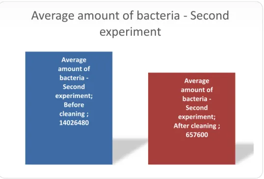
In the second experiment, after the teeth was contaminated with E. faecalis, the modified disinfection protocol method used to sterilize the surfaces gave a reduction of bacterial DNA with 95 % (fig. 2.)
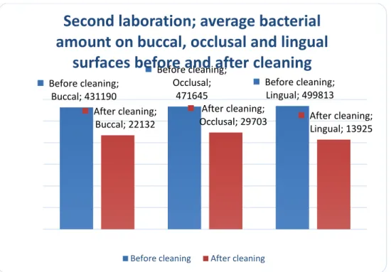
In addition to the fact that there was a difference between the amounts of detectable bacterial DNA in all the ten teeth in the second experiment, the result also shows a difference between the surfaces (fig. 3). Even though the lingual surfaces (2IS-L) had the most bacterial amount before the cleaning process, it was also the surfaces that had the greatest reduction of bacteria after cleaning.
There was no major difference in detectible DNA between the undrilled buccal surfaces (2ISB) and the drilled lingual surfaces (2IS-L) before cleaning (fig. 3). However, the results shows that more bacterial DNA was detected on the drilled lingual surfaces compared to the undrilled buccal surfaces before cleaning; there was 16 % more bacterial DNA in the lingual surfaces (drilled to the enamel-dentine junction). After cleaning, the drilled lingual surfaces
DISCUSSION
In this study, it was evident that despite the modified disinfection protocol we used, it was not possible to eliminate all bacterial DNA from the teeth surfaces. As mentioned in the results, the samples that were cultured with conventional technique showed no bacterial growth, while the PCR technique detected DNA. This can be explained by technical differences between the two methods; where conventional culture technique only detects viable bacteria while PCR detects both viable and dead bacterial DNA.
Analyzing PCR data, it was found that the modified disinfection protocol gave a bacterial DNA reduction of 95 %. The experiment also showed a difference in amount of bacteria between the different surfaces; in average the lingual surfaces were most infected, yet they had the greatest reduction after using the modified disinfection protocol. When comparing undrilled buccal surfaces and drilled lingual surfaces there was no major difference in amount of DNA (16 % more bacterial DNA on the drilled lingual surfaces). Although the drilled lingual surfaces had slightly higher amount of bacteria before cleaning comparing to the buccal undrilled surfaces, the modified disinfection protocol gave a reduction of 37% less amount of bacteria on the drilled lingual surfaces compared to the undrilled buccal surfaces.
To ensure that we did not contaminate the teeth while taking samples, all instruments that were used during the experiment were sterile. Also, the mini-foams that were used to take samples were analyzed for contaminating DNA and the results showed that they contained 100 copies of DNA (data not shown) - that can be explained by contamination during the distribution of the mini foams, for example when packing them on the dental trays. The amount of DNA that was detected in the mini-foams can be transcribed as background noise.
When comparing, the teeth stored in ethanol/glycerol to the teeth that were cleaned according to the modified disinfection protocol (fig. 1) the results confirms that using the cleaning routine the teeth surfaces will be 21 times cleaner. Even if this is a big progress of reducing the bacterial DNA, there is still a big amount of DNA still present after cleaning; 6×10⁵ copies of bacterial DNA after using the cleaning protocol compared with 1.4×10⁷ which was the average amount of bacterial DNA before cleaning the teeth. The reason why the result did not show entirely cleaned teeth with no bacterial DNA may be explained by some various factors. One of them is the molecular technique method itself. It turned out that the PCR machine naturally gives off some background noise that may be disturbing to the real data. Another factor may be that there were four laboratories executing this experiment, meaning that even
though there was a detailed protocol to be followed, human factors lead to some divergence from it. It must also be mentioned that the tools that were used, such as the instruments and the clinical gloves were sterile, but that does not mean that they were entirely free from DNA.
Another aspect we acknowledged was whether there was a difference between the drilled and undrilled sites. Does the affinity of the bacterial DNA get affected by the surface roughness? Is there a difference between a smooth (undrilled) and a rough (drilled) surface? According to our results there was a small difference in the amount of detected bacterial DNA when
comparing drilled and undrilled sites before using the cleaning protocol. Even though the lingual (drilled) sites (2IS-L) had an average of more bacterial DNA than the buccal
(undrilled) sites (2IS-B)when infected before using the cleaning protocol (fig. 3), our results show that we managed to get the lingual (dentin) site more free from bacterial DNA compared to the buccal (enamel) by cleaning the teeth. For the buccal (undrilled) site the amount of bacterial DNA was 431 190 before cleaning according to the protocol and 2×10⁴ after cleaning. Corresponding for the lingual (drilled) was 499 813 before using the cleaning protocol and 1.3 × 10³ after wash. The theory behind why more bacterial DNA was initially detected on the drilled site, compared with the smooth undrilled site, may be due to the rougher surface of the dentine which with its tubules provide a greater retention for bacteria (Stolovitzky and Cecchi, 1996). Also by drilling a cavity into the enamel dentine junction, we created a site that the bacteria can bind to much easier. Studies have shown that bacterial DNA has a strong capability of binding to the main inorganic component in enamel,
hydroxyapatite (Brundin et al., 2014). As mentioned above the post-washing results for the lingual (drilled) surface contained less bacterial DNA compared to the buccal surface. That can be explained by the limited area the lingual samples were taken from. When sampling the buccal surface, the mini-foams that was used could be rubbed against a bigger area and collect a larger amount of DNA. On the lingual surface, we could only collect bacteria/DNA from a small cavity which was approximately 1/3 of the buccal area, leading the mini-foam to be exposed to less DNA. Another reason due to the result can be explained by the construction of the tooth; enamel contains more hydroxyapatite compared to dentine (which contains other organic materials such as collagen) (Slimani et al. 2017). This may affect the affinity between the dentine and the bacterial DNA, making it less attachable to dentine than enamel.
wash (2SS-O). Compared to the lingual and buccal surfaces, the occlusal surface was the area that was hardest to get clean. This is explained by the biological factor of the anatomy of the premolars and molars; the fissures (Preservation and Restoration of Tooth Structure, 2016), that provides the bacteria with shelter and makes it harder for the medicaments to eliminate the bacteria.
Even with a strict modified disinfection protocol it is not possible to eliminate all bacterial DNA from the teeth. Although it must be mentioned that the results were analyzed using molecular technique; which can detect both viable and dead bacteria (Josephson et al., 1993). When it comes to endodontic treatment, only viable bacteria are relevant for the outcome of the treatment (Siqueira et al., 2000). This means that there is a slight chance that the results from our samples may represent a false-positive result when analyzed by PCR.
However, one must remember the limitations of conventional culture techniques such as the difficulty to culture all species of bacteria due to their growth being demanding, so called not-yet-cultivable species. There may be some fastidious bacteria in the samples that cannot be cultured because of their sensitivity. Therefore, there is a possibility that our samples may also represent a false-negative result when analyzed by culture.
Proceeding from here we propose that the next planned study should approach the methods used to collect data, more aggressively. A way to do this is by using DNA-eraser during the molecular technique to eliminate irrelevant DNA i.e. from dead bacteria. This could give a more authentic result.
ACKNOWLEDGEMENTS
The authors thanks tutor Malin Brundin for your great tutorial, all support and time. Mrs Carina Öhman for invaluable technical assistance, Dr Frida Hagberg for the assistance in the experiment, and Dr David Figdor for having a participating part in this study.
REFERENCES
Bernardi G (1965). Chromatography of nucleic acids on hydroxyapatite. Nature 206:779–83. Brito PR, Souza LC, Machado de Oliveira JC, Alves FRF, De-Deus G, Lopes HP et al.
(2009). Comparison of the effectiveness of three irrigation techniques in reducing intracanal Enterococcus faecalis populations:an in vitro study. J Endod 35:1422–7.
Brundin M, Figdor D, Johansson A, Sjögren U (2014). Preservation of bacterial DNA by human dentin. J Endod 40:241–5.
Brundin M, Figdor D, Roth C, Davies J.K., Sundqvist G, Sjögren U (2010). Persistence of dead-cell bacterial DNA in ex vivo root canals and influence of nucleases on DNA decay in vitro.
OOOE 110:789-794.
Brundin M, Figdor D, Sundqvist G, Sjögren U (2013). DNA binding to hydroxyapatite: a potential mechanism for preservation of microbial DNA. J Endod 39:211–6.
Burger J, Hummel S, Hermann B, Henke W (1999). DNA preservation: a microsatellite-DNA study on ancient skeletal remains. Electrophoresis 20:1722–8.
Figdor D, Brundin M (2016). Contamination Controls for Analysis of Root Canal Samples by Molecular Methods: An Overlooked and Unsolved Problem. J Endod 42:1003-8.
Graham JM, Wyatt RH, Hien CN, Mark SW (2016). Preservation and Restoration of Tooth Structure. 3rd rev. ed. London: Mosby-Wolfe.
Josephson KL, Gerba CP, Pepper IL (1993). Polymerase Chain Reaction Detection of Nonviable Bacterial Pathogens. Microbiol 59;10:3513-5.
Kim SY, Kim SH, Cho SB, Lee GO, Yang SE (2013). Different Treatment Protocols for Different Pulpal and Periapical Diagnoses of 72 Cracked Teeth. J Endod 39:449-52.
Nalawade TM, Bhat KG, Sogi S (2016). Antimicrobial Activity of Endodontic Medicaments and Vehicles using Agar Well Diffusion Method on Facultative and Obligate Anaerobes. Int J
Clin Pediatr Dent 9:335-341.
Opel KL, Chung D, McCord BR (2010). A study of PCR inhibition mechanisms using real time PCR. J Forensic Sci 55:25–33.
Ricucci D, Siqueira JF (2010). Biofilms and apical periodontitis: study of prevalence and association with clinical and histopathologic findings. J Endod 36:1277–88.
Ridell K, Petersson A, Matsson L, Mejàre I (2006). Periapical status and technical quality of rootfilled teeth in Swedish adolescents and young adults. A retrospective study. Acta Odontol
Scand 64:104-10.
Siqueira JF Jr, Rocas IN (2009). Diversity of endodontic microbiota revisited. J Dent Res 88:969–81.
Siqueira JF Jr, Rocas IN (2003). PCR methodology as a valuable tool for identification of endodontic pathogens. J Dent 31:333-9.
Siqueira JF Jr, Rocas IN, Favieri A, et al. (2000). Chemomechanical reduction of the bacterial population in the root canal after instrumentation and irrigation with 1%, 2.5%, and 5.25% sodium hypochlorite. J Endod 26:331–4.
Sjogren U, Hagglund B, Sundqvist G, Wing K (1990). Factors affecting the long-term results of endodontic treatment. J Endod 16:498-504.
Slimani A, Nouioua F, Desoutter A, Levallois B, Cuisinier FJG., Tassery H et al. (2017). Confocal Raman mapping of collagen cross-link and crystallinity of human dentin–enamel junction. JBO 22:0860031-8.
Stolovitzky G, Cecchi G (1996). Efficiency of DNA replication in the polymerase chain reaction. PNAS 93:12947–52.
Sundqvist G (1976). Bacteriological studies of necrotic dental pulps (odontologial dissertation). Umeå University, Umeå, Sweden.
Sundqvist G, Figdor D, Persson S, Sjögren U (1998). Microbiologic analysis of teeth with failed endodontic treatment and the outcome of conservative re-treatment. OOOE 85:86-93.
FIGURES
Figure 1. Chart showing the difference (mean) between number of bacterial DNA analyzed with PCR, on all surfaces (IS-B1, IS-O1, IS-R1) before and after cleaning (SS-B1, SS-O1, SS-R1), in the first experiment.
Figure 2. Amount (mean) of detectable bacterial DNA by qPCR on all surfaces after infecting the teeth with E. faecalis. Left bar shows amount of bacteria detected before cleaning the surfaces (Before cleaning). The right bar shows amount of bacterial DNA detected after cleaning the surfaces (After cleaning).
Average amount of bacteria - First experiment; Before cleaning ; 17745 Average amount of bacteria - First experiment; After cleaning ; 7152
Average amount of bacteria - First
experiment
Average amount of bacteria -Second experiment; Before cleaning ; 14026480 Average amount of bacteria -Second experiment; After cleaning ; 657600Average amount of bacteria - Second
Figure 3. A chart representing the ten teeth in the second experiment, amount (mean) of bacterial DNA for each surface before cleaning (2IS-B, 2IS-O, 2IS-L) and after cleaning (2SS-B, 2SS-O, 2SS-L). Before cleaning; Buccal; 431190 Before cleaning; Occlusal; 471645 Before cleaning; Lingual; 499813 After cleaning; Buccal; 22132 After cleaning;
Occlusal; 29703 After cleaning; Lingual; 13925