swedish dent al journ al supplement 226, 20 1 2. d oct or al dissert a tion in odont ol og y h elen a Fr ansson malmö universit y malmö university
helena Fransson
on the repair oF
the dentine barrier
isbn/issn 978-91-7104-390-0/0348-6672 o n the r ep air o F the dentine b arrier
Swedish Dental Journal Supplement 226, 2012
© Helena Fransson 2012 ISBN 978-91-7104-390-0 ISSN 0348-6672 Holmbergs, Malmö 2012
HELENA FRANSSON
ON THE REPAIR OF
THE DENTINE BARRIER
Departments of Endodontics and Oral Biology,
Faculty of Odontology,
Malmö University, Malmö, Sweden 2012
The summary of this publication can be downloaded from se www.mah.se/muep
CONTENTS
ABSTRACT ... 9 POPULÄRVETENSKAPLIG SAMMANFATTNING ... 11 (Summary in Swedish) PREFACE ... 13 THESIS AT A GLANCE ... 14 VOCABULARY/ABBREVIATIONS ... 15 INTRODUCTION ... 17 AIMS ... 28MATERIAL AND METHODS ... 29
Systematic review (I) ...29
Clinical trial (II) ...30
Immunohistochemical studies (II and III) ...32
Cell culture study (IV) ...34
RESULTS ... 37
Systematic review (I) ...37
Effect of EMDgel on experimentally performed pulp exposures (II) ...47
Characterization of the newly formed hard tissue (III) ...50
Effects of bacterial products on the activity of odontoblastlike cells and their formation of Col I (IV) ...54
DISCUSSION ... 57
CONCLUSIONS ... 67
CONTRIBUTORS TO THE STUDIES ... 69
ACKNOWLEDGEMENTS ... 70
ABSTRACT
The overall aim of this thesis was to study some aspects of the re-pair of the dentine barrier, especially in conjunction with dental pulp capping. Understanding the events leading to the healing of the dentine and pulp, and hence successfully preserving the vitality and functions of the tooth, would lead to a scientific basis for a less invasive treatment of pulp exposures than performing root canal treatments.
The surfaces of the body have physiological barrier functions aimed at protecting the body from external noxious agents. In the tooth, the odontoblasts, which line the outermost part of the pulp and are responsible for the formation of dentine, play a central role in the barrier function and thus in the defence mechanisms of the tooth. The micro-organisms in the caries lesion can reach the pulp via the dentinal tubules. However, the barrier function helps to prevent microbial invasion and thereby avoid deleterious inflam-mation and subsequent necrosis of the pulp. Dentine repair is an important part of the barrier function. There are however doubts as to whether the repair also leads to restitution of the function and the ability to withstand bacterial influx over the longer term.
Pulp capping is a treatment method used when the pulp has been exposed in order to stimulate healing of the pulp and dentine. The evidence for repair of the dentine after pulp capping in humans has been studied by means of a systematic review. The focus of the lit-erature search was studies performed in humans where hard tissue formation had been studied with the aid of a microscope. We
con-cluded, based on the limited evidence available, that calcium hy-droxide based materials but not bonding agents promote formation of a hard tissue bridge. Scientific evidence was lacking as to whether MTA was better than calcium hydroxide based materials in this regard.
A gel (Emdogain®Gel) containing amelogenin, known to be
in-volved in dentinogenesis, was evaluated with regard to formation of hard tissue in a clinical study. A greater amount of hard tissue was formed after application of the gel compared to the control. Characterization of the tissue concluded it to be dentine, based on its content of type 1 collagen and dentine sialoprotein, although it was not formed as a continuous bridge covering the pulp wound.
Beneath a deep caries lesion an important part of the barrier func-tion is the odontoblasts´ response to bacteria with the formafunc-tion of new dentine. A cell model with odontoblasts was used to study the effects of clinical isolates from a deep carious lesion on their viabil-ity and production of type 1 collagen, the major component of the dentine in the early stages of its formation. There were bacteria that negatively affected the viability of the odontoblast-like cells and different bacteria varied in their effects on type 1 collagen pro-duction, suggesting that some bacteria may have a direct influence on the odontoblasts´ ability to form dentine.
In summary; Emdogain®Gel initiated dentine formation, though
not in a form that could constitute a barrier and there are indica-tions that bacteria may differentially affect the odontoblasts´ ability to repair the dentine barrier.
POPULÄRVETENSKAPLIG
SAMMAN-FATTNING (SUMMARY IN SWEDISH)
Det övergripande målet för avhandlingen har varit att studera någ-ra aspekter av läkningen av tandens huvudsakliga hårdvävnad, dentinet. Vid mycket djupa kariesangrepp där dentinet förstörts och pulpan därmed blottats, rotbehandlas ofta tanden vilket inne-bär att pulpan tas bort och att rotkanalen fylls med ett rotfyll-ningsmaterial. Djupare kunskaper om dentinets läkningsförmåga kan leda till att andra mindre invasiva och kostsamma behand-lingsmetoder än rotbehandlingar skulle kunna användas vid myck-et djupa kariesangrepp.
Kroppens ytor har barriärfunktioner för att skydda kroppen mot skadliga ämnen. I tanden svarar pulpans yttersta cell-lager för en viktig del i barriärfunktionen. Dessa celler, odontoblasterna, bildar dentinet och verkar spela en central roll i de skyddsmekanismer som tanden har. När ett kariesangrepp bryter ned tandens hård-vävnader kan bakterier eller deras produkter få möjlighet att tränga in till pulpan vilket leder till ett inflammatoriskt och immu-nologiskt svar som kan leda till vävnadsdöd av pulpan. Under vissa omständigheter verkar dock pulpan ha förmåga att reparera hård-vävnadsbarriären på ett sådant sätt att den fysiologiska funktionen kvarstår så att vävnadsdöd och därmed invasion av mikroorganis-mer undviks. Det finns emellertid studier som antyder att den repa-rerade hårdvävnadens barriärfunktion ger vika och att den inte kan stå emot ny mikrobiell belastning.
Pulpaöverkappning är en behandling som används när pulpan bli-vit blottad i ett försök att bibehålla pulpans bli-vitalitet och funktion. Faktorer som påverkar hårdvävnadsbildningen vid pulpaöver-kappningar har studerats i en systematisk litteraturöversikt. Base-rat på det begränsade vetenskapliga stödet visade resultaten att kalciumhydroxidbaserade material men inte bondingmaterial ger en hårdvävnadsbildning som täcker pulpasåret då de används som överkappningsmaterial. Det finns inget vetenskapligt stöd för att kunna fastslå att mineraltrioxidaggregat (MTA) skulle ge mer hårdvävnadsbildning jämfört med kalciumhydroxidbaserade mate-rial när dessa används som överkappningsmatemate-rial.
En gel (Emdogain®Gel) som innehåller amelogenin som man vet är
inblandat i processen då dentinet börjar bildas, utvärderades i en klinisk studie med syfte att studera hårdvävnadsbildningen. En större mängd hårdvävnad bildades efter appliceringen av gelen jämfört med kontrollmaterialet. Hårdvävnaden kunde karaktärise-ras som att vara likt det ursprungliga dentinet, men den bildades inte i en struktur som skulle kunna utgöra en fysiologisk barriär.
Under ett kariesangrepp bör odontoblasterna svara på närvaro av bakterier med försvarsreaktioner såsom bildande av nytt dentin, men kvalitén på det dentinet verkar ibland bli sämre än det ur-sprungliga dentinet. Produkter från bakterier tagna från ett djupt kariesangrepp användes för att studera dess effekter på odontob-lastliknande cellers aktivitet och förmåga att bilda en typ av kolla-gen som är den huvudsakliga beståndsdelen i nybildat dentin. Vissa bakterier hade en negativ påverkan på odontoblasternas aktivitet och bakteriernas effekt på kollagenproduktionen varierade, vilket skulle kunna tyda på att bakterier kan ha en direkt effekt på odon-toblasternas förmåga att upprätthålla dentinets barriärfunktion.
Sammanfattningsvis kan man säga att Emdogain®Gel initierade
dentinbildning, men inte i en struktur som skulle kunna utgöra en barriär och det förefaller som om bakterier i olika grad kan påver-ka odontoblasternas förmåga att bilda en dentinbarriär.
PREFACE
This thesis is based on the following papers, which are referred to in the text by their Roman numerals.
I Olsson H, Petersson K, Rohlin M. Formation of a
hard tissue barrier after pulp cappings in humans. A systematic review. Int Endod J 2006; 39: 429-442.
II Olsson H, Davies JR, Holst KE, Schröder U,
Peters-son K. Dental pulp capping: effect of Emdogain®Gel
on experimentally exposed human pulps. Int Endod J 2005; 38: 186-194.
III Fransson H. Petersson K, Davies JR. Dentine
sialo-protein and Collagen I expression after experimental
pulp capping in humans using Emdogain®Gel. Int
Endod J 2011; 44: 259-267.
IV Fransson H. Petersson K, Davies JR. Effects of
bac-terial products on the activity of odontoblast-like cells and their formation of type 1 collagen. Submit-ted to J Endod.
The papers I, II and III are reprinted with permission of the copy-right holder John Wiley & Sons Ltd.
THESIS A
T A g
LAN
CE
VOCABULARY/ABBREVIATIONS
There are several terms used in the literature concerning dentine formation and different modes of pulp capping. The following ap-plies to this thesis:
Dentine
Primary dentine formed by primary
odonto-blasts until the root is fully formed
Secondary dentine that is formed at a highly
re-duced rate after the root has been fully formed
Tertiary dentine formed in response to injury.
This might further be characterized as reactive when the primary odonto-blasts have been triggered or repara-tive when the primary odontoblasts have been replaced by odontoblast-like cells
Pulp capping application of a material to an
ex-posed pulp (direct capping) in order to allow the pulp to recover and main-tain its normal vitality and function
Partial pulpotomy a superficial pulp amputation and
ap-plication of a pulp capping material as in pulp capping
Indirect pulp capping a treatment in which infected dentine is intentionally left under a restoration in order to avoid pulp exposure
CENTRAL the Cochrane Controlled Trials
Col I type 1 collagen
DAB 3, 3-Diaminobenzidine
DMEM Dulbecco´s Modified Eagle Medium
DSP dentine sialoprotein
DSPP dentine sialophosphoprotein
ELISA enzyme-linked immunosorbent assay
EMD enamel matrix derivative
EMDgel enamel matrix derivative in a
formula-tion with propylene glycol alginate
FBS foetal bovine serum
LPS lipopolysaccharide
LTA lipoteichoic acid
MDPC-23 a spontaneously immortalized cell line
from the dental papillae of mouse mo-lars
MeSH Index Medicus: Medical Subject
Headings
MTT methol-thiazolyl-diphenyltetrazolium bromide
PGA propylene glycol alginate
RCT randomized controlled trial
SE standard error
SIBLING Small Integrin-Binding LIgand
N-linked Glycoprotein
TBS tris-buffered-saline
INTRODUCTION
The barrier function of dentine
The surfaces of the body including the skin, the gastro-intestinal tract and the oral mucosa have physiological barrier functions aimed at protecting the body from external noxious agents (Turner 2009; Elias 2008). In the tooth the enamel is, by virtue of its hard-ness and structure, an effective mechanical barrier while the physiological barrier function of the tooth lies mainly within the pulp-dentine complex.
In the pulp-dentine complex, the dentine is traversed by tubules filled with fluid in which the odontoblast processes are located. The odontoblasts are the cells responsible for the formation of den-tine. They align at the periphery of the pulp in close proximity to the dentine with their cellular processes situated within the dentine tubules. As dentine is being formed, a layer of collagenous network with associated non-collagenous proteins is laid down by the odon-toblasts. This zone of so-called “predentine” is thus to be found between the fully mineralised dentine and the odontoblast.
The physiological barrier function of the pulp-dentine complex consists of several key elements:
Dentinal fluid
Formation of peri-tubular dentine Formation of tertiary dentine
There is a continuous outward flow of dentinal fluid through the dentine tubules and in exposed dentine, the outward fluid flow is
the first barrier against the inward diffusion of noxious agents such as bacteria or bacterial components (Pissiotis & Spångberg 1994). Consequently, any noxious agent migrating towards the pulp must do so against a pressure gradient (Mjör 2009) and in addition, the dentinal fluid dilutes the noxious agents. The dentinal fluid, which is considered to be plasma-derived, contains serum proteins and globulins which can agglutinate or bind noxious agents and thus
play a protective role in the barrier function (Knutsson et al. 1994).
A range of cytokines and chemokines such as 1, 6, 8,
IL-10 and TNFcan be found in the dentinal fluid (Geraldeli et al.
2012), though their role has not yet been elucidated. In addition, the dentinal fluid also contains beta-defensins which have
antim-icrobial properties and can kill bacteria directly (Dommisch et al.
2005). The substances found in the dentinal fluid do not fully cor-respond to those in plasma and thus dentinal fluid composition and flow thus appear to be regulated by the odontoblasts
(Geral-deli et al. 2012).
The formation of peri-tubular dentine by the odontoblasts is an-other part of the physiological barrier function. This reduces the diameter of the dentinal tubules and reduces the chance of noxious agents penetrating into the pulp (Bjørndal & Mjör 2002). The se-cretion of peri-tubular dentine by the odontoblasts as well as the precipitation of crystals dissolved during the caries process may block the dentinal tubules, reducing the transport of fluids and
matter (Tagami et al. 1992).
Slow formation of secondary dentine is seen throughout life. It is a continuum of the primary dentine, though the secondary dentine is formed at a much reduced rate once the root has been fully formed. In cases of disruption of the hard tissues, the odontoblasts may again, very locally, increase the rate of dentine production to form what is referred to as tertiary dentine. If the primary odontoblasts have been triggered, the tertiary dentine is denoted as reactive, whereas reparative dentine is the term used for tertiary dentine that has been formed by differentiated stem cells often called odontoblast-like cells, which have replaced the original odontoblasts. As reparative dentine is not formed by the same
odontoblasts as those which formed the primary dentine, the con-tinuum of the tubules is interrupted and the structure of reparative dentine thus described as atubular (Barber & Massler 1964; Mjör 2009). The formation of tertiary dentine is part of the physiologi-cal barrier function through the increase of the dentine thickness between the site of the insult and the pulp. In case of reparative dentine, the atubular structure may reduce the transport of fluids and matter to the pulp. However the function of the dentinal fluid in the dentinal tubules and thereby the odontoblasts is affected.
These barrier functions of the dentine are considered to be under control of the odontoblasts. Strategically positioned, the odonto-blasts with their processes within the dentinal tubules are the first cells to encounter and respond to noxious agents such as bacteria or bacterial components within the dentinal tubules. Odontoblasts are highly specialized cells and are involved in a similar defence system to that of epithelial cells regulating the host’s response to
injury (Dommisch et al. 2008; Goldberg et al. 2008). Apart from
forming tertiary dentine, the odontoblasts are proposed to be in-volved in several defence mechanisms which protect the tooth from bacterial invasion. The odontoblasts are known to express several
Toll-like receptors which recognise bacterial antigens (Staquet et al.
2011). Upon activation of Toll-like receptors the odontoblasts may release substances that will affect angiogenesis and the regulation
of blood flow, key elements of the inflammatory process (Botero et
al. 2006; Soden et al. 2009). Through activation of Toll-like
recep-tors it seems possible that the odontoblasts, by releasing different substances, may negatively or positively regulate the inflammatory and immune response within the pulp and thus affect the
forma-tion of dentine (Durand et al. 2006; Farges et al. 2011; Hahn &
Liewehr 2007). Odontoblasts may also release antibacterial
pep-tides which can kill bacteria directly (Dommisch et al. 2005) and
appear to amplify the responses to the presence of bacteria through
a self-feedback system (Horst et al. 2011). To sum up; the
physio-logical functions of the odontoblast-dentine unit play an important role in protecting the pulp from microbial influence when exposed to the oral environment and hence constitute an active barrier function as described for the skin and gastro-intestinal tract.
Injury to the dentine
Apart from trauma, attrition or restorative procedures, caries is the most evident process in which the dentine barrier function is acti-vated and if overloaded, the dentine will eventually be destroyed leading to exposure of the pulp. Movement of bacterial products within the dentinal tubules will affect the odontoblasts, even though the bulk of the micro-organisms are situated some distance from the pulp.
Investigations of the micro-flora in deep caries lesions have pro-posed that the prevailing species are Gram-positive organisms such
as Lactobacillus, Actinomyces, Eubacterium, Proprionibacterium
and Streptococcus species even though Gram-negative organisms
including Fusobacterium, Veillonella and Bacteroides may be
found at lower levels (Edwardsson 1974; Hoshino 1985). More re-cent studies using 16S rRNA gene sequencing have shown a
domi-nance of Lactobacillus species in some caries lesions (Chhour et al.
2005; Munson et al. 2004). Bacteria have a strong tendency to
at-tach to surfaces and form biofilms (Costerton et al. 1995) and
sev-eral oral bacteria express surface adhesins which allows them to
bind to collagen within the dentine matrix (McGrady et al. 1995;
Love et al. 1997). Secondary colonizers that do not express these
surface adhesins can co-aggregate to the primary colonizers allow-ing the formation of polymicrobial biofilms within the dentinal tu-bules (Love & Jenkinsson 2002). Bacteria in biofilms may express phenotypes which differ from those of the same species growing in planktonic state due to the closeness of the bacterial cells and the presence of micro-niches in the biofilm (Stewart & Franklin 2008;
Svensäter et al. 2001). Physiological adaptations to surface
attach-ment include changes in the production of extracellular polymeric substances as well as modifications in cell morphology.
In in-vitro models of biological events within the pulp due to
car-ies, it is common to use commercially available solutions of known bacterial pathogenic components such as lipoteichoic acid (LTA) or
lipopolysaccharide (LPS) (Durand et al. 2006; Farges et al. 2011;
Oliveira & Santos 2011; Soden et al. 2009; Telles et al. 2003). The
studies are made easy, but is unlikely to reflect the situation during the caries process as the pathogenic effect of the bacteria may not solely be attributed to LTA or LPS. There is scant information con-cerning the biological events within the pulp using fresh isolates in biofilm models.
Dentine reactions and repair
As mentioned above, the odontoblasts should be seen as a barrier entity such as the epithelium lining of the oral cavity, the gastro-intestinal tract and the skin. The odontoblasts may be injured and die during restorative procedures or as a sequel to a rapidly pro-gressing caries lesion. Unlike epithelial cells, odontoblasts are post-mitotic and are therefore incapable of cell division in order to re-place the lost cells (Arana-Chavez & Massa 2004; Tziafas & Kodonas 2010). The fact that the odontoblast is post-mitotic might be of importance since the death of other post-mitotic cells within the brain or heart can lead to irreversible loss of function of the tis-sues. The ultimate aim of repair is to restore the original structure and the biological functions of the damaged tissues (Schmalz & Gal-ler 2011); however as the dead odontoblasts need to be replaced by differentiated odontoblast-like pulpal stem cells, the repair process does not completely fulfil this ultimate aim. How the recruitment of the odontoblast-like cells actually takes place is not fully elucidated, though it seems likely that multipotent pulpal stem cells migrate to the site of the injury, where they proliferate and differentiate into
odontoblast-like cells (Balic et al. 2010; Shi & Gronthos 2003). In
cases of exposure of the pulp, healing with hard tissue has been ob-served although the morphology is similar to reparative dentine with an atubular structure and with cellular inclusions (Arana-Chavez & Massa 2004). It is not known whether this reparative hard tissue ac-tually constitutes a functional recovery as it is questionable whether the odontoblast-like cells responsible for the formation of the hard tissue possess the same protective barrier functions as those seen in primary odontoblasts. Since the tubular structure of dentine is mostly missing, the pathway for agents to induce reactions in the odontoblasts is more or less hindered. Nevertheless, healing of the hard tissue structure with the physiological functions seems to be of great importance in keeping the integrity and vitality of the tooth.
The odontoblasts may be affected to various extents during the caries process. Theoretically this means that the dentine reaction beneath a caries lesion may involve anything from up-regulating primary odontoblasts to the replacement of the entire odontoblast layer. In the reaction to minor injuries, the pulp tissue is not ex-posed but dentine production is up-regulated underneath what is clinically observed as an intact layer of dentine. Beneath some car-ies lesions it seems to be rather similar to the primary dentine, whereas beneath others it appears to be structurally different or even absent (Bjørndal 2001). One explanation for these varying states might be that different consortia of micro-organisms lead to different degrees of repair. Thus invasion of the pulp by micro-organisms is probably dependent on both the nature of the bacteria present and the barrier function of the pulp-dentine complex. When the pulp has been exposed, and thus the odontoblasts have been destroyed, a pulp capping procedure in which a protective agent is placed on the pulp tissue may result in the recruitment of odontoblast-like cells and hard tissue repair (Nyborg 1958; Schröder & Granath 1972).
As reparative dentine is sometimes quite irregular with cellular in-clusions and tunnel defects not seen in the primary dentine, it is sometimes referred to as “osteodentine”. As a consequence of the morphological differences observed, the use of the terms “dentine-like” and “odontoblast-“dentine-like” have gained popularity when denot-ing hard tissue and cells involved in the repair, especially since the repair involves replacement of post-mitotic cells. Using a biochemi-cal approach to characterize the odontoblast phenotype, a wide range of differentiation markers such as members of the SIBLING-family (Small Integrin-Binding LIgand N-linked Glycoprotein) has been suggested. Dentine sialoprotein (DSP) is a member of this family and together with type 1 collagen (Col 1) is suggested to be a late marker for odontoblast formation. These markers are fre-quently used to characterize the odontoblast and the dentine
(But-ler & Ritchie 1995; Nakashima et al. 2002; Narayanan et al.
2001). However members of the SIBLING-family are also
ex-pressed to some extent by osteoblasts (Qin et al. 2002). Using a
to solely morphological observations aimed at increasing the possi-bility of identifying whether newly formed hard tissue is dentine. However it does not actually elucidate whether all cellular func-tions are identical to those of the primary odontoblasts.
Treatment of pulp exposures
A pulp exposure is defined in the MeSH browser (Index Medicus: Medical Subject Headings) as the result of pathological changes in the hard tissue of a tooth caused by carious lesions, mechanical factors, or trauma, which render the pulp susceptible to bacterial invasion from the external environment. Two basically different types of treatment of teeth with pulp exposures are used in order to avoid extraction: pulp capping with the ultimate goal of preserving the pulp and thereby the vitality of the tooth and pulpectomy where the inflamed or injured pulp is removed and the root canal obturated with a root filling material.
During the pulp capping procedure, a protective agent is applied to an exposed pulp in order to allow it to recover and maintain its vi-tality and function. It is difficult to state the survival or success rate of teeth that have been pulp capped as most studies are performed retrospectively. The concepts “pulp survival” or “success” are typically used for teeth that are sensitive to electric pulp testing and show no signs of apical periodontitis. Partial pulpotomies, a type of deep pulp capping, have been carried out in traumatized teeth with complicated crown fractures with calcium hydroxide as a pulp capping agent and success was reported in 96% of the teeth after an average observation period of 31 months (Cvek 1978). Partial pulpotomies performed in young permanent teeth with carious pulp exposures were reported as successful in 93% of cases after an average observational period of 56 months (Mejàre &
Cvek 1993). Al-Hiyasat et al. (2006) found a success rate of 92.2%
after mechanical pulp exposures and 33.2% after caries pulp expo-sures at least three years after pulp capping with calcium hydroxide
performed by dental students. In a study by Hørsted et al. (1985) it
was demonstrated that the survival of the pulp after capping with calcium hydroxide in teeth that had pulp exposures mainly due to cavity preparation and some due to caries decreased over time. The
survival rate was 82% after 5 years and 73% after 10 years.
Barthel et al. (2000) has published a study with a long observation
period of pulp capping performed with calcium hydroxide in teeth that had carious pulp exposures. Five years after the pulp capping procedure had been performed there was a 37% success rate and after 10 years the success rate had decreased to 13%. Results from another recent study using MTA as a pulp capping material in teeth with carious pulp exposures show a one-year pulp survival of
67.7% and a two-year survival of 56.2% (Miles et al. 2010). In a
prospective, multi-centre study in which patients with very deep caries (involving 75% or more of the dentine thickness) leading to pulp exposures were randomized to direct pulp capping or partial pulpotomy. After 1 year no difference in pulp survival (31.8% and
34.5% respectively) was seen (Bjørndal et al. 2010). The
observa-tion that the failure rate increases over time might imply that the newly formed hard tissue is not able to act as a functional barrier protecting the pulp against bacterial micro-leakage along the
resto-ration margins (Barthel et al. 2000; Hørsted et al. 1985; Miles et
al. 2010). The preoperative pulp status of the teeth also seems to
influence the pulp survival since high success rates have been ob-served for pulp capping of traumatic or mechanical pulp exposures where most of the pulps are not inflamed, compared to some very low success rates observed for caries pulp exposures with
some-times severely inflamed pulps (Al-Hiyasat et al. 2006; Barthel et al.
2000; Bjørndal et al. 2010).
Pulpectomy means that pulp tissue is irreversibly removed and the root canal obturated with a root filling material. Because of the varying outcome of pulp capping, pulpectomy with a subsequent root filling that has a well-documented high success rate (Petersson
et al. 1982; Gesi et al. 2006) is a common treatment for teeth with
pulp exposures due to caries in adults. It is also the treatment rec-ommended by The National Board on Health and Welfare in Swe-den (National Guidelines for Adult Dental Care) in spite of this treatment being more invasive and more technically difficult to per-form than pulp capping. Even if high success rates are observed for pulpectomy, the root filled tooth may be considered to be a tooth at risk of developing complications. Studies show that 10 - 19% of
root filled teeth will be extracted within 5 years after treatment
(Alley et al. 2004; Tilashalski et al. 2004) and extractions of root
filled teeth are more common than extractions of teeth without
root fillings (Eckerbom et al. 1992).
Nearly a quarter of a million root canal treatments were reported to the Swedish Health Service (Försäkringskassan) during 2010 with reference costs from the Dental and Pharmaceutical Benefits Agency (Tandvårds- och läkemedelsverket) varying from 3180 to 5095 SEK per root filled tooth depending on the number of canals treated in each tooth. Thus, considerable resources are spent on root canal treatments in Sweden. A low estimation would mean that at least one billion SEK is spent solely on the fee for root canal fillings reported to the Swedish Health Service. Avoidance of some of these treatments in favour of pulp capping would be of benefit for the individual, oral health and society. Understanding the bio-logical events leading to the healing of functional dentine and the successful preservation of the vitality of the tooth would give a sci-entific basis for a less invasive and more cost-effective treatment of human pulp exposures.
Dentine repair by mimicry
There have been many attempts to find a technique and an agent that will predictably induce repair of the hard tissue barrier (Hell-ner 1930; Nyborg 1958; Schröder & Granath 1972). Much atten-tion has been given to calcium hydroxide as a pulp capping agent as it is considered to promote a superficial necrosis which will act as scaffold for the odontoblast-like cells (Schröder & Granath 1971). The cellular events in this process are poorly understood, but it is thought that bio-active dentine matrix components are re-leased from the dentine in contact with calcium hydroxide which
stimulate new hard tissue formation (Graham et al. 2006). There
have been reports of irregularities in the newly formed hard tissue after pulp capping with calcium hydroxide (Schröder & Granath 1972). Cox has studied the formation of tunnel defects in dentine bridges on rhesus monkeys and reported that 90% of the bridges
Much effort has been placed on finding strategies other than the ones in clinical use for the induction and regulation of the repara-tive or regenerarepara-tive process after pulp exposure. These include the application of growth or cell differentiation factors to try to mimic dentinogenesis during embryonic development in the hope of achieving healing that resembles the original structure and
func-tions of the lost dentine (Nakashima et al. 2002; Rutherford et al.
1993; Sloan et al. 2000; Smith et al. 2001).
Amelogenins and amelin are proteins that have been proposed to participate in the final differentiation of odontoblasts and
subse-quent dentine formation during dentinogenesis (Inai et al. 1991;
Papagerakis et al. 2003; Spahr et al. 2002; Tompkins et al. 2005).
Amelogenins are detectable in the odontoblasts during tooth for-mation at the time that they are synthesizing and secreting dentine sialophosphoprotein (DSPP), a protein involved in the
mineralisa-tion of dentine (Inai et al. 1991). An amelogenin-rich fraction of
porcine enamel matrix derivative (EMD) that also contains amelin has been used to induce cementum formation and periodontal
ligament regeneration in monkeys (Hammarström et al. 1997). The
mechanism(s) by which EMD promotes periodontal ligament re-generation is still largely unknown, although the amelogenins are thought to self-assemble into nanospheres that create an extracellu-lar matrix. Within the body, this matrix will slowly be digested by specific extracellular proteolytic enzymes (matrix metallopro-teinases) releasing bioactive peptides to the surrounding tissues for weeks after application. These will stimulate the secretion of local growth factors and cytokine expression in the treated tissues, prompting a regenerative process mimicking odontogenesis
(Lyng-stadaas et al. 2009). Nakamura et al. (2001; 2002; 2004) used a
formulation of EMD as a pulp capping agent in two studies per-formed on miniature swine. The amount of hard tissue per-formed in the EMD-treated teeth was twice that of the control group capped with calcium hydroxide cement. The biological responses to vari-ous treatments may differ between species, and prior to this thesis the effects of EMD used as a pulp capping agent on the formation of hard tissue and pulp status in humans had not been studied.
Today it is not known whether the unfavorable long-term results of pulp capping in teeth with deep caries are due to a dysfunctional barrier and a secondary infection at the exposure site or a with-standing inflammation originally caused by the caries lesion. Nev-ertheless, as the barrier function in the dentine seems to be im-portant in protecting the pulp from microbial influence it would be beneficial to gain knowledge about factors that influence the repair of the dentine after dental pulp capping.
AIMS
Repair of the hard tissue after pulp capping has been observed in human experimental studies; however the clinical outcome of pulp capping is regarded as uncertain. The overall aim of this thesis was to study some aspects of the repair of the dentine barrier, especially in conjunction with dental pulp capping and to ascertain if, and under what conditions, the exposed pulp is able to heal with a hard tissue barrier. This, in order to obtain a scientific basis for a less invasive and perhaps more cost-effective treatment of pulp expo-sures.
The specific aims were:
To evaluate the evidence for the formation of a hard tissue barrier after pulp capping in humans by conducting a sys-tematic review
To study the effect of an enamel matrix derivative
(Em-dogainGel) as a pulp capping agent on experimentally
exposed human dental pulps with regard to hard tissue formation and pulp inflammation
To characterize the hard tissue formed after pulp capping
with EmdogainGel using DSP and type 1 collagen as
den-tine markers
To investigate how extracellular products from biofilms of bacteria found in a deep caries lesion affect the activity of odontoblast-like cells and their formation of type 1 colla-gen.
MATERIALS AND METHODS
Systematic review (I)
A systematic review of the English literature on the formation of a hard tissue barrier after dental pulp capping was performed using specific indexing terms. In order to achieve a systematic approach, the literature search was conducted in four major steps using the method described by Goodman (1993). These steps were to: (1) specify the problem; (2) formulate a plan for the literature search; (3) conduct a literature search and retrieve publications; and (4) interpret and assess the evidence from the literature retrieved.
The following questions defined the problem:
Can a pulp exposure heal, i.e. form a hard tissue barrier
with pulp tissue that is free of signs of inflammation after pulp capping?
Under which circumstances does a hard tissue barrier form?
What happens to the pulp and the newly formed hard tis-sue over time?
Literature search
Publications were retrieved from PubMed with publication dates from 1 January 1966 to 1 January 2005. As several new articles on the subject have been published since the publication of the review in 2006, the systematic review was supplemented with a literature search extended until 1 October 2010. The searches were per-formed using the MeSH (Medical Subject Heading) term “Dental Pulp Capping” and was limited to publications with abstracts.
Animal studies and case reports were excluded. An additional search was performed in CENTRAL (Cochrane Controlled Clinical Trials Register) and the reference lists of included publications were hand searched to find additional original scientific articles that had not been found through PubMed.
Interpretation and assessment
The interpretation and the assessment of the quality of the studies were performed independently by the authors using pre-tested pro-tocols. The quality of each included original scientific article was rated as high, moderate or low according to criteria modified after
Guyatt et al. (1993; 1994). The data extraction and assessment
were performed without blinding and disagreements were solved by discussion to reach consensus. The overall evidence grade, based on the quality of each included article, was rated to be strong, moderately strong, limited or insufficient as proposed by Centre for Evidence Based Medicine, University of Oxford.
Clinical trial (II)
Subjects and pulp capping procedure
The subjects were recruited from the Public Dental Health Service; Skåne County Council, Sweden and were eligible to be included in the study if they had contralateral premolars without clinically evi-dent caries scheduled for extraction on orthodontic grounds. Eight subjects, aged between 12 and 16 years, with a total of 9 pairs of premolars were included in the study.
Following an experimental superficial pulp amputation, either
Em-dogainGel (EMDgel) or a mix of calcium hydroxide and saline
was placed at random in contact with the pulp wound. A disc of Teflon was placed over the pulp capping material and the tooth was restored with a temporary restoration. Each subject received both test and control in a split mouth design. After 12 weeks the teeth were extracted. Adverse events were recorded.
Outcome measures
Frequency of any symptoms Evaluation of pulpal status
Evaluation of hard tissue formation
The subjects made records of any symptoms using a Visual Ana-logue Scale (VAS) (Huskisson 1983) over the first 10 days after the pulp capping procedure. They were also asked to report any ex-perience of spontaneous pain or use of analgesics. A blinded exam-iner carried out telephone interviews with the subjects about pain or discomfort. The structured interviews were performed at 1 day, 2 weeks, 6 weeks and 12 weeks after the treatment. At 12 weeks, just prior to extraction, the teeth were clinically examined for:
tenderness to percussion tenderness to palpation mobility
and the temporary filling was assessed for risk of leakage. The teeth had been examined similarly prior to the experimental treat-ment.
Histological preparations and analysis
The extracted teeth were fixed in formaldehyde for 7 days and thereafter demineralized in EDTA and subsequently embedded in paraffin. After longitudinal serial sectioning (5 μm), every fifth sec-tion was stained with haematoxylin and eosin. All stained secsec-tions covering the wound area were studied with the aid of a light mi-croscope equipped with a camera and computer with a software programme in order to perform histometry. The sections were blinded regarding the subject and type of treatment that had been performed and were analysed by the operator performing the pulp capping procedure. The inflammation in the pulp was analysed and
classified after criteria modified after Heyeraas et al. (2001) (Table
1). The inflammation was classified in three different locations within the pulp; in the central part, just apically to where the pulp had been amputated or within the proliferated pulp (if such a pro-liferation had occurred). The newly formed hard tissue was ana-lysed with regard to: if and how it covered the wound area, the
area of hard tissue formed and the thickness of the hard tissue bridge if such a hard bridge had been formed covering the wound area.
Table 1 Criteria used for classification of the pulp status with re-gard to inflammation, modified after Heyeraas et al. (2001).
0 None: Fibroblasts but no inflammatory cells are found.
Capillar-ies but no extravasated red blood cells can be found.
1 Slight: Increased number of cells, predominately fibroblasts. A
few inflammatory cells are involved. An increased number of capil-laries are noted, and a few extravasated red blood cells may be found.
2 Moderate: Predominantly characterised by more cells in the area
than in the slight reaction. Increased number of capillaries and ves-sels are found.
3 Severe: Marked cellular infiltration, including local abscess
for-mation. Numerous blood vessels are found in the tissue surround-ing the intense cellular infiltration.
4 Abscess formation or extended lesions not localised only to the tissue beneath the cavity floor.
Ethical approval and informed consent
The study was approved by the Committee on Investigations In-volving Human Subjects at Lund University, Sweden (LU 297-01) and all the participants and their guardians completed a consent form.
Immunohistochemical studies (II and III)
Localisation of EMD in EMDgel treated teeth
As the control material in the clinical study was calcium hydroxide and not the propylene glycol alginate (PGA) which was used as the vehicle for EMD, there were concerns regarding whether the effects
seen could be attributed to EMD. In order to see if the hard tissue formed in the teeth treated with EMDgel was located in close prox-imity to the EMD, immunohistochemical localisation was carried out using a polyclonal rabbit anti-EMD antibody. The antibody
has been described previously (Gestrelius et al. 1997b) and was
supplied by Biora AB. Endogenous peroxidase activity was quenched and non-specific binding was blocked using normal goat serum. Endogenous biotin was blocked with the DAKO biotin blocking kit (Dako, Glostrup, Denmark) according to the enclosed instructions. Sections were incubated overnight at 4°C using the antibody. Antibody binding was visualized using DAB in TBS con-taining hydrogen peroxide and sections were counterstained with haematoxylin. Using a light microscope, evaluation was made by the same operator who assessed the histological sections stained with haematoxylin and eosin.
Localisation and assessment of DSP and Col I
In order to characterize the newly formed hard tissue in the teeth pulp capped with EMDgel or calcium hydroxide, two relatively specific markers for dentine were used; DSP and Col I. Representa-tive sections from the clinical study were used, and at least one sec-tion from each tooth was used for each antiserum. The polyclonal DSP antibody, raised in rabbit, was provided by Dr. Qin, Depart-ment of Endodontics, University of Texas-Houston Health Science Center Dental Branch, Houston, TX, USA and the polyclonal rab-bit anti-Col I was purchased from Nordic Biosite AB, Täby, Swe-den. The same procedures were undertaken as for the localisation of EMD. Using a light microscope, two calibrated observers inde-pendently evaluated the reactivity of anti-DSP and anti-Col I in dif-ferent locations within the area that had been pulp capped:
newly formed hard tissue
areas with less mineralised hard tissue, corresponding to predentine
lining cells
areas of inflammation
The immunostaining was classified as moderate when the intensity was comparable to the staining of dentine and predentine in the
root of the same tooth examined, as well as in a stained tooth which had been extracted prior to placement of a pulp capping ma-terial. In other cases the staining was classified as “not detectable” or “strong”. The sections were coded during the evaluation and disagreements among the observers were solved by discussion so that consensus was reached.
Staining of micro-organisms
In order to examine the possible occurrence of micro-organisms in the wound area, a modified Brown and Brenn technique (Chu-rukian & Schenk 1982) was used to stain sections as well as the
LIVE/DEAD® BacLight™ Bacterial Viability Kit according to the
manufacturer’s instructions (Molecular Probes Inc., Eugene, OR, USA).
Cell culture study (IV)
An established cell-line of spontaneously immortalized cells from the dental papillae of mouse molars was provided by Professor Nör, University of Michigan, Ann Arbor, USA and was used in this study. The cells of MDPC-23 express both DSP and Col I and are therefore considered to be of the odontoblast lineage. The cells were grown in Dulbecco´s Modified Eagle Medium (DMEM) con-taining 10% foetal bovine serum (FBS) and supplemented with penicillin, streptomycin and L-glutamine at 37°C in a humidified
atmosphere of 5% CO2 in air.
Collection and isolation of bacteria
A dentine sample was taken from a tooth with a very deep caries lesion. Further excavation lead to a pulp exposure, with bleeding pulp tissue. Bacteria were cultured and identified on the basis of aerobic and anaerobic growth. Isolated strains were Gram stained and classified with regard to colony morphology into genus.
Sub-culturing of Lactobacillus spp was done using Rogosa SL medium
and the isolates were kept frozen. Further identification using 16S rRNA gene sequencing was done for the isolates used further in the study. The gene sequencing was performed by Associate Pro-fessor Ann-Catherine Petersson, Skåne University Hospital, Lund, Sweden.
Biofilm supernatants
Fresh isolates from the caries lesion (Lactobacillus rhamnosus,
Lac-tobacillus casei and Shuttleworthia satelles) and from a clinical
iso-late of Enterococcus faecalis from an infected root canal were
al-lowed to form biofilms in tissue flasks containing DMEM supple-mented with 1% FBS and L-glutamine. After 96 hours of
incuba-tion in 5% CO2 in air, the supernatants were collected and
centri-fuged to remove the bacteria. After concentration, the supernatant was filtered using a 0.2 µm filter. The total protein content was measured to lie between 2.8 and 3.2 mg/ml.
Cell activity
In order to investigate whether the biofilm supernatants influenced the MDPC-23 cells activity, a methol-thiazolyl-diphenyltetrazolium bromide (MTT) assay was used. MDPC-cells were grown and then exposed to the different biofilm supernatants or to the known bac-terial antigens lipoteichoic acid (LTA) or lipopolysaccharide (LPS). After 96 hours of exposure, MTT was added. Yellow MTT is re-duced to purple formazon crystals by enzymes in the mitochondria of cells and may therefore be used for assessment of metabolic ac-tivity but is also frequently used to assess viability or proliferation of cells. After incubation, the reduced MTT was dissolved and the absorbance was read. Absorbance values were divided by the value for the control and expressed as a percentage.
Col I production
To determine whether bacterial components affected the produc-tion of Col I, the major component of the non-mineralised matrix of tertiary dentine, MDPC-23 cells were grown as above. After 96 hours the cell layers were dissolved using guanidium hydrochlo-ride, containing 0.1% CHAPS. The amount of Col I was assessed using enzyme-linked immunosorbent assay (ELISA) with a bioti-nylated monoclonal rat-anti-mouse Col I antibody (Chondrex Inc., Redmond, WA, USA). Absorbance values were divided by the value for the control and expressed as a percentage.
Statistical method
All data from study IV were analysed using GraphPad Prism 5 (GraphPad Software Inc., La Jolla, CA, USA). A one-sample t-test was used to determine whether the mean percentage changes were significantly different from the control value of 100%. Values p <0.01 were considered to be significant.
RESULTS
Systematic review (I)
The search strategy yielded 107 articles that were retrieved in full text of which 21 original scientific studies were included in the published systematic review. The reasons, in descending order, for exclusions were: animal experiment no histology full pulpotomy no pulp exposure review
no light microscopic evaluation no intervention
impossible to assess data
no examination of the hard tissue case reports
non-English language
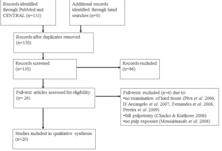
The extended literature search is presented in fig 1. Twenty articles in full text were included in the extended literature. Hand search-ing of the reference lists of the reviews and articles included in this extended literature search did not result in any additional publica-tions. Table 2-4 shows the summary of data from the extended sys-tematic review, corresponding tables for the previously published systematic review can be found in paper I.
Figure 1 The extended literature search, reported according to
PRISMA (Moher et al. 2009).
Assessment of quality
The results from the published systematic review are combined here with data from the new literature search. One of the studies
included was judged to have a high quality (Olsson et al. 2005), six
were judged to be of moderate quality (Accorinte et al. 2008;
Hør-sted-Bindslev et al. 2003; Kiatwateeratana et al. 2009; Lu et al.
2008; Min et al. 2008; Silva et al. 2006) and the remaining 34
studies were considered to have a low quality. The large number of studies with low quality was predominantly due to deficiencies in the study design, sample size or the manner in which the micro-scopic evaluation had been executed or reported.
Study designs
The studies were heterogeneous regarding their designs and the pulp capping procedures. Sixteen were classified as being
prospec-tive and randomised (Accorinte et al. 2009; Accorinte Mde et al.
a 2 N 2 eq b 2 S n 1 S st (B C T S ti fr
P
M y re te A th b m p H re w b to al. 2006; De 2006; Kiatwa Nair et al. 20 2006; Sübay qual numbe but without 2008; Accori ouza Costa nat 2005; H 980; Parolia übay & Dem tudies, were Bhaskar et Clarke 1971; Takagi 1991; everal studie ional period rom one to 2Pulp cappin
Most studies ounger heal eports of pa eeth with carA variety of hough the m bonding mat materials ofte periods of sh However, the esulted in ha where bondi based materia o the bondin marco et al ateeratana et 008; Olsson et al. 199 er of studies, a randomis inte Mde et et al. 2001; Hebling et al a et al. 2010 mirci 2005; e solely obse al. 1972; C Cowan 196 ; Sari et al. 1 es had divid ds which me 20 teeth per
g procedur
used a dire thy teeth wi artial pulpot rious exposu materials w most frequen terials. The en resulted i horter than 3 e use of bon ard tissue fo ing material al, the calciu ng material i l. 2001; Elia et al. 2009; L et al. 2005; 5; Hørsted-, sixteenHørsted-, we sation proce al. 2005; 20 do Nascime al. 1999, Jer 0; Pereira et Turner et a ervational st Caicedo et a 66; Fitzgeral 1999; Stanleyded the samp eant that the observation
res
ect pulp cap ithout signs omy and a ures.
was used for nt were calc use of diffe in hard tissu 3 weeks rev nding mater rmation onl l was comp um hydroxid n all studies as et al. 200 Lu et al. 200 Sawicki et a Bindslev et ere prospect edure (Acco 008; Aeineh ento et al. 2 rrell et al. 1 al. 2000; Sü al. 1987). Th udies with s al. 2006; Ce d & Heys 19 y & Lundy 1
ple size into, ere were sam
al period. pping proced of caries, al few studies r the pulp c cium hydrox erent calcium ue bridges, t vealed no ha ials as the p y in single c pared to a c de based ma . 07; Iwamoto 8; Min et al al. 2008; Silv al. 2003) a
tive and con
orinte et al.
hchi et al. 20
2000; Ersin & 1984; Negm übay & Asci he remainde several singl ehreli et al. 991; Kashiw 1972). , up to, 6 ob mple sizes r dure, perform lthough ther were condu capping proc xide based o m hydroxide though obser ard tissue br pulp capping cases. In the calcium hyd aterial was su o et al. l. 2008; va et al. and an ntrolled 2008; 003; de & Ero-m et al. i 1993, er, nine le cases 2000; wada & bserva-ranging med in re were ucted in cedure; or resin e based rvation ridging. g agent studies droxide uperior
Table 2 Data from the extended literat
ure search re
garding studies using othe
r pulp capping materials
than bonding
or MTA, or
studyin
g cert
ain operative procedures.
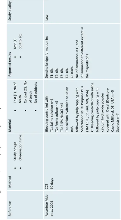
Refer ence Method Study design Obser vation time Material Test (T), No of teeth Cont rol (C ), N o of teeth No of subje cts Repo rted r esul ts Test (T) Control (C ) Study quality Acco rinte Mde et al . 200 5 CCT 60 d ays Bl eedi ng c ontrolled w ith T1 : Saline solution n= 5 T2 : Ferr ic sulfate n= 5 T3: 2. 5% N aOCl n= 5 T4 : c alc ium hy dr ox ide solution
n=5 followed by pulp capping with Scotchbond
M ulti P urpose P lus (3 M E SPE, St Paul, MN, USA ) C: B leeding c ontr
olled with saline
followed by pulp capping with calc
ium h ydroxide powder cove red wi th D yca l ( Dent spl y-Caulk, M ilford, D E, U SA ) n =5 Subjects n=? Dentine br idge form ation in: T1: 0% T2: 0% T3: 0% T4: 0% C: 10 0% No inflam ma tion in C, and inflam ma tion to di ffer ent extent in the m ajor ity of T Low
de L ourd es Rodrigues Acco ri nt e et al. 20 06 RCT 2 o bs ervation intervals : 3 0 a nd 6 0 days T1: adhesive resin, rubbe r dam n=10 C1: adhesive resin, no rubb er da m n=10 T2: Calc ium hy dr oxide powder cove red wi th D yca l ( Dentspl
y-Caulk, Milford, DE,
USA ), ru bber dam n=10 C2: Calc ium hy dr oxide powder cove red with Dyca l, no rubbe r dam n=10 Subjects n=? Dentine br idge form ation in: T1: 0 of 10 C1: 0 of 10 T2 : 10 o f 10 C2: 1 0 o f 1 0 No inflammation in T2 o r C2 and some i nfl ammat io n i n t he groups tr
eated with adhesive r
esin Low Parolia et al . 20 10 CCT 2 o bs ervation intervals : 1 5 a nd 4 5 days T: Pr opolis ( Ec uador ian R ainfor est LLC, U SA) mixed w ith e thyl a lco hol n=12 C1: ProRootMTA (Dentsply Caulk , Milford, DE, US A) n=12 C2: Dycal (D entsply Caulk) n =12 Subjects n=? At 45 da ys: T: 6 of 6 ha rd t issu e bri dg e C1: 6 of 6 ha rd ti ssue bri dg e C2: 5 of 6 ha rd ti ssue bri dg e Mor e i nfl am m ation in C 2 t han in T and C1 Low Kiatwateeratana et al . 200 9 RCT 6 m onths T: Emdogain ® Gel (B ior a AB , Malmö, Sw eden) n =1 5 C: Calcium hydroxi de mixed with water n =15 Subjects n=15 T: No com plete ha rd tissue bridge, infl ammat ion C: 10 of 13 co mpl et e ha rd t issue bridge, n o o r minimal infl ammat ion Moderate Olsson et al . 20 05 RCT 12 wee ks T: Emdogain ® Gel (B ior a AB , Malmö, Sw eden) n =9 C: Calcium hydroxi de mixed with saline n= 9 Subjects n=8 Com plete har d tissue br idge T: 0 of 9 C: 9 o f 9 Mor e i nflam m ation in T t han in C , though o ne tooth in C w as n ecrotic High
Table
3
Data from the extended literat
ure search r
egarding studies using
mineral trioxide
aggregate as
the pulp capping material.
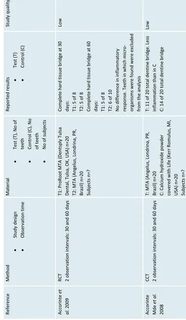
Ref er en ce Method Study des ign Obs er va tion ti me Material Tes t (T), N o o f teeth Co ntr ol (C), N o of teeth N o o f s ubjects Rep orted resu lts Test (T) Co ntr ol (C) St udy qual ity A cco ri nte et al. 2009 RCT 2 o bservatio n interva ls: 30 and 60 days T1: P roRo ot MTA ( Den tsp ly Tu lsa Dental , Tul sa, OK , US A) n=20 T2: MTA (A ngel us , Lo ndr in a, PR , Brazil) n =2 0 Subjects n=? Co mpl ete har d ti ss ue br id ge at 30 days : T1: 5 o f 8 T2: 5 o f 8 Co mpl ete har d ti ss ue br id ge at 60 days : T1: 5 o f 8 T2: 6 o f 1 0 N o di ff er ence i n i nf lammato ry res po ns e. T eet h i n w hi ch m ic ro-org ani sm s we re fo und w ere e xc lu ded
from the analysis
Low A cco ri nte Mde et al. 2008 CCT 2 o bservatio n interva ls: 30 and 60 days T: M TA (A ngelus , L ondr in a, P R, Brazil) n =2 0 C: Calc iu m h ydr oxid e p owd er co ver ed w ith Li fe (K er r R omul us , MI, USA) n=20 Subjects n=? T: 1 1 o f 2 0 t ota l d entine b ridge. Le ss inflammation than in C C: 14 o f 20 to tal dent in e br id ge Low
A cco ri nte et al. 20 08 CCT 2 o bservatio n interva ls: 30 and 60 days
T: MTA (Dentsply Cau
lk, Milford, DE, USA) n=20 C: Li fe (K er r, R omul us , MI, US A) n=20 Subjects : n=? A t 60 days: T: 5 o f 10 co mplete b ridge C: 6 o f 10 co mplete b ridge N o di ff er ence i n i nf lammati on between T and C Low Caicedo et al. 2006 Series o f c ases Typical ly a fter 6 m onths Pr oR oot MT A ( Den tsp ly Tu lsa Den tal, Tul sa, OK , US A) n=10 Subjects : n=12 7 o f 8 denti ne br id ge. The majo ri ty o f teeth s ho w inf lammati on. Low Iw am oto et al. 2006 RCT 136 +/- 24 days T: Wh ite P roRoot MT A ( paten t pendi ng) mi xe d w ith st er ile s ali ne n=22 C: Dycal (Dents pl y-Ca ul k, Mi lfo rd, DE, USA) n=23 Subjects n=? T: 20 o f 22 bridging, 17 o f 22 no su pe rfic ial in flam ma tion C: 18 o f 23 bridging , 14 o f 23 no su pe rfic ial in flam ma tion Low Min et al. 2008 RCT 2 mo nths T: MTA P roR oot ( Den tsp ly, Tu lsa, O K, USA) n=10 C: Dycal (Dents pl y-Ca ul k, Mi lfo rd, DE, USA) n=10 Subjects n=16 T: 9 o f 9 c ompl ete b rid ge C: 6 o f 10 co mplete b ridge No sig nif ic an t d iffere nc e in reg ar d to inflammation, thoug h 1 o f 1 0 i n C sh owed severe in fla mma tion Moderate Nair et al . 2008 RCT 3 o bservatio n interva ls: 1 w eek, 1 and 3 mo nths T: W hite M TA (P roro ot ® , Dentsply, Tul sa Dental , OK , US A) n=20 C: Dycal
® Ivory (Dentsply Caulk,
Milf ord , DE, USA) n =13 Subjects n=23 At 1 mo nth: T: 3 o f 6 co mpl ete br id ge C: 1 o f 5 co mpl ete br id ge At 3 mo nths : T: 4 o f 5 co mpl ete br id ge C: 0 o f 4 co mpl ete br id ge N o or mi ni mal inf lammati on in T, mo re inf lammati on in C Low Sawic ki et al. 2008 RCT 47-609 days, in average 138 da ys T: Wh ite P roRoot MT A ( Den tsp ly
Tulsa Dental, Tulsa, OK, USA) n=32 C: Lif
e n=16 Subjects n=23 T: 2 8 o f 3 0 co mpl ete denti ne b ri dg e and l es s i nf lammati on than in C C: 11 o f 14 co mpl ete denti ne br id ge Low
Table 4 Data from the extended literat
ure sear
ch regarding studies using bonding adhesives
as pulp capping mat
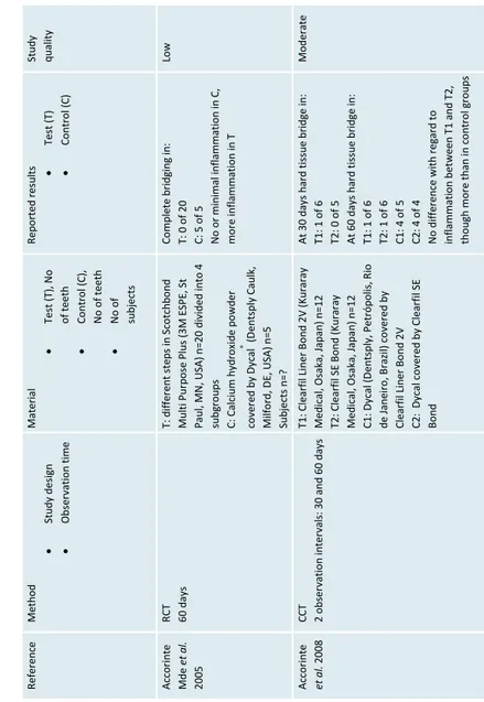
erials. Ref er en ce Method Study des ign Obs er va tion ti me Material Tes t (T), N o of teeth Co ntr ol (C), N o of teeth N o of subjects Rep orted resu lts Test (T) Co ntr ol (C) St udy qual ity A cco ri nte Mde et al. 2005 RCT 60 d ays T: d iffe re nt s teps in S co tchbo nd Mul ti Pur po se Pl us (3 M ES PE, S t Pa ul , MN, USA) n =20 d ivid ed in to 4 subgr oups C: Calc iu m h ydr oxid e p owd er co ver ed b y D ycal ® (D ents pl y C au lk, Milf ord , DE, USA) n =5 Subjects n=? Co mpl ete br id gi ng i n: T: 0 o f 2 0 C: 5 o f 5 N o or mi ni mal inf lammati on in C, mo re inf lammati on in T Low A cco ri nte et al. 2008 CCT 2 o bservatio n interva ls: 30 and 60 days T1: Clearf il Liner B ond 2V (Kuraray Medi cal , Os aka, Japa n) n= 12 T2: C lear fil S E B ond (K ur ar ay Medi cal , Os aka, Japa n) n= 12 C1: Dycal (Dentsply, Petrópolis, Ri o de Janei ro , B razi l) co ver ed by Cl ear fil Li ner Bo nd 2V C2: Dycal co ver ed by Cl ear fil S E Bon d A t 3 0 d ays h ar d t issu e b ri dg e i n: T1: 1 o f 6 T2: 0 o f 5 A t 6 0 d ays h ar d t issu e b ri dg e i n: T1: 1 o f 6 T2: 1 o f 6 C1: 4 o f 5 C2: 4 o f 4 N
o difference with regard to inflammati
on b etween T1 a nd T 2, tho ugh mo re than i n co ntr ol gr oups Moderate
Elias et al. 2007 RCT 2 o bservatio n interva ls: 30 and 90 days T: Cl ear fil S E Bo nd (K ur ar ay Medi cal , Os aka, Japa n) n= 16 C: Cal ci um hydr oxi de po wd er n=10 Subjects n=? A t 30 days denti ne b arri er form ati on in : T: 0 o f 8 C: 5 o f 5 A t 90 days denti ne b arri er form ati on in : T: 1 of 8 C: 5 o f 5
Less inflammation in C than T.
Low
Ersin & Eronat 2005
CCT 2 o bs er vati on i nter val s: 7 and 9 0 days T: P ri m e& Bo nd 2 .1 (C aul k, D ents pl y, Milf ord , DE, USA) n =10 C: Cal ci um hydr oxi de mi xe d w ith water n = 1 0 Subjects : n=? A t 90 days: T: 0 o f 5 d enti ne b ri dge, inf lammati on C: 5 o f 5 denti ne br id ge, no inf lammati on Low Lu et al. 2008 RCT 3 o bservatio n interva ls: 7, 30 an d 90 days T: Cl ear fil S E Bo nd (K ur ar ay Medi cal , Os aka, Japa n) n= 21 C: Dycal (Dents pl y, W eybr idge, UK ) n=20 Subjects n=27 A t 90 days: T: 0 of 7 fu ll b rid ge C: 5 of 6 fu ll b rid ge N o di ff er ence i n i nf lammati on between T and C Moderate Silva et al . 2006 RCT 4 o bservatio n interva ls: 1, 3, 7 and 30 days T1: 37% pho spho ri c aci d n=26 T2: 10% pho spho ri c aci d n=26 fol low ed by pul p cap pi ng w ith Si ngl e B ond A dhes ive S ys tem (3M Dental Pr oducts , S t P aul , MN , US A) C: Calc iu m h ydr oxid e p owd er co ver ed w ith Dycal (Dents pl y Ind e Com L tda , R J, B razil) n=2 6 Subjects n=? At 30 days denti ne br id ge depo si tio n in: T1: 0 o f 5 T2: 0 o f 5 C: 5 o f 5 So mew hat mo re inf lammati on in T1 and T2 t han i n C Moderate Sű bay & Demirci 2005 CCT 40 d ays T1: S co tchbo nd M ul ti P ur po se P lu s (3M ES PE, S t Paul , MN , US A) n=10 C: Dycal ® (Dents pl y C aul k, Mi lfo rd,
DE, USA) n=6 Subjects
n=4 Den tin e b rid ge in : T: 0 o f 1 0 C: 3 o f 6 More in flam m ati on in T th an C. Low
Concluding evidence grade
Combining the results from the published systematic review with the new literature search leads to these conclusions regarding the pulp capping materials:
The use of calcium hydroxide based materials as pulp capping agents frequently results in the formation of hard tissue covering the pulp exposure (limited scientific evi-dence).
The use of Emdogain®
Gel as a pulp capping agent does not result in the formation of hard tissue covering the pulp exposure (limited scientific evidence).
The use of bonding materials as pulp capping agent does not result in the formation of hard tissue covering the pulp exposure (limited scientific evidence).
Scientific evidence is lacking as to whether teeth treated with MTA as a pulp capping material show hard tissue formation more frequently than calcium hydroxide based materials.
Effect of EMDgel on experimentally performed pulp
exposures (II)
All subjects completed the study and no adverse events related to the treatment were recorded.
Symptoms and clinical assessment
No teeth were reported to give severe symptoms during the tele-phone interviews. The teeth treated with EMDgel gave fewer symp-toms compared to the teeth treated with calcium hydroxide. At the examination, just prior to extraction, all temporary fillings were judged to be adequate. Two teeth treated with EMDgel were sensi-tive to percussion. The reported symptoms in each tooth are shown in table 5.
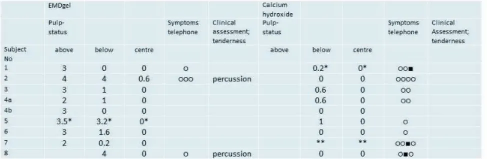
Table 5 Pulpal status with regard to inflammation, classified from 0 (None) to 4 (Abscess formation). The mean number of classifica-tions from the representative central secclassifica-tions (n = 5) from each ex-perimental tooth is given. The inflammation was judged above the exposure in the proliferated pulp tissue, directly below the expo-sure and in the centre of the pulp. Mild (•) and moderate (•) symp-toms reported at any of the telephone interviews after the pulp capping procedure. No severe symptoms were reported. At the clinical assessment only mild sensitivity to percussion was reported.
Pulpal status
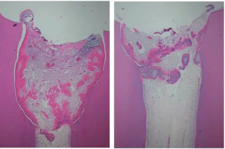
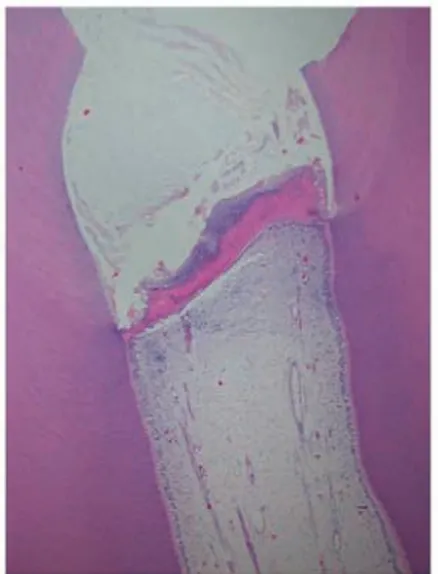
The pulp had proliferated into the space initially occupied by the EMDgel (fig 2). Therefore the soft tissue reactions were judged in three different areas of the pulp; the proliferated pulp, just below the amputation site and in the central part of the pulp. Within the proliferated pulp tissue there were areas where the inflammation was judged to be moderate or severe. One tooth showed an ab-scess. Two teeth showed abscesses or extended lesions not only lo-calized to the proliferated pulp. In the calcium hydroxide treated teeth there was no proliferation of the pulp tissue (fig 3). The ma-jority of teeth showed no or minimal inflammation, though one tooth exhibited total necrosis. The pulpal status in each tooth is shown in table 5.
Figure 2 Micrographs of teeth from subject no. 1 and subject no.
4a who twelve weeks earlier had received a partial pulpotomy and had been treated with EMDgel. Staining with haemotoxylin and eosin.
Figure 3 Micrograph of a tooth from subject no. 8 who twelve weeks before had received a partial pulpotomy and had been treated with calcium hydroxide. Staining with haemotoxylin and eosin.
Hard tissue formation
The hard tissue in the EMDgel treated teeth was not formed as a bridge covering the pulp tissue, but hard tissue was formed along-side the dentine walls which restricted the area which the pulp had proliferated into. There was also hard tissue formed in patches within the proliferated pulp tissue. In one tooth treated with EM-Dgel that showed an abscess, there was no hard tissue formed. In the calcium hydroxide treated teeth the hard tissue was formed as a bridge covering the pulp tissue.
The amount of hard tissue in the EMDgel treated teeth was consid-erably larger than in the teeth treated with calcium hydroxide. The thickness of the hard tissue bridge that had been formed in the cal-cium hydroxide treated teeth was on average 148 µm, though the dentine bridge was considerably less thick in the junction with the predentine of the primary dentine.
Localization of EMD
Detection of EMD was made in all EMDgel treated teeth. It was localized within the patches of newly formed hard tissue as well as alongside the dentine walls which restricted the area into which the pulp had proliferated.
Characterization of the newly formed hard tissue (III)
EMDgel treated teeth
In the teeth treated with EMDgel there was hard tissue formed alongside the dentine walls which restricted the area into which the pulp had proliferated, however this did not appear to be attached to the dentine wall. The areas corresponding to predentine in this hard tissue did appear to be continuous to the predentine and odontoblastic layer in the pulp. Staining with the DSP and Col I-specific antibodies revealed the proteins to be both present in the primary dentine and more abundantly in the predentine. They were also localized in diffuse areas of the newly formed hard tissue. The cells located in the close proximity to the hard tissue formed were also stained for DSP and Col I (fig 4, table 6).
Figure 4 Micrographs of a tooth from subject no. 7 who twelve weeks earlier had received a partial pulpotomy and been treated with EMDgel. (a) New hard tissue is observed alongside the ex-posed dentine surfaces and in isolated masses within the prolifer-ated pulp (arrows). A dentine chip (DC) is seen below the amputa-tion site. Staining with haematoxylin and eosin. (b) Detail of (a) at a higher magnification showing the dentine (D), predentine (PD), the hard tissue formed alongside the exposed dentine surfaces (HTA) and patches within the proliferated pulp (HTP). (c) DSP staining (brown) is observed in the newly formed hard tissue and is marked in areas corresponding to predentine (arrow). (d) Col I staining (brown) was observed in the newly formed hard tissue,
es-pecially in the areas corresponding to predentine (arrow).The
den-tine (D) and predenden-tine (PD) were used as positive controls for each antibody in each tooth section.
Calcium hydroxide treated teeth
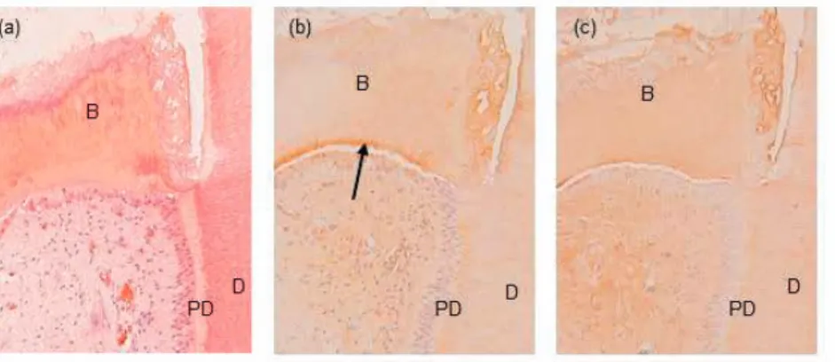
In the teeth treated with calcium hydroxide the same appearance regarding the staining with DSP and Col I in the root was seen; it was stronger in the predentine than in the primary dentine. Diffuse Col I staining was seen throughout the hard tissue bridge. The DSP staining was also observed in the bridge, though with a more patchy appearance and with the same intensity as seen in the pre-dentine areas. A layer of columnar cells, positive for DSP and Col I lined the hard tissue bridge (fig 5, table 6).
No bacteria could be detected in the wound area after staining with the modified Brown and Brenn technique or with the
LIVE/DEAD® BacLight™ Bacterial Viability Kit.
Figure 5 Micrographs of a tooth from subject no. 4a who twelve weeks earlier had received a partial pulpotomy and been treated with calcium hydroxide. (a) The hard tissue was formed as a bridge (B) covering the pulp. (b) DSP staining (brown) is observed in the bridge (B) and is marked in areas corresponding to predentine (ar-row). (c) Col I staining (brown) was observed in the bridge (B). The dentine (D) and predentine (PD) were used as positive controls for each antibody in each tooth section.