33
DOI: 10.2174/1874210601913010033, 2019, 13, 33-40
The Open Dentistry Journal
Content list available at: https://opendentistryjournal.com
CASE REPORT
Surgical Management of Granular Cell Tumor of the Orbit: Case Report and
Literature Review
Jahan Abtahi
*, Iman Malakuti and Aida Ajan
Department of Oral and Maxillofacial Surgery, Linköping University Hospital, SE-581 85 Linköping, Sweden
Abstract:
Introduction:
Granular Cell Tumors (GCTs) of the orbit are rare-entity soft-tissue tumors, and few reports have been published in the literature. The treatment of the choice is total excision. Early diagnosis prior to surgery is valuable for the distinction of malignant from benign tumor.
Case presentation:
We report a case of a 55-year-old woman with a solitary slow-growing mass in the right orbit with the involvement of the rectus inferior muscle, and present a review of the recent literature. The lesion had a diameter of 1 cm and was noticed 2 years before the examination. Excisional biopsy confirmed the diagnosis of GCT. The tumor was resected through a retroseptal transconjunctival approach. The final histological examination revealed findings characteristic of GCT, including positive reaction for protein S-100, SOX10, and calcitonin and negative reaction for desmin, myogenin, Smooth Muscle Antigen (SMA), Melan-A, and HMB-45. There were no signs of malignancy in this sample. Disturbance of motility was not noted by the patient after surgery.
Conclusion:
GCT should be included in the differential diagnosis of intraorbital lesions, particularly those that involve the orbit muscles. A biopsy is recommended before surgical resection, to exclude malignancy and prevent radical resection.
Keywords: Granular cell tumor, Orbit, Protein S-100, Inferior rectus muscle, Magnetic resonance imaging, Biopsy.
Article History Received: December 7, 2018 Revised: December 14, 2018 Accepted: December 27, 2018
1. INTRODUCTION
Granular Cell Tumor (GCT) is mostly a benign soft-tissue tumor with a Schwann cell phenotype that can arise in any site in the body [1]. The most common sites are arm, chest wall, and tongue [2]. GCT was described by Abrikossoff in 1926 as a tumor of the tongue with granular cells derived from striated muscle [3]. This tumor was initially called granular cell myoblastoma or myoblastic myoma due to the location. How-ever, this terminology is now considered to be controversial due to the histogenesis of the tumor [3].
The occurrence of GCT in the orbit is very rare, and to date, only a few cases have been reported in the literature in English. GCT of the orbit presents as a slow-growing, solitary, well-demarcated unilateral lesion. Common clinical manifestat-* Address correspondence to this author at the Department of Oral and Maxillofacial Surgery, Linköping University Hospital, SE-581 85 Linköping, Sweden; Tel: +46 101038551; Fax: +46 101038565;
E-mail: Jahan.abtahi@regionostergotland.se
ions are swelling and pain, alone or in combination [1]. The condition is most common in women between the fourth and the sixth decades of life [4]. The presenting symptoms are diplopia, proptosis, and ocular dysmotility [4].
GCT should be considered in the differential diagnosis of orbital mass, especially when the extraocular muscles are affected [1, 4, 5]. Involvement of ocular muscles and proptosis has been reported in 84.6% of orbital GCT cases [6]. Several imaging modalities have been used in patients with GCT of the orbit, including Computed Tomography scan(CT-scan) and Magnetic Resonance Imaging (MRI) [7, 8]. Radiological examinations reveal information such as tumor size, extent of the tumor, and metastatic spread [9]. Positron Emission Tomo-graphy and Magnetic Resonance Imaging (PET/ MRI) are more accurate than CT for the description of tumor invol-vement and soft tissue invasion [10]. However, microscopic analysis of the tumor is regarded as the gold standard for distinguishing between malignant tumor and benign tumor [11]. Although GCTs are mostly benign tumors, Malignant
differentiation of MGCTs from benign GCTs is crucial for treatment outcome. The most characteristic histological criteria for MGCT are mitotic activity, necrosis, increase in nuc-lear/cytoplasmic ratio, and pleomorphism [12].
Although GCT is a neoplasm derived from neural crest cells, many pathologists routinely employ immunohis-tochemical nuclear staining, such as for S-100 and Sox-10, for diagnostic purposes [13, 14]. The treatment of choice for orbital GCT is surgical excision. However, following surgery, the risk of diplopia is high due to damage to extraocular muscles [4, 5].
Here we present a case of orbital GCT with the involvement of the rectus inferior muscle, along with a review of the recent literature. The importance of excisional biopsy and immunohistochemical analysis for correct diagnosis and treatment strategies are also discussed in this article.
2. CASE PRESENTATION
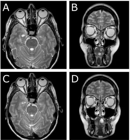
A 55-year-old woman was referred to the Department of Otorhinolaryngology, Head and Neck Surgery, Linköping University Hospital, Sweden, for evaluation of swelling at the lateral border of the right eye (Fig. 1a). No pain or visual disturbances were experienced by the patient. She had a 2-year history of swelling without any family history of malignancy or chronic disease. By palpation, a cartilaginous-like tumor with good motility and a smooth surface was felt at the conjunctive side of the right lower lid. Further investigation with slit-lamp biomicroscopy showed the presence of a subconjunctival mass adjacent to the right inferior rectus muscle. Force duction test did not reveal any restriction of muscle movement. Complete ophthalmological examination revealed intraocular pressure of 17 mm Hg on the right side and 15 mm Hg on the left side, with normal ocular motility. Visual acuity was 20/20 in both eyes. CT-scan showed a homogeneous soft-tissue mass of 14 × 14 × 8 mm in the right retrobulbar space, with bone erosion (Fig. 1b). Further MRI examination showed a solitary soft-tissue mass adjacent to the rectus inferior muscle (Figs. 1c &
d). Excisional biopsy was performed, and histological
examination identified the lesion as GCT (Fig. 2); H&E, magnification × 20). The histological pattern included lymphoid cells, fibrous stroma, and large polygonal eosi-nophilic cells with abundant granular cytoplasm and round nuclei. There were no signs of malignancy.
The tumor was resected through a transconjunctival approach using general anesthesia (Figs. 3a & 3b). The lower eyelid was everted using two stitches of 5-0 nylon through the tarsal plate. The incision was made using retroseptal approach through mucous membrane tissue in the right inferior fornix of the periorbital soft tissue (3 mm below the tarsus). The excised tumor measured 19 ×16 × 8 mm; it was firm and avascular (Figs. 3a & 3b). The tumor was infiltrated within the inferior rectus muscle, which made it difficult to excise completely.
Macroscopic appearance of the tumor revealed a gray-tan-colored to brownish mass measuring 19 × 16 × 8 mm (Fig. 3c). The specimen was fixed in 10% buffered neutral formalin, routinely processed, and embedded in paraffin wax. Sections were stained with Hematoxylin and Eosin (H & E). Final microscopic examination revealed a homogeneous population of large, round tumor cells with small nuclei and abundant eosinophilic granular cytoplasm infiltrating dense fibroco-nnective tissue (H & E, magnification ×40) (Fig. 3d). There were no signs of cell atypia, necrosis, or increased mitotic activity at higher magnification (H&E, magnification ×40) (Fig. 3d). On immunohistochemical staining, the granular cells were immunereactive for the proteins S-100 (Fig. 3e), SOX10, and calcitonin and also negative for desmin, myogenin, Smooth Muscle Antigen (SMA), Melan-A, and HMB-45. Proliferation activity of tumor cells for Ki67 was 1-2%.
Fig. (1). A-D. Clinical photograph and radiological imaging of patient
with granular cell tumor. A. Clinical photograph with swelling at the lateral border of the right eye. B. preoperative CT-scan. C and D. Preoperative MRI.
Postoperatively and until two months after surgery, the patient experienced diplopia in sight direction laterally downwards. The ophthalmological examination showed some reduction in elevation and lateral movement of the right eye. Six months later, MRI radiography revealed postoperative fibrosis with no signs of inferior rectus muscle entrapment (Figs. 4a & 4b). At 1.5-year follow-up, no diplopia was recorded. Ophthalmological examination revealed intraocular pressure of 15 mm Hg on the right side and 14 mm Hg on the left side, normal ocular motility, and visual acuity. In this case, no recurrence was noted at 1.5-year follow-up, either clini-cally or radiologiclini-cally (Figs. 4c & 4d).
Fig. (2). Histological image of the excisional biopsy (H & E, magnification ×20).
Fig. (3). A-E. A. Clinical photograph of surgical resection of tumor by the transconjunctival approach. B. Gross specimen of tumor mass. C. Gross
sections of tumor mass. D. Histological photograph of GCT (H & E, magnification ×40). E. Immunohistochemical staining with antibodies that were immune-reactive to S100.
3. LITERATURE REVIEW
A PubMed search was performed up until April 2018 on the terms “Granular Cell Tumor” and “Orbit”. Case reports written in English were included. Cases written in other languages and those with ocular adnexa involvement were
excluded.
Demographic data on age, gender, symptoms, duration of symptoms, ophthalmological findings and restriction in ocular movement were registered. We also analyzed treatment stra-tegies and recurrence rate. The present paper is authored as a narrative review contribution.
Fig. (4). A and B. MRI six months postoperatively. C and D. One-year follow-up with MRI.
3.1. Demographic Outcome and Clinical Manifestations
A PubMed search gave 35 articles in total. We excluded five articles (six cases) due to being written in German. Altogether, 30 articles (involving 38 cases, 15 males and 20 females with a mean age of 44.4 years (range 4-75 years) were included in the study. Demographic data are given in Table 1.
3.2. Treatment Strategies and Recurrence Rate
The different treatment strategies presented in previous studies included total resection, subtotal resection, biopsy and follow-up, and lastly additional treatment with radiotherapy and systemic chemotherapy. Recurrence rates for GCTs accor-ding to treatment, age, and site of origin are presented in Table
2. Table 1. Demographics and clinical features of GCTs.
Authors and Years
No. of Cases
Age Sex Symptoms Duration of
Symptoms
Ophthalmological Findings Restriction in
Ocular Movement
Benign/Malignant
Abtahi et al. 2018*
1 55 F Swelling Not stated Increased intraocular pressure No Benign
Yang et al. 2017 1 54 M Diplopia Sudden onset Ptosis Lateral Benign
Ullivieri et al.2017
1 36 F Diplopia, proptosis 1 year Ptosis Not stated Benign
Zhang et al. 2016 3 71 F Swelling 1 month Ptosis, lateral deviation of the globe
Not stated Benign
68 F Not stated Not stated Not stated Not stated Benign
15 M Headache, pain Sudden onset Not stated Not stated Benign
Yuan et al. 2016 1 37 F Diplopia, photophobia 6 months Ptosis, abnormal light reflex, visual acuity affected
All directions Benign
Morita et al. 2015 1 40 F Not stated Not stated Not stated Not stated Malignant
Germano et al. 2015
1 49 F Diplopia, proptosis, exophthalmos
2 months Ptosis, partial occlusion of the lid, visual acuity affected
Authors and Years
No. of Cases
Age Sex Symptoms Duration of
Symptoms
Ophthalmological Findings Restriction in
Ocular Movement
Benign/Malignant
De La Vega et al. 2015
1 37 M Diplopia, headache 2 months Ptosis, lid retraction Elevation and depression
Benign Salour et al. 2013 1 50 F Diplopia,
displacement of the eye globe
4 years Ptosis, visual acuity affected, afferent pupillary defect
Elevation and depression
Benign
Fernandes et al. 2012
1 53 F Proptosis Slow progression Upward displacement of the eye globe
Not stated Benign Ribeiro et al.
2012
1 74 M Diplopia, proptosis, ptosis
3 years Ptosis, superior displacement of the eye globe
Elevation, abduction, adduction Benign Guerriero et al. 2011
1 65 F Sudden blindness 4 years Ptosis, no light perception, forward displacement of the
eye globe
All directions Benign
Poyraz et al. 2009 1 53 F Diplopia, swelling Long-standing Superior displacement of the eye globe
Elevation Benign Golio et al. 2006 1 49 F Proptosis,
displacement of the eye globe
2 years Visual acuity affected Elevation Benign
Ahdoot et al. 2005
1 56 F Asymptomatic Not stated Choroidal striae Elevation Benign
Callejo et al. 20001
1 72 M Diplopia, proptosis Not stated Ptosis, visual acuity affected Abduction and adduction
Malignant Allaire et al. 1995 1 35 F Diplopia, swelling 2 months Swelling of lower lid Elevation and
depression
Benign Rodriguez et al.
1993
1 56 M Diplopia 1 year Limitation of downward
vision, torticollis
Elevation and depression
Benign McNab et al. 1991 4 4 F Proptosis left
divergent squint
3 months Ptosis, visual acuity affected, reduced color perception
Elevation and adduction
Benign
27 F Ptosis 1 year Ptosis No Benign
54 M Diplopia, displacement of the
eye globe
10 months Hypertropia Depression Benign
37 M Diplopia 8 months Ptosis, visual acuity affected, lateral displacement of the eye
globe
Abduction Benign
Moseley et al. 1991
4 36 M Diplopia, pain 1 year Ptosis, afferent pupillary defect and edema
No Benign
51 M Diplopia, swelling 1 year Palpable mass in lower lid, superior displacement of the
eye globe
Not stated Benign
29 F Ptosis, lid lag 1 year Not stated Not stated Benign
4 F Proptosis, left divergent squint
Rapid progression Visual acuity affected Not stated Benign
Jaeger et al. 1987 1 71 F Swelling 1 month Not stated Not stated Benign
Dolman et al. 1987
1 44 M Diplopia 11 months Visual acuity affected, superior displacement of the eye globe
Elevation, abduction
Benign Ueda et al. 1986 1 18 F Diplopia, pain,
exophthalmos
2 years Ptosis, visual acuity affected Not stated Benign Singleton et al.
1983
1 42 F Diplopia,
displacement of the eye globe
2 years Not stated Not stated Benign
Karcioglu et al. 1983
1 65 F Proptosis, visual acuity affected
2 years Visual acuity affected, superior displacement of the eye globe
Downward and inferior
Benign Goldstein BG et
al. 1982
1 51 F Diplopia 1 months Visual acuity affected, superior displacement of the eye globe
Downward Benign
Drummond et al. 1979
1 43 F Proptosis, visual acuity affected
3 months Visual acuity affected, anterior displacement of the eye globe
No Benign
Müller et al. 1978 1 75 M Left-sided deafness, loss of sight
6 years Left-sided blindness, atrophy of optic nerve
Not stated Malignant
Gonzalez et al. 1975
1 8 M Epiphora,
conjunctival hyperemia, pain
7 months Visual acuity affected Not stated Benign
Shang-Hsien et al. 1955
1 23 M Diplopia, proptosis, headache
2 months Visual acuity affected, upper lid dropped Downward, outward Benign Dunnington et al. 1948 2 36 F Proptosis, Drooping of the lid
19 months Ptosis Upward Malignant
40 M Redness and swelling of the lid
Not stated Upward and lateral displacement of the eye globe
Not stated Benign *The present study
Table 2. Recurrence rate of GCTs according to treatment, age, and site of origin.
Variable Recurrence No.
(frequency)
No recurrence No. (frequency)
Unknown No. (frequency)
Total Length of Follow-up Treatment
Total resection Subtotal resection Other treatment* Biopsy and observation
3 (10%) 3 (3.7%) 2 (100%) 0 (0%) 19 (63.3%) 2 (2.5%) 0 (0%) No growth 1 (100%) 8 (26.7%) 3 (3.7%) 0 (0%) 0 (0%) 30 8 2 1 0-6 y 0-2 y 2 y 2 y Age, y < 35 ≥ 35 1/6 5/6 4/22 18/22 3/11 8/11 39 39 – Site of origin Inferior orbit Inferomedial orbit Inferolateral orbit Superior orbit Medial orbit Retrobulbar Orbital apex Inferior, medial, lateral orbit
Superiolateral orbit Orbital apex Orbital fissure 2 (18%) 1 (50%) 0 (0%) 1 (16.7%) 1 (25%) 0 (0%) 0 (0%) 0 (0%) 0 (0%) 0 (0%) 1 (50%) – 5 (45.5%) 1 (50%) 1 (33.3%) 4 (66.7%) 3 (75%) 3 (100%) 1 (100%) 1 (100%) 1 (100%) 1 (100%) 1 (100%) 0/2 7 (63.6%) 0 (0%) 2 (66.7%) 1 (16.7%) 0 (0%) 0 (0%) 0 (0%) 0 (0%) 0 (0%) 0 (0%) 1 (50%) – 14 2 3 6 4 3 1 1 1 1 1 2 unknown-6 y unknown-1 y unknown-4y unknown-2y 6m-8y 2-3y 2y 6m 15m 2y 2y 2y 9m Total 6 22 11 39 –
* Radiotherapy and chemotherapy in combination with surgical treatment.
4. DISCUSSION
We have presented a case of GCT within the right orbit in a 55-year-old woman, along with a review of 38 cases in the recent literature [4, 6, 14 - 37, 40]. In the case presented, the tumor was successfully resected with no recurrence or postoperative sequelae during the 18 months of follow-up. Physical examination revealed no other abnormalities or visual disturbances. Slit-lamp biomicroscopy showed a subcon-junctival mass adjacent to the right inferior rectus muscle. In this case report, a pre-surgical biopsy was necessary to differentiate between a benign and a malignant lesion. In the literature, other lesions such as lymphoma, lacrimal cyst, meningioma, schwannoma, and glioma might be considered as differential diagnoses to GCT in this area [4, 5]. In the present study, the patient was referred with a 2-year history of swelling at the lateral border of the right eye without any other symptoms. Previous case reports and case series on GCT of the orbit showed that the most common symptoms were diplopia (55%) and proptosis (21%), the mean duration of symptoms being 15.6 months (range 1-72 months) (Table 1).
MRI is superior to CT-scan for evaluation of ocular
muscles and soft tissue within the orbit in relation to the tumor mass [18]. The present case showed characteristic features of GCT on MRI: An oval lesion with circumscribed borders which was isointense to gray matter relative to extraocular muscles on T1-weighted images and hypointense on T2-weighted images relative to fat, and showed strong contrast enhancement. CT-scan and MRI reveal information such as tumor size, extent of the tumor, and metastatic spread. These modalities cannot differentiate between GCT and other benign or malignant tumors of the orbit.
Histological analysis of a tumor is regarded as the gold standard for distinguishing malignant tumors from benign. In the literature, malignant presentation accounts for appro-ximately.
1-3% of all GCTs [18]. Fanburg-Smith et al., put forward six histological criteria for malignant diagnosis: (1) necrosis, (2) spindling, (3) vesicular nuclei with large nucleoli, (4) increased mitotic activity (> 2 mitoses per 10 high-power fields at 200× magnification), (5) high nuclear to cytoplasmic ratio, and (6) pleomorphism [12]. The presence of three or more of these criteria confirms the diagnosis of malignant lesion. In the
case of pleomorphism only, without any of the other criteria, the tumor is classified as benign. We found a homogeneous population of large round tumor cells with small nuclei and abundant eosinophilic granular cytoplasm, infiltrating dense fibroconnective tissue. There were no signs of cell atypia, cell pleomorphism, necrosis, or increased mitotic activity. This is important to consider during the pre-surgical evaluation and highlights the importance of excisional biopsy in treatment strategies for GCT. In the present literature review, four of 39 GCTs in the orbit were malignant (10.5%), which is a considerably higher frequency of MGCT than previously reported from other sites of the body.
The immunohistochemical characteristics of GCTs have been discussed in the literature and their probable origin from Schwann cells has also been considered [8, 17, 38]. These tumors are positive for expression of protein S-100, which supports the neural origin of GCTs [19]. Furthermore, it has been found that the granular cells are also positive for inhibin-a (a peptide hormone that participates in the regulation of the pituitary gland) and CD68 (a macrophage marker) [39]. Expression of CD68 in GCTs is related to the intracytoplasmic accumulation of phagolysosomes.
Total surgical removal of the tumor with tumor-free margins is the optimum treatment option. This may be difficult to achieve in cases with extraocular muscle involvement. In the literature, the most affected extraocular muscles are the inferior rectus (26%) and medial rectus (38%) [4]. At the time of diagnosis, almost 80% of patients show diplopia, and after surgical resection, the diplopia persists in 73% of cases [4, 40]. Previous studies have shown a recurrence rate of 16% for all treatment approaches (Table 2). After total surgical resection and subtotal resection, the recurrence rate has been 10% and 37%, respectively [27].In the present case, MRI examination showed a solitary soft-tissue mass with involvement of the rectus inferior muscle. The surgical removal of the tumor in our case was challenging, due to the risk of permanent extraocular dysfunction. We removed the tumor with approx-imately 1 mm of the right lateral border of the inferior rectus muscle. This was necessary to avoid recurrence, and when there is diplopia various strabismus procedures would have to be used for primary ocular alignment.
MGCTs are rare, and only a few cases have been reported in the literature. Fanburg-Smith et al. presented 28 cases of MGCT and found local recurrences in 32% and metastases in 50% of the cases [12]. Eleven of the 28 patients with MGCT (39%) died of the disease after a median time interval of 3 years. Again, these findings highlight the importance of excisional biopsy for correct diagnosis and treatment strategies. In malignant cases, monotherapy with pazopanib (a tyrosine kinase inhibitor) in combination with radiotherapy and wide surgical excision of the tumor would be the treatment of choice [19]. However, more studies are needed to confirm these findings.
CONCLUSION
The occurrence of GCT within the orbit is a rare event and a PubMed search gave a total of 39 cases. GCT should be considered in the differential diagnosis of orbital tumors and a
biopsy will often be required to exclude malignancy. The choice of treatment for GCTs is complete surgical resection, and the recurrence rate is low.
ETHICS APPROVAL AND CONSENT TO PART-ICIPATE
Not applicable.
HUMAN AND ANIMAL RIGHTS
No animals/humans were used for studies that are the basis of this research.
CONSENT FOR PUBLICATION
Written informed consent was obtained from the patient for her anonymized information to be published in this article.
CONFLICT OF INTEREST
The authors declare no conflict of interest, financial or otherwise.
ACKNOWLEDGEMENTS
Declared none.
REFERENCES
Jaeger MJ, Green WR, Miller NR, Harris GJ. Granular cell tumor of [1]
the orbit and ocular adnexae. Surv Ophthalmol 1987; 31(6): 417-23. [http://dx.doi.org/10.1016/0039-6257(87)90033-6] [PMID: 30 39674] Evans M, Chang E, Yu DL, Rao NA. Granular cell tumour: A rare [2]
caruncle lesion. Br J Ophthalmol 2006; 90(2): 246-7. [http://dx.doi.org/10.1136/bjo.2005.083790] [PMID: 16424 546] Abrikosov AA. Über Myome ausgehend von der quergestreiften [3]
willkürlichen Muskulatur. Virchows Arch 1926; 260: 215-33. Ribeiro SF, Chahud F, Cruz AA. Oculomotor disturbances due to [4]
granular cell tumor. Ophthal Plast Reconstr Surg 2012; 28(1): e23-7. [http://dx.doi.org/10.1097/IOP.0b013e3182141c54] [PMID: 214647 81]
Callejo SA, Kronish JW, Decker SJ, Cohen GR, Rosa RH Jr. [5]
Malignant granular cell tumor metastatic to the orbit. Ophthalmology 2000; 107(3): 550-4.
[http://dx.doi.org/10.1016/S0161-6420(99)00135-9] [PMID: 107118 94]
Salour H, Tavakoli M, Karimi S, Rezaei Kanavi M, Faghihi M. [6]
Granular cell tumor of the orbit. J Ophthalmic Vis Res 2013; 8(4): 376-9.
[PMID: 24653826]
Morimura T, Hayashi H, Kohchi N, Ozaki I, Tani E. MR appearance [7]
of intraorbital granular cell tumor: A case report. Am J Neuroradiol 1991; 12(4): 714-6.
[PMID: 1882750]
Ghassibi MP, Ulloa-Padilla JP, Dubovy SR. Neural tumors of the [8]
orbit: What Is New? Asia Pac J Ophthalmol (Phila) 2017; 6(3): 273-82.
[http://dx.doi.org/10.22608/APO.2017157] [PMID: 28558180] Silva M, Zambrini EI, Chiari G, et al. Pre-surgical assessment of [9]
mandibular bone invasion from oral cancer: Comparison between different imaging techniques and relevance of radiologist expertise. Radiol Med (Torino) 2016; 121(9): 704-10.
[http://dx.doi.org/10.1007/s11547-016-0654-1] [PMID: 2726 2579] Huang SH, Chien CY, Lin WC, et al. A comparative study of fused [10]
FDG PET/MRI, PET/CT, MRI, and CT imaging for assessing surrounding tissue invasion of advanced buccal squamous cell carcinoma. Clin Nucl Med 2011; 36(7): 518-25.
[http://dx.doi.org/10.1097/RLU.0b013e318217566f] [PMID: 216370 51]
Masamatti SS, Gosavi AV. Histopathological study of malignant oral [11]
tumous: A five-year study. Int J Sci Stud 2016; 4: 30-4.
Fanburg-Smith JC, Meis-Kindblom JM, Fante R, Kindblom LG. [12]
Karamchandani JR, Nielsen TO, van de Rijn M, West RB. Sox10 and [13]
S100 in the diagnosis of soft-tissue neoplasms. Appl Immunohistochem Mol Morphol 2012; 20(5): 445-50.
[http://dx.doi.org/10.1097/PAI.0b013e318244ff4b] [PMID: 224953 77]
Rodríguez-Ares T, Varela-Durán J, Sánchez-Salorio M, Varela-Nuñez [14]
R, Capeans-Tomé C, Urdiales-Viedma M. Granular cell tumor of the eye (myoblastoma): Ultrastructural and immunohistochemical studies. Eur J Ophthalmol 1993; 3(1): 47-52.
[http://dx.doi.org/10.1177/112067219300300109] [PMID: 838 7364] Yang D, McLaren S, Van Vliet C, deSousa JL, Gajdatsy A. [15]
Progressive orbital granular cell tumour associated with medial rectus. Orbit 2017; 36(5): 356-8.
[http://dx.doi.org/10.1080/01676830.2017.1337181] [PMID: 287002 58]
Ulivieri S, Muscas G, Lavalle L, et al. Granular cell tumor of the orbit: [16]
Pathological features and treatment. J Neurosurg Sci 2017; 61(3): 342-3.
[PMID: 28417615]
Zhang ML, Suarez MJ, Bosley TM, Rodriguez FJ. Clinicopathological [17]
features of peripheral nerve sheath tumors involving the eye and ocular adnexa. Hum Pathol 2017; 63: 70-8.
[http://dx.doi.org/10.1016/j.humpath.2017.02.006] [PMID: 28 235631] Yuan WH, Lin TC, Lirng JF, Guo WY, Chang FP, Ho DM. Computed [18]
tomography and magnetic resonance imaging findings of intraorbital granular cell tumor (Abrikossoff’s tumor): A case report. J Med Case Reports 2016; 10(1): 119.
[http://dx.doi.org/10.1186/s13256-016-0896-5] [PMID: 271 76551] Morita S, Hiramatsu M, Sugishita M, et al. Pazopanib monotherapy in [19]
a patient with a malignant granular cell tumor originating from the right orbit: A case report. Oncol Lett 2015; 10(2): 972-4.
[http://dx.doi.org/10.3892/ol.2015.3263] [PMID: 26622607] Germanò D, Elbadawy HM, Ponzin D, Ferro D, Priore L. Surgical [20]
excision of orbital progressive granular cell tumour. Case Rep Ophthalmol Med 2015; 2015: 420490.
[http://dx.doi.org/10.1155/2015/420490] [PMID: 26090251] de la Vega G, Villegas VM, Velazquez J, et al. Intraorbital granular [21]
cell tumor ophthalmologic and radiologic findings. Neuroradiol J 2015; 28(2): 140-4.
[http://dx.doi.org/10.1177/1971400915576657] [PMID: 259 63156] Fernandes BF, Belfort Neto R, Odashiro AN, Pereira PR, Burnier MN [22]
Jr. Clinical and histopathological features of orbital granular cell tumor: Case report. Arq Bras Oftalmol 2012; 75(2): 137-9.
[http://dx.doi.org/10.1590/S0004-27492012000200014] [PMID: 227608 08]
Guerriero S, Giancipoli G, Sborgia A, Fiore MG, Rossi R, Piscitelli D. [23]
Orbital granular cell tumor in a patient with Churg Strauss syndrome: The importance of biopsy. Orbit 2011; 30(1): 30-3.
[http://dx.doi.org/10.3109/01676830.2010.535645] [PMID: 212810 77]
Golio DI, Prabhu S, Hauck EF, Esmaeli B. Surgical resection of [24]
locally advanced granular cell tumor of the orbit. J Craniofac Surg 2006; 17(3): 594-8.
[http://dx.doi.org/10.1097/00001665-200605000-00037] [PMID: 167702 06]
Ahdoot M, Rodgers IR. Granular cell tumor of the orbit: magnetic [25]
resonance imaging characteristics. Ophthal Plast Reconstr Surg 2005;
Allaire GS, Laflamme P, Bourgouin P. Granular cell tumour of the [26]
orbit. Can J Ophthalmol 1995; 30(3): 151-3. [PMID: 7627903]
McNab AA, Daniel SE. Granular cell tumours of the orbit. Aust N Z J [27]
Ophthalmol 1991; 19(1): 21-7.
[http://dx.doi.org/10.1111/j.1442-9071.1991.tb00317.x] [PMID: 16455 57]
Moseley I. Granular cell tumour of the orbit: Radiological findings. [28]
Neuroradiology 1991; 33(5): 399-402.
[http://dx.doi.org/10.1007/BF00598611] [PMID: 1749468]
Dolman PJ, Rootman J, Dolman CL. Infiltrating orbital granular cell [29]
tumour: A case report and literature review. Br J Ophthalmol 1987; 71(1): 47-53.
[http://dx.doi.org/10.1136/bjo.71.1.47] [PMID: 3028469]
Ueda N, Yoshida A, Yokota T, Mochizuki T, Fukunishi R. Granular [30]
cell tumor of the orbit. Appl Pathol 1986; 4(3): 179-85. [PMID: 3036188]
Singleton EM, Nettleship MB. Granular cell tumor of the orbit: A case [31]
report. Ann Ophthalmol 1983; 15(9): 881-3. [PMID: 6318639]
Karcioglu ZA, Hemphill GL, Wool BM. Granular cell tumor of the [32]
orbit: Case report and review of the literature. Ophthalmic Surg 1983; 14(2): 125-9.
[PMID: 6302618]
Drummond JW, Hall DL, Steen WH Jr, Maxey SA. Granular cell [33]
tumor (myoblastoma) of the orbit. Arch Ophthalmol 1979; 97(8): 1492-4.
[http://dx.doi.org/10.1001/archopht.1979.01020020154014] [PMID: 223530]
Müller W, Dahmen HG. Granular cell tumor of the optic nerve. [34]
Albrecht Von Graefes Arch Klin Exp Ophthalmol 1978; 207(3): 181-8.
[http://dx.doi.org/10.1007/BF00411052] [PMID: 213985]
González-Almaraz G, de Buen S, Tsutsumi V. Granular cell tumor [35]
(myoblastoma) of the orbit. Am J Ophthalmol 1975; 79(4): 606-12. [http://dx.doi.org/10.1016/0002-9394(75)90800-4] [PMID: 16 4119] Dunnington JH. Granular cell myoblastoma of the orbit. Arch [36]
Ophthalmol 1948; 40(1): 14-22.
[http://dx.doi.org/10.1001/archopht.1948.00900030017002] [PMID: 18110004]
Hsu SH, Fu KC, Pang SH. Granular cell myoblastoma of the orbit. [37]
Chin Med J 1955; 73(5): 436-40. [PMID: 13277044]
Rejas RA, Campos MS, Cortes AR, Pinto DD, de Sousa SC. The [38]
neural histogenetic origin of the oral granular cell tumor: An immunohistochemical evidence. Med Oral Patol Oral Cir Bucal 2011; 16(1): e6-e10.
[http://dx.doi.org/10.4317/medoral.16.e6] [PMID: 20526269] Le BH, Boyer PJ, Lewis JE, Kapadia SB. Granular cell tumor: [39]
immunohistochemical assessment of inhibin-alpha, protein gene product 9.5, S100 protein, CD68, and Ki-67 proliferative index with clinical correlation. Arch Pathol Lab Med 2004; 128(7): 771-5. [PMID: 15214825]
Poyraz CE, Kiratli H, Söylemezoğlu F. Granular cell tumor of the [40]
inferior rectus muscle. Korean J Ophthalmol 2009; 23(1): 43-5. [http://dx.doi.org/10.3341/kjo.2009.23.1.43] [PMID: 193374 79]
© 2019 Abtahi et al.
This is an open access article distributed under the terms of the Creative Commons Attribution 4.0 International Public License (CC-BY 4.0), a copy of which is available at: (https://creativecommons.org/licenses/by/4.0/legalcode). This license permits unrestricted use, distribution, and reproduction in any medium, provided the original author and source are credited.
View publication stats View publication stats