Swedi S h dent al journ al, S upplement 2 1 5, 20 1 1 . d oct or al di SS ert a tion in odont ol og y . mi K ael S one SS on malmö uni V er S it y malmö uniVerSity
MIKAEL SONESSON
ON MINOr SALIvAry
gLANd SEcrEtION IN
chILdrEN, AdOLEScENtS
ANd AduLtS
isbn/issn 978-91-7104-385-6 / 0348-6672 on min or gl and S ecretion in c hildren, adole S cent S and adul t SO N M I N O R S A L I V A R Y G L A N D S E C R E T I O N I N C H I L D R E N , A D O L E S C E N T S A N D A D U L T S
Swedish Dental Journal, Supplement 215 , 2011
© Copyright Mikael Sonesson 2011
Photo: Sara Schlyter, Illustrations: Bo Veisland ISBN 978-91-7104-385-6
MIKAEL SONESSON
ON MINOR SALIVARY
GLAND SECRETION IN
CHILDREN, ADOLESCENTS
AND ADULTS
This publication is also available online see: www.mah.se/muep
CONTENTS
PREFACE ... 9 ABSTRACT ... 10 POPULÄRVETENSKAPLIG SAMMANFATTNING ... 12 INTRODUCTION ... 14 Saliva ...14 Salivary glands ...14Salivary gland regulation...16
Salivary secretion ...16
Protective components in minor gland saliva ...17
Age-dependent differences in salivary secretion ...20
Measurement of minor gland secretion ...21
Final remarks ...21
AIMS ... 23
SUBJECTS AND METHODS ... 24
Subjects ...24 Papers I-II ...24 Papers III-IV ...25 Methods ...25 Papers I-II ...25 Papers III-IV ...27 Statistical methods ...29 RESULTS ... 31 Paper I ...31
Minor gland salivary secretion rates ...31
Paper II ... 32
Mucins in minor gland saliva ... 32
Paper III ... 33
Salivary IgA in minor gland saliva ... 33
Salivary IgA in whole saliva ... 33
Paper IV ... 33
Gp-340 in minor gland saliva ... 33
Gp-340 and total protein in whole saliva ... 36
Sialic acid in minor gland saliva ... 36
Sialic acid in whole saliva... 36
DISCUSSION ... 37
Methodological aspects ... 38
Minor glands ... 40
Secretion rates (Paper I) ... 40
Numerical density (Paper I) ... 41
Innate components in minor gland saliva (Papers II, IV) ... 42
Adaptive component in minor gland saliva (Paper III) ... 44
Whole saliva ... 45
Adaptive and innate immune components (Papers III-IV) ... 45
CONCLUSIONS ... 49 ACKNOWLEDGEMENTS ... 51 REFERENCES ... 54 PAPER I PAPER II PAPER III PAPER IV
PREFACE
This thesis is based on the following papers, which are referred to in the text by their Roman numerals.
I. Sonesson M, Eliasson L, Matsson L. Minor salivary gland secretion in children and adults. Arch Oral Biol. 2003;48:535-539. II. Sonesson M, Wickström C, Kinnby B, Ericson D, Matsson L. Mucins MUC5B and MUC7 in minor salivary glands of children and adults. Arch Oral Biol. 2008;53:523-527.
III. Sonesson M, Hamberg K, Lundin-Wallengren ML, Matsson L, Ericson D. Salivary IgA in minor-gland saliva of children, adolescents and young adults. Eur J Oral Sci. 2011;119:15-20. IV. Sonesson M, Ericson D, Kinnby B, Wickström C. Glycoprotein 340 and sialic acid in minor gland and whole saliva of children, adolescents and adults. In press, Eur J Oral Sci.
The papers I to IV are reprinted with kind permission from the copyright holders.
ABSTRACT
The minor salivary glands are of great importance for maintenance of homeostasis in the oral cavity. These glands continuously secrete substances which lubricate and protect the oral tissues, contributing to comfort and health. The minor salivary glands contribute approximately 7-8 per cent of the total volume of saliva. Flow rate and composition seem to vary according to anatomical location. Current knowledge about the minor salivary glands is derived primarily from studies on adults. The overall aim of this thesis was to study age-related changes in minor gland saliva, from childhood to adulthood. By increasing the knowledge of minor gland secretion, we hopefully better understand how different mucosal locations are lubricated and protected in individuals of different ages and various health statuses. The project comprises four papers.
In Paper I, the flow rate and numerical density of the labial and buccal minor glands of pre-school children, adolescents and adults were investigated. Saliva was collected on filter paper discs and the
flow rate was measured by the Periotron-method®
. The numerical density was assessed by PAS-staining.
Key findings: The flow rate of the buccal glands was significantly lower in children than in adults and the number of labial glands was significantly higher in children than in the other age-groups.
In Paper II, the composition of minor gland saliva of the three age groups (Paper I) was analysed (by ELISA-technique), with reference
to the mucins MUC5B and MUC7, representing some of the major components of innate salivary immunity.
Key findings: Children did not differ from adolescents and adults with respect to MUC5B content in labial gland saliva, but had less MUC7 than the adults. In the buccal gland saliva, detectable amounts of the mucins were found in only a few of the participants.
In Paper III, the content of the adaptive immune component (salivary IgA) in minor gland saliva of pre-school children, adolescents and adults was measured by the ELISA technique. The salivary IgA-concentration in whole saliva of the three age-groups was also estimated.
Key findings: The IgA-concentration was significantly lower in the labial glands and the whole saliva of the children than in the adults.
In Paper IV, age-dependent differences of other innate components were studied in pre-school children, adolescents and adults, by analysing the amount of glycoprotein 340 (gp-340) in minor gland and whole saliva, using the ELISA technique. The content of sialic acid, a common terminal structure of glycoproteins, was analysed using the ELLA technique.
Key findings: With respect to minor gland saliva, no differences were disclosed among pre-school children, adolescents and adults. However, the gp-340 content of whole saliva was significantly higher in the children than in the adults.
The above investigations of properties of minor salivary glands in children, adolescents and adults seems to be the first to present data on age-dependent variations in gland density and secretions from healthy individuals. The results show high gland density, mature innate immunity and an ongoing maturation of adaptive immunity in the saliva of children. The report provides a reference for further comparative studies on minor gland saliva of younger individuals in health and disease.
POPULÄRVETENSKAPLIG
SAMMANFATTNING
Det finns få studier om de små spottkörtlarna, men deras främsta funktion anses vara att skydda individen genom att utsöndra ett skyddande sekret som utgör en slags barriär mot främmande och sjukdomsframkallande substanser. Från observationer på vuxna är det känt att körtlarna producerar en rad antimikrobiella ämnen, tillhörande det specifika och det ospecifika immunförsvaret. Vidare anses de små körtlarna ha en smörjande effekt, vilket troligen har stor betydelse för välbefinnandet. Inga studier av unga växande individers sekretion från små salivkörtlar tycks finnas. Studierna inriktar sig främst på att öka kunskaperna om grundläggande egenskaper hos de små salivkörtlarna och olika immunologiska faktorer i saliven från dessa körtlar. Studierna baseras på obser-vationer på förskolebarn, tonåringar och yngre vuxna.
Avhandlingen söker svar på följande frågeställningar:
Studie I. Finns det åldersrelaterade skillnader i flöde från små salivkörtlar och finns det åldersrelaterade skillnader i antal småkörtlar?
Studie II. Finns det åldersrelaterade skillnader i mängden av framträdande ospecifika försvarskomponenter (mucinerna MUC5B och MUC7) i saliv från små körtlar?
Studie III. Finns det åldersrelaterade skillnader i koncentrationen av specifika försvarskomponenter (saliv-IgA) i småkörtel- och helsaliv?
Studie IV. Finns det åldersrelaterade skillnader i mängden av ytterligare en viktig ospecifik försvarskomponent (Gp-340) samt
mängden kolhydrater (främst sialinsyra) som företrädesvis sitter på ospecifika försvarskomponenter, i småkörtel- och helsaliven? Huvudfynden i studierna är:
· Barnen hade lägre flöde från små körtlar i kindslemhinnan jämfört med de vuxna. Vidare uppvisade barnen fler körtlar i läppen jämfört med de vuxna.
· Barnen och de vuxna hade samma innehåll av MUC5B, men barnen hade mindre innehåll av MUC7 i saliv från små körtlar i läppen. Endast ett fåtal individer uppvisade muciner i saliven från små körtlar i kinden.
· Barnen hade lägre koncentration av saliv-IgA i saliv från små körtlar i läppen och i helsaliv, jämfört med de vuxna.
· Barnen och de vuxna uppvisade liknande mängder av gp-340 och sialinsyra i småkörtelsaliven men barnen hade större mängd gp-340 i helsaliven. De vuxna hade större mängder av gp-gp-340 och sialinsyra i saliven från små körtlar i kinden jämfört med körtlarna i läppen.
Förskolebarnens småkörtelsaliv innehåller samma mängd av några viktiga komponenter tillhörande det ospecifika immun-försvaret som de vuxnas, medan det specifika immunimmun-försvaret tycks fortfarande vara under utveckling hos förskolebarnen. Vidare har förskolebarnen lägre salivflöde från de små körtlarna i kinden och tätare mellan körtlarna i läppen än de vuxna.
Genom denna grundläggande kunskap kan genomförandet av nya jämförande studier av hur saliven fungerar hos yngre medicinskt eller odontologiskt belastade individer och av omhändertagande av patienter med störningar i saliven utformas. Skillnader i salivsekretion mellan barn och vuxna är också viktiga att utreda bland annat som eventuell förklaringsmodell för åldersvariationer i hur orala sjukdomstillstånd mellan åldrarna uttrycks.
INTRODUCTION
Saliva
Saliva has multiple roles: it is important not only for protection of the oral cavity, but also for general health, digestion and well-being. Saliva is secreted by exocrine glands, anatomically located in different parts of the maxillofacial region: the two main sources are the major and the minor salivary glands. Whole saliva, or oral fluid, is a mixture of glandular secretions and gingival crevicular fluid.
Salivary glands
The major salivary glands are the parotid, submandibular and sublingual glands, which together produce more than 90 per cent of saliva (1). The parotid glands are bilateral, located anterior to the external auditory meatus; the saliva drains into the oral cavity
via ducts located buccal to the maxillary second molars. The
submandibular glands are located beneath the tongue, with ducts lateral to the lingual frenulum. The sublingual glands are also located beneath the tongue, anterior to the submandibular glands;
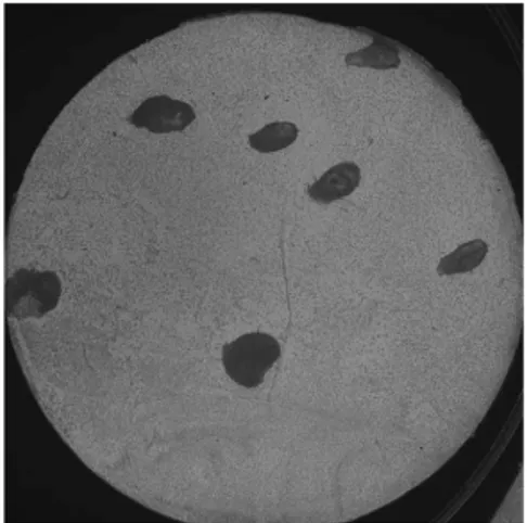
saliva drains into the oral cavity via ducts which terminate in rows
of minor orifices (Fig. 1) (2).
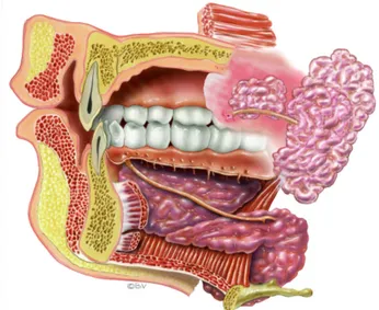
The minor salivary glands, also called mucosal glands, are located in the labial, buccal, palatal and lingual regions of the oral cavity and account for 7-8 per cent of the total volume of saliva (1, 3). The glands are surrounded by blood vessels, nerve and muscle fibres (1). Each individual gland comprises a cluster of cells connected by a duct to the oral cavity (Fig. 2). Like the major
glands, the minor salivary glands are formed during the first trimester of pregnancy: oral epithelium proliferates into the underlying ectomesenchyme and forms ductal and terminal secretory end pieces (1).
Similar minor exocrine glands are found in most mucosal
surfaces of the human body i.e. the eyes and the respiratory,
uterine, urinary and gastrointestinal tracts.
Fig. 1. Schematic picture of major salivary glands. Illustration: Bo Veisland.
Salivary gland regulation
Salivary secretion is influenced by several physiological conditions and is regulated by nervous systems (4), hormones (5) and neuropeptides (6). The salivary glands are innervated by the autonomic nervous system and both the major and minor glands are stimulated by parasympathetic nerve fibres of the seventh (facial) and the ninth (glossopharyngeal) cranial nerves. Unlike the minor glands, the major glands are also directly innervated by sympathetic nerve fibres from the second thoracic segment of the spinal cord (6-8).
When a neurotransmitter adheres to a receptor on an acinar cell,
e.g. when acetylcholine adheres to a muscarinic receptor, the gland
starts to synthesize isotonic primary saliva, by transforming capillary blood into interstitial fluid. As it flows through the glandular duct, the tonicity of the primary saliva is modified, from isotonic to hypotonic, before draining into the oral cavity (6, 7, 9-11).
Salivary secretion
The daily volume of secreted saliva is approximately 0.6 to 1.0 L in adults and 0.5 L in children (12-14). Saliva constantly bathes the oral cavity, lubricating and moistening the tissues and delivering components of the innate and adaptive immune system, such as mucins and immunoglobulins (15-17) (Table 1). Several of these components are incorporated into the pellicle on the hard and soft tissues (18, 19).
The saliva from major glands is rich in phosphate-binding proteins, buffering bicarbonates and calcium and has several functions considered to be important for the maintenance of homogeneity of dental tissues. Saliva is also important for the digestive process: mastication of food stimulates secretion of saliva from the major glands, causing an increase in salivary flow and in amylase concentration. The increased flow and the elevated amylase concentration facilitate clearance of food remnants and digestion, respectively (9).
The minor glands continuously secrete saliva (3) rich in organic substances such as proteins and glycoproteins (20-23) which have gel-forming properties and create a protective lubricating layer of
mucus (24-26), which is important for oral comfort. The composition of saliva from different mucosal areas seems to vary (21, 27), and the glandular cell types differs in different locations (28, 29).
Protective components in minor gland saliva
The adaptive component of the immune system in saliva consists of different classes of immunoglobulins (IgA, IgE, IgG, IgM), mainly immunoglobulin A (IgA) (20, 30, 31). The buccal glands are reported to have particularly high concentrations of IgA (21, 27). Innate immune protection is often mediated by various mucins or other classes of glycoproteins (24, 32), which bind and agglutinate
bacteria, mostly via their carbohydrate moieties. Glycoproteins
contains a large number of oligosaccharides covalently bound to the protein core, often exposing sialic acid as a terminal structure (33). Notably, these sialylated structures act as free radical scavengers and are also responsible for interactions with microorganisms and host ligands (34-36).
Analogous microbial interactions have been attributed to the salivary glycoproteins MUC5B and MUC7, due partly to the presence of sialic acids and fucose on the terminal part of their carbohydrate chains, making them receptors for micro-organisms (37-39). Glycoprotein 340 (gp-340), also known as salivary agglutinin, is one of the bacterial-binding glycoproteins (40, 41) and has been shown to bind to oral streptococci (42).
Mucins
All mucosal surfaces are coated with a film comprising different types of mucins. The main mucins in the oral cavity are MUC5B (formerly referred to as MG1) and MUC7 (formerly referred to as MG2) (26, 39, 43-45).
MUC5B is a high-molecular-mass oligomeric glycoprotein, with a molecular mass of up to 44 MDa (26), characterized by
serine-threonine-proline (STP)-rich domains, highly substituted with O
-linked oligosaccharides. The carbohydrates comprise almost 80 per cent of the weight and contribute to the structure of the molecule and protect the protein core against proteolysis (33). The carbohydrate structure and terminal epitopes of MUC5B have a
high degree of heterogeneity, which might have important implications for the biological properties of the molecule (37, 46). Between the carbohydrate fractions, the polypeptide backbone consists of unglycosylated hydrophobic cysteine-rich domains (47).
MUC5B is found on both soft (24) and hard surfaces (18, 48) and has lubricating properties, attributed to its water-binding capacity. It has been suggested that MUC5B acts as a matrix-retainer on the oral surfaces for other protective proteins such as IgA, lactoferrin and lysozyme, (49, 50), which facilitates the elimination of foreign substances and organisms.
MUC7 is a smaller unit, with a molecular mass of 150 to 250 KDa, also containing STP-rich domains substituted with approximately 70 per cent carbohydrates (39, 45). It is suggested that MUC7 interacts with oral micro-organisms (36, 51), through its ability to self-associate (52, 53), forming larger complexes which agglutinate and eliminate the micro-organisms.
Gp-340
Gp-340 is present in tears, respiratory tract fluids and in the gastrointestinal tract (54, 55). It is a glycoprotein with a molecular mass of 300-400 kDa and a carbohydrate content of approximately 45 per cent (40). Gp-340 seems to be identical to salivary agglutinin and shows great similarities with Deleted in Malignant Brain Tumours 1 (DMBT1), and is encoded by the same
gene, the dmbt1 gene (55, 56). It has been shown that gp-340 may
modulate the bacterial composition due to its ability to inhibit
colonization of Streptococcus mutans (42). Gp-340 also interacts
with other salivary components, e.g. salivary IgA, creating
complexes which agglutinate different types of bacteria (57, 58).
Glycosylation
Glycoproteins in humans consist of a protein core decorated with oligosaccharides (glycans). The oligosaccharides are covalently
attached, most often viaN- or O-linkages, to the protein core. The
N-glycans are covalently linked to an asparagine residue,
commonly involving an N-Acetylglucosamine (GlcNAc) residue
and the O-glycans are frequently linked via N-Acetylgalactosamine
the protein core. The O-linked oligosaccharides have three sections: core, backbone and peripheral regions. The core section consists of permutations of Gal/GlcNAc/GalNAc and the backbone of repeating sequences of Gal-GlcNAc (33). The peripheral region consists of different types of carbohydrates such as sialic acid, fucose, constituting sometimes blood group determinants, or sulfate (37, 38, 59, 60).
The proteins undergo glycosylation primarily in the rough
endoplasmic reticulum (N-linked) and Golgi apparatus of the cells
(O-linked). The oligosaccharide pattern differs, probably due to the
diversity in available glycosyltransferases (33).
Salivary IgA
IgA is involved in mucosal immunity on all the mucosal surfaces of the body (17), mostly in the form of secretory IgA, a dimer connected by a polypeptide (J chain) and surrounded by a proteolysis-resistant secretory component (SC). Monomeric IgA may also be present (mostly originating from the gingival exudate) (16, 61). Approximately 30–35 per cent of all salivary IgA in adult whole saliva is secreted by the minor salivary glands (20).
Salivary IgA binds to various micro-ogranisms, disrupting adhesion (61-63). The concentration seems to vary in different mucosal areas. Some studies have reported high concentrations in particularly buccal and palatal gland saliva in adults (21, 27).
The formation of IgA consists of a series of events. Briefly, the micro-organism is engulfed and digested into small peptides which are absorbed by specialized membranous cells (M cells) on the mucosal-associated lymphoid tissues (MALT) in tonsils and/or adenoids (Waldeyer´s ring) and in the small intestine (Peyer´s patches). After absorption, the substance is transported by antigen-presenting cells (APC) which process and present the antigen to regulatory T cells. These cells stimulate B cells to differentiate into precursors of IgA-secreting plasma cells which then differentiate into plasma cells, which produce IgA antibodies against the original antigen (17). Secretory IgA is then formed when the IgA molecule adheres to the secretory component in the membrane of the glandular cell, thus the molecule becomes more resistant to proteolytic degradation (17).
Age-dependent differences in salivary secretion
Saliva flow rate seems to increase in the growing individual (64), but for the ageing individual, there are divergent data: some studies report a decrease in flow rate in middle-age (65-67), while other investigators have failed to find any differences in rates between young and older adults (68).
Some of the whole saliva components are reported to change in concentration early in life. For example an increase of MUC5B and a decrease of MUC7 have been observed during infancy (69). In ageing individuals, however, the total quantity of mucins seems to decline (70, 71).
The salivary IgA-concentration in whole saliva and serum is reported to increase during infancy and childhood (30, 72, 73), reaching adult levels at four to twelve years of age (16, 74). In later life (60–80 yrs), a gradual decrease in concentration is reported (75-78).
Age, gender and site-dependent differences in minor glands
In adults, the data on minor salivary gland flow rates in relation to age are contradictory: some studies report comparable flow rates from minor glands in young and older individuals (21, 79-82), while others report a decrease with age (83, 84).
There are also conflicting reports about age-dependent differences in salivary IgA, the major adaptive immune component in saliva. Eliasson et al. (21) observed that the concentration in minor gland saliva was higher in older than younger subjects (> and <65 yrs respectively), while the opposite was reported by Smith et al. (83): lower values in older than in younger subjects (>55 yrs and 17-24 years respectively).
Minor salivary gland flow rates and IgA-concentration are reported to be lower in women than in men (21, 79, 84). However, some studies report comparable IgA-concentrations in men and women (83).
Furthermore site-dependent differences, with lower flow rates from labial than buccal glands have also been observed (21, 27, 79).
There are no corresponding studies of minor gland saliva in children. Thus little is known about the salivary secretion rates or
the innate and adaptive immune components of minor gland saliva in children, or in comparison with corresponding values in adults.
Measurement of minor gland secretion
Minor gland saliva is viscous and secreted only in small volumes. Sampling is thus a challenge. Estimations of salivary volume have been made by different absorption techniques, using filter paper discs (85, 86), synthetic discs (87) or surgical sponges (83) in combination with gravimetric, electromagnetic methods
(Periotron-method®
) or semiquantitative methods. Syringes and capillary tubes have also been used (88, 89). Volumetric measurements have also been made from photographs of droplets of saliva formed on mucosa, or saliva-coloured dots, depicting droplets of saliva, on chromatology papers (80, 90). For subsequent qualitative analysis of samples, various biochemical techniques are applied, such as radial immunodiffusion (RIA) (20, 85), enzyme-linked immunosorbent assay (ELISA) (27, 31) and sodium dodecyl sulfate polyacrylamide gel electrophoresis (SDS-PAGE) (25).
The Periotron-method®
was originally developed for measuring crevicular fluid (91). The method has high precision and reproducibility in estimating small volumes and has been modified for research on saliva (79, 81, 86, 92). Briefly, this electromagnetic instrument creates a voltage between two plates on the measuring instrument and measures the resistance of the salivary molecules in the filter paper, corresponding to a loss in voltage, and transforms the loss in voltage to relative values shown on the instrument display. These figures constitute the basis for calculation of the volumes secreted. The method provides both an estimation of flow rate by measuring the salivary volume in the filter paper and biochemical analysis of the salivary components collected on the paper. An important advantage of the technique is that the equipment is easy to handle and thus appropriate for studies of mucosal gland secretions in children.
Final remarks
Biological and pathological responses in children have frequently been extrapolated from the findings of studies on adults. As reported above, no published studies on minor salivary glands in
children, seem to be available. Documentation of minor salivary gland secretion in young individuals is fundamental to our understanding of the maturation of the salivary protective systems. Based on the hypothesis that the secretions change between childhood and adulthood, the present clinical investigations have been designed to study flow rate, gland density, and some of the major innate and adaptive immune components in the saliva of children and adults. The studies have been conducted under similar, controlled conditions, using the same methods. Furthering our knowledge of minor gland secretion should lead to improved understanding of the mechanisms by which the oral tissues are lubricated and protected in individuals of different ages and states of health.
AIMS
As data on minor salivary glands in children seem to be unavailable, the overall objective of this thesis was to investigate various salivary factors in 3-yr-old children, 14-yr-old adolescents and 20 to 25 yr-old adults. The specific aims were:
- To compare the secretion rate of labial and buccal glands in children and adults (Paper I).
- To compare the labial and buccal gland density in children and adults (Paper I).
- To compare the content of MUC5B and MUC7, commonly occurring components of the innate immune system, in labial and buccal gland saliva in children and adults (Paper II).
- To compare the concentration of salivary IgA, the major component of the adaptive immune system, in labial and buccal gland saliva in children and adults (Paper III).
- To compare salivary IgA-concentrations in whole saliva of children and adults (Paper III).
- To compare the content of gp-340, a bacterial-binding component of the innate immune system, in labial, buccal and whole saliva of children and adults (Paper IV).
- To compare the content of sialic acid, a terminal carbohydrate of glycoproteins, in labial, buccal and whole saliva in children and adults (Paper IV).
SUBJECTS AND METHODS
Subjects
Papers I-II
In all, 90 participants were recruited to the study. There were three groups: 3-yr-olds, 14-yr-olds and 20 to 25-yr-olds. Each age-group comprised 30 subjects, equal numbers of males and females. The subjects were healthy and not taking any medication at the time of examination, except for five adult females using contraceptives. Three subjects occasionally used antihistamines, but not during the week before they participated in the study. All subjects, with the exception of two Asian children, were of Caucasian origin. The 3-yr-olds and the 14-yr-olds were randomly selected from the patient stock at the School of Dentistry, University of Malmö, Sweden and the adult group comprised students at the University of Malmö.
Saliva sampling and measurements were carried out between February and July 2001. The participants were instructed, either directly or through their parents, not to eat, drink or brush their teeth for at least 1 h prior to sampling. In order to reduce the influence of circadian variations (75), all measurements were taken between 9 and 12 a.m. The adolescent and adult subjects were instructed not to use tobacco on the day of the appointment. Before study start, all participants of legal age, and the parents of the children and adolescents, gave written informed consent to participation.
Papers III-IV
Another saliva sample was collected, under the same conditions as for Papers I and II.
The subject selection procedure, instructions to the participants, saliva collection time and oral hygiene were the same as for the previous collections. Before study start, all participants of legal age, and the parents of the children and adolescents, gave written informed consent to participation. Eighty-seven of the 90 subjects were Caucasians and three, one in each age group, were of Asian origin. Saliva sampling and measurements were undertaken between March and July 2005.
Studies I-IV were approved by the Ethics Committee of the University of Lund, Sweden (Dnr. LU 437-00 and LU 766-02).
Methods
Papers I-II
Measurement of salivary secretion from minor glands
Unstimulated (resting) saliva was collected from the left labial and buccal mucosal areas of the oral cavity. In the labial mucosal area, measurements were made near the mid-line of the lower lip, halfway between the vermillion border and the vestibule. In the buccal mucosal area, measurements were made at the level of the parotid gland duct and approximately midway between the duct and the angle of the lips.
The mucosa in the area of measurement was gently dried with a small cotton pad and then immediately covered with a filter paper (SialoPaper 8 mm, Oraflow Inc., Smithtown, NY, USA), the buccal area for 5 s and the labial area for 15 s. To ensure mucosal contact, the paper was held under light finger pressure; the surgical glove on the examiner’s hand protected the paper. To measure the
volume of collected saliva, a Periotron 8000®
instrument (Oralflow Inc.) was used. Before measurement of the volume of saliva, the instrument was adjusted by placing the filter paper between the sensors. After harvesting, the paper was immediately placed in the device and the reading (0-199 units) was recorded. Secretion,
expressed as µl cm-2 min-1,was then calculated from the regression
formula y = 0.011x-0.036 where y is the volume in µl and x the
estimated by creating a standard curve made up of different
volumes of distilled water, aspirated by a Hamilton SyringeTM
(model 7001KH), onto filter papers. The formula describes a linear relationship between aspirated volumes and the readable digits. The reproducibility of the instrument was tested regularly during the saliva collection period.
Measurement of numerical density of minor glands
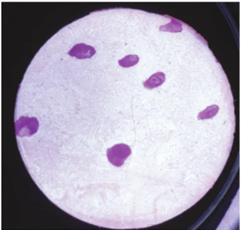
The number of active glands was assessed according to Gaubenstock et al. (90). After measurement of salivary secretion, the labial and buccal mucosa on the right side was dried with a cotton pad and covered with a pre-cut circular disc (Ø 10 mm) of chromatography filter (Millipore Corporation, Bedford, MA, USA). The disc was immediately removed and the mucosa was left exposed for 20 s, allowing droplets of saliva to form. A new disc was applied under light pressure and then immediately removed and transported to the laboratory for analysis.
The discs were submerged in 0.104 M periodic acid, followed by Schiff´s reagent and washed in 0.04 M sodium metabisulphite solution. The glycoproteins of the salivary droplets took on a dark red stain (Fig. 3). The number of secretory glands was assessed under light microscopy (16x) and expressed as the number of
secretory minor salivary glands per cm2.
Despite a number of pre-study tests of varying intervals for salivary droplet formation and different staining techniques, reliable numerical assessments were not achieved for the buccal mucosal area.
Fig. 3. Disc with salivary dro-plets treated with periodic acid and Schiff´s reagent, re-presenting gland density. Pho-to: Hans Herrlander.
Assessment of minor gland secretion of MUC5B and MUC7
After measurement of minor gland flow rate (Paper I) the filter papers were placed in Eppendorf tubes™ with 150 µl 4 M guanidinium chloride (GuHCl) and stored at below -80°C until biochemical analysis. Four papers from the labial and buccal mucosal areas, respectively, were used to obtain an adequate volume of saliva for analysis.
The amount of MUC5B was assessed by ELISA, using the polyclonal antiserum LUM5B-2. The antiserum was raised against a sequence within the cysteine-rich domain, in the central exon of MUC5B and outside the glycosylated domain, to avoid epitope shielding (93). The amount of MUC7 was estimated by the use of the polyclonal antiserum LUM7-1, raised against specific amino acid motif (94). The MUC5B used as a standard, was purified using density-gradient centrifugation of whole saliva and the MUC7 standard was also pooled out from a density gradient, although these fractions contained a mixture of proteins. The standard curves for MUC5B and MUC7 were made by serial dilutions and showed a linear relationship between the serially diluted standard and the absorbance.
Papers III-IV
Minor gland and whole saliva collection
In papers III and IV both minor gland saliva and whole saliva were collected.
Unstimulated (resting) minor gland saliva was collected from lower labial and buccal mucosal areas, as described above. The collection times were 120 s (labial) and 60 s (buccal). After volume measurements as described above, the filter papers were placed in Eppendorf tubes™ containing 150 µl phosphate buffered saline (PBS), pH 7.2 and stored at below -80°C until biochemical analysis. One paper per area was used. All samples were collected by the same investigator as in previous saliva sampling, assisted by a biochemical technician.
At the same appointment, minor gland saliva sampling was followed by collection of whole saliva. The participants were seated, leaning forward slightly, so that the saliva could drain passively for 1-5 min. into a polypropylene tube (Sarstedt,
Nümbrecht, Germany). Children sat in their parents’ laps. After collection, the samples were stored at below -80°C.
Assessment of salivary IgA, glycoprotein 340 (gp-340) and total protein in saliva
To assess the concentrations of salivary IgA in small sample volumes, a modified sandwich ELISA using polyclonal antibodies was carried out (95). The gp-340 content was determined through ELISA, using polyclonal antiserum (96). Total protein concentration of whole saliva was assessed by a Protein Assay (Bio-Rad). The protein assay is a colorimetric assay, based on a shift in color reflecting protein concentration. An absorbance microplate reader (ELx800; BioTek Instruments, Winooski, VT, USA) was used to measure the reactivity of the salivary IgA, total protein and gp-340. The results for salivary IgA and total protein were plotted against specific standard curves of colostrum or bovine serum albumin, respectively. Gp-340 was pooled out of a density gradient, containing a mixture of proteins, serially diluted and used as standard curve. Measurements were performed within the linear range of the standard curves.
Assessment of carbohydrates
Salivary samples were analyzed by ELLA, using a mixture of the
lectins Sambucus nigra, recognizing sialic acid (NANA-2-6) and
Maakia amurensis, recognizing sialic acid (NANA-2-3) (97). The results of carbohydrate reactivity were plotted against a standard curve made by serial dilution of purified MUC5B, analyzed for carbohydrate content, to check that the absorbance values were within the range of the standard.
In a supplementary analysis of whole saliva, fucose content was
analysed using the Ulex europaeus agglutinin I lectin, recognizing
fucose (α-fuc).
Recovery from the filter papers
To maximize the recovery of mucins, disulphide bonds were abolished by adding 10 mM dithiothreitol (DTT), in 6 M GuHCl, 0.1M Tris-HCl buffer (pH 8.0), for 1 h at 37°C, followed by
alkylation by addition of iodacetamide (IAA). The reduced samples were then centrifuged (18.000 x g, 10 min) and analyzed.
To estimate the recovery of mucins, equal volumes of whole saliva from one individual were placed on filter papers and in tubes containing 150 µl 4 M GuHCl. The filter papers were then placed
in the Periotron 8000® for volume assessment. The filter papers,
together with the saliva in the tubes, were then treated according to the above protocol. Recovery from the filter papers was 73 and 72 per cent for MUC5B and MUC7, respectively.
Pilot studies were also undertaken on salivary IgA, to reduce the absorption of the protein in the filter papers. The highest recovery was achieved by preparing the papers with 0.05% polysorbate 20 (Tween20) in PBS before use. 4M GuHCl was tested but was shown to interfere with the biochemical analysis. The recovery differed by only 1-2 per cent between saliva containing low and high concentrations of salivary IgA, respectively. The mean recovery was 35 per cent.
Statistical methods
Sample size calculation
As no data on salivary secretion and mucins in minor gland saliva in children were available, sample size calculation in papers I and II were based on an assumption that a mean difference between age-groups of approximately 50 per cent might be of clinical relevance. Assuming a standard deviation within the group of approximately 50 per cent and a power of 90 per cent at a significance level of 5 per cent, the sample size was estimated to n = 30. In Paper III a
mean difference in salivary IgA concentration of 3.5 mg 100 ml -1
was assumed to be clinically relevant. Assuming a SD of 4.0 mg
100 ml -1 within the age-groups, the resultant sample size, at 5%
significance and a power of 90% was calculated as n = 28. With reference to analysis of gp-340 and sialic acid in paper IV, there were no existing data on age-dependent differences. After pre-study tests, an assumption on sample size was made. To achieve a power of 90 per cent, the required sample size was calculated to be 25 individuals. A mean difference of 100 per cent between the age-groups and a SD of 100 per cent were chosen.
Descriptive data
The data were analyzed by the GraphPad Prism (GraphPad Software Inc. San Diego, CA, USA) version 4.0 software program and SPSS (SPSS, SPSS Inc., Chicago, IL, USA). For numerical data the arithmetic mean and standard deviations (SD) were calculated.
In papers I and II, a two-way analysis of variance, with comparisons by Tukey´s post-hoc test, was used to test the influence of the factors age and gender on secretion, the number of secretory glands and MUC5B and MUC7 content. Differences at the 0.05 level of significance were considered to be statistically significant. In Papers III and IV, a one-way analysis of variance was applied, with comparisons by Tukey´s test, to calculate the effects of age on the concentration of salivary IgA and total protein.
To investigate the effect of gender on salivary IgA-concentrations or the gp-340 and the sialic acid contents within each age-group
(papers III, IV), Mann-Whitney U-tests were used because of the
lower number of individuals in the subgroups.
Paired t-tests were carried out to analyze the intra-individual
differences in concentrations between the mucosal sites (papers I, III, IV). Intra-individual correlations were tested by Pearson´s
correlation test. P-values below 0.05 were considered to be
RESULTS
Paper I
Minor gland salivary secretion rates
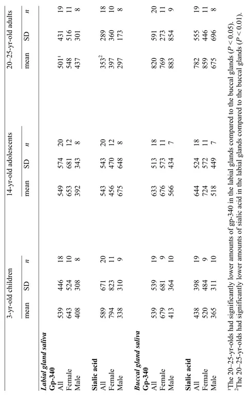
The flow rate from buccal glands was statistically significantly lower in the 3-yr-olds than in the 20 to 25-yr-olds. There were no age-dependent differences in the salivary secretion of the labial glands, nor were there any statistically significant intra-group gender differences with respect to either buccal or labial gland secretions. In all three age groups, the labial secretion rate was significantly lower than the buccal rate (Table 1).
Numerical density
The mean density of the labial secretory glands was significantly higher in the 3-yr-olds than in the 14 and the 20 to 25-yr-olds. Moreover, the density in the 14-yr-olds was significantly higher than in the 20 to 25-yr-olds (Fig. 4).
Fig. 4. Plots of numerical density of glands in the three age-groups (dark line = mean, glands/cm2). The
3-yr-olds had significantly higher numbers than the 14-yr-olds and the 20-25 yr-olds (P < 0.01).
Supplementary calculation of labial gland secretion rates in relation to gland density showed a significantly lower secretion rate per gland in the 3-yr-olds than in the adults. These calculations are based on the assumption of a bilateral symmetry of secretion from labial glands (82). No intra-group gender differences were disclosed.
Paper II
Mucins in minor gland saliva
No significant age-related differences in MUC5B content of the labial gland saliva were noted (Table 1). The subgroup 3-yr-old males had the lowest MUC5B content, significantly lower than that of the 3-yr-old females. However, compared to the adults, the labial gland saliva of the 3-yr-old group as a whole contained significantly less MUC7.
In contrast, analysis of buccal gland saliva disclosed measurable amounts of MUC5B in only nine 3-yr-olds, two 14-yr-olds, and none of the adults. Measurable amounts of MUC7 were found in only nine 3-yr-olds, three 14-yr-olds, and two of the adults.
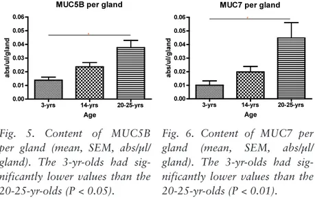
Assuming bilateral symmetry in secretion from labial glands, as mentioned above (paper I), further calculation of MUC5B and MUC7 in relation to numerical gland density showed a significant increase with age in both MUC5B and MUC7 secretion from individual glands (Figs. 5, 6).
*
*
Fig. 5. Content of MUC5B per gland (mean, SEM, abs/µl/ gland). The 3-yr-olds had sig-nificantly lower values than the 20-25-yr-olds (P < 0.05).
Fig. 6. Content of MUC7 per gland (mean, SEM, abs/µl/ gland). The 3-yr-olds had sig-nificantly lower values than the 20-25-yr-olds (P < 0.01).
Paper III
Salivary IgA in minor gland saliva
The concentration of salivary IgA in labial gland saliva was significantly lower in the 3-yr-olds (Table 1) than in the other age-groups. In the adult group, females had significantly lower concentrations than males.
In buccal gland saliva, no statistically significant inter-group differences in mean salivary IgA concentration were observed. In the adult group, the buccal gland concentration was significantly lower in females than in males.
Comparisons of salivary IgA concentrations in the buccal and labial glands revealed no statistically significant differences, except in the group of 3-yr-olds as a whole, where the concentration in the labial glands was significantly lower.
There were no statistically significant intra-individual correlations in salivary IgA between the different types of saliva in any of the age groups.
Salivary IgA in whole saliva
In whole saliva, the lowest concentration of salivary IgA was detected in the 3-yr-olds (Table 2); the difference between this group and both the older age-groups was statistically significant. Salivary IgA-concentration expressed as a ratio of total protein was also significantly lower in the 3-yr-olds than in the 14-yr-olds.
Paper IV
Gp-340 in minor gland saliva
The gp-340 content of labial and buccal gland saliva respectively, did not differ significantly among the age-groups (Table 1). This applied not only to the total group of subjects, but also to the subgroups of males and females.
Comparison of the gp-340 content of buccal and labial glands revealed a statistically significant difference in the adults, with statistically significantly higher values for the buccal glands.
There was no intra-individual correlation in the gp-340 content of the different types of minor gland saliva.
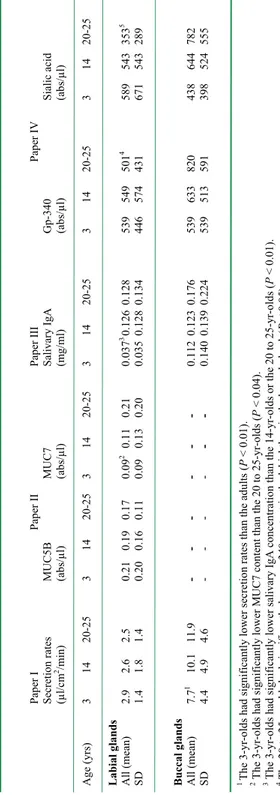
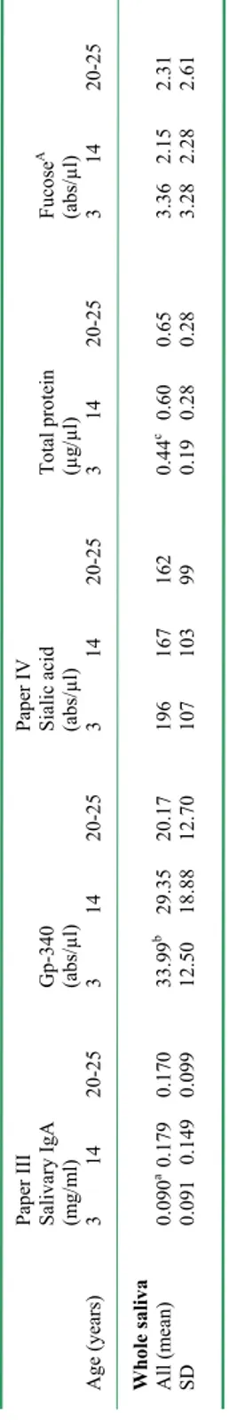
Tabl e 1. Sum m ary o f a ge-de pe nde nt fi nd in gs o n m inor sal iv ary gl an d co m ponent s (Pa pers I-IV ). Pap er I Pap er II Pap er II I Pap er IV Secr etion ra tes MU C5 B MU C7 Saliv ar y IgA G p-340 Sialic acid (µl/c m 2/m in ) (ab s/µl) (ab s/µl) (m g/ ml) (ab s/µl) (ab s/µl) Age (yr s) 3 14 20-25 3 14 20-25 3 14 20-25 3 14 20-25 3 14 20-25 3 14 20-25 Labial glands Al l (m ean ) 2. 9 2. 6 2. 5 0. 21 0. 19 0. 17 0. 09 2 0. 11 0. 21 0. 03 7 3 0. 126 0. 128 539 5 49 5 01 4 589 543 3 53 5 SD 1. 4 1. 8 1. 4 0. 20 0. 16 0. 11 0. 09 0. 13 0. 20 0. 03 5 0. 12 8 0. 13 4 44 6 57 4 43 1 67 1 54 3 28 9 Buccal gl an ds All (m ean) 7. 7 1 10 .1 11 .9 - - - - - - 0. 11 2 0. 12 3 0. 17 6 539 6 33 8 20 438 6 44 782 SD 4. 4 4 .9 4 .6 - - - - - - 0. 14 0 0. 13 9 0. 22 4 539 5 13 5 91 398 5 24 555 1 Th e 3-yr -o ld s h ad si gn ifi ca nt ly lo w er se cr et io n ra te s th an th e ad ul ts (P < 0. 01 ). 2 The 3-y r-ol ds ha d si gni fica nt ly lo wer M UC 7 cont en t t ha n th e 20 to 25 -y r-ol ds (P < 0. 04) . 3 Th e 3-yr-o ld s h ad si gn ificantly lo wer sali vary Ig A con cen tration th an th e 14 -yr-o ld s or th e 20 to 25 -y r-o ld s ( P < 0. 01) . 4 Th e 20 to 25-yr -o ld s h ad sign ifican tly low er g p-340 c on te nt in th e la bia l th an in th e bu cc al gla nd s( P < 0. 05 ). 5 The 20 to 25-yr-ol ds ha d significantly lowe r sialic acid co ntent in the la bial than in the buccal gla nds (P < 0. 01 ).
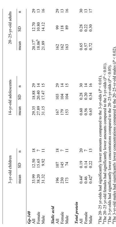
Tabl e 2. C om pone nt s of w hol e sal iv a (Pape rs II I-IV ) Pap er II I Pap er IV Saliv ar y IgA G p-340 Sialic acid To tal pr otei n Fu co se A (m g/m l) (abs/µl) (abs/µl) (µg/µl) (abs/µl) A ge (years) 3 14 20-2 5 3 14 20-2 5 3 14 20-2 5 3 14 20-2 5 3 14 20-2 5 Who le sa liva All (m ean) 0. 09 0 a 0.179 0 .1 70 33 .99 b 29 .3 5 20 .1 7 19 6 1 67 1 62 0. 44 c0. 60 0. 65 3. 36 2. 15 2. 31 SD 0. 09 1 0. 14 9 0. 09 9 12 .5 0 18 .8 8 12 .7 0 10 7 10 3 99 0. 19 0. 28 0. 28 3. 28 2. 28 2. 61 A Supplem entary analysis a T he 3 -y r-ol ds ha d si gn ifi ca nt ly lo w er c on cen tra tio ns o f saliv ary Ig A th an th e 20 -2 5 yr-old s (P<0.05). b The 3 yr-ol ds ha d si gni fica nt ly hi ghe r am ount s o f gp -3 40 th an th e 20 -2 5 yr-ol ds (P< 0. 01) . c T he 3 -y r-ol ds ha d si gn ifi ca nt ly lo w er c on cen tra tio ns o f total pr otein th an th e 20 -2 5 yr-old s (P<0.05).
Gp-340 and total protein in whole saliva
Compared with the older age-groups, the gp-340 content of whole saliva was statistically significantly higher in 3 yr-olds (Table 2). Moreover, the ratio of gp-340 to total protein in whole saliva was significantly higher in the 3-yr-olds than in the adults. The total protein concentration was significantly lower in the 3-yr olds than in the adults (Table 2).
Sialic acid in minor gland saliva
The sialic acid content of labial and buccal gland saliva respectively did not differ significantly among the age-groups (Table 1). This applied to analysis of the total age-group as well as to the subgroups of males and females.
Comparison of the sialic acid content of buccal and labial gland saliva revealed a statistically significant difference within the adult group, with statistically significantly higher values for buccal gland saliva (Table 1). No intra-individual correlations were observed in the sialic acid content of the different types of minor gland saliva.
Sialic acid in whole saliva
The sialic acid content of whole saliva did not differ significantly among the age-groups (Table 2). However, expressed as a ratio of total protein, a decrease with age was noted and the difference between the 3-yr-olds and the adults was statistically significant. When gp-340 was expressed as a ratio to sialic acid in minor gland and whole saliva, the proportion of gp-340 did not change.
Whole saliva was further analyzed with respect to the content of fucose, a common terminal carbohydrate of gp-340, revealing a trend towards higher values in the 3-yr-olds than in the other age-groups. Expressed as a ratio of total protein, statistically significantly higher values were disclosed in the 3-yr-olds than in the older age-groups (Table 2).
DISCUSSION
This series of studies was undertaken in order to produce scientific evidence to support clinical observations that the saliva coating the oral mucosa in young (pre-school) children is more viscous than in adults. A search of the literature for published studies on the secretion of the minor salivary glands disclosed that most of the available data originate from studies on adults, primarily on flow rates in the elderly and patients undergoing medical treatment. No clinical study on the secretion of minor salivary glands in healthy, growing individuals was found in the literature.
As described in the introduction, a variety of methods has been applied to determine the flow rates of minor salivary glands in adults. Most studies have been conducted on the readily accessible labial glands. However, because of the variety of methods used, it
is difficult to compare the results of the studies. The Periotron®
method is now applied in studies of salivary flow rates (21, 27, 79, 86, 92, 98). It is uncomplicated to use and involves no discomfort for the subjects.
As the youngest age-group in the studies, 3-yr-olds were selected on the basis of children's ability to comply during the measurement procedures: 3-yr-olds, but not younger children, are generally able to co-operate during an oral examination. The selection of 14-yr-old adolescents met the requirement of an age-group between childhood and adulthood, with an established permanent dentition. The 20 to 25 yr-olds represented young adults.
Methodological aspects
Collection of whole saliva from pre-school children is truly a challenge. It is difficult for young children to comply with instructions to refrain from swallowing the saliva and not to move the tongue or other muscles in the lower facial region. Thus the risk that the samples in fact comprised slightly stimulated, rather than resting saliva, could not be excluded, and this might have influenced the present results. This risk has also been discussed in studies on adults (99).
During measurement of minor salivary glands, the filter paper was held against a well-defined mucosal area, under fingertip pressure. To avoid contamination, the investigator wore a surgical glove, in accordance with Eliasson et al. (79). This method of assessing mucosal flow rates might involve some risk of glandular stimulation: it has previously been shown that mechanical stimulation increases flow rates from the glands (100). The magnitude of stimulation of minor glands is unknown, but as one investigator undertook all measurements, the degree of stimulation should have been constant throughout.
Whether the estimated flow rates using the Periotron® reflect the
“true” unstimulated secretion rate has been questioned. Compared to other studies, the values are relatively high, especially for buccal areas (101). It has been suggested that when different areas of the mucosa are being investigated, variations in density of minor glands throughout the mucosa might influence the flow rates (102). These authors also suggested that while the flow rates estimated by
the Periotron®-method should definitely not be extrapolated to give
the total salivary flow rate, the method was appropriate for comparative studies of flow rates at specific mucosal sites (102).
Associated with the filter paper method of sampling saliva is the risk of complex bindings between salivary components, or retention of salivary molecules within the filter papers. Different extraction buffers were used to improve recovery. To extract the glycoproteins, the disulphide bonds were abolished by DTT and to prevent the mucins from reclaiming their molecular structure, IAA was added (Paper II). To enhance recovery of IgA (Paper III), the papers were soaked in Tween20, thus saturating the binding areas and reducing the surface tension of the papers.
In the mucin study (Paper II), recovery was approximately 72 per cent, which is marginally lower than data on protein recovery (27, 98). In the IgA study (Paper III) the yield was lower (~35 per cent), but recovery from papers with low or high salivary IgA-concentrations showed negligible differences (1-2 per cent), thus making the comparisons between age-groups feasible: the primary aim of all the investigations was to make group comparisons.
Another potential risk associated with the use of filter papers is leakage of interstitial fluid through the epithelia onto the paper (103). However, as the epithelia of the lip and cheek consist of several dense cellular layers and basement membranes, it seems reasonable to assume that the volume of fluid passing out of the tissues is minimal.
In the studies of mucins and gp-340 (Paper II and Paper IV) the polyclonal antisera LUM5B-2, LUM7-1 and anti-gp-340 were used to detect MUC5B, MUC7 and gp-340, respectively (93, 94, 96). All antibodies were raised against specific peptide sequences in unglycosylated stretches of the glycoproteins. The specificity of the polyclonal antibodies and the treatment of the samples with different extraction buffers before analysis have reduced the risks of carbohydrate masking the mucins and gp-340. Polyclonal antibodies were also used in the salivary IgA study (Paper III). The antibodies were raised against peptide epitopes on the α1- and α2-chains on the heavy α2-chains of the IgA molecules. The use of polyclonal antibodies, interacting with several specific sites on the molecules, increases the chances of detecting the target molecules.
To our knowledge there are no previous studies on sialic acid in minor gland saliva. In some studies on agglutinins in minor gland
saliva, antibodies against e.g. specific carbohydrate epitopes, such
as sialic acid on the glycoprotein, were used (104, 105). This might indirectly describe the presence of sialic acid. Moreover, other components, such as secretory-IgA, contain sialic acid, but only in minor amounts; sialic acid has been shown to constitute the smallest part of the carbohydrates present in secretory IgA (106). To maximize the detection of sialic acid (Paper IV), a mixture of
the lectins Sambucus nigra and Maakia amurensis which recognize
Minor glands
Secretion rates (Paper I)
The secretion rate of buccal glands was significantly lower in 3-yr-olds than in adults. In all age-groups, the calculated values for flow rates in buccal glands were about three times as high as in labial glands.
The cause of age-dependent differences only in buccal secretion rates is not clear. One explanation for the lower rates in children might be an age-dependent difference in sensitivity to glandular stimulation: compared to adults, the flow rates of labial glands in 3-yr-olds might be stimulated more readily in response to application of the filter paper to the mucosa. This issue has been addressed in general discussions of advantages and disadvantages of filter paper measurements (80, 82).
Another explanation for the lower flow rate in children might be the lower numerical density of buccal glands. However, as the density of minor glands in the lower labial mucosa was higher in the 3-yr-olds than in the adults (Paper I), it is more likely that these
glands are not fully developed in young children, i.e. the glandular
acini and ducts are smaller and have yet to reach maximum secretory capacity (Paper I). This has been discussed for major glands (14).
The site-dependent differences in secretion rates is in line with a previous report (79) might reflect differences in gland density, secretion pattern or responsiveness to the measurement procedures in the different mucosal areas. These variations in minor gland secretion may reflect variations of protection in different regions of the oral cavity.
No differences in flow rates were observed with respect to gender. This finding is in accordance with a study by Won and co-workers (98). Other studies have reported lower secretion rates among women (79, 84). However, the group characteristics, in these two studies are different and the lower flow rates applied to women in the older age-groups.
In the present studies, the flow rates for adults are within the
range of earlier studies using the Periotron. However, the reported
data on labial flow rate vary five-fold (21, 79, 81, 82, 86, 98). There may be several explanations. The different measurement
techniques cause varying degrees of glandular stimulation. Moreover, even when the anatomical area to be measured is well-defined, the investigators may have applied the paper at different sites of the mucosal area; as discussed above, variations in the distribution of glands over the area may result in differences in flow rates. Seasonal variations may also have influenced the results: Kavanagh et al. (107) have reported seasonal fluctuations in the flow rates of whole saliva. However, as the measurements of flow rate in Paper I were carried out in accordance with some of
the previous Periotron-based studies (21, 27, 79), comparison of
the results of Paper I with these studies should disclose the process of maturation of minor gland secretion from childhood to old age (Table 1).
Numerical density (Paper I)
The numerical density of the labial glands decreased with age. In adults, the average density was approximately 5.4 secretory glands
per cm2. The density of secretory glands in the adolescents (8.2)
was also significantly lower than in the children (14.4), but significantly higher than in the adults. The values for adults are in accordance with previously presented data (80). Hypothetically, the higher degree of active secretory glands in children might be attributable to differences in sensitivity of the glands to stimulation, with higher sensitivity in children than in adolescents and adults, as discussed above. However, it seems more reasonable that the actual number of glands is constant and the decrease in numerical density is related to the general growth of the oral cavity, increasing the distance between the glands.
Using periodic acid-Shiff (PAS) staining it was not possible to calculate the numerical density of glands in the buccal area. This might be attributable to the low concentration of mucins in buccal gland saliva (Paper II): periodic acid oxidizes the carbohydrates on the glycoproteins collected on the papers, thus almost no mucins are available for staining. An immunohistochemical study by Riva et al. (29) showed that the buccal glands contain more sero-mucous cells than the labial glands; under such conditions, the saliva would be expected to have lower concentrations of glycoproteins as mucins. Also different concentrations of
Coomassie Brilliant Blue staining (108) were tested but no distinct dots were shown. Another explanation for the absence of distinct dots in the papers used in the buccal area could be the texture of the mucosa. The epithelium of the buccal mucosa is reported to be thicker than the labial mucosa, with more pronounced fissures (109), thus allowing the saliva to spread more extensively over the buccal area, before being soaked into the filter paper.
Innate components in minor gland saliva (Papers II, IV)
In labial gland saliva, the 3-yr-olds had approximately the same amount of MUC5B as the adolescents and adults, but significantly lower amounts of MUC7 than the adults (Paper II). With respect to the buccal glands, detectable amounts of MUC5B and MUC7 were observed only in a minority of the participants.
The results with respect to MUC5B from labial glands (Paper II) might imply that the secretion has reached adult levels as early as the age of three years. However, calculations of secreted amounts of MUC5B and MUC7 in relation to labial gland density revealed a significantly lower mucin secretory capacity of individual labial glands in the 3-yr-olds than in the adults. An age-dependent increase in mucin secretion by the individual gland seems reasonable, as gland density decreases and concurrently the total
area to be “lubricated and protected” increases: from 118 cm2 in
5-yr-olds to 215 cm2 in adults (14, 110). The finding that MUC7
content of labial gland saliva is lower in children than in adults suggests that MUC7 is produced primarily by the palatal, the submandibular and sublingual glands.
Detectable amounts of MUC5B in buccal gland saliva were found in only nine of the children, two of the adolescents and none of the adults; detectable amounts of MUC7 were found in nine of the children, three of the adolescents and two of the adults. These findings suggest that MUC7 secretion by buccal glands decrease with age in contrast to labial glands were the content of MUC7 increase with age. These results might indicate that the secretion of MUC5B and MUC7 differ between the different glands, during growth.
Only limited data are available about the MUC5B and MUC7 content of saliva from different types of minor glands. MUC5B has
been detected in palatal saliva and MUC7 in both labial and palatal gland saliva (22, 23, 25, 26, 46, 111, 112).
As mentioned above, mucins were detected in the buccal gland saliva of only a few subjects (Paper II): this indicates site-dependent differences in production of these glycoproteins. As the mucins are reported to have anti-microbial properties (36, 51, 113-115), it is reasonable to assume that intra-oral variation in bacterial
colonization might occur (116). Interestingly, in vitro studies have
shown that adsorption of MUC5B to different surfaces increases with increased concentration (18). This tends to support the above-mentioned concept of intra-oral variation in biological activities.
Gp-340 was detected in labial and buccal gland saliva (Paper IV), a finding consistent with earlier reports on minor gland saliva (105). Although no age-dependent differences were observed with respect to the gp-340 content of labial or buccal gland saliva (Paper IV), in the adult group the gp-340 content was greater in buccal than in labial saliva. This is not readily explained, but as mucins in buccal saliva were detected in only a few adults, the observation indicates a regional variation in glycoprotein secretion by the minor glands; the differences in composition provide protective properties specific to the area in which the minor gland is located. Moreover, agglutinins are more frequent in premolar than incisor pellicle (104); this supports the findings in Paper IV of higher gp-340 content of buccal than labial gland saliva. While the lower gp-340 content of adult labial saliva might imply a weaker defence against microbes in this area, the lower gp-340 content might be compensated for by higher content of other components, such as mucins (Paper II).
The sialic acid content of labial and buccal gland saliva did not differ significantly among the age-groups or between the subgroups of males and females. In adults, the buccal gland values were higher than for the labial glands. The MUC5B, gp-340 and sialic acid content (Paper II, IV) of labial gland saliva did not differ significantly between children and adults, which might indicate that the content of sialic acid reflects the total glycoprotein content of minor gland saliva. In buccal gland saliva, mucins were detected in only a few subjects, suggesting that the glycoproteins which accommodate the sialic acids, and also glycosylation, might vary.
In females, the glycosylation of glycoproteins might fluctuate, as the amount of carbohydrates has been shown to increase in whole saliva and in other mucosal secretions during ovulation (117, 118). In the present studies, the menstrual cycle in the female subgroups of adolescents and adults was not recorded and this may have influenced the results.
An increase of sialic acid in overweight and obese children and a decrease in individuals with gingivitis and periodontitis have been reported (119, 120). The physiques of the participants, determined from their weights, seem to be normal. However, the samples were not analyzed with respect to bacterial and bacterial enzyme content. As bacteria and bacterial neuraminidases hydrolyze sialic acid (120), a high content of bacteria and bacterial enzymes might have reduced the levels of bound sialic acid. On the other hand, the use of filter papers should have reduced microbial contamination of the minor gland saliva samples.
Adaptive component in minor gland saliva (Paper III)
The major finding was a lower concentration of salivary IgA in labial saliva of children than in adolescents and young adults (Paper III). No differences were observed between adolescents and adults. In the children, the concentration in the labial glands was lower than in the buccal glands. No such difference was observed in the other age-groups.
The results indicate that the major components of the adaptive salivary immune system in minor gland saliva are still under development in children but had reached maturity in adolescents. Similar findings are reported for tears (121) and whole saliva (16, 74). As mentioned above, there are conflicting data on IgA-concentration in the ageing individual (21, 83), but other immunoglobulins might develop differently; the IgG-concentration has been reported to be relatively unchanged during ageing (83). The IgG-concentration in minor gland saliva in children has yet to be determined.
No differences were observed in the IgA-concentration of buccal and labial gland saliva in adolescents and adults. There are previous reports of higher concentrations in buccal than in labial saliva of adults (27). As the concentration was also reported to
increase with age (21), one explanation for the differences between the present and previous reports on adults might be that the concentration in buccal gland saliva continues to increase with age, more so than in the labial saliva; compared with the subjects in the present studies, the participants in the previous studies were older and thus the site differences might be more pronounced. The higher IgA-concentrations in buccal than in labial gland saliva in the 3-yr-olds might be attributable to higher antigenic loading and antigenic stimulation (122) in the buccal than in the labial area, leading to an increase in concentration in buccal saliva.
In the present study, the intra-individual correlation of salivary IgA-concentration between different minor gland saliva was tested, but no correlation was found. Similar observations have been reported in studies on palatal and labial gland saliva, together with parotid saliva. One explanation proposed by the authors was differences in lymphocyte homing between the glands (31) which also might explain the results of the present study.
In the adult group, gender differences were observed for salivary IgA-concentration (Paper III): women had significantly lower concentrations than men in both labial and buccal gland saliva. This is in agreement with previous studies on minor gland saliva (21, 27), but in other types of secretions such as tears, no such gender differences in IgA concentration are reported (121). While all gender comparisons should be interpreted with caution because of the low number of subjects in the subgroups, the differences in the present study might reflect a hormonal influence on IgA secretion in minor glands.
Whole saliva
Adaptive and innate immune components (Papers III-IV)
The present study disclosed significantly lower IgA-concentration in whole saliva of children than in adolescent and adults, in accordance with the results for minor glands (Paper III). The concentration of IgA in saliva and serum increases rapidly during the first year of life (72, 123) and the increase in salivary IgA-concentration seems to continue until at least six to twelve years of age (16, 74, 78). Later, during ageing, the concentration in whole saliva seems to be unaffected or decreased (75).
The salivary IgA-concentrations in minor gland saliva and whole saliva were similar with those reported in a study on stimulated saliva by Eliasson et al. (27). Stimulation might have decreased the salivary IgA-concentration (124). However, whole saliva is a mixture of fluids, originating mainly from major and minor glands, but also harbouring not only a wide range of micro-organisms but also components of crevicular fluid and damaged mucosa, which together might have influenced the concentration. It is therefore difficult to compare concentrations found in whole and minor gland saliva.
The importance of salivary IgA in caries protection is controversial: there are as many studies reporting an anti-caries effect as there are studies reporting the opposite (125, 126). In young children, a maturation of the secretory immune system with impact on cariogenic microorganism, was seen during one year follow-up (127). However, IgA might have besides functions than caries protection, such as the defence of the upper respiratory tract (128). It has been shown that unstimulated (resting) saliva of children suffering from protein energy malnutrition has impaired immunological and agglutinating defence components, which might weaken defence against infections (72, 129). A decrease in IgA has also been reported among passive-smoking young children (130).
In the present study no statistically significant difference in salivary IgA-concentrations between genders was established, but there was a trend towards lower concentrations in women, which is in agreement with previous reports on IgA concentrations in saliva and serum, respectively (131, 132). There may be fluctuations in concentration in women; a correlation between estradiol and IgA concentrations in saliva has been reported (133). No data on the menstrual cycle were recorded in the present study, as mentioned above, but such a correlation might have influenced the results. The total protein content of whole saliva was significantly lower in the 3-yr-olds than in the adults, which is in agreement with the results presented by other observers (73).
The lower content of gp-340 in the whole saliva of adults but not in minor gland saliva might be a result of bacterial-specific binding or more thorough digestion than in children, as it is