ACTA UNIVERSITATIS
Digital Comprehensive Summaries of Uppsala Dissertations
from the Faculty of Science and Technology
1527
Studies of the two redox active
tyrosines in Photosystem II
Dissertation presented at Uppsala University to be publicly examined in Room 2001, Ångströmlaboratoriet, Lägerhyddsvägen 1, Uppsala, Wednesday, 14 June 2017 at 13:15 for the degree of Doctor of Philosophy. The examination will be conducted in English. Faculty examiner: Professor Robert Burnap (Oklahoma State University, USA).
Abstract
Ahmadova, N. 2017. Studies of the two redox active tyrosines in Photosystem II. Digital
Comprehensive Summaries of Uppsala Dissertations from the Faculty of Science and Technology 1527. 72 pp. Uppsala: Acta Universitatis Upsaliensis. ISBN 978-91-554-9933-4.
Photosystem II is a unique enzyme which catalyzes light induced water oxidation. This process is driven by highly oxidizing ensemble of four Chl molecules, PD1, PD2, ChlD1 and ChlD2 called,
P680. Excitation of one of the Chls in P680 leads to the primary charge separation, P680+Pheo-. Pheo
-transfers electrons sequentially to the primary quinone acceptor QA and the secondary quinone
acceptor QB. P680+ in turn extracts electrons from Mn4CaO5 cluster, a site for the water oxidation.
There are two redox active tyrosines, TyrZ and TyrD, found in PSII. They are symmetrically
located on the D1 and D2 central proteins. Only TyrZ acts as intermediate electron carrier
between P680 and Mn4CaO5 cluster, while TyrD does not participate in the linear electron flow
and stays oxidized under light conditions. Both tyrosines are involved in PCET.
The reduced TyrD undergoes biphasic oxidation with the fast (msec-sec time range) and the
slow (tens of seconds time range) kinetic phases. We assign these phases to two populations of PSII centers with proximal or distal water positions. We also suggest that the TyrD oxidation and
stability is regulated by the new small lumenal protein subunit, PsbTn. The possible involvement of PsbTn protein in the proton translocation mechanism from TyrD is suggested.
To assess the possible localization of primary cation in P680 the formation of the triplet state
of P680 and the oxidation of TyrZ and TyrD were followed under visible and far-red light. We
proposed that far-red light induces the cation formation on ChlD1.
Transmembrane interaction between QB and TyrZ has been studied. The different oxidation
yield of TyrZ, measured as a S1 split EPR signal was correlated to the conformational change of
protein induced by the QB presence at the QB-site. The change is transferred via H-bonds to the
corresponding His-residues via helix D of the D1 protein.
Keywords: Photosystem II, Tyrosine Z and D, proton-coupled electron transfer
Nigar Ahmadova, Department of Chemistry - Ångström, Molecular Biomimetics, Box 523, Uppsala University, SE-75120 Uppsala, Sweden.
© Nigar Ahmadova 2017 ISSN 1651-6214 ISBN 978-91-554-9933-4
List of Papers
This thesis is based on the following papers, which are referred to in the text by their Roman numerals.
I Sjöholm, J., Ho, F.*, Ahmadova, N., Brinkert, K., Hammarström,
L., Mamedov, F.*, Styring, S. (2016). The protonation state
around TyrD/TyrDin photosystem II is reflected in its biphasic
oxidation kinetics. Biochim. Biophys. Acta, 1858(2):147-155 II Ahmadova, N., Ho, M.F., Styring, S., Mamedov, F.* (2017)
Ty-rosine D oxidation and redox equilibrium in Photosystem II. Bi-ochim. Biophys. Acta, 1858(6):407-417
III Pavlou, A., Jacques, J., Ahmadova, N., Mamedov, F.*, Styring,
S.* The triplet state of the primary donor, P
680, in Photosystem II
is not formed by far-red light at 5 K; implications for the locali-zation of the primary radical pair. Manuscript
IV Ahmadova, N., Mamedov, F.* Formation of tyrosine radicals in
Photosystem II under far-red light illumination. Submitted man-uscript
V Ahmadova, N., Schröder, P.W.*, Mamedov, F. Role of the
PsbTn, a small luminal protein in Photosystem II, in the redox reactions of Tyrosine D. Manuscript
VI Ahmadova, N., Mamedov, F.* The donor-acceptor side
interac-tions in Photosystem II. Manuscript
Contents
Introduction ... 11
Solar energy... 11
Overview of Photosynthesis ... 13
Photosystem II: The biological machinery of water splitting ... 16
Protein constituents of Photosystem II ... 16
Redox-active cofactors of electron transfer chain ... 17
WOC – water oxidizing complex ... 20
TyrZ and TyrD: two redox active tyrosines in Photosystem II ... 22
Protein environment ... 22
Functional differences ... 23
Interaction of tyrosines with S-states of WOC ... 25
Methods ... 27
Electron Paramagnetic Resonance ... 27
Fluorescence ... 29
Thermoluminescence ... 30
The protonation state around TyrD/TyrD and redox equilibrium in Photosystem II ... 32
Far-red light triggered induction of P680+ and tyrosine radicals formation ... 41
Role of the PsbTn subunit in steering H+ from Tyr D ... 46
The Donor-Acceptor side interactions in Photosystem II ... 50
Sammanfattning ... 51
Xülasə ... 57
Acknowledgements ... 61
Abbreviations
ATP adenosine triphosphate Car β-carotene Chl chlorophyll Cyt cytochrome DCBQ 2,5 dichloro-1,4-benzoquinone DCMU 3-(3,4-dichlorophenyl)-1,1-dimethylurea DMBQ dimethyl 1,4-benzoquinone
DMSO dimethyl sulfoxide DQ duroquinone
EPR electron paramagnetic resonance
FNR FAD-containing ferredoxin-NADP+ reductase FTIR Fourier transform infrared
HQ hydroquinone
QM/MM quantum mechanics/molecular mechanics LHC light harvesting complex
NADPH nicotinamide adenine dinucleotide phosphate P680 the reaction center chlorophyll
PCET proton coupled electron transport Pheo pheophytin PpBQ phenyl p-benzoquinone PQ plastoquinone PQ-10 decylplastoquinone PSI photosystem I PSII photosystem II
QA primary quinone acceptor in photosystem II QB secondary quinone acceptor in photosystem II TL Thermoluminescence
WOC water oxidizing complex TyrD tyrosine 160 on the D2 protein TyrZ tyrosine 161 on the D1 protein
Introduction
The biological machinery to efficiently capture, convert and store solar energy in the chemical bonds of energy-rich molecules was invented by nature more than 3 billion years ago [2, 3]. This process, called photo-synthesis, led to the explosion of biological activity on Earth with an enormous diversity of living organisms. The accumulation of a large amount of biomass is an undeniable evidence of such event. The carbon stored in this biomass has provided us with fuels, food and other useful chemicals to maintain our everyday life.
Solar energy
Currently, the rate of global energy consumption is about 16.3 TW, 40 % of which used by the EU and the USA [4]. This value constantly rises owing to continuous industrialization and the increase of world popula-tion. It is projected to reach 20 TW or more by 2030, and more than double that by 2050 [5, 6]. At the present time, about 85 % of consumed global energy is derived from fossil fuels [4, 7]. The remaining 15 % is nuclear, hydroelectric, and renewable energy sources, such as wind, bio-mass, solar panels, etc. [4]. The availability of fossil fuels lead to the suppression of development non-fossil energy sources. Estimates of the global oil, gas and coal reserves indicate they are more than sufficient to meet energy demands for at least a century [8]. Therefore, the current problem in the global arena is not the future lack of fossil fuels but, rather the consequences of its usage [9]. If we burn all fossil fuel reserves, the total amount of CO2 accumulated in the atmosphere and oceans will be
equivalent to the level of CO2 which existed before life appeared on Earth
[10]. To tackle this problem, we have to develop new technologies based on principles that the energy source must be abundant, renewable, safe and clean. One such source available to us is solar radiation [11]. The solar energy reaching our planet annually is approximately 100 000 TW,
far exceeding our present energy demands. In other words, the solar en-ergy absorbed by the earth during about 2 hours is equal approximately to the energy used by mankind in an entire year [12]. Nature has mastered the task of solar energy conversion by the process of photosynthesis [13]. The biomass produced annually during photosynthesis is estimated to be more than 100 billion tons of dry weight, which corresponds to about 100 TW of energy [12]. The success of this natural phenomenon, photosyn-thesis, on such a significant scale is due to the infinite amounts of raw materials and energy required to drive it, i.e. solar energy, water and car-bon dioxide. The key of this process is the oxidation of water into oxy-gen, electrons and protons. The oxygen is released into the atmosphere and creates the living environment for all aerobic organisms. The elec-trons and protons extracted from water during photosynthesis are used to make energy rich molecules.
Our goal would be to make an artificial light-driven system that can convert solar energy into energy rich products. If we could succeed in mimicking photosynthesis in efficient artificial systems, it would assist in solving the energy demands of mankind. Artificial photosynthesis could provide fuels with high energy density such as hydrogen, methane etc., meanwhile significantly reducing the amount of CO2 in the
atmos-phere [12]. The more we learn about natural photosynthesis the greater possibility we will have in achieving success in making biomimetic ro-bust artificial systems.
Overview of Photosynthesis
Photosynthesis is a large-scale process of solar energy conversion into the energy rich chemical products in the biosphere [12]. Photosynthesis is carried out by both fundamental domains of life, bacteria and eukarya [14]. Bacterial photosynthesis, based on the released byproduct, is di-vided to two types: anoxygenic (non-oxygen evolving) and oxygenic (oxygen evolving). Oxygenic photosynthesis is performed by cyanobac-teria along with eukaryotic organisms: plants and algae. O2 as a
byprod-uct of this process is released into the atmosphere to support aerobic life [14]. Oxygenic photosynthesis is a complex process that can be simpli-fied to the following reaction:
6CO
2+ 6H
2O
hν
6(CH
2O) + 6O
2This reaction of photosynthesis occurs within the photosynthetic cells in specialized organelles known as chloroplasts (Fig. 1). Chloroplasts are comprised of two envelope membrane systems: the outer membrane and the inner membrane. The fluid-filled space of the chloroplast is called stroma. Within the stroma there is a third membrane system called thylakoid membrane. The thylakoid membrane is a network of flattened disks arranged in stacks called grana which are interconnected by the stroma lamellae membranes. The in-ner part of thylakoid membrane is called lumen.
Photosynthesis consists of two sets of reactions: the light dependent and light independent reactions. In oxygenic photosynthetic organisms, the light dependent reactions are performed by the chain of three large pro-tein supercomplexes embedded in the thylakoid membrane: Photosystem II (PSII), Cytochrome b6f (Cyt b6f) and Photosystem I (PSI) (Fig. 2).
The electron transfers through these supercomplexes was first proposed by Hill and Bendal and implemented in the form of a Z-scheme by Duysens (Fig. 3) [15, 16].
Figure 3. The Z-scheme of photosynthesis. The diagram indicates the major component of the light driven electron transport chain on an energy scale. The process is initiated by absorption of light by antenna complexes of the two pho-tosystems, PSII and PSI.
The light reaction of photosynthesis is initiated in PSII. After the light absorption the reaction center chlorophyll of PSII loses an electron to an electron transport chain. The electron generated by PSII is replaced in the process of water splitting. The electrons leaving PSII are shuttled to PSI via Cyt b6f complex. In PSI the low energy electrons are reenergized
and pass through electron transfer cofactors of PSI to reduce Feredoxin (Fd) in the stroma. Fd in turn passes an electron to the FAD-containing ferredoxin-NADP+ reductase (FNR) where the reduction of NADP+ to
NADPH takes place. As electrons pass through the electron transport Figure 2. Photosynthetic electron transfer chain of the thylakoid membrane.
chain, their energy is used to translocate protons from stroma to lumen forming transmembrane proton gradient. The transmembrane proton gra-dient drives the protein complex called ATP synthase to generate ATP. The light independent, or dark reactions, occur in the stroma of chlo-roplasts. These reactions are fueled by two molecules that are called the energy currency of the cells produced during the light reactions, NADPH and ATP. The dark reactions consist of several enzymatic steps collec-tively called Calvin-Benson-Bassham cycle to synthesize carbohydrates from CO2. The carbon fixation reactions represent one of the nature´s
Photosystem II: The biological machinery
of water splitting
Photosystem II is a unique oxygen-evolving enzyme which catalyzes the water oxidation reaction in the thylakoid membranes of oxygenic organ-isms [17]. Nature had to fulfill two imperative functional goals to enable living organisms to split water by sun light: (i) formation of light-induced oxidizing cofactors that could be a driving force for water splitting and (ii) assembly of a catalytic site where water splitting could occur step-wise to store charges and prevent the generation of harmful intermedi-ates. This chapter focuses on the overall structure of PSII and the electron transfer chain involved in oxidative water splitting in plants, algae and cyanobacteria.
Protein constituents of Photosystem II
PSII is a pigment-protein complex composed of more than 25 protein subunits, 105 chlorophylls, 28 carotenoids, chain of the redox cofactors and water oxidizing complex (WOC) per monomer [1]. Active PSII ex-ists in a dimeric form located in the stacked regions of grana thylakoids (Fig. 4A) [1, 18, 19].
PSII dimer is 105x205x110 Å with a molecular mass of 700 kDa. The top view of PSII dimer surrounded by pigment-antenna complexes is de-picted in Fig. 4B. The monomeric PSII core complex consists of four large intrinsic proteins (D1, D2, CP43 and CP47), twelve small mem-brane-spanning subunits (PsbE, PsbF, PsbH, PsbI, PsbJ, PsbK, PsbL, PsbM, PsbTc, PsbW, PsbX and PsbZ) and four extrinsic protein subunits (PsbO, PsbP, PsbQ and PsbTn) [1] . Most redox cofactors of PSII are housed by the D1 (psbA) and D2 (psbD) heterodimer. CP43 and CP47 are the inner light-harvesting antenna proteins of PSII, bind Chla and
Apart from the inner antenna complex, each PSII dimer is surrounded by number of periferal light-harvesting antenna complexes, denoted CP29, CP26, CP24 and LHCII trimers. Contrary to CP43 and CP47, LHCII, CP24, CP26 and CP29 bind not only Chla but also Chlb and xanthophylls
(lutein, violaxanthin and neoxanthin). The other twelve low-molecular intrinsic subunits surround the PSII core. Only two of them (PsbE and PsbF) bind the cofactor of the electron transport chain, namely the heme group of cytochrome b559 (Cyt b559) which plays an essential role in PSII
assembly and protection against photodamage [20, 21]. However, the re-maining small proteins were found to be an essential for assembly, sta-bilization and dimerization of the PSII core complexes (PsbH, PsbI, PsbJ, PsbK, PsbL, PsbM, PsbTc, PsbW, PsbX and PsbZ) [20].
The three extrinsic subunits, namely PsbO, PsbP, PsbQ cover WOC complex on the lumenal side. Their absence leads to the loss of water oxidation activity. The function of the forth extrinsic subunit, PsbTn was unknown at the time [1].
In addition to the above listed subunits, over 1300 water molecules were found in each PSII monomer, some directly interacting with WOC, as revealed in the 1.9 Å resolution structure from T. vulcanus [22].
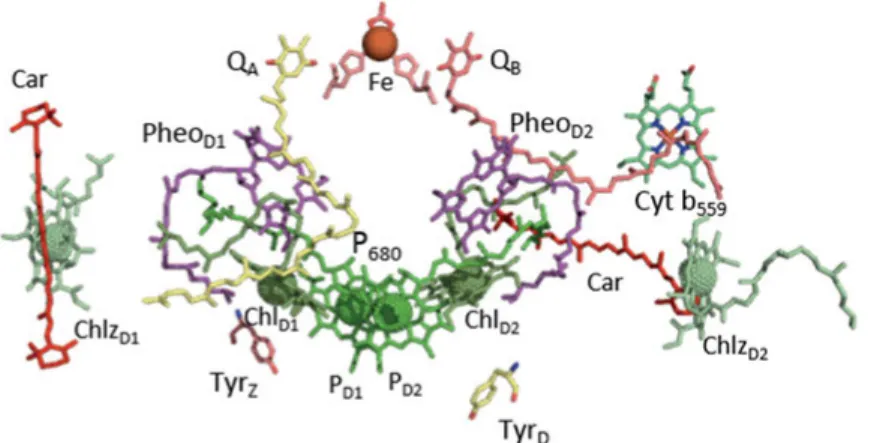
Redox-active cofactors of electron transfer chain
The D1 and D2 heterodimer houses the redox-active components in-volved in the charge separation, water splitting and reduction of the ter-minal electron acceptor of PSII, plastoquinone QB. The electron transfer
Figure 4. (A) Side view of the dimer of Photosystem II (PDB-4UB6, 1.9 Å res-olution) colored by monomers. (B) Top view of spinach PSII–LHCII supercom-plex (PDB-3JCU, 3.2 Å).
chain of PSII is divided into the donor and acceptor side (Fig. 5) [18]. The oxidation of water occurs at the donor side of PSII. The acceptor side is a part of the electron transfer chain from P680 to the terminal
elec-tron acceptor, plastoquinone QB. The acceptor side cofactors are
ar-ranged around the pseudo-two-fold axis of transmembrane helices of D1 and D2 proteins. The axis passes through the middle of Chl dimer PD1
and PD2 to the non-heme Fe. The primary and secondary quinones, QA
and QB are located on each side of the non-heme Fe on the D2 and D1
proteins respectively.
The light induced reaction starts from photoexcitation of antenna-pig-ment complexes (the peripheral and the inner antenna complexes) and exciton migration towards the reaction center Chls, P680. P680 is the
pri-mary electron donor of PSII which consists of 4 chlorophyll molecules PD1, PD2, ChlD1 and ChlD2. The PD1 and PD2 are positioned relatively close
to each other forming a weak excitonic interaction between their tetrap-yrolle head groups. After photoexcitation of one of the chlorophylls in P680, the reaction center loses an electron to the nearby Pheo. Pheo is a
chlorophyll molecule without a central Mg+2 ion in the porphyrin ring.
The primary charge separation between P680 and PheoD1 is a very rapid
process, happening in 3-4 psec. Reduced PheoD1- transfers an electron to
the immobile primary quinone acceptor, QA in 300 psec [23]. All these
steps are one electron transfer reaction. QA- in turn transfers an electron
to the secondary quinone acceptor, QB [17]. After two subsequent
elec-tron transfer reactions, QB gets protonated from the stromal side and
leaves the QB site, making place for the new quinol molecule to bind.
QBH2 diffuses towards the binding site on the Cyt b6f complex, and, upon
binding, transfers electrons to Cyt b6f and releases protons to the lumen,
thus contributing to the transmembrane proton gradient. PSII is the only known protein complex in nature capable of splitting water. The driving force of this energetically challenging reaction is P680+. The primary
do-nor P680+, has the highest oxidizing potential among any cofactors
re-ported in biological systems, 1.25 eV [24, 25].
P680+ oxidizes a redox active tyrosine residue TyrZ (D1-Tyr161). Then,
oxidized TyrZ extracts electrons stepwise from Mn4CaO5-cluster. The
catalytic site of WOC, Mn4CaO5-cluster has to go through a series of five
intermediate Sn-states (n = 0, 1, 2, 3, 4), called S (Kok) cycle to oxidize
protons to the lumen and transferring four electrons via TyrZ to P680+ [26,
27].
PSII contains a second redox active tyrosine homologous to TyrZ on
the D2 protein, denoted as TyrD (D2-Tyr160). TyrD is not involved in the
linear electron transfer reaction from WOC and stays fully oxidized un-der the light.
The only redox component in PSII not bound by D1/D2 is cytochrome b559 (Cyt b559) [28]. It is comprised of α and β subunits (PsbE and PsbF)
coordinating the heme. Cyt b559 together with ChlZ and β-carotene form
a side electron pathway to P680. Cyt b559 can also serve as acceptor of
electrons from reduced QB. Cyt b559 is not a competitor with WOC due
to the slow kinetics timescale, but serve as additional electron donors to P680 to protect the system during destructive high light conditions [29].
WOC – water oxidizing complex
The WOC is an inorganic cluster bound in the protein pocket of mostly D1 and also CP43 proteins on the luminal side of thylakoid membrane where water oxidation takes place [18]. It is composed of four Mn, one Ca and five oxygen atoms. Four oxygen atoms bridge among three Mn and one Ca atoms, forming an irregular cubical cluster, Mn3CaO4 which,
in turn, connects to the fourth Mn (Mn4) via μ-oxos bridge (O4 and O5) (Fig. 6A).
The shape of the whole cluster looks like a distorted chair, with the cu-bical part Mn3CaO4 serving as the chair base, and the fourth Mn (Mn4)
as the back of the chair. According to the current knowledge based on a variety of spectroscopic techniques and calculations, the distorted chair shape of the cluster has an advantage during water splitting reaction, al-lowing structural rearrangements during the S-state cycle advancement. In the dark stable S1 state of a Mn4CaO5 cluster, four water molecules
(W1-W4) were found directly ligated to the cluster [18]. W1 and W2 are associated with Mn4 and the other two W3 and W4 with the Ca atom. Since there were no other water molecules found ligated to the cluster, it was suggested that some of these water molecules serve as the substrate for the water splitting reaction [30].
The Mn4CaO5 cluster is coordinated by seven amino acids where six
of them are carboxylate residues and only one is His (D1-His332) which ligated to Mn1 (Fig. 6A) [18, 31]. All of the carboxylate residues serve as bidentate ligands (D1-Asp170, D1-Glu333, D1-Asp342, D1-Ala344, and CP43-Glu354) except for D1-Glu189, which is a monodentate ligand Figure 6. Structure (A) and protein environment (B) around the Mn4CaO5
to Mn1. Together with water molecule and oxo-bridges, these amino ac-ids bring about a saturating ligand environment for the Mn4CaO5 cluster,
with six ligation to of the each manganese ions and seven to the calcium. WOC must pass through five oxidation states, Si, of so called Kok
cycle, where i denotes the number of oxidizing equivalents stored after each charge separation in order to extract 4 electrons and 4 protons (H+)
and, finally, oxidize two molecules of water to O2 (Fig. 7) [27, 32]. The
S transitions of WOC repre-sent single electron oxidation process, caused by reduction of P680+ through TyrZ. The
Kok cycle starts with the most reduced S0 state of the
Mn4CaO5 cluster. Photon
ab-sorption by P680 drives S0-S1
transition. S1 is the most
sta-ble state of Mn4CaO5 cluster
in the dark and the dark-adapted PSII centers, the clus-ter is found mostly in the S1 state (75 %) [33-35]. Subsequent photon
absorptions lead to the S1-S2, S2-S3 and S3-S4 transitions where O2 is
re-leased. S4 is a transient state that has not yet been detected [26].
Tyr
Zand Tyr
D: two redox active tyrosines
in Photosystem II
Tyrosyl radicals play crucial role in the oxidative reactions of many en-zymes. One such enzyme is Photosystem II. There are two redox active tyrosines, TyrZ and TyrD, involved in light-induced electron transfer
re-actions [36]. By site-directed mutagenesis it was shown that TyrZ is
ty-rosine 161 of the D1 protein, and TyrD is tyrosine 160 of the D2 protein
[37]. Both redox active tyrosines are symmetrically positioned in the D1 and D2 proteins respectively. However, only one of them, namely TyrZ,
is directly involved in the water oxidation [3]. The other redox active tyrosine, TyrD, is in slow redox equilibrium with the WOC but can also
be oxidized by P680+ at certain conditions [38]. The structural and
func-tional difference between the two redox active tyrosines, as well as the redox reactions involving them, will be discussed in this chapter.
Protein environment
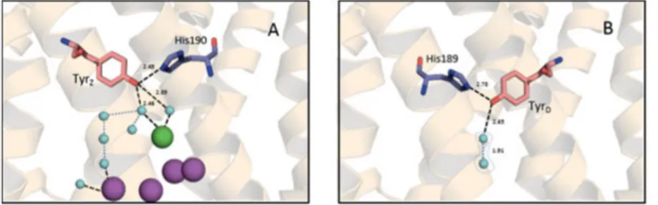
TyrZ (Tyr-161) is located in the D1 protein between the Mn4CaO5 cluster
and P680. As it was confirmed by 1.9 Å resolution crystal structure, TyrZ
is hydrogen bonded to D1-His190 (2.5 Å distance) (Fig. 8A) [18]. It is assigned as a proton acceptor to TyrZ by several methods (site-directed
mutagenesis, FTIR and structural modeling) [36, 39-45]. Additionally, TyrZ is connected either directly to W4 or through the additional water
molecules to W3, W2 and W1. D1-His190 is connected through the se-ries of amino acids and water molecules to the luminal bulk solution. The amino acid residues lying along the way of this channel to the lumen belong to the D1, Cp43 and PsbV subunits. The charged amino acid res-idues (D1-Arg323, D1-His304 and PsbV-Lys129) surround the exit of this channel [18].
Contrary to TyrZ, TyrD (Tyr-160) is deeply buried in the hydrophobic
environment of the D2 protein. It is positioned symmetrically to TyrZ in
the D2 protein. The homologous to D1-His190, D2-His189 was identi-fied in the vicinity of TyrD (Fig. 8B). A single water molecule was found
in the hydrogen bond distance to TyrD. It is not completely occupied in
the structure, but there are indications for the two possible positions of this water molecule in the vicinity of TyrD [18, 46]. The hydrogen
bond-ing patterns of both tyrosines determined their functional differences and mechanistic aspects of oxidation which will be discussed in the section below.
Functional differences
TyrZ bears a highly oxidizing potential (Em TyrZ•/TyrZ > 900–1000 mV)
which makes oxidation of water at the Mn4CaO5 cluster possible [47].
Under illumination, TyrZ acts as a relay between highly oxidative P680+
and Mn4CaO5 cluster. It is a two steps process: i) oxidation of TyrZ by
P680+ in the nanosecond to microseconds time scale, ii) the reduction of
TyrZ• by the Mn4CaO5 cluster in the microseconds to millisecond time
regime [45, 48-51].
TyrZ is a neutral radical in the oxidized state. Upon TyrZ oxidation,
phenolic proton is transferred to the nearby D1-His190 residue. This pro-ton is transfered back after TyrZ reduction. The process does not require
significant rearrangement in the system, which is why the TyrZ oxidation
can occur in a fast nsec/µsec timescale. However, the oxidation of TyrZ
is S-state dependent, multiphasic and a very fast process. It is 20-40 ns in the S0 and S1 states while, at higher S-states, it slows to 50 ns in S2 and
260 ns in S3 states respectively [48, 52, 53]. In 20 % of PSII centers, TyrZ
undergoes oxidation in a microsecond time scale [49, 53, 54]. For a long time it was thought that the slow microsecond kinetics reflects inactive PSII centers. But later it was assigned to the centers with less defined hydrogen bonding environment around TyrZ, which restricts very fast
deprotonation reaction. TyrZ oxidation is pH independent between pH
5.5 and 8.0. The rate and amplitude of TyrZ oxidation decreases towards
acidic pH with pKa 4.5-5.3, which was attributed to the titration of
D1-His190, causing disordering of the essential hydrogen bonding [55, 56]. Its symmetrical counterpart in the D2 subunit, TyrD, does not undergo
such fast oxidation behavior [57]. It is not involved in the water oxida-tion, but still was conserved throughout evolution. The role of TyrD in
PSII has been the subject of continual research [38, 58-61]. TyrD is
oxi-dized by S2 or S3 state with a half-time tens of sec at pH range between
5 and 7.5, forming a long-lived tyrosine radical (TyrD) [47, 62]. It slowly
decays over hours in the dark. TyrD has a slightly less positive oxidation
potential Em = 700-800 mV if compared to TyrZ [36, 63]. Both tyrosines
are positioned the same distance to P680 (12.4 Å away), and can be
oxi-dized by P680+ [18]. However, due to the slow oxidation at physiological
pH TyrD is not competitive to TyrZ as an electron donor to P680+. TyrD
oxidation rates becomes efficient, comparable to those seen for TyrZ, at
elevated pH with a pKa~7.6 (t1/2 ≈ 190 ns) [38]. Like TyrZ, TyrD is
in-volved in PCET reaction, getting protonated or deprotonated upon reduc-tion or oxidareduc-tion respectively. Earlier publicareduc-tions suggested that D2-His189 is the proton donor to the reduced state of TyrD as the
symmet-rical counterpart D1-His190 to TyrZ [45, 64-67]. However, the recent
crystal structure at 1.9 Å identified also a water molecule in the hydrogen bonded distance from TyrD, which can occupy proximal or distal
posi-tions [18, 46]. As has been reported independently by spectroscopic stud-ies and calculations, the proximal water molecule accepts phenolic pro-ton upon oxidation of TyrD [46]. The mechanism of TyrD oxidation will
be discussed in detail in papers I and II.
It can be concluded that the key to the functional difference of TyrZ
and TyrD is determined by the fate of the proton upon tyrosine oxidation.
The oxidation of TyrZ in nsec timescale is due to the proton-rocking
mechanism of the hydrogen bond between D1-His190 and TyrZ, while
the slow oxidation of TyrD is due to the movement of water molecule,
serving as a proton acceptor with subsequent release of proton to the bulk. The crucial role of TyrZ in PSII has been demonstrated in many
postulated roles of TyrD in PSII: i) it acts as an electron acceptor from
Mn4CaO5 cluster in the lowest state of S-cycle (S0), it stabilizes the
clus-ter to the higher and more stable state in the dark (S1); ii) it has a role in
the oxidation of over-reduced forms of Mn during photoassembly of WOC [64, 68, 69]; iii) it has an electrostatic effect to the localization of cation in the P680 moiety [57, 70, 71]. Regardless of the proposed possible
roles of TyrD in PSII, the undeniable fact is that TyrD is conserved in all
photosynthetic species, and the growing TyrD-less mutants have some
difficulties [57].
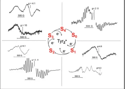
Interaction of tyrosines with S-states of WOC
TyrZ• is able to oxidize all S-states of WOC. The fast oxidation and
re-duction of TyrZ• in the presence of WOC makes it difficult to study. One
of the important probes in this respect are the metalloradical EPR signals [72]. When two paramagnetic species are located at a short distance (5-10 Å) from each other, their spins magnetically interact, forming very distinct set of EPR signals. The nature of such interaction is due to a spin exchange interaction or between the magnetic dipoles of the electrons [73]. The interaction of the TyrZ and Mn4CaO5 cluster is one of them.
The prerequisite for this interaction is the formation of TyrZ radical under
continuous illumination at cryogenic temperatures at which WOC is not able to turn over, as this will cause the appearance of so called EPR split signals. Except for the transient S4 state, interaction of all S-states with
TyrZ gives rise to the Split signals (Fig. 9) [74, 75]. Interaction of TyrZ
with the S1 state of Mn4CaO5 cluster was a subject of study in Paper VI.
Contrary to TyrZ, TyrD can only oxidize S0 state of WOC. It is very
slow process taking from minutes to hours at room temperature [35]. The oxidation of TyrD occurs by only the S2 and S3 states. It is pH dependent
reaction accelerating towards higher pH [47]. The oxidation of TyrD by
the S2 or S3 states is biphasic process with fast and slow phases governed
by water position in the vicinity of TyrD. The population of PSII centers
with water in the proximal position give rise to the fast, pH independent oxidation phase and the population of PSII centers with water in the dis-tal position induce the slow, pH-dependent oxidation phase. The biphasic kinetics was observed at pH<7.3 and one phase for pH>7.3 [47].
Figure 9. Summary of the Split EPR signals from different S-states induced in PSII by illumination at low temperature. The middle part of each spectrum from overmodulation TyrDis removed for clarity.
Methods
Electron Paramagnetic Resonance
Electron paramagnetic resonance is a technique for the detection of un-paired electrons in a molecular system. The method has been notably used in the detection of transition metals, radicals, triplets, etc., and, therefore, is extremely informative in studying the electron transfer re-action. The system with unpaired electrons is called paramagnetic, and without unpaired electrons, diamagnetic. An electron has a magnetic mo-ment induced by the two properties: spin and charge. In the presence of a magnetic field, the magnetic moment can be oriented parallel or anti-parallel to the magnetic field. Each orientation requires specific energy due to the Zeeman effect:
E = m
sg
eμ
BB
0ge – known as the electron´s g-factor, or Lande´s factor, ge= 2.0023
for free electron; μB is the Bohr magneton. If microwave energy matches
with the energy difference of these two states, an unpaired electron ab-sorbs energy hν and changes spin. The absorption leads to the rise of an EPR signal.
Commoner observed an EPR signal from PSII first time in 1956, which later was known as a Signal IIslow or Signal IID [76]. It was a stable
radical signal with g=2.0046 and decay halftime in min-hours time range [58]. Later it was identified that signal comes from tyrosine radical and was called TyrD (index D derived from the word “dark” due to its
stabil-ity in the dark). TyrD signal is considered to be a robust signature for PSII
quantification. Afterwards, by improved EPR spectrometer, similar to Signal IIslow, signal with fast decay kinetics was observed in Mn-depleted
PSII [77]. The signal has msec-sec decay halftime and was called Signal IIfast. Further studies assigned Signal IIfast to the TyrZ radical of PSII.
Soon, the third type of Signal II, Signal IIvery fast, was detected in intact
PSII samples, with a very fast decay halftime (μsec-msec) [78].
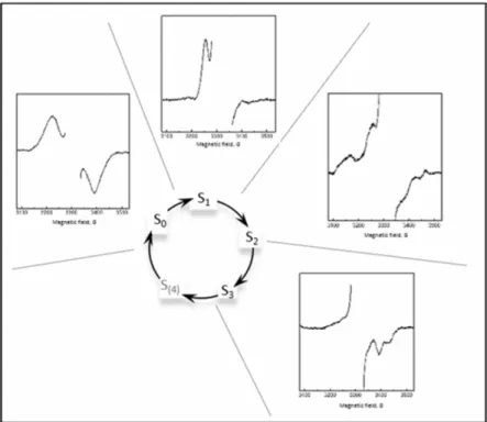
In 1981, EPR signal from Mn4CaO5 cluster was reported in thylakoid
membranes [79]. It showed a period of four oscillations with every ap-plied laser flash. The signal appeared after the first and fifth flash. It was concluded that this is a signal from S2 state of the cluster, the so-called
S2 multiline signal (Fig.10). Later, signals from other S-states of
Mn4CaO5 cluster were also discovered, except for the transient S4-state
(Fig.10).
Figure 10. EPR signals from different S-states of the Mn4CaO5-cluster.
Another cofactor of the electron transport chain detected by EPR spec-troscopy is Cyt b559. Cyt b559 exhibit rhombic signals with anisotropic g
values (gz approx. 3.0, gy approx. 2.2, and gx approx. 1.5), varying in
dif-ferent preparations. The anisotropic g values represent the orientation of the heme relative to the membrane plane which, in turn, affect the redox potential of Cyt b559. The heme was found to be perpendicular to the
membrane plane in the oxidized and reduced states of Cyt b559. Slight
variations in the g-values of the redox form of Cyt b559 reflects changes
The high spin signal originated from QA-Fe2+ interaction has a
characteristic EPR signal with g value around 1,82 [80]. QA-Fe2+QB-
in-teraction signal is detected with g=1,66 [81].
Photoxidizible Chl and carotenoids molecules can also be detected by EPR spectroscopy. Photoinduced P680+ has g=2,0027 with line width 10
G. The spin-polarized triplet signal from 3P
680 with the zero-field splitting
parameters |D| = 0.0286 cm−1, |E| = 0.0044 cm−1 is another detectable
EPR signal from PSII. It is formed due to Pheo-P
680+ recombination [82].
Fluorescence
The light energy absorbed by Chls molecule can undergo one of the three possible pathways: i) it can be used to drive photochemistry (water split-ting reaction); ii) it can be dissipated as heat; or iii) be re-emitted as light (so called chlorophyll fluorescence) [83]. These pathways are compet-ing, the increase of one will lead to the decrease of the another two path-ways. Re-emission of excess energy as heat or fluorescence is a protec-tive mechanism of the system against high light conditions. Hence, meas-urements of the yield of chlorophyll fluorescence has been used to obtain information about the integrity and changes in the photosynthetic elec-tron transport chain [84, 85]. In 1931, Kautsky and coworkers observed for the first time an increase in the yield of chlorophyll fluorescence for approximately 1 sec after exposing dark adapted leaves to light [86]. The rise was later assigned to the reduction of acceptors in PSII, namely QA.
In the dark adapted the “open” PSII centers exhibit very low fluorescence yield, QA is able to accept one electron, and, after illumination, an
elec-tron is on QA during this period, and the reaction center is called “closed,”
and is highly fluorescent [87, 88]. After an electron passes further in the chain to QB, the decay of fluorescence is observed [89, 90]. The
flash-induced variable fluorescence is one of the fluorescence techniques to follow this decay. After a saturating flash, the rise of fluorescence is ob-served from F0 level when all reaction centers are open, and low
fluores-cent to Fmax when all QA is reduced and reaction centers are closed and
highly fluorescent. The following dark adaptation leads to the subsequent decay of fluorescence. The difference in changed fluorescence yield is called variable fluorescence. The observed decay of fluorescence reflects the reoxidation of QA- [89, 91]. The kinetics consists of three exponential
scale, and can vary depending on PSII material. The fast phase is as-signed to electron transport from QA- to QB or QB- , which is bound to the
QB-site. The middle phase in msec time scale is attributed to those centers
which did not have QB bound before the flash, and requires time for new
QB to bind. The third slow phase originates from centers where QB is not
available and QA- recombines with the S2 state of Mn4CaO5-cluster.
When the forward electron transfer is blocked by the herbicide addition, for example DCMU, an electron stays on QA for a few seconds and
slowly decays via the QA-S2 recombination [90, 92].
Thermoluminescence
Thermoluminescence is a weak light emission from irradiated materials with trapped charge separated states upon being heated. The light emis-sion comes from a recombination of photoinduced charge pairs when the thermal energy of the environment matches with the activation energy barrier of two separated charges. Thermoluminescence is widely used as a dating technique in archeology. Thermoluminescence from photosyn-thetic systems were first observed by Arnold and Sherwood in 1957 [93]. They observed light emission peak at 30-40 oC from dried chloroplast
samples, illuminated prior to the heating, which they explained as the release of stored energy upon heating. Later, it was assigned to the re-combination of different cofactors in PSII [94, 95].
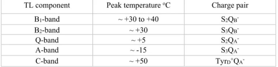
Table 1. The temperature peak and origin of TL bands
TL component Peak temperature oC Charge pair
B1-band ~ +30 to +40 S2QB-
B2-band ~ +30 S3QB-
Q-band ~ +5 S2QA-
A-band ~ -15 S3QA-
C-band ~ +50 TyrD+QA-
The rate of recombination can be brought down to a negligible level if samples are cooled down immediately after illumination; then heating up progressivly reveals the appearance of different thermoluminescence bands. Photoinduced charge pairs are stabilized by the activation energy barriers (ΔG) limiting back flow. Thermoluminescence from PSII origi-nates mostly by the recombination of the following charge pairs: S2QB-,
S3QB-, S2QA-. Thermoluminescence bands detected in PSII samples are
The protonation state around Tyr
D/Tyr
Dand redox equilibrium in Photosystem II
The proton coupled electron transfer reaction is a fundamental property of the energy conversion in biological systems [96]. In many enzymes, processes like catalysis and charge transport are often underpined due to the coupling of a proton transfer to an electron. Such coupling helps bi-ological systems to stay neutral, avoid high energy pathways, and the formation of unfavorable species in the oxidation-reduction processes. One example of PCET reactions is the oxidation and reduction of the two tyrosines (TyrZ and TyrD) in PSII [97]. These tyrosines represent an ideal
model system to study PCET reaction of two identical species in a dif-ferent protolytic environment.
In Paper I, the mechanism of PCET of TyrD was studied by using CW
EPR and deuterium isotope effect on the TyrD oxidation behavior under
different pH conditions. In Paper II, in addition to the TyrD oxidation
kinetics at different pH, the competing recombination reactions, which involved the acceptor side of PSII, were investigated by using the varia-ble flash-induced fluorescence and thermoluminescence techniques.
TyrD oxidation kinetics consists of two phases: fast and slow. A fast
phase is essentially pH-independent both in amplitude and lifetime, while a slow phase exhibits a strong pH-dependence [47]. The biphasic behavior of the oxidation kinetics of TyrD was previously assigned to two
distinct proton equilibria associated with the oxidation of TyrD by the S2
state. One of these proton equilibria was suggested to be in the vicinity of TyrD, and the other near the Mn4CaO5 cluster [47]. According to this
model, biphasic kinetics are observed when the TyrD oxidation process
is faster than the proton equilibria near TyrD and the Mn4CaO5 cluster.
The deprotonated and protonated populations in this case are independ-ent from each other. Contribution of each population to the fast and slow phases respectively, have a pH-independent lifetime. However, the am-plitude of each phase would be pH-dependent due to the changes in the
relative degree of protonation. It changes the relative size of protonated and deprotonated TyrD populations. Contrary, if the proton equilibrium
is faster than TyrD oxidation, it causes the reestablishment of the
equilib-rium prior to oxidation of TyrD and a monophasic kinetic behavior is
ob-served. We further advanced the results obtained in [47] with incorpora-tion of some important differences.
The TyrD oxidation kinetics were followed in a deuterated buffer over
a wide pD range (4.7-8.7). The time resolved kinetics in a deuterated buffer at pD 6.3 differed significantly both in amplitude and in half-time compared to non-deuterated. The kinetics were observed to be slower by more than a 3–fold for the fast phase and almost by a 10-fold for the slow phase. The full kinetic analysis of amplitudes and half-times is summa-rized in Fig.11
The kinetics were fitted by two exponential phases, for pH < 7.3 and pD < 8.0, and one phase for pH > 7.3 and pD > 8.0. Hence, at lower pL the oxidation of TyrD was biphasic (fast and slow phases), whereas at high
pL only monophasic oxidation behaviour was observed. The amplitude of the slow phase was strongly pL-dependent and decreased towards lower pL values from about 90% to 20-30% (Fig. 11A). Contrary to the slow component, the fast component was independent at pL below 7.5 (Fig. 11B). The half-times of the fast phase were almost pH independent, moderate pD-dependence was observed (Fig. 11D). The half-time of slow phase was again pL dependent. It decreased towards higher pH/pD value and eventually merged with the half-times of the fast phase by Figure 11. Amplitudes and half-times of the two kinetic phases in the oxidation of TyrD in pH (blue) or pD (red) buffers. A and B show the amplitudes of the
slow (filled circles) and fast (open circles) kinetic phases as a function of pL. C and D show the half-times of the two phases as a function of pL.
forming monophasic TyrD oxidation kinetics (Fig. 11C). The half-time
of the slow phase ranged from 23 sec at pH 4.7 to <1 sec at pH 8.7 in non-deuterated buffers, while in deuterated buffers it changed from 50 sec at pD 4.7 to 1-2 sec at pD 8.7.
The rate constants for the slow phase were calculated as a function of pL (kH for non-deuterated and kD for deuterated buffers) (Fig. 12). The
KIE for whole pL range was calculated by taking into account the degree of protonation/deuteration. This was accomplished using the pK values from the data in Fig. 12. For the detailed explanation of the fit-ting parameters, see Paper I.
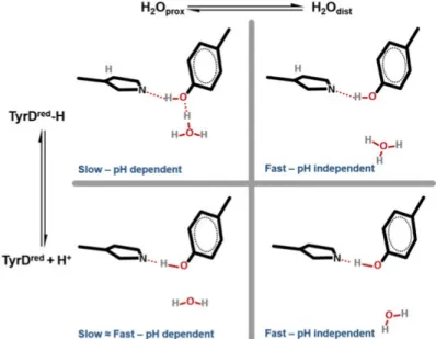
The recent crystal structures of PSII revealed a water molecule in the vicinity of the phenol group of TyrD which can occupy two
posi-tions [18]. The proximal position of the water molecule is 2.6-3.2 Å away from the phenolic oxygen of TyrD and the distal position is
4.3-4.5 Å. In Paper I, we proposed that the equilibrium between these two possible water molecule positions gives rise to two distinct populations of PSII centers. At low pH, the fast phase in the TyrD oxidation
corre-sponds to those centers where a water molecule occupies the proximal position, and hydrogen bonded to the phenolic proton of TyrD which is
released during its oxidation (Fig. 13). According to the DFT-QM/MM calculations [46], this water molecule accepts a proton released upon TyrD oxidation and moves to the distal position. Then proton accepted by
the water molecule transferred further to the D2-Arg190 and is released via proton channel out to the lumen. This proton release to the lumen after TyrD oxidation has been recently shown by FTIR experiments [98].
However, at higher pH values, the phenolic proton of TyrD is already
titrated away, and only electron transfer process takes place. This is the reason for the pH-independency of the fast component of TyrD oxidation
kinetics (Fig. 13). In contrast, the slow pH-dependent kinetic phase is assigned to the centers with the water molecule already in the distal po-sition. In this case, the water position is the rate-limited step for the TyrD
Figure 12. pL dependence of the rate constants for TyrD formation in the
slow phase in pH (blue) with pK 7.9 and pD (red) buffers with pK 8.9.
oxidation process. TyrD does not have a direct hydrogen bond to the
wa-ter molecule, and deprotonation of TyrD can only happen after the distal
water comes close enough to accept the proton through thermal motion without being in the fully occupied proximal position (Fig. 13). Since water is not readily available to accept the released proton, it makes the oxidation of TyrD slower. Towards higher pH values, the amount of the
deprotonated TyrD populations are increasing, which makes convergence
of the slow component with the fast component of TyrD oxidation
kinet-ics. It leads to the higher amplitude and eventually only monophasic TyrD
oxidation kinetics, with pK 7.9 in H2O and 8.9 in D2O. Therefore, the
overall rate of TyrD oxidation increases with pH as well.
Equations 2-6 in Paper I shows the model of TyrD oxidation with the
fast proton equilibrium where kprot and kdep are the oxidation rate
con-stants of protonated and deprotonated TyrD, respectively.
The calculated kinetic isotope effect (KIE) was around 2.3-2.5. The origin of KIE was attributed to the proton/deuterium, which is in the strong hydrogen bond between TyrD and D2-His189.
Figure 13. The changes of the hydrogen bond prior the TyrD oxidation
depend-ing on protonation state (up/down) and water position (left/right) affectdepend-ing the kinetics of TyrD oxidation.
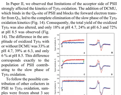
In Paper II, we observed that limitations of the acceptor side of PSII strongly affected the kinetics of TyrD oxidation. The addition of DCMU,
which binds in the QB-site of PSII and blocks the forward electron
trans-fer from QA, led to the complete elimination of the slow phase of the TyrD
oxidation kinetics (Fig. 14). Consequently, the total yield of the oxidized TyrD was also altered, and only 18% at pH 4.7, 24% at pH 6.3 and 72%
at pH 8.5 was observed (Fig. 14). The difference in the am-plitude of oxidized TyrD with
or without DCMU was 33% at pH 4.7, 39% at 6.3, and only 6 % at pH 8.5. This difference corresponds exactly to the population of PSII contrib-uting to the slow phase of TyrD oxidation.
To follow the possible con-tribution of other cofactors in PSII to TyrD oxidation,
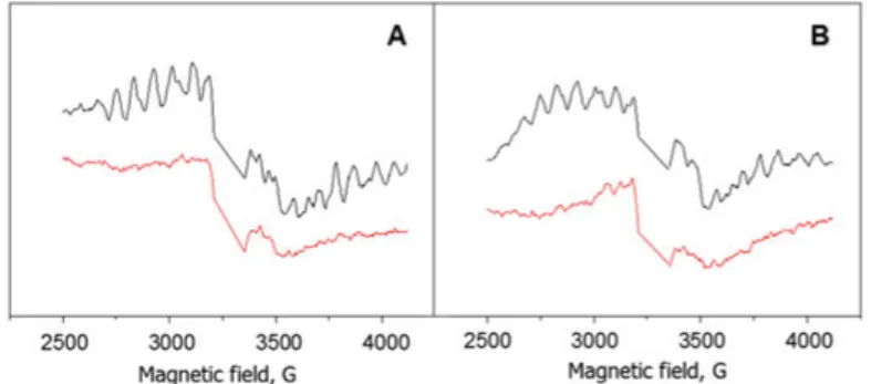
sam-ples were frozen about 3 sec after the laser flash (indicated by the green arrow in Fig. 14, and EPR measurements were performed at low temperature. In the samples without DCMU addition, within 3 sec after the flash, 100 % of the S2 state
multiline EPR signal was de-tected at pH 6.3 (Fig. 15).
However the presence of DCMU lowered the S2 state multiline signal to
82 % (Fig. 15). The decrease in the multiline signal was assigned to S2QA
-state recombination which occurred before the freezing. Complete decay of the signal was observed after 80 sec in the presence or absence of DCMU. The similar pattern was observed at pH 4.7. Only 90 % of the S2
state multiline was detected within 3 sec after the flash in the absence of DCMU. As was reported before, the decrease in the amplitude is due to the pH-dependence of the S2 state multiline signal. In the presence of
DCMU, 70 % of the S2 multiline signal was observed at 3 sec after the
flash. At pH 8.5 no S2 multiline signal was detected.
Figure 14. Oxidation of TyrD after a single
flash at different pH values at RT. Kinetic traces (left: A, C, E) and field-swept (right: B, D, F) in the absence (red line) or presence of 1.75 mM DCMU (blue line) at pH 4.7 (A and B), pH 6.3 (C and D) and pH 8.5 (E and F).
The oxidation of TyrD occurs via the WOC, namely by the reduction
of the S2 state to the S1 state via equilibrium with P680+ and TyrZ.
This was first suggested in [47], but was never experimentally proven. Our results showed indeed verify the rise in the amplitude of TyrD
oxi-dation accompanied by the decay of the S2 state multiline signal (Fig.
15).
Figure 15. The S2 state multiline EPR signal in samples with reduced TyrD
fro-zen 3 sec (black spectrum) or 80 sec (red spectrum) of laser flash at pH 6.3 in the absence (A) or presence of 1.75 mM DCMU (B).
Another redox component in PSII is Cyt b559 (Fig. 16). The flash
in-duced oxidation of the high potential form of Cyt b559 at pH 4.7 and 6.3
was observed (Fig. 16, g=3.05). We observed 10 % at pH 6.3 and 16 % at pH 4.7 oxidation of the Cyt b559 3 sec after the flash. An additional 10
% at pH 6.3 and 6 % at pH 4.7 oxidation of the Cyt b559 was detected 80
Figure 16. EPR spectra of the gz
re-gion of oxidized Cyt b559 in samples
with reduced TyrD at pH 6.3 in the
ab-sence (A, B) or preab-sence of 1.75 mM DCMU (C, D). Spectra in A and C were recorded from samples before laser flash (black spectra) or after 6 min illumination at 77 K (red spec-trum). Spectra in B and D were rec-orded after 3 sec (black spectrum) or 80 sec (red spectrum) of laser flash.
sec after the flash. No oxidation of Cyt b559 was observed in the presence
of DCMU. Interestingly, there were no changes in the redox state of Cyt b559 at pH 8.5.
To follow the competing acceptor side recombination with TyrD
oxi-dation for the S2 state recombination, the flash-induced fluorescence
de-cay kinetics were measured. Fig.17 shows the flash induced fluorescence kinetics at three pH values in samples with TyrD in the oxidized (black
traces) and reduced form (red traces). No difference was observed in the kinetics of fluorescence decay between TyrD oxidized and TyrD reduced
samples at pH 4.7 and 6.3. Contrary, at pH 8.5, samples with TyrD in
reduced form demonstrated a non-decaying slow phase of the fluores-cence kinetics. This non-decaying phase was assigned to the centers where TyrD oxidation outcompetes the S2QA- recombination reaction.
The non-decaying phase was more pronounced in the presence of DCMU (Fig. 17). We observed the difference between samples with TyrDox and
TyrDred at all three pH when the forward electron transfer from QA- was
inhibited, and re-oxidation of QA- is only possible via S2 state
recombi-nation. The difference in amplitude of the slow phase between TyrD
ox-idized and reduced samples was increasing at higher pH (Table 2 in Pa-per II). Interestingly, the difference can reach 40 % of the total variable fluorescence at pH higher than 8.0 (Paper II, Suppl. Fig. 2).
Figure 17. Flash-induced variable fluorescence decay traces from samples with TyrD reduced (red traces) and oxidized (black traces) in the absence (A, B, C) or
presence of 20 μM DCMU (D, E, F) at pH 4.7 (A, D), pH 6.3 (B, E) and pH 8.5 (C, F).
Our fluorescence measurements indicated that the redox state of TyrD
does not have any effect on the forward electron transfer in PSII. How-ever, the inhibition of the forward electron transfer (with DCMU addi-tion) gave rise to the appearance of a difference between PSII samples with oxidized and reduced TyrD. The difference correlated well with the
amplitude of the fast phase of TyrD oxidation kinetics. Therefore, it can
be concluded that the population of TyrD which undergoes the fast
oxi-dation clearly wins the competition with the S2QA- state recombination.
We observed an even more pronounced effect at pH 8.5 where the fast phase of TyrD oxidation was dominant.
The recombination reaction, in addition to fluorescence measure-ments, was studied by thermoluminescence (Fig. 18) The B1-band which reflects S2QB- recombination was found at 39 oC at all three pH values in
the TyrD oxidized PSII samples. However, in the TyrD reduced PSII
sam-ples, the peak of the B1 band was shifted down to 31 oC and amplitude
was 4 times lower at pH 4.7, and 2.5 times lower at pH 6.3. The ampli-tude of the B1 band in TyrD oxidized samples was significantly decreased
at pH 8.5 compared to the lower pH, and completely absent in the TyrD
reduced samples. The observed difference in the amplitude and temper-ature peak between samples with TyrD in oxidized and reduced state
re-flects the changes in the free energy gap, ΔG between the two charge separated component QB- and the S2 state. The lower free energy gap ΔG
between the charge separated state at lower temperature the recombina-tion will take place. The shift of the peak detected in TyrD reduced
sam-ples could be caused by an absence of the positive charge on TyrD. In
addition, the loss in the amplitude of the TyrD reduced samples can also
be assigned to the competing TyrD oxidation reaction. The presence of
DCMU induces the Q-band peak around 10 oC, which is attributed to the
S2QA- recombination. In the TyrD oxidized PSII samples, the Q-band
glowed at 9 oC at pH 4.7 and 6.3. PSII samples with Tyr
D in reduced state
showed the upshift of temperature peak to 13 oC at pH 4.7 and 6.3. The
Q-band of TyrD oxidized samples at pH 8.5 was shifted to 34 oC, with
significant lower amplitude than at pH 4.7 or 6.3, and was almost absent in TyrD reduced samples.
The kinetics model for TyrD oxidation and competing electron transfer
steps is summarized in Fig. 19. There are six possible acceptor/donor side reaction pathways that lead to TyrDformation: in two steps after
electron transfer to QB (k1/k3; k1/k4; k2/k3; k2/k4), or in one step with
the acceptor-side electron still on QA (k3/k4). The results of this kinetic
modeling are summarized in Fig. 7 Paper II, which correspond well with the experimental data. Our
exper-imental data, kinetic modeling of the TyrD oxidation and competing
recombination reaction in Paper II confirmed the existence of two distinct populations of PSII cen-ters with respect to the redox state of TyrD: the slow oxidized
popu-lation which is in the competition due to similar halftime with re-combination reactions, and the fast oxidized population of TyrD
which is always prevalent over
recombination reactions. Figure 19. Kinetics models for electron transfer after initial transition of PSII to the S2 state.
Figure 18. Thermoluminescence bands obtained in samples with TyrD in
re-duced (red traces) and oxidized (black traces) in the absence (A, B, C) and presence of 40μM DCMU (D, E, F) at pH 4.7 (A, D), pH 6.3 (B, C) and 8.5 (C, F).
Far-red light triggered induction of P
680+and tyrosine radicals formation
PSII photochemistry is a complex process affected by several factors, of which the light quality is the most important. Absorption of light by Chl promotes an electron from the ground state (S0) to the singlet excited
state (Si). The greater the energy in the absorbed photon, the higher the excited singlet state transition will be initiated. The type of chlorophyll molecules and their arrangement defines the energy required for this transition. There are at least five different chlorophyll found in oxygenic photosynthetic organisms: Chl a, b, c, d, f. These chlorophylls are pre-sented with differences in their absorption wavelength. This difference is not only tuned by the protein environment but also by the distinct chemical composition of each of the chlorophyll molecules respectively. [24]. Nearly all chlorophylls in the photosynthetic organisms are associ-ated with specialized proteins: antenna proteins or reaction center pro-teins.
The reaction center of PSII, P680, consists of four Chl molecules bound
by the D1/D2 heterodimer denoted as PD1, PD2, ChlD1 and ChlD2, (Fig.
24). PD1 and PD2 are weakly excitonically coupled to the central Chl pair,
and are situated at a 30 angle to the horizontal plane. The other two Chls, ChlD1 and ChlD2, are located symmetrically on either side of PD1 and PD2
respectively (Fig. 23). The excitation energy transfer to the reaction cen-ter Chls of PSII initiates the primary charge separation. The formation of the first stable Chl cation after the primary charge separation reaction, has been monitored extensively by various spectroscopic methods [99-104]. There are indications that the first Chl electron donor is different when PSII is excited by the far-red light (720-800 nm) if compared to visible light (400-700 nm).
To investigate the localization of the primary Chl donor under far-red light, the spin polarized triplet state of 3P
680 and the difference in the
ox-idation efficiency of TyrZ and TyrD were used as probes in Paper III and
IV respectively.
The prerequisite for the formation of the spin polarized triplet 3P 680
state is the double reduction of QA. The double reduction of QA was
achieved by a prolonged incubation of PSII samples in the presence of a reducing agent, sodium dithionite and a mediator, benzyl viologen. Un-der these conditions, the double reduced QA becomes protonated and
pre-sumably released from its binding site. As a consequence, the long-lived charge-separated state P680+ Pheo1- decays via slow recombination to
al-low spin dephasing to the spin-polarized triplet state.
The illumination of the reduced samples with visible light at cryo-genic temperatures (5 K), induces the formation of a characteristic EPR signal arising from the spin polarized 3P
680 with high yield (Fig. 20). We
have attempted the induction of the triplet signal under far-red light illu-mination (>730 nm) however, no spin polarized 3P
680 EPR signal was
de-tected under these conditions, even after meticulous accumula-tion (Fig. 20). Several reasons have been postulated to explain the above observation: I) The far-red light is not efficient to drive photochemistry. However, this can be ruled out because the effi-cient far-red photochemistry of PS II has been demonstrated before. In addiiton we have observed the accumulation Pheo- signal under
these conditions [99]. II) The formed 3P
680 signal decays faster
than can be detected. This possi-bility was tested by the decay ki-netic measurements of the 3P
680 signal induced at different wavelengths
(Paper III, Fig. 6). No differences were observed in the decay kinetics under different wavelength. III) We propose that the primary electron donor to Pheo is different under far-red light illumination compared to visible light. Most studies suggest that the primary cation formed under visible light is PD1. This well explains the triplet formation under visible
Figure 18. Light-dark difference EPR spectra of the spin-polarized 3P
680 in PS
II samples with (a) white light and (b) far-red light (732nm) at 5 K
Figure 20. Light-dark difference EPR spectra of the spin-polarized 3P
680 in PS
II samples with (a) white light and (b) far-red light (732nm) at 5 K
light, since the recombination between Pheo- and P
D1 is slow enough to
allow spin dephasing. In contrast, we suggest that under far-red light il-lumination, reduction of Pheo occurs from ChlD1. It has been previously
experimentaly observed that, the ChlD1+ is close enough to oxidize TyrZ,
but too far for the oxidation Cyt b559/ChlZ/Car pathway [100]. The close
proximity of ChlD1 to Pheo allows for fast recombination without spin
dephasing, which essentially prevents the formation of the spin polarized
3P
680 (Fig. 20).
To test this hypothesis at room temperature, the oxidation of two ty-rosines in Mn-depleted samples were followed by EPR spectroscopy un-der visible and far-red light conditions, at different pH values (Fig. 21). The reduction of TyrD was achieved by prolonged dark incubation at
room temperature. Steady state oxidation of TyrZ and TyrD under
contin-uous illumination with white or far-red light resulted in a different yield of Tyr oxidation depending on pH. The yield of Tyr oxidation increased upon increase of pH, with the maximum at pH 8.5. The yield of Tyr ox-idation was lower at all measured pH under the far-red light illumination, compared to white light illumination. The decreased yield of Tyrunder
far-red light illumination could be due to either i) less effective charge separation; or ii) the different primary electron donor which drives a less efficient far-red photochemistry. The first hypothesis can be ruled out because the accumulation of Pheo- indicates that most of the PSII centers
undergo charge separation [99]. Oxidation of both tyrosines were ob-served under white light illumination at pH 8.5, while the far-red light induced only TyrDformation. The difference between these two
wave-lengths can be attributed to either a different tyrosine oxidation pathway (TyrD and TyrZ) or the different nature of the primary donor.
Figure 21. Kinetics of tyrosines oxidation recorded at 3465 G under continuous white light (black traces) or far-red illumination (red traces) at pH 4.7 (A), pH 6.3 (B), pH 8.5 (C) in Mn-depleted PSII samples.
To distinguish between the TyrZ and TyrD signal formation at all
three pHs, a sequence of flash-induced measurements in the absence and presence of an effective electron donor to TyrZ (DPC) and acceptor from
QB-site (ferricyanide) was performed (Paper IV Fig. 5, 6, 7). TyrZ
oxida-tion was observed at all three pH values and under both 532 and 732 nm flashes (Paper IV Fig. 3). The amplitude of TyrZwas both pH and
wave-length dependent. However, the calculated decay half-time values of TyrZhave shown significant pH dependence (Paper IV, Table 1).
There are three possible donors to TyrZ: TyrD, DPC (if present), or
recombi-nation with the acceptor side (Fig. 22). In the presence of DPC, the amplitude of the TyrZradical formation was significantly
inhibited, especially under 732 nm flashes at pH 4.7 and 6.3 (to 3 and 25 % respec-tively). This resulted in significantly less formation of TyrDespecially with the 732
nm flashes. Based on the above, it can be concluded that, at pH 4.7 and 6.3, oxida-tion of TyrD occurs via the TyrZradical
according to the following reaction:
Contrary to the low pH, at pH 8.5 and in the precense of DPC, a sig-nificant amount of TyrDwas still formed. The absence of the fast decay
on the first flash (Paper IV Fig. 7) at pH 8.5 indicates the direct TyrD
donation to P680+ under both light conditions.
Our results also confirm that the far-red light induced photochemistry occurs in the majority of PSII centers. Interestingly, the far-red light re-sults in decreased TyrZ formation at normal and low pH values at room
temperature. We attribute the latter observation to the higher recombina-tion rate from the acceptor side under far-red light illuminarecombina-tion. We pro-pose that increase in the recombination rate is due to the different cation localization in P680+ (Fig. 23).
Both the formation of the spin polarized 3P
680, as well as the oxidation
of the two tyrosines in PSII, indicate the existence of a difference in the primary photochemisty between these two wavelengths. The proposed Figure 22. Electron transfer events upon TyrZ and TyrD
different cation localization is based on the following structural differ-ences between the Chl of P680 ensemble: The special Chl pair of P680, PD1
and PD2 has a weak electronic coupling due to the greater physical
sepa-ration, a slight difference in the tetrapyrrole ring orientation and a smaller dipole strength of the Qy transition [105-107]. The latter results in a pair
that does not represent the lowest energy sink for the excitation energy. Since the monomeric ChlD1 does not have such coupling, it is believed to
be the lowest energy sink for the excitation energy [107]. Therefore, the migration of the far-red exciton among the four Chl molecules in P680
would not be an energetically favorable process. Thus, we conclude that, under far-red light excitation, the primary hole is localized on ChlD1.
Figure 23. Primary charge separation in PSII under far-red (732 nm) and visible light (532 nm)
Role of the PsbTn subunit in steering H
+from Tyr
DThe recent single particle CEM structure revealed four extrinsic protein subunits, namely PsbO, PsbP, PsbQ and PsbTn , on the luminal side of the eukaryotic PSII complex [1]. The PsbO, PsbP, and PsbQ subunits form the triangular crown shape structure interacting with the luminal domain of the C-terminal of the D1 protein and CP43. These shield the WOC from any exogenous reductant on the luminal side. The PsbO, PsbP, PsbQ subunits can be easily removed by “washing” the PSII prep-arations with various reagents and their loss leads to the deactivation of WOC, with subsequent release of a Mn4CaO5 cluster from a site.
Mean-while, as a tightly bound protein PsbTn remains bound after the washing procedures [108-110]. The role of the PsbTn subunit has been unknown [111, 112]. In Paper V, we investigated the role of the PsbTn subunit in the redox reactions of TyrD.
In our experiments we used PSII membrane preparations from Ara-bidopsis thaliana WT and three deletion mutants, ΔPsbTn1, ΔPsbTn2
Figure 24. TyrD oxidation kinetics at 3445 G at pH 4.7 (A), 6.3 (B) and 8.5 (C)
measured in PSII membranes with the reduced TyrD from the WT (black traces),
ΔPsbTn1 (green traces), ΔPsbTn2 (blue traces) and ΔPsbTn1+2 double mutant (red traces). Arrows indicate the time position of the laser flash.
and ΔPsbTn1+2. TyrD was chemically reduced prior to measurements
according to [47, 113]. The TyrD oxidation kinetics were followed at
three pH values by using time-resolved EPR spectroscopy (Fig. 24). The TyrD oxidation kinetics of the WT sample were observed as biphasic at
pH 4.7 and 6.3, accelerating at higher pH values, leading to monophasic oxidation kinetics being observed at pH 8.5 (Fig. 24). These observa-tions corresponds to the oxidation behavior of TyrD reported earlier
[113-115]. The TyrD oxidation kinetics in ΔPsbTn1, ΔPsbTn2 and ΔPsbTn1+2
followed the same kinetics as TyrD in WT samples at pH 4.7 and 8.5 (Fig.
24A, C). Interestingly, at pH 6.3 the kinetics of TyrD oxidation were
dif-ferent from the WT in all three mutants (Fig. 24B). The kinetics slowed down from ΔPsbTn1 to ΔPsbTn2 and ΔPsbTn1+2, respectively. Changes in the amplitude and half-time of both phases were observed (Table 2). Table 2. The total yield of TyrD and the amplitude of the fast and slow phases
(t½, sec and amplitude (%)) were obtained after two exponential growth fitting
of traces shown in Fig. 24.
Wild Type ΔPsbTn1 ΔPsbTn2 ΔPsbTn1+2
TyrD 100% 71% 89% 89%
Fast phase 1.64 (51%) 3.77 (45%) 4.80 (35%) 5.48 (27%) Slow phase 8.89 (49%) 18.80 (55%) 21.29 (65%) 52.20 (73%) It is known that the TyrD radical slowly decays in the dark by reduction
from S0 state of the Mn4CaO5 cluster and other components. The decay
Figure 25. The total yield of TyrD induced by continuous illumination at pH
6.3 (A) and with subsequent decay during the dark adaptation (B) in PSII membranes from the WT (black), ΔPsbTn1 (green), ΔPsbTn2 (blue) and ΔPsbTn1+2 double mutant (red)