MicroRNAs as biomarkers in
some cardiovascular diseases
A bioinformatics and review study
Zina Ahmed
Degree Thesis in Pharmacy 30 ECTS
Master’s Programme in Pharmaceutical Science Report passed: Spring 2017
Supervisor: Samaneh Tousi Examiner: David Andersson
Abstract
Introduction
MicroRNAs (miRNA) are small noncoding RNA sequences with gene regulation abilities by binding to specific regions of messenger RNAs (mRNA) and silence their expression. The importance of miRNAs in cardiovascular development has been underscored and studies suggest that dysregulation of miRNAs can contribute to pathological conditions, including cardiovascular diseases (CVDs). As a result of the aberrant expression there is a suggestion for their possible use as novel diagnostic biomarkers. However, studies point out obstacles in extraction and detection methods, which may limit this possibility.
Objective
The aim of this study was to map a comprehensive list of miRNAs that would be considerable as novel biomarkers for diagnosis of some CVDs. Furthermore, due to reports pointing out difficulties in detection of miRNAs, it was of value to review which extraction and detection methods that had been utilized in desired circulating miRNAs and also in which diseases. Moreover, since the interaction among miRNA and mRNA might predispose for other disorders in terms of CVDs, an investigation of target and host gene interactions was carried out.
Method
This study was achieved by using databases such as miRWalk, mirBase, HOCTAR and GeneMANIA to map and outline desired information. Additionally, 39 articles were reviewed from Pubmed in terms of disease, miRNA regulation, biological fluid and extraction and quantification method.
Result
Results from miRWalk showed a variety of miRNAs involved in different CVDs, mainly atherosclerosis and hypertension, such as mir-10a-5p and mir-638. Different target and host genes were detected for each CVD specific miRNA with different regulations. Furthermore, the most common chromosomal positions were on chromosome number 13 and 1. The review of articles indicated detection of miRNAs and their regulation in several diseases, specifically various cancerous diseases and also some CVDs. Moreover, different interactions among target and host genes were detected such as co-expression and genetic interaction.
Discussion
A variation of miRNAs, their host and target genes were detected using a combination of databases, which made the process flexible but also limited amount of collected data. Some CVD specific miRNAs that can be utilized as a potential biomarker for diagnosis of atherosclerosis are, based on the findings of this study, mir-10a-5p and mir-638 due to their un-simulated regulations in non-CVDs. On the other hand, considering the different regulations of many CVD specific miRNAs in multiple diseases, it might be of value to exclude non-CVDs in comorbid patients in order to enable diagnosis. Furthermore, GeneMANIA showed interactions among genes leading to predisposed patients of primarily ruptured vascular development and triglyceride homeostasis.
Conclusion
This study further confirms the statement that miRNAs can be utilized as novel biomarkers considering their detection and quantification. Specific miRNAs include mir-10a-5p and mir-638.
1
Introduction
Biogenesis and function of miRNAs
Micro ribonucleic acids (miRNAs) are small regulatory ribonucleic acids (RNAs) in the human genome that, unlike some RNAs, do not code for any proteins. These non-coding RNA molecules contain of about 21-25 nucleotides and have the ability to regulate posttranscriptional gene expression by switching off some of the genetic information, which is one of the reasons for the development of different cell types in, for example, the human body (1).
The miRNA biogenesis initiates in the cell nucleus where a transcription by RNA polymerase II occurs, forming a typical hairpin loop structure and resulting in the primary miRNA. The double stranded stem is recognized by the protein DGCR8, which associates with an enzyme called DROSHA to form a complex and together are able to cut the RNA into a smaller precursor miRNA. Exportin 5 is a transporter molecule that assists the precursor miRNA to exit the nucleus through the nucleus pore and enter the cytoplasm where other processes take place. Firstly, the precursor miRNA is recognized by an enzyme called DICER that cleaves the stem loop and forms a short double stranded miRNA molecule. This is followed by interaction with another protein, AGO2, that helps to unwind the double stranded molecule and releases one of the strands leaving the remaining strand to form RISC, RNA induced silencing complex, which can be guided to its targets and inactivate one or several genes. This inactivation occurs by a complementary sequence base pairing between the miRNA and messenger RNA (mRNA) of the host gene (1), although the base pairing does not always have to be completely perfect. Normally, the miRNA binds to the 3 prime untranslated region (3’UTR) of the mRNA to silence it, but it can also bind to the 5 prime untranslated region (5’UTR) of the mRNA, resulting in a complex regulatory effect with several binding sites for miRNAs and regulation of mRNA by many miRNAs (2). Once bound to the target mRNA, there are a few ways to induce the gene silencing: translational inhibition and mRNA degradation. During the translational inhibition, the ribosomal subunits responsible for protein synthesis, is prevented from binding to the miRNA-mRNA complex, leading to inhibition of protein formation. The miRNA-mRNA can also be degraded where the miRNA-protein complex simply cuts the mRNA leading to further degradation by the cell (1).
MicroRNA target prediction and databases
The complex regulatory potential between miRNAs and mRNAs indicates the necessity of computational prediction tools and datasets that aids to facilitate the gene target prediction, which currently also is used. A number of miRNA target prediction programs are used nowadays and are mainly based on to identify complementary sequence base pairing in the 3’UTR with the seed region of the miRNA (3). One of these programs is the “host gene oppositely correlated targets” (HOCTAR), which collects lists of target genes by studying the behavior of the host genes for a number of miRNAs in humans (4). This database also provides information on host genes of certain miRNAs.
Another program, miRWalk, enables target prediction in other regions beside the 3’UTR. This program, like most others, is also an algorithm and contains predicted and experimentally validated information on miRNA binding site and target interaction in humans, rat and mouse. This is possible by combining validated information from Pubmed database and several already existing miRNA-target prediction programs. The combination of programs results in reduction of false positivity and a more specific outcome (5).
The database miRBase contains information on the genomic context, sequence and predicted hairpin structure of miRNAs. Information on predicted and experimentally
2 validated targets is also provided, through crosslinking to other databases (2). When entering a miRNA sequence, primary references and links that describe the discovery and support proof of the finding is provided.
Diagnostic markers in CVDs
Cardiovascular diseases (CVDs) are the main leading causes of mortality in the world today, with over 17 million deaths every year due to different risk factors such as unhealthy lifestyles including lack of physical activity, use of tobacco and unhealthy diets (6). This has led to primary and secondary preventions being the standard health priority, which includes medication and change of lifestyle among others. To enhance clinician’s ability to identify high-risk individuals for cardiovascular diseases, different tools and assessment measurements are frequently used. One of these tools is biomarkers (“measurable and quantifiable biological parameters”), which help to accurately and immediately diagnose or indicate diseases or health characteristics. Aside from simplification for clinicians to point out endangered patients, biomarkers also need to be easy to interpret and, if possible, noninvasive for the convenience of the patient (7).
A number of ways to identify biomarkers include for example biosamples such as blood, urine, plasma and tissue test. The currently available biomarkers for detecting CVDs via blood sample are well established and frequently used in healthcare. A few examples of these biomarkers include the highly sensitive and specific heart muscle-specific troponins in myocardial infarction and B-type natriuretic peptide (BNP) in heart failure (7). However, there are a few limitations to be taken into consideration regarding these biomarkers. For starters, it is a known fact that during the initial hours of myocardial infarction, the cardiac troponins are often not significantly elevated, leading to a posterior diagnosis (8). Limitation with BNP blood test is that renal function plays a role as BNP is cleared from plasma via glomerular filtration. A decease in renal functions leads to accumulation of BNP and a higher value will therefore be obtained (9).
Additionally, limitations of electrocardiogram, ECG, which is a part of diagnosis of many CVDs, are that it does not give a deviants result when the patient is symptom-free and may also show non-specific abnormal patterns that gives a wrongful result (10). Furthermore, there are limitations within blood pressure measurements as well. These include position of cuff and monitor, movement by patient, irregular heartbeats due to abnormal cardiac function and white-coat effect among others (11). Thus, the exploration for a new, or possibly a complementary, biomarker remains an area of interest.
Despite previous report of intracellular presence of miRNAs, circulating miRNAs have been detected in serum/plasma (12) with ability to resist degradation (13). This suggests the potentiality of miRNAs as a diagnostic tool for detection of CVDs. However, reports indicate the difficulty of this task for small RNA molecules. Obstacles such as inadequate pre-analytical steps that affect the detection and quantification process (14) and hemolysis process that causes altered miRNA content may result in bias (15).
Extraction methods
Since circulating miRNAs are considered as potential diagnostic marker using body fluids, the way to approach the extraction process is important for the analysis of the desired body fluid. There are numerous commercial kits available at the market and although an important part is to find suitable collection tubes (16) for each sample of biofluid, it is also important to find the right commercial extraction kit. A study compared different extraction methods and concluded that the miRNeasy kit was best in terms of yield, recovery and detection (17).
3
Quantification and profiling methods
One of the most commonly used methods for quantification and detection of miRNAs is polymerase chain reaction, PCR, which is a technique used to amplify copies of DNA aided by primers, nucleotides and a mixture of chemicals (18). The primers will attach to separate ends of the DNA and act as starting point for the enzyme DNA-polymerase to copy the single strand until a large number of copies remains and can be detected. Usually fluorescence dye is imprinted to the sample to enable detection via a connected computer that shows the result in a diagram, so called real-time PCR. When dealing with RNA molecules, reverse transcriptase PCR (RT-PCR) can be applied. The method includes transcription of RNA into complementary DNA (cDNA) (19). A limitation with RT-PCR is the variation of reaction conditions due to sequence specific primer annealing (20).
Another established method is microarrays that involves combining cDNA from each sample (disease and control), fluorescently labeled red and green and applied to a microarray chip where the fluorescently labeled will stick to a corresponding gene feature. The chip is subsequently scanned with laser to activate the fluorescence dye so the data can be captured on a computer screen and gives the information for which genes are expressed in the control and disease samples (20). Downsides to microarrays are the low sensitivity and dynamic range.
The role of miRNA in cardiovascular development
The process of heart development is quite complex, involving several cell derivations that connects this vital organ to surrounding vascular system. Abnormalities in heart formation can cause different defects such as congenital heart diseases, which may result in lethal consequences. The contribution of miRNAs to developmental processes vary between embryonic stem cell differentiation, cardiomyocyte proliferation, ion-channel regulation, cardiac conduction and contractibility (21) and therefore play a significant role in the development, function and diseases of the myocardial muscle by affecting transcriptional processes and cardiac signaling. Dysregulation of miRNAs can have serious complications leading to heart diseases among others. Some miRNAs, such as miR-1 and miR-133, cooperate to promote differentiation of a specific embryonic stem cell responsible for cardiac-, skeletal, - and smooth muscle cells as well as red blood cells (21).
Moreover, MiRNAs have also been revealed to aid processes that contribute to the formation and function of vascular systems. One example is the endothelial-cell specific miR-126 that controls cell migration and angiogenesis, the formation of new blood vessels from already existing vasculature. The lack of this miRNA leads to fragile and dysfunctional vascular system shown in mice (21).
The role of miRNA in cardiovascular diseases
Since miRNAs are important regulators of proliferation and differentiation of cardiac cells, it is possible to hypothesize that miRNAs are implicated in cardiovascular pathologies. This hypothesis was confirmed after developments of high sensitive analyze methods detecting deviant expressed miRNAs in hypertrophic hearts and heart failure. The expression of miR-24, miR-125b, miR-195, miR-199a and miR-214 are significantly increased in comparison to normal functioning hearts (22).
Furthermore, the muscle specific miR-1 is significantly upregulated in ischemic heart tissue. This miRNA also has a role in cardiac arrhythmias where experiments have shown that injection of miR-1 aggravates arrhythmias whereas an elimination of this miRNA has an anti-arrhythmic effect (22).
4
Genes and their functions
As previously mentioned, there is an interaction among target and host genes of miRNAs through binding of a complementary sequence of mRNA (target). The more expressed miRNAs the greater possibility to inhibition of target genes and vice versa. The target genes in turn code for proteins that have different functions in the cell and an alteration of these may have consequences for the cardiovascular system in particular and lead to CVDs (22). Some of these genes and their functions include: Epidermal growth factor like 7 (EGFL7) is a miRNA target gene highly expressed in endothelial cells and acts as an important controller of angiogenesis. An inhibition of this protein/gene leads to suppression of migration, proliferation and sprouting of epithelial cells (23).
Cellular repressor of E1A-stimulated genes (CREG) promotes endothelial cell restoration as well as modulation of vascular smooth muscle cells. When targeted by increased amount of miR-31, the CREG expression and function decreases (24).
Serum paraoxonase 1 (PON1) acts atheroprotective including decrease of inflammation. Homeobox A1 (HOXA1) has a function in embryonic development, morphogenesis and also processes such as cellular proliferation and apoptosis (25). Meanwhile, Homeobox B3 (HOXB3) is paralog with HOXA1, which means they have developed from the same ancestral gene but developed different functions, acts as a transcription factor that has a role in development (26).
Lipoprotein lipase (LPL) is expressed in adipose, heart and muscle tissues and has a function in hydrolysis of triglycerides and participates in removal of these from the blood circulation (27).
Objective
Despite several articles reporting on miRNAs in CVDs, a comprehensive study on miRNAs as diagnostic markers still remains undone. Therefore, the aim of this study is to report a comprehensive list of miRNAs that could be considered as novel biomarkers for diagnosis of different CVDs. This study can be a valuable contribution as a survey for miRNAs as potential novel biomarkers and also in linkage and association studies by mapping potential causative variants of miRNAs to their target sites (28).
Furthermore, by studying the interaction between miRNA host and target genes, it might be predictable that a patient may be predisposed to another CVD in proceeding from a certain disease.
Additionally, since reports on the difficulty of extraction and detection of miRNAs have emerged, it is of value to review which extraction and detection methods that has been utilized in desired circulating miRNAs and also in which diseases.
The main questions include:
How many miRNAs and their target genes are involved in each of the remaining CVDs?
How many miRNAs involved in the CVDs are located in each human chromosome?
Are there the same miRNAs involved in the CVDs in both women and men? What other diseases have expressed the same miRNAs as the ones considered as
5 Can the miRNAs that are selected as exclusive markers, be utilized as a
diagnostic circulating miRNA in terms of appearance in other diseases, miRNA extraction and quantification methods?
What relationship is there between the different CVDs regarding miRNA target and host genes that are involved?
Method
When searching for CVD-specific miRNAs, the miRWalk 2.0 database (29) was applied. Under the “validated target module” tab, disease-based miRNAs were chosen followed by selection of the eligible diseases. For the selection process, the list of the disease specific miRNAs was gone through and all CVD related disorders were simply collected. In this study, only human miRNAs (hsa-miR) were desirable since the interest lay in human CVDs and all non-human miRNAs were therefore excluded1. In this study,
hypertension and atherosclerosis were considered as CVDs since these are major risk factors for some CVDs. The CVD specific miRNAs were later extracted so that the miRNAs that were present in only one type of CVD remained. If a miRNA appeared in two or more different CVDs, then this miRNA did not proceed towards becoming a potential biomarker.
The validated target genes were obtained from articles that were linked to the miRNAs with a specific voucher number and where following the link would bring you to Pubmed and the article. By eventually reading through the articles, the target gene for most miRNAs could be identified.
Subsequently, for detection of host genes, the HOCTAR database (4) was applied. The miRNAs that appeared in only one CVD were selected under the “select miRNA” tab, which lead to the exposure of the host gene.
Furthermore, the database miRBase (30) was used to obtain chromosomal positions of the different host genes of the miRNAs. By typing the desired miRNA in the search field, moving on to “ID” and later the “stem-loop” miRNA, one would be directed to the stem-loop sequence and “genome context” where chromosomal position could be found. The detection of stem-loop precursor miRNAs also helped to facilitate the search for which miRNA to search for in HOCTAR database.
Later on, to answer some of the question formulations, an article search in Pubmed was carried out. The ambition was to compare different articles regarding the detection of the miRNAs extracted from miRWalk in different diseases and to compare which extraction and quantification methods that were used in order to detect miRNAs obtained from either serum, plasma or other biological fluids. The search words that were used in order to find relevant articles can be viewed in table 1 below.
Table 1. Article search in PubMed in terms of date, search word, limitations, number of hits and chosen references.
Date Search word Limitations Number of
items Chosen reference(s)
161214 circulating mir-124 Humans 7 2,3,4,5,7
161215 Circulating mir-320 Humans 4 1,2,3
161215 Circulating mir-135a Humans 2 2
161216 Circulating mir-20a-5p Humans 3 1,2
161216 Circulating mir-19b-3p Humans 6 2,3,4,5,6
1 MiRWalk is regularly updated with new disease specific miRNAs. In retrospect, it is clear that
new CVD-specific miRNAs have been added. The last miRWalk search occurred in November of 2016.
6 161216 Circulating mir-19a-3p Humans 4 1,3,4
161217 Circulating mir-17-5p NOT
review Humans 12 2,6,7,9,11,12
161217 Circulating mir-9-5p Humans 0 0
161217 Circulating mir-802 Humans 1 0
161217 Circulating mir-31-5p None 3 1,2
161217 Circulating mir-126-3p
NOT review Humans 7 2,3,5,6
161218 Circulating mir-181a-5p Humans 4 1,2,3 161218 Circulating mir-195-5p Humans 4 1,3,4 161218 Circulating mir-29a-3p Humans 6 2,5,6
161218 Circulating mir-146a-5p Humans 2 2
161218 Circulating mir-34a-5p Humans 1 0
161219 Circulating mir-10a-5p Humans 5 1,3,4
161219 Circulating mir-638 Humans 4 2,3,4
161219 Circulating mir-616-3p None/Humans 0 0
Reasons for exclusion of certain articles included not enough method explanation, non-disease specific articles, prognosis, animal studies, combination of several non-diseases, dose dependent miRNA profiling and tissue samples.
To further expand this study, host and target genes were introduced into GeneMANIA (31) to study the interaction amongst them. The different genes obtained from HOCTAR and miRWalk were written in the search field, which subsequently showed the interaction results among them. Only the genes that were recognized to have a function according to the database were included in the result. The results were later used to interpret the interaction between the genes in order to identify any predisposed patients.
7
Results
A total of about 14 different CVDs were found in the miRWalk database with a variation of 1-14 miRNAs per CVD and 11 of which appeared in at least two CVDs (table 2). Atherosclerosis and hypertension were the ones with the largest number of miRNAs respectively. Another observation was the occurrence of hsa-miR-1 in seven different CVDs.
Table 2. The presence of miRNAs in different CVDs according to miRWalk.
Disease miRNA (hsa-miR) Disease miRNA (hsa-miR)
Aortic aneurysm 26a-5p* Coronary disease 21-5p* Aortic valve stenosis 1* Death sudden
cardiac 1*
Arrhythmias cardiac 1* Heart defects
congenital 155-5p* Atherosclerosis 616-3p, 802 638 Heart failure 199b-5p* 21-5p* 1* 216a-5p* 9-5p 217* 133a-3p* 10a-5p 133b* 34a-5p Hypertension 155-5p* 26a-5p* 17-5p 155-5p* 19a-3p 146a-5p 19b-3p 29a-3p 20a-5p 155-5p* 92a-3p* 195-5p 124-3p 181a-5p 135a-5p Cardiomegaly 1* Long QT syndrome 133a-3p* 126-3p 133b* 199b-5p* Myocardial infarction 92a-3p* Coronary artery disease 1* 1* 216a-5p* 424-5p* 217* Myocardial ischemia 320a 31-5p Peripheral vascular diseases 424-5p* *Occurrence in more than one CVD.
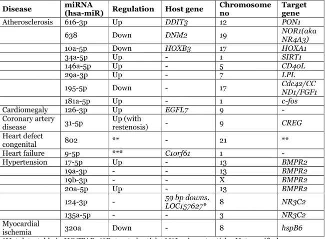
Out of the 14 diseases, only 7 of them remained after screening the miRNAs out of the appearance of a miRNA in at least two CVDs. These diseases were atherosclerosis, cardiomegaly, coronary artery disease, heart defect congenital, heart failure and hypertension (table 3). The remaining miRNAs varied from 1-8 miRNAs per CVD and host genes, target genes, up or down regulation and chromosome numbers for some miRNAs may also be viewed from the same table. HOCTAR database did not cover all the miRNAs for extracting their host genes, leaving a total of 13 miRNAs without the knowledge of their host gene. On the other hand, six of host genes were detected by the database. Regarding the target genes, a few were not attached to any article validating the target gene. The chromosomal positions vary between the different miRNAs as can be viewed in table 3. The most common chromosomal positions are chromosomes number 13, 9, 1 and 17. No miRNAs located on the Y chromosome were found for these diseases according to mirBase. However, hsa-mir-19b-3p was located on the X-chromosome.
8
Table 3. The remaining miRNAs in each CVD after screening (exclusive miRNAs in each CVD). The miRNA targets, host genes and chromosomal positions are also presented.
Disease miRNA (hsa-miR) Regulation Host gene Chromosome no Target gene
Atherosclerosis 616-3p Up DDIT3 12 PON1
638 Down DNM2 19 NOR1(aka NR4A3)
10a-5p Down HOXB3 17 HOXA1
34a-5p Up - 1 SIRT1 146a-5p Up - 5 CD40L 29a-3p Up - 7 LPL 195-5p Down - 17 Cdc42/CCND1/FGF1 181a-5p Up - 1 c-fos Cardiomegaly 126-3p Up EGFL7 9 - Coronary artery
disease 31-5p Up (with restenosis) - 9 CREG
Heart defect
congenital 802 ** - 21 **
Heart failure 9-5p *** C1orf61 1 -
Hypertension 17-5p Up - 13 BMPR2 19a-3p - - 13 BMPR2 19b-3p - - X BMPR2 20a-5p Up - 13 BMPR2 124-3p - 59 bp downs. LOC157627* 8 NR3C2 135a-5p - - 3 NR3C2 Myocardial
ischemia 320a Down - 8 hspB6
*Not detectable in HOCTAR. **Retracted article. ***Irrelevant article.-Not specified.
Review of articles
A total of about 39 articles, based on the miRNA filtration from miRWalk, were reviewed in terms of disease, miRNA regulation, biological fluid and extraction and quantification method (table 4). As can be seen from the table, the articles indicated detection of miRNAs and their regulation in several diseases, specifically various cancerous diseases and also some CVDs. Furthermore, the most common biological fluids and miRNA extraction kits were plasma and miRNeasy commercial kit.
Table 4. Summarization of articles for circulating miRNAs.
Hsa-mir Disease/health
condition and regulation (↑/↓)
Biological fluid miRNA extraction
kit
638 SLE, SSc ↑ (32) Plasma Norgen Total RNA
Purification
PCOS ↑ (33) Serum TRIzol + miRNeasy
SEOC (34) Serum mirVana
10a-5p Chronic hepatitis B ↑
(35) Serum miRNeasy
Osteoporosis ↑ (36) Serum miRNeasy Acute myeloid
leukemia ↑ (37)
Serum TRIzol reagent
146a-5p Hypertrophic
cardiomyopathy ↑
(38)
Plasma Qiazol lysis reagent + miRNeasy
HIV ↑1 (39) Plasma Qiazol lysis reagent +
miRNeasy
29a-3p Hypertrophic
cardiomyopathy ↑
(38)
Plasma Qiazol lysis reagent + miRNeasy
9
PCOS ↓ (33) Serum TRIzol + miRNeasy
WEMD (40) Plasma High Pure miRNA
isolation
195-5p Breast cancer ↓ (41) Plasma + PBMC miRNeasy (plasma) +
mirVana (PBMC) Osteosarcoma ↑ (42) Plasma mirVana
Gastric cancer ↓ (43) Serum High Pure miRNA isolation
181a-5p Breast ↓, colorectal,
lung, thyroid and melanoma ↑ tumors
(44)
Plasma and serum miRNeasy
Pre-eclampsia ↑2 (45) Plasma RNeasy Min Elute
spin spin columns
HTAP ↓ (46) Serum RNeasy mini
126-3p Diabetes3 ↓ (47) Plasma RNA purification
CAV ↑ (48) Plasma mirVana
Diabetes type 2 ↓
(49) Plasma RNA purification
STEMI ↓ (50) Serum Trizol reagent
31-5p Obesity and
overweight ↑ (51)
Plasma miRNeasy
Celiac disease ↓ (52) Plasma miRNeasy
17-5p Hypertrophic
cardiomyopathy ↑
(38)
Plasma Qiazol lysis reagent + miRNeasy
Esophageal Adenocarcinoma ↓
(53)
Serum miRNeasy
Endometriosis ↓ (54) Plasma mirVana ESNCLC ↓4 (55) Serum and plasma Total RNA
purification Gastric cancer ↑ (56) Plasma mirVana
WEMD ↑ (40) Plasma High pure miRNA
isolation
19a-3p Pre-eclampsia ↓ (57) Plasma miRNeasy
Crohns disease ↓ (58) Serum
Astrocytoma ↑ (59) Serum mirVana
19b-3p AMI ↑ (60) Plasma Trizol reagent
Pre-eclampsia ↓ (57) Plasma miRNeasy Hypertrophic
cardiomyopathy ↑
(38)
Plasma Qiazol lysis reagent + miRNeasy
Crohns disease ↓ (58) Serum miRNeasy Gastric cancer ↓ (61) plasma mirVana
20a-5p WEMD ↑ (40) plasma High pure miRNA
isolation Astrocytoma ↑ (59) serum mirVana
124 DD ↓ (62) Buffy coat miRNeasy
AIS (63) Serum Trizol reagent
Smokers ↑ (64) Plasma miRNeasy
PCOS (33) Serum Trizol + miRNeasy
Prostate cancer ↑
(65) Whole blood Ribopure
135a Endometriosis ↓ (66) Serum mirVana isolation
320 CIN (67) Plasma miRNeasy
AKI ↓ (68) Plasma Masterpure RNA
purification Diabetes type 2 ↓
(69)
Plasma miRNeasy
Arrows indicate up or downregulation in comparison to controls. No arrow indicates unaltered miRNA or unknown. Systemic lupus erythematosus (SLE), systemic sclerosis (SSc), polycystic
10 ovarian syndrome (PCOS), serous epithelial ovarian cancer (SEOC), human immunodeficiency virus (HIV), wet age-related macular degeneration (WEMD), hypertriglyceridemia induced acute pancreatitis (HTAP), cardiac allograft vasculopathy (CAV), ST-segment elevation myocardial infarction (STEMI), early stage non-small cell lung cancer (ESNCLC), acute myocardial infarction (AMI), diastolic dysfunction (DD), acute ischemic stroke (AIS), contrast induced nephropathy (CIN), acute kidney injury (AKI). 1Upregulated in elite compared to
chronic, although not significant. 2Upregulated at the time of delivery. 3With and without
complications. 4Downregulated in serum and no difference in plasma.
Results GeneMANIA
Results from GeneMANIA showed a variation of different interactions among the introduced genes (miRNA host and target genes) that might imply to potential role of a gene regulatory network among CVDs. After selecting the functions recognized by the database, found on the left corner of the site, the following interactions were selected:
Blood vessel development/blood vessel morphogenesis:
HOXA1: shared protein domains and genetic interaction with HOXB3. EGFL7: genetic interaction with HOXB3 and CCND1.
HOXB3: Genetic interaction with HOXA1, EGFL7 and PON1. Co-expression with LPL and shared protein domain with HOXA1.
SIRT1: Co-expression with CREG1.
Positive regulation of cholesterol transport/regulation of cholesterol efflux/positive regulation of lipid transport:
PON1: Genetic interaction with HOXB3.
SIRT1: Co-expression with CREG1, physical interaction and “predicted” with FOS.
Triglyceride homeostatis/acylglycerol homeostasis:
SIRT1: Co-expression with CREG1.
LPL: Physical interaction with CCND1. Co-expression with HOXB3 and CREG1.
Transcription corepressor activity:
SIRT1: Co-expression with CREG1.
CREG1: Co-expression with LPL, SIRT1 and DDIT3. DDIT3: Co-expression with CREG1.
CCND1: Physical interaction with LPL and genetic interaction with EGFL7.
Discussion
Choice of method
This has been a data collection and processing study along with a review study where data has been collected from various available databases. Initially, the database miRWalk was applied and the results for how many miRNAs and their target genes can be viewed in table 3 in the result section.
Collecting information from different databases like miRWalk, mirBase and HOCTAR was an advantage and made the process rather flexible due to the publicly open access and organized collection of data. Some difficulties were however encountered. Despite the experimentally validated information provided by miRWalk via a voucher number linked to articles in Pubmed, some links did not provide an accurate tie to neither the miRNA nor the disease in question. Therefore, information on target genes and regulation could not be provided in some cases, hence the blank squares in table 3. This also questions the ability of the software to find relevant articles. In order to make sure that a relevant article was being referred to, the issue was avoided by basically reading through the articles and using common sense to make sure the link was relevant, otherwise it was left out. Since one single miRNA can target several mRNAs (2) the miRWalk database was chosen since it provides validated information on some of those targets. Of course other databases could have been used if it weren’t for the fact that these only provide predicted targets and not validated ones like miRWalk.
11 Furthermore consequences of informatics studies are that the information is taken from available databases, which in turn are obtained from various sources, and is not confirmed experimentally by one. An example worth pointing out is that one’s own interpretations were used to figure out target genes and regulations through articles, which can be a source of uncertainty. Although these sources are rather reliable and seem to be referred to in articles, the information is still being passed on from one source to another and is finally being summarized in this study. This leads to the possibility that the information might be taken out of their context.
The database used for review of articles, Pubmed, was considered a suitable source to apply due to its extensive supply of sources. Of course other databases could have been utilized, yet Pubmed was the one that felt easier to handle and frequently used. Although other search words could have been used to reach the same results, the chosen ones were thought to be convenient since they gave enough results to cope with.
Chromosomal positions
In table 3, the location of miRNAs in human chromosomes can be viewed. The results, obtained from mirBase, show that a majority of the miRNAs are located on chromosome number 1 and 13 that predisposes for usefulness in linkage and association studies by knowing or predicting the chromosomal positions and proceed from it. Additionally, since there was no sign of miRNAs located on the Y chromosome, however one hit of hsa-mir-19b-3p was located on the X-chromosome. This led to the conclusion that, based on the chosen miRNAs, there might not be any distinguishable differences between women and men in terms of miRNA chromosome location, and existence in general. Naturally, one would need more research to truly further confirm this statement such as expression pattern study of miRNAs and their host and target genes between women and men.
Expression of miRNAs in CVDs and other diseases
A range of various diseases were detected in the article search that can be viewed in table 4. The low variation of types of CVDs found in the articles implies the need for further more research in the field of those miRNAs found from the miRWalk database as diagnostic markers in various CVDs.
Another matter noticed was the different regulations of the miRNAs found from the miRWalk database compared to the other diseases in the review part. For example, mir-638 seems to be downregulated in atherosclerosis (table 3) and unaltered or upregulated in the other diseases in the review. The same result can be seen for mir-10a-5p. On the other hand, mir-146a-5p seems to be upregulated in atherosclerosis and also upregulated in other diseases such as hypertrophic cardiomyopathy from the review.
In some cases there seems to be an up or downregulation in table 3 and a variety of up-and downregulations in other diseases. Some examples would be mir-29a-3p, which is upregulated in atherosclerosis and both up and downregulated in other diseases such as hypertrophic cardiomyopathy, downregulated mir-195-5p also in atherosclerosis, and upregulated mir-181a-5p in atherosclerosis, upregulation of mir-126-3p in cardiomegaly and upregulation of mir-31-5p in coronary artery disease. This will be more discussed further down.
Furthermore, the CVDs found in the review illustrate other miRNAs than the CVDs obtained from miRWalk even though this was tried to be avoided in the filtering of the miRNAs from the beginning. Examples are mir-146a-5p, which is altered in hypertrophic cardiomyopathy according to the review section and in atherosclerosis according to miRWalk, instead of cardiomegaly that is similar to hypertrophic cardiomyopathy. Similarly, mir-181a-5p is expressed in atherosclerosis according to miRWalk and in preeclampsia and hypertriglyceridemia induced acute pancreatitis based on the review. A side note here is that hypertriglyceridemia and atherosclerosis
12 are connected (70). A further example is mir-17-5p that is expressed in cardiomyopathy (review) and atherosclerosis (miRWalk-method).
However, there were similarly expressed miRNAs. An example would be mir-19a-3p that is altered in preeclampsia and, although not sure, altered in hypertension. Similar reasoning can be made for mir-19b-3p that also appears in preeclampsia and, not to mention, myocardial infarction and hypertrophic cardiomyopathy.
Another matter worth mentioning is since a dysregulation of mir-124 in smokers has been detected, a suspicion as to what other factors might affect the alteration of miRNAs was raised. Although, according to table 3, this miRNA is altered in hypertension, which can be a cause of smoking. In the other work (64), during the recruitment of patients, there was no exclusion of hypertensive volunteers, which in turn strengthens the statement that there might have been individuals with hypertension involved.
As a comparison of this study to (71), where mir-92a among others is being discussed and linked to angiogenesis whereas here, according to miRWalk, this miRNA is linked to hypertension and myocardial infarction. Similarly, mir-126 is associated to atherosclerosis and angiogenesis (71) and linked to cardiomegaly according to miRWalk.
In cardiomyocyte proliferation, mir-133 is involved, which makes sense considering its association in heart failure and long QT-syndrome according to miRWalk. Similarly, mir-29 is associated with atherosclerosis according to miRWalk and tissue fibrosis in (71).
Alteration of miRNAs in various diseases
Due to the fact that miRNAs are found in different tissues and are dysregulated in several types of diseases it is important to underscore that an increase or decrease of a certain miRNA connected with a CVD may also advocate other types of diseases. Occurrence of miRNAs in other types of diseases was not taken into consideration when information was collected from miRWalk. This proposes that the dysregulation of a certain miRNA in a comorbid patient may not indicate a CVD. According to the results where the miRNAs that were listed as exclusive miRNAs for a specific CVD (table 3) also appeared in a variety of other diseases (review). This suggests that a specific miRNA cannot be considered as an exclusive diagnostic marker unless all other diseases are excluded during diagnosis or invasively analysis of biological tissues are performed in order to receive a correct result, which is not very practical. Of course, if there is knowledge that a miRNA exists in different types of diseases where it is upregulated in a CVD and downregulated in another disease, then this miRNA could be utilized as a potential biomarker through analysis of biofluids. An example is hsa-miR-10a-5p, which is downregulated in atherosclerosis and upregulated in chronic hepatitis, osteoporosis and acute myeloid leukemia. An exclusion of diseases would otherwise have to be made for patients in order to exclusively limit the miRNA for one CVD only. On the other hand, it might be worth mentioning that most articles obtained from miRWalk had performed analysis on tissue based samples and not circulating miRNAs received from body fluids where the concentration of miRNA may differ. This deviation remains to be evaluated.
Extraction and detection
In the review part, a majority of the articles used miRNeasy isolation kit as a miRNA extraction method that was followed by mirVana miRNA isolation kit for serum or plasma. Most of the studies used the commercial extraction kits according to manufacturer’s protocols while some added slight modifications that may have affected the result of collected and detected miRNA. Of course there are other factors that may have influenced the results as well such as which inclusion and exclusion criteria were
13 used (some did not check for presence of other diseases for example, which can affect the result negatively), the lab process, handling of samples and so on.
An agreement as to whether plasma, serum or any other biological fluid, using any of the commercial extraction kits, is superior is difficult to come by since miRNAs from different types of body fluids actually have been detected. Furthermore, there was no real miRNA level measurement methods such as different primer designing protocols and some chemical modifications on nucleotides (such as Exiqon's LNA) to improve the sensitivity and specificity compared to traditional chemistries that made it difficult to determine altered miRNA(s) in each disease.
The large extent of qRT-PCR usage indicates the superiority of this method for quantification and profiling of miRNAs. In some cases a combination of qRT-PCR and microarray was performed or vice versa. This might have further confirmed the results despite limitations of microarray such as lower accuracy, low to medium sensitivity and dynamic range unlike qRT-PCR, which typically has more accuracy and sensitivity (20).
Interpretation of GeneMANIA results and the relation between CVDs regarding miRNA target and host genes
The results from GeneMANIA shows various types of connections between the introduced genes, both host and target genes. As mentioned in the method section, only the genes recognized to have a function in GeneMANIA is included.
Firstly, a genetic interaction and shared protein domain is noticed between HOXA1, which is the target gene of hsa-miR-10a-5p that is downregulated in atherosclerosis, and HOXB3 that is the host gene. HOXA1 seems to have participation in blood vessel morphogenesis and development according to GeneMANIA, which also seems to be a similar case according to the literature (25). A downregulation of this miRNA would naturally imply an upregulation in target HOXA1 that in turn would lead to some defect in the blood vessel morphogenesis. A downregulation of hsa-miR-10a-5p has not been detected in any of the diseases found in table 5 where it seems to be upregulated in all the found articles. This does not necessarily mean this miRNA could be used as one exclusive biomarker, but rather implies the need for further research in this field to confirm it.
Moreover, there seems to be a genetic interaction between EGFL7, the host gene of hsa-miR-126-3p upregulated in cardiomegaly, and HOXB3. EGFL7 has, according to both the literature and GeneMANIA, a role in blood vessel development (23). HOXB3 is, as mentioned before, the host gene of hsa-miR-10a-5p, downregulated in atherosclerosis. This gene also has a function in blood vessel development according to GeneMANIA and some sort of development function according to the literature (26). The genetic interaction among these could imply that a perturbation in any of these two genes might predispose for a defect in the vasculature system.
Furthermore, HOXB3 also has a genetic interaction with PON1, which is the target gene of hsa-miR-616-3p that is upregulated in atherosclerosis. This gene is a positive regulator of cholesterol transport (GeneMANIA) and downregulated in atherosclerosis that indicates less positive regulation of cholesterol transport. Therefore, high cholesterol levels can lead to clogged arteries and atherosclerosis, since the two genes have a genetic interaction.
A co-expression exists between HOXB3 and LPL. If HOXB3 is downregulated then LPL should also be downregulated due to the co-expression connection. This can also be figured out from table 3. Therefore, a dysregulation of HOXB3 can rupture the triglyceride homeostasis (function of LPL).
SIRT1, the target gene of hsa-miR-34a-5p that is upregulated in atherosclerosis (miRWalk-method), has according to GeneMANIA a function in blood vessel
14 development, transcription corepressor activity, positive regulation of cholesterol transport and triglyceride homeostasis. CREG1 also has a transcription corepressor activity and together with SIRT1 is co-expressed. CREG1 is the target gene of hsa-miR-31-5p that is upregulated in coronary artery disease (miRWalk-method) and thereby implies the downregulation of CREG1. This makes sense with the downregulation of SIRT1 since there is an existing co-expression. Other genes possessive of transcription corepressor activity are DDIT3, CCND1 and MECP2. CREG1 has a co-expression connection with DDIT3. A dysregulation of any of these genes would predispose for diseases in each of the connecting genes. As an example, if SIRT1 has a co-expression connection with CREG1, then a patient with atherosclerosis might be predisposed for coronary artery disease.
Additionally, CREG1 also has a co-expression with LPL, which has a function in triglyceride homeostasis according to GeneMANIA and the literature (27). LPL is the target gene of hsa-miR-29a-3p that is upregulated in atherosclerosis (miRWalk-method) and acknowledges thereby the downregulation of LPL. This might indicate that a person with dysfunction of triglyceride homeostasis might be predisposed to coronary artery disease.
Meanwhile, LPL has physical interaction with CCND1 that, in GeneMANIA, has transcription corepressor activity and is upregulated since the miRNA targeting this gene is downregulated. An upregulation implies more interaction with LPL leading to further repression of transcription activity of LPL and therefore less triglyceride homeostasis.
Conclusion
Based on the literature there is potential for miRNAs to be considered as novel CVD biomarkers. This statement was further confirmed in this study due to the possibility of extraction and detection of miRNAs through commercial kits and modern technology. However, due to miRNA alteration in non-CVDs, there needs to be an exclusion of other diseases in order to diagnose a patient if the miRNA regulation simulates the desired one, for example mir-146a-5p, which is upregulated in atherosclerosis as well as hypertrophic cardiomyopathy and HIV from the findings. On the other hand, based on the findings of this study, mir-10a-5p can be utilized as a potential biomarker for diagnosis of atherosclerosis.
Furthermore, GeneMANIA showed interactions among genes leading to patients with primarily ruptured vascular development and triglyceride homeostasis deficiencies might be predisposed to other CVDs.
Acknowlegement
I would like to thank my supervisor, Dr. Samaneh Tousi, for her support and guidance throughout this project.
15
References
1. Liu N, Olson EN. MicroRNA regulatory networks in cardiovascular development. Dev cell. 2010 April; 18(4): 1-15.
2. Kunz M, Xiao K, Liang C, Viereck J, Pachel C, Frantz S et al. Bioinformatics of cardiovascular miRNA biology. J mol cell cardiol. 2015 Dec; 89(Pt A): 3-10.
3. Shen E, Diao X, Wei C, Wu Z, Zhang L, Hu B. MicroRNAs target gene and signaling pathway by bioinformatics analysis in the cardiac hypertrophy. Biochem biophys res commun. 2010 July; 397(3): 380-5.
4. Gennarino VA, Sardiello M, Mutarelli M, Dharmalingam G, Maselli V, Lago G, et al. HOCTAR database: a unique resource for microRNA target prediction. Gene. 2011 July; 48D(1-2):51-8.
5. Dweep H, Sticht C, Pandey P, Gretz N. Mirwalk database prediction of possible miRNA binding sites by “walking” the genes of three genomes. J Biomed inform. 2011 Oct;44(5):839-47.
6. World health organization. New initiative launched to tackle cardiovascular disease, the world’s number one killer. Cited 161103. Brought from:
http://www.who.int/cardiovascular_diseases/global-hearts/Global_hearts_initiative/en/
7. Vasan RS. Biomarkers of cardiovascular disease: molecular basis and practical
considerations. Circulation. 2006 May; 113(19):2335-62.
8. Twerenbold R, Jaffe A, Reichlin T, Reiter M, Mueller C. High-sensitive troponin T measurements: what do we gain and what are the challenges? Eur Heart J. 2012 Mar;33(5):579-86.
9. Polachini do Valle A, José Fortes Villas Boas P, Iglesias de Oliveira Vidal E, Bono Fukushima F. B-type natriuretic peptide in the evaluation of cardiac function.
10. Kuick DL. Electrocardiogram (ECG or EKG). 2016 [Updated 2016-07-15; cited
2016-11-23]. Obtained from:
http://www.medicinenet.com/electrocardiogram_ecg_or_ekg/page3.htm
11. NPS Medicinewise. Limitations of in-clinic BP measurements. 2015 [Updated 2015-02-25; cited 2016-11-25]. Obtained from: http://www.nps.org.au/conditions/heart-blood- and-blood-vessel-conditions/blood-pressure/for-health-professionals/measuring-blood-pressure/limitations-of-in-clinic-bp-measurements
12. Gupta Sk, Bang C, Thum T. Circulating MicroRNAs as Biomarkers and Potential Paracrine Mediators of Cardiovascular Disease. Circ Cardiovasc Genet. 2010 Oct;3(5):484-8.
13. Gilad S, Meiri E, Yogev Y, Benjamin S, Lebanony D, Yerushalmi N, et al. Serum miRNAs are promising novel biomarkers. PLoS One. 2008 Sep;3(9):e3148.
14. McDonald JS, Milosevic D, Reddi HV, Grebe SK, Algeciras-Schimnich A. Analysis of circulating microRNA: preanalytical and analytical challenges. Clin Chem. 2011 Jun;57(6):833-40.
15. Kirschner MB, Kao SC, Edelman JJ, Armstrong NJ, Vallely MP, van Zandwijk N et al. Haemolysis during sample preparation alters microRNA content of plasma. PLoS One. 2011;6(9):e24145.
16. http://www.lcsciences.com/discovery/microrna-sample-preparation-from-biofluid-samples/
16 17. Duy J, Koehler JW, Honko AN, Minogue TD. Optimized microRNA purification from
TRIzol-treated plasma. BMC Genomics. 2015 Feb 18;16:95.
18. Statens veterinärmedicinska anstalt. PCR-metoden. 2016 [Updated 2016-05-19; cited 2016-11-19]. Obtained from: http://sva.se/analyser-och-produkter/pcr-metoden 19. Thermofisher scientific. Basic Principles of RT-qPCR. 2017 [Cited 2017-01-09]. Obtained
from: https://www.thermofisher.com/se/en/home/brands/thermo- scientific/molecular-biology/molecular-biology-learning-center/molecular-biology-resource-library/basic-principles-rt-qpcr.html
20. Pritchard CC, Cheng HH, Tewari M. MicroRNA profiling: approaches and considerations. Nat Rev Genet. 2012 Apr 18;13(5):358-69.
21. Small EM, Olson EN. Pervasive roles of microRNAs in cardiovascular biology. Nature. 2011 Jan;469(7330):336-4.
22. Zhang C. MicroRNAs: role in cardiovascular biology and disease. Clin Sci (Lond). 2008 Jun;114(12):699-706. http://www.scielo.br/pdf/jbpml/v49n4/v49n4a02.pdf
23. Nichol D, Stuhlmann H. EGFL7: a unique angiogenic signaling factor in vascular development and disease. Blood. 2012 Feb 9;119(6):1345-52.
24. Greer JM, Puetz J, Thomas KR, Capecchi MR. Maintenance of functional equivalence during paralogous Hox gene evolution. Nature. 2000 Feb 10;403(6770):661-5.
25. Zha TZ, Hu BS, Yu HF, Tan YF, Zhang Y, Zhang K. Overexpression of HOXA1 correlates with poor prognosis in patients with hepatocellular carcinoma. Tumour Biol. 2012 Dec;33(6):2125-34.
26. Greer JM, Puetz J, Thomas KR, Capecchi MR. Maintenance of functional equivalence during paralogous Hox gene evolution. Nature. 2000 Feb 10;403(6770):661-5.
27. Pingitore P, Lepore SM, Pirazzi C, Mancina RM, Motta BM, Valenti L et al. Identification and characterization of two novel mutations in the LPL gene causing type I hyperlipoproteinemia. J Clin Lipidol. 2016 Jul-Aug;10(4):816-23.
28. Patnala R, Clements J, Batra J. Candidate gene association studies: a comprehensive guide to useful in silico tools. BMC Genet. 2013 May 9;14:39.
29. Dweep, H et al. miRWalk2.0: a comprehensive atlas of microRNA-target interactions. Nature Methods. 2015;12(8):697-697.
30. Kozomara A, Griffiths-Jones S. miRBase: annotating high confidence microRNAs using deep sequencing data. Nucleic Acids Res. 2014 Jan;42(Database issue):D68-73.
31. Warde-Farley D, Donaldson SL, Comes O, Zuberi K, Badrawi R, Chao P et al. The GeneMANIA prediction server: biological network integration for gene prioritization and predicting gene function. Nucleic Acids Res. 2010 Jul;38(Web Server issue):W214-20.
32. Steen SO, Iversen LV, Carlsen AL, Burton M, Nielsen CT, Jacobsen S et al. The circulating cell-free microRNA profile in systemic sclerosis is distinct from both healthy controls and systemic lupus erythematosus. J Rheumatol. 2015 Feb;42(2):214-21. 33. Ding CF, Chen WQ, Zhu YT, Bo YL, Hu HM, Zheng RH. Circulating microRNAs in patients
with polycystic ovary syndrome. Hum Fertil (Camb). 2015 Mar;18(1):22-9.
34. Kan CW, Hahn MA, Gard GB, Maidens J, Huh JY, Marsh DJ et al. Elevated levels of circulating microRNA-200 family members correlate with serous epithelial ovarian cancer. BMC Cancer. 2012 Dec 28;12:627.
35. Tan Y, Ge G, Pan T, Wen D, Gan J. Serum MiRNA panel as potential biomarkers for chronic hepatitis B with persistently normal alanine aminotransferase. Clin Chim Acta. 2015 Dec 7;451(Pt B):232-9.
17 36. Weilner S, Skalicky S, Salzer B, Keider V, Wagner M, Hildner F et al. Differentially circulating miRNAs after recent osteoporotic fractures can influence osteogenic differentiation. Bone. 2015 Oct;79:43-51.
37. Zhi F, Cao X, Xie X, Wang B, Dong W, Gu W et al. Identification of circulating microRNAs as potential biomarkers for detecting acute myeloid leukemia. PLoS One. 2013;8(2):e56718.
38. Fang L, Ellims AH, Moore XL, White DA, Taylor AJ, Chin-Dusting J. Circulating microRNAs as biomarkers for diffuse myocardial fibrosis in patients with hypertrophic cardiomyopathy. J Transl Med. 2015 Sep 24;13:314.
39. Reynoso R, Laufer N, Hackl M, Skalicky S, Monteforte R, Turk G et al. MicroRNAs differentially present in the plasma of HIV elite controllers reduce HIV infection in vitro. Sci Rep. 2014 Aug 1;4:5915.
40. Ertekin S, Yıldırım O, Dinç E, Ayaz L, Fidancı SB, Tamer L. Evaluation of circulating miRNAs in wet age-related macular degeneration. Mol Vis. 2014 Jul 29;20:1057-66. 41. Mishra S, Srivastava AK, Suman S, Kumar V, Shukla Y. Circulating miRNAs revealed as
surrogate molecular signatures for the early detection of breast cancer. Cancer Lett. 2015 Dec 1;369(1):67-75.
42. Lian F, Cui Y, Zhou C, Gao K, Wu L. Identification of a plasma four-microRNA panel as potential noninvasive biomarker for osteosarcoma. PLoS One. 2015 Mar 16;10(3):e0121499.
43. Gorur A, Balci Fidanci S, Dogruer Unal N, Ayaz L, Akbayir S, Yildirim Yaroglu H et al. Determination of plasma microRNA for early detection of gastric cancer. Mol Biol Rep. 2013 Mar;40(3):2091-6.
44. Ferracin M, Lupini L, Salamon I, Saccenti E, Zanzi MV, Rocchi A et al. Absolute quantification of cell-free microRNAs in cancer patients. Oncotarget. 2015 Jun 10;6(16):14545-55.
45. Murphy MS, Casselman RC, Tayade C, Smith GN. Differential expression of plasma microRNA in preeclamptic patients at delivery and 1 year postpartum. Am J Obstet Gynecol. 2015 Sep;213(3):367.e1-9.
46. An F, Zhan Q, Xia M, Jiang L, Lu G, Huang M et al. From moderately severe to severe hypertriglyceridemia induced acute pancreatitis: circulating miRNAs play role as potential biomarkers. PLoS One. 2014 Nov 3;9(11):e111058.
47. Olivieri F, Spazzafumo L, Bonafè M, Recchioni R, Prattichizzo F, Marcheselli F et al. MiR-21-5p and miR-126a-3p levels in plasma and circulating angiogenic cells: relationship with type 2 diabetes complications. Oncotarget. 2015 Nov 3;6(34):35372-82.
48. Singh N, Heggermont W, Fieuws S, Vanhaecke J, Van Cleemput J, De Geest B. Endothelium-enriched microRNAs as diagnostic biomarkers for cardiac allograft vasculopathy. J Heart Lung Transplant. 2015 Nov;34(11):1376-84.
49. Olivieri F, Bonafè M, Spazzafumo L, Gobbi M, Prattichizzo F, Recchioni R et al. Age- and glycemia-related miR-126-3p levels in plasma and endothelial cells. Aging (Albany NY). 2014 Sep;6(9):771-87.
50. Hsu A, Chen SJ, Chang YS, Chen HC, Chu PH. Systemic approach to identify serum microRNAs as potential biomarkers for acute myocardial infarction. Biomed Res Int. 2014;2014:418628.
51. Iacomino G, Russo P, Stillitano I, Lauria F, Marena P, Ahrens W et al. Circulating microRNAs are deregulated in overweight/obese children: preliminary results of the I.Family study. Genes Nutr. 2016 Mar 21;11:7. doi: 10.1186/s12263-016-0525-3.
18 52. Buoli Comani G, Panceri R, Dinelli M, Biondi A, Mancuso C, Meneveri R et al.
miRNA-regulated gene expression differs in celiac disease patients according to the age of presentation. Genes Nutr. 2015 Sep;10(5):482.
53. Chiam K, Wang T, Watson DI, Mayne GC, Irvine TS, Bright T et al. Circulating Serum Exosomal miRNAs As Potential Biomarkers for Esophageal Adenocarcinoma. J Gastrointest Surg. 2015 Jul;19(7):1208-15.
54. Jia SZ, Yang Y, Lang J, Sun P, Leng J. Plasma miR-17-5p, miR-20a and miR-22 are down-regulated in women with endometriosis. Hum Reprod. 2013 Feb;28(2):322-30.
55. Heegaard NH, Schetter AJ, Welsh JA, Yoneda M, Bowman ED, Harris CC. Circulating micro-RNA expression profiles in early stage nonsmall cell lung cancer. Int J Cancer. 2012 Mar 15;130(6):1378-86.
56. Tsujiura M, Ichikawa D, Komatsu S, Shiozaki A, Takeshita H, Kosuga T et al. Circulating microRNAs in plasma of patients with gastric cancers. Br J Cancer. 2010 Mar 30;102(7):1174-9.
57. Sandrim VC, Luizon MR, Palei AC, Tanus-Santos JE, Cavalli RC. Circulating microRNA expression profiles in pre-eclampsia: evidence of increased miR-885-5p levels. BJOG. 2016 Dec;123(13):2120-2128.
58. Lewis A, Mehta S, Hanna LN, Rogalski LA, Jeffery R, Nijhuis A et al. Low Serum Levels of MicroRNA-19 Are Associated with a Stricturing Crohn's Disease Phenotype. Inflamm Bowel Dis. 2015 Aug;21(8):1926-34.
59. Zhi F, Shao N, Wang R, Deng D, Xue L, Wang Q et al. Identification of 9 serum microRNAs as potential noninvasive biomarkers of human astrocytoma. Neuro Oncol. 2015 Mar;17(3):383-91.
60. Wang KJ, Zhao X, Liu YZ, Zeng QT, Mao XB, Li SN et al. Circulating 19b-3p, MiR-134-5p and MiR-186-5p are Promising Novel Biomarkers for Early Diagnosis of Acute Myocardial Infarction. Cell Physiol Biochem. 2016;38(3):1015-29.
61. Zhang J, Song Y, Zhang C, Zhi X, Fu H, Ma Y et al. Circulating MiR-16-5p and MiR-19b-3p as Two Novel Potential Biomarkers to Indicate Progression of Gastric Cancer. Theranostics. 2015 Apr 5;5(7):733-45.
62. Nair N, Kumar S, Gongora E, Gupta S. Circulating miRNA as novel markers for diastolic dysfunction. Mol Cell Biochem. 2013 Apr;376(1-2):33-40.
63. Jia L, Hao F, Wang W, Qu Y. Circulating miR-145 is associated with plasma high-sensitivity C-reactive protein in acute ischemic stroke patients. Cell Biochem Funct. 2015 Jul;33(5):314-9.
64. Banerjee A, Waters D, Camacho OM, Minet E. Quantification of plasma microRNAs in a group of healthy smokers, ex-smokers and non-smokers and correlation to biomarkers of tobacco exposure. Biomarkers. 2015 Mar;20(2):123-31.
65. Medina-Villaamil V, Martínez-Breijo S, Portela-Pereira P, Quindós-Varela M, Santamarina-Caínzos I, Antón-Aparicio LM et al. Circulating MicroRNAs in blood of patients with prostate cancer. Actas Urol Esp. 2014 Dec;38(10):633-9.
66. Cho S, Mutlu L, Grechukhina O, Taylor HS. Circulating microRNAs as potential biomarkers for endometriosis. Fertil Steril. 2015 May;103(5):1252-60.
67. Gutiérrez-Escolano A, Santacruz-Vázquez E, Gómez-Pérez F. Dysregulated microRNAs involved in contrast-induced acute kidney injury in rat and human. Ren Fail. 2015;37(9):1498-506.
19 68. Lorenzen JM, Kielstein JT, Hafer C, Gupta SK, Kümpers P, Faulhaber-Walter R et al. Circulating miR-210 predicts survival in critically ill patients with acute kidney injury. Clin J Am Soc Nephrol. 2011 Jul;6(7):1540-6.
69. Zampetaki A, Kiechl S, Drozdov I, Willeit P, Mayr U, Prokopi M et al. Plasma microRNA profiling reveals loss of endothelial miR-126 and other microRNAs in type 2 diabetes. Circ Res. 2010 Sep 17;107(6):810-7.
70. Glueck CJ, Fallat RW, Tsang R. Hypercholesterolemia and hypertriglyceridemia in children. A pediatric approach to primary atherosclerosis prevention. Am J Dis Child. 1974 Oct;128(4):569-77.
71. Schulte C, Zeller T. microRNA-based diagnostics and therapy in cardiovascular disease-Summing up the facts. Cardiovasc Diagn Ther. 2015 Feb;5(1):17-36.
1 Department of Chemistry S-901 87 Umeå, Sweden Telephone +46 90 786 50 00 Text telephone +46 90 786 59 00 www.umu.se