PERSONAL USE
ONLY
Prolonged survival and expression of neural markers
by bone marrow-derived stem cells transplanted into
brain lesions
Esteban Alberti
1ABCDEFG, Marek Los
2,3,4DEF, Rocío García
1BC, Jorge L. Fraga
5BC,
Teresa Serrano
1BC, Elizabeth Hernández
1BC, Thomas Klonisch
2BC,
René Macías
1BC, Lísis Martínez
1BC, Lázara Castillo
1BC, Karelys de la Cuétara
1BC
1 Department of Neurobiology, International Center of Neurological Restoration, CIREN, Havana, Cuba 2 Department of Human Anatomy and Cell Science, Univ. Manitoba, Winnipeg, Canada3 Manitoba Institute of Cell Biology, and Department of Biochemistry and Medical Genetics, Univ. Manitoba, Winnipeg, Canada
4 BioApplications Enterprises, Winnipeg, Manitoba, Canada 5 Department of Parasitology, “Pedro Kouri” Institute, Havana, Cuba
Source of support: Interdisciplinary Center of Clinical Research, Tübingen (IZKF; 01KS9602)
Summary
Background:
Bone marrow-derived stem cell transplantation is a potentially viable therapeutic option for the treatment of neurodegenerative disease.Material/Methods:
We have isolated bone marrow stem cells by standard method. We then evaluated the survival of rats’ bone marrow mononuclear cells implanted in rats’ brain. The cells were extracted from rats’ femurs, and marked for monitoring purposes by adenoviral transduction with Green Fluorescent Protein (GFP). Labeled cells were implanted within the area of rats’ striatum lesions that were in-duced a month earlier employing quinolinic acid-based method. The implants were phenotyped by monitoring CD34; CD38; CD45 and CD90 expression. Bone marrow stromal cells were extract-ed from rats’ femurs and cultivatextract-ed until monolayer bone marrow stromal cells were obtainextract-ed. The ability of bone marrow stromal cells to express NGF and GDNF was evaluated by RT-PCR.Results:
Implanted cells survived for at least one month after transplantation and dispersed from the area of injection towards corpus callosum and brain cortex. Interestingly, passaged rat bone marrow stromal cells expressed NGF and GDNF mRNA.Conclusions:
The bone marrow cells could be successfully transplanted to the brain either for the purpose of trans-differentiation, or for the expression of desired growth factors.key words:
bone marrow stromal cells • CD34 • CD38 • CD45 • CD90 • hematopoetic stem cells • trans-differentiation • transplantationAbbreviations:
CNS – central nervous system; GDNF – glial-derived neurotrophic factor; GFP – green fl uorescent protein; NGF – nerve growth factor; PBS – phosphate buffered saline; RT-PCR – reversetranscriptase-polymerase chain reaction
Full-text PDF:
http://www.medscimonit.com/fulltxt.php?ICID=869551Word count:
3282Tables:
1Figures:
6References:
38Author’s address:
Marek Los, BioApplications Enterprises, Winnipeg, MB, R2V 2N6, Canada, e-mail: bioappl@gmail.comAuthors’ Contribution: A Study Design B Data Collection C Statistical Analysis D Data Interpretation E Manuscript Preparation F Literature Search G Funds Collection Received: 2007.12.08 Accepted: 2008.04.25 Published: 2009.02.01 Basic Research
PMID: 19179961
BR
•
•
•
•
•
•
•
PERSONAL USE
ONLY
B
ACKGROUNDApoptosis is the common mechanism of cell demise in cen-tral nerve system and in other organs, and apoptosis inhi-bition is the principle of several experimental methods of treatment [1–3]. Transplantation of pluripotent stem cells that have the capacity to differentiate into neural tissue com-ponents is a viable treatment alternative for neurodegen-erative diseases. Embryonic stem cells have been used in a regenerative medicine with variable success, but their use attracts signifi cant ethical controversy in may countries [4]. Currently, pluripotent neural stem cells are not available, and surgical removal of neural tissues for the sole purpose of isolation of neural stem cells is not a feasible option. The aim of the described experiments was to test if bone-mar-row derived cells could technically be used to treat neuro-degenerative lesions. The stem cells are an attractive cel-lular source for the treatment of illnesses of the Central Nervous System (SNC). Under certain conditions they are capable of self-renewal for long periods of time – remain in totipotent state until they receive appropriate differen-tiation signals [5,6].
Bone marrow is the only well-known organ containing a het-erogeneous mixture of different types of stem cells [6,7]. These populations include the hematopoietic system and stromal cells that interact in a reciprocal way through inter-cellular contacts and secretion of cytokines or growth fac-tors [8]. The hematopoietic system contains stem cells that are responsible for the formation of all the types of blood cells. Bone marrow stromal cells generate bone, cartilage, adipocytes, connective fi bers and a reticular net that con-stitutes the formation of these cells [7,9,10].
Plasticity of bone marrow stem cell populations gives them the capacity to generate specialized cells of a various tis-sues [5,6]. Thus, bone marrow hematopoietic stem cells are able to differentiate to microglia and macroglia once transplanted into the brain of mature mice [9]. In anoth-er expanoth-erimental system when transplanted into the para-ventricular area of neonatal mice, they differentiated to as-trocytes and neurons [11]. Furthermore, when these cells were given intravenously to rats with traumatic brain inju-ry, they migrated toward the damaged brain area within 15 days, some of them differentiated to neurons, and improved neural functions [12].
The development of transplants in experimental models de-mands implanted cells to be detected in situ once the ani-mal is sacrifi ced. One of the more frequently used methods for marking of these cells is genetic modifi cation that may include viral vectors expressing Green-Fluorescent Protein (GFP) [13,14], or b-galactosidase [15].
Bone marrow stromal stem cells are multipotent and easily available from whole bone marrow aspirates which can be enriched by adherence [7]. Recent reports demonstrated that bone marrow stromal stem cells are able to migrate ex-tensively, and have the potential for a neuronal differentia-tion after transplantadifferentia-tion into the brain parenchyma [8]. Neurotrophic factors promote neuronal survival and stim-ulate axonal growth. It has been proposed that under some clinical conditions, neurons fail to obtain a suffi cient
quan-tity of necessary neurotrophic factors and die by apoptosis [16–18]. Trophic support provided by transplanted cells could counteract cell death in damaged tissue. Therefore, we have tested whether bone marrow stem cells (both hema-topoetic and stromal) could differentiate and produce neu-rotrophic factors. Furthermore, using bone-marrow derived stem cells, tagged by two different methods we have tested the survival and migratory potential of these cells once im-planted into brain. Our paper communicates for the fi rst time that rat bone marrow derived stromal cells could pro-duce GDNF and NGF.
M
ATERIALANDM
ETHODSLaboratory animals
Male rats of the Sprague-Dawly line were used with a body weight between 200–250 g (CENPALAB, Havana, Cuba). Experimental animals were kept in cages with free access to water and standard chow, with light/obscured cycles of 12 h. The animals were randomly divided into three exper-imental groups: (i) animals that received the transplant of mononuclear cells genetically modifi ed to express GFP (GFP, n=10), (ii) animals that received the transplant of Hoechst labeled mononuclear cells (HOE, n=10), and (iii) n=10 an-imals used as control (mock-transplanted with cell culture medium DMEM).
Quinolinic acid induced neuronal lesions
The animals were anesthetized with chloral hydrate (420 mg/kg of body weight) and placed in the stereotactic surgi-cal devices for rodents. Lesion inductions were carried out with 1.2 μl of quinolinic acid solution (112.5mM, pH=7.4) in the right striatum. (AP=+1.2; L=+2.8; V=5.5) volume was injected at 1 μl/min [19–21].
Isolation and immunocytochemical characterization of bone marrow-derived cells
The animals (20 Sprague-Dawly rats) weights between 250 and 300 g, were anesthetized with chloral hydrate and sac-rifi ced [14]. Femurs were used for cell extractions. Bone marrow was obtained by passing with a syringe and fl ushing with sterile phosphate buffered saline (PBS) (NaCl 8 g/L; KCl 0.2 g/L; Na2HPO4 1.09 g/L; KH2PO4 0.26 g/L, pH 7.2) through the femur. The mononuclear cells were isolated us-ing Ficoll-Hypaque™-Plus gradient (Amershan-Pharmacia Biosciences, Sweden) and then cultured in DMEM supple-mented with 20% FCS, and 50 mg/mL gentamicine. Cells viability was evaluated by staining with Trypan Blue [22,23]. For immunocytochemical evaluation, the isolated cells were seeded in a 12-well-plates, fi xed in 4% paraformaldehyde in PBS, pre-incubated in a blocking solution (5% skim milk in PBS for 1 h at RT) and then incubated on ice with mono-clonal antibodies against CD34, CD38, CD45 and CD90 (Sigma-Aldrich, Oakville, ON, Canada). Cells were then washed 3 times with cold PBS and incubated with anti-mu-rine IgG and IgM, biotinylated secondary antibodies for 1 h at RT, then washed 3 times in cold PBS and fi nally the staining was developed using the ABC system and diami-nobenzidine (DAB) substrate reagent (PA-ABC system, DAKO, Mississauga, ON, Canada). The staining was detect-ed by light microscopy. Cells staindetect-ed with secondary
anti-PERSONAL USE
ONLY
bodies only were used as negative control [24,25]. The pri-mary antibodies were diluted as follows: CD34 1:40, CD38 1:200, CD45 1:50, and CD90 of 1:20 (Table 1). The opti-mal working dilutions for our experimental purposes were obtained empirically.
Bone marrow stromal cells culture
Bone marrow stromal cells were obtained from the femurs of adult male Sprague-Dawley rats (as described above). The extraction was carried out using MEM. The cells were then cultured in the same medium supplemented with 20% FCS, 2 mM L-glutamine and 100 mg/ml streptomycin. Non-adherent cells were removed by 3× washing with PBS and subsequent replacment of the medium. Bone marrow stro-mal cells were subcultured 6 times prior to performing ex-periments [26].
Labeling of bone-marrow-derived cells with GFP or with Hoechst 33342
The bone marrow-derived cells were infected with an ade-novirus type 5 (MOI=10) carrying a GFP under a human cy-tomegalovirus early promoter. Cells were then cultured for 5 days at 37°C, 5% CO2. Next, the cells were washed with PBS, cell viability was determined by Trypan blue exclusion, and used for transplantation [22,23]. Hoechst 33342 label-ing (1 μg/ml) was carried out in DMEM medium. The cells were incubated for 24 h at 37°C and 5% CO2. The cellu-lar viability was determined by MTT-assay as described pre-viously [27].
Stereotactic cell transplantation
The transplant was carried out by stereotactic surgery into the lesions area, one month after lesion induction, follow-ing the same procedure as described previously for the le-sion development. The cells were grafted into two sites in the striatum (AP=+0,7; L=+2,8; V=-5,5 and AP=+0,7; L=+2,8; V=–4,6) [28]. The cellular sample had a concentration of 1.5×10⁵ cells/μl and 1 μl was infused in each site. The
max-imum surgical time was 5 h and during this time; the cellu-lar suspensions remained at 4°C. The cellucellu-lar viability was determined/confi rmed again after the surgical procedure was concluded [19,20,22].
Histologic evaluation of transplantation results
The animals that were transplanted with cells genetically modifi ed to express GFP were sacrifi ced (n=4 each time) at day 3 and 30 after transplantation; whereas animals implant-ed with markimplant-ed cells with Hoechst reagent were sacrifi cimplant-ed
(n=4 each time) at day 10, 20 and 30 post-transplantation. The animals were anesthetized with chloral hydrate prior to killing and then perfused with 4% paraformaldehyde (PFA) through the ascending aorta. The extracted brains were then maintained for 2 h at 4% PFA and then passed through sucrose gradient solutions at 15 and 30% respec-tively for 24 h and subsequently frozen. The specimens were sectioned at 20 μm thickness according to the stereotactic atlas [28], and were randomly selected for further study [22]. GFP- and Hoechst-labeled cells were then detected by fl uorescence (Hoechst lem=420 nm, GFP lem=590 nm), by microscopy (Digital Microscope Leica DM4000/5000). Some sections were processed for conventional violet cre-sil histology-staining to identify glial and astrocyte cells by immunocytochimistry [19,20].
Reverse transcriptase-polymerase chain reaction (RT-PCR)
To detect mRNA expression, total cellular RNA from bone-marrow-derived cells at passage 7, was isolated using Trizol method (Gibco). For GDNF RT-PCR amplifi cation, the fol-lowing primers were used: 5’-gactctaagatgaagttatgggatgtc-‘3 and 5’-gggtcagatacatccacaccgtttagc-‘3. To amplify the NGF message we used 5’-tcaacaggactcacagg-‘3 and 5’-cgcggatcct-tatctcacagccttcctgctgag-‘3 primers. To amplify the BDNF mes-sage we used 5’-ttggcctacccagctgtgcggac-3’ and 5’-ctcttcgat-cacgtg ctcaaaagtg-3’ primers. The RT-PCR was carried out using “Access RT-PCR Systems” (Promega). The reaction included 10 mM of a dNTP mix, 50 pmol of each primer, 25 mM MgSO4, 31 ng of RNA, and 3U DNase. The reac-tion mixtures were maintained for 45 min at 48°C, 2 min at 94°C, then cycled 40 times through a program of 30 sec at 94°C, 1 min at 62°C for GDNF or 1 min at 50°C for NGF and 1 min at 60°C for BDNF, and 2 min at 68°C, fi nally the reaction was incubated for an extra 7 min at 68°C. The RT-PCR DNA products were electrophoresed on 1.8% aga-rose gel and stained with ethidium bromide. To confi rm the integrity of isolated mRNA a fragment of the b-actina mRNA was amplifi ed (primers: 5’-tca cgc acg att tcc ctc tca g-‘3 and 5’-att tgg cac cac act ttc tac a-‘3). The PCR was car-ried out as described above, except that the annealing tem-perature was 51°C [29,30].
R
ESULTSCharacterization of bone marrow derived cells used for transplantation
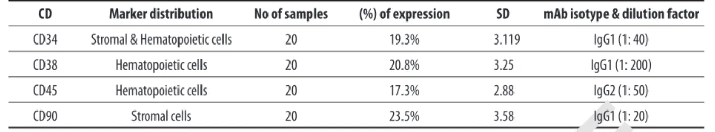
Bone marrow cells were extracted from femurs of 20 Sprague-Dawly rats. We next used hematopoetic stem cell markers (CD38, CD45), stromal cells marker CD90, and CD34 that
CD Marker distribution No of samples (%) of expression SD mAb isotype & dilution factor
CD34 Stromal & Hematopoietic cells 20 19.3% 3.119 IgG1 (1: 40)
CD38 Hematopoietic cells 20 20.8% 3.25 IgG1 (1: 200)
CD45 Hematopoietic cells 20 17.3% 2.88 IgG2 (1: 50)
CD90 Stromal cells 20 23.5% 3.58 IgG1 (1: 20)
Table 1. Detection of surface markers on bone marrow derived cells (see also Figure 1).
PERSONAL USE
ONLY
is expressed on subpopulations of both cell types, to as-sess the composition of the bone marrow-derived cells. For each case, the corresponding negative control was utilized (Figure 1A–E). The mean percentages of cells positive for each marker were as follows: CD34=19%, CD38=21%, CD 45=17% and CD90=24% (Table 1).
Bone marrow stromal cells have signifi cant trans-differ-entiation capacity and migratory potential, even in CNS [11,12,18]. Thus, using RT-PCR we next tested these cells for the expression of neural mRNAs. We extracted the to-tal RNA by Triazol method, and assessed its quality by RNA denaturating gel electrophoresis. The electrophoregram shown in fi gure 2A shows good quality of RNA preparation (quality control). RT-PCR signals indicate that bone marrow stromal rat cells, after 7 passages, expressed NGF, (395 bp) (Figure 2B, panel 2, lane 3), BDNF (Figure 2B, panel 3, lane 3), and GDNF (648 bp) amplifi ed message fragments (Figure 2B, bottom, panel lane 3).
Transplanted bone-marrow-derived stem cells remain viable within neural tissue for at least 30 days
To evaluate the experimental model, we microscopically as-sessed the degree of brain tissue injury induced by the injec-tion of quinolinic acid (Figure 3A–C). The injured animals showed loss of neuronal cells and abundant gliosis. The in-jured groove differed from the intact groove; it was thicker in the right lateral ventricle ipsilateral area.
The cells meant for transplantation were marked with two different methods (GFP, Hoechst 33342). Unless indicated differently, the viability of bone marrow-derived cells was above 90% during all experimental procedures. The adeno-viral transduction to express GFP showed maximum (~50%) infection effi ciency after 5 days of incubation with the virus (Figure 4). The prolonged incubation caused 25–43% of cell death within the virus treated cell population. However, when the bone marrow-derived cells were stained with Hoechst 33342 (24 h incubation in Hoechst-containing medium), 100% of cells had incorporated the nuclear marker and cell viability continued to be always above 90%.
Stereotactic transplantations were carried out one month after the injury induction, and it was done as described in the method section. Microscopy studies of tissues derived from animals transplanted with bone marrow derived- GFP-expressing cells, transplanted into groove-area, were per-formed 3 days, and 30 days post transplantation. After 3 days, the GFP-expressing cells were easily detected within striatum, but not in other areas of the brain. 30-days post-ransplantation many cells still expressed the GFP, but their number diminished considerably (Figure 5AB). The cell viability within the transplanted areas, as assessed morpho-logically, was about 70–75%.
The implanted bone marrow-derived cells marked with Hoechst 33342 were easily and abundantly detectable at the site of injury, by fl uorescent microscopy between 10–30 days after transplantation to the groove (Figure 6A–C). The anteroposterior transplant’s extension was 2–2.2 mm as
de-Figure 1. Detection of CD34, CD38, CD45, and CD90 markers in bone
marrow derived mononuclear cells. (A) Negative control (secondary antibody only), (B) CD34 positive cells, (C) CD38 positive cells, (D) CD45 positive cells, and (E) CD90 positive cells. Triplicate samples for each staining were blindly evaluated by two independent scientists. At least 400 cells per each sample were counted. Mean values of the percentage of positive cells for each one of the antigens are indicated in Table 1. A B C D E
PERSONAL USE
ONLY
termined by morphometry. The transplanted cells spread markedly from the point of injection, indicating signifi cant migratory potential of these cells. The viability of transplant-ed cells at day 30 after transplantation was above 75% as assessed morphologically. Cells that shrunk and had con-densed nuclei were considered apoptotic.
D
ISCUSSIONThis project was aimed to test the feasibility and migratory potential of bone marrow derived stem cells as donor cells for the treatment of brain lesions. We have used a quinolinic acid based method to induce neuronal lesions. Upon stereo-tactic injection of quinolinic acid into the brain a notorious gliosis was evident in the corresponding groove in the
ipsilat-eral ventricle beside the lesion (product of the shrinkage of the groove). The characteristics described in this report coin-cide with those reported by other authors within the pattern of striatal lesions induced with quinolinic acid [20]. For tagging of the cells that we have transplanted, we have used either adenoviral-vector based GFP gene trans-fer to mark the bone marrow-derived mononuclear cells [22,23,25,29] or a Hoechst 33342 based tagging (see below). The adenoviral tagging was only moderately effective; upon 5 days incubation with the adenovirus carrying the GFP-gene, only about 50% of cells expressed the tag. Furthermore, the adenovirus genome is not integrated to the genome of the host cell, thus each cell division dilutes the adenoviral gene and the marker gene (GFP). The Hoechst 33342
re-Figure 2. Expression of neurotrophic factors by bone marrow-derived
stem cells. (A) mRNA isolated from rat bone marrow stromal cells in passage 7 (quality control). (B) Detection of NGF, BDNF and GDNF mRNA by RT-PCR from bone marrow stromal cells. β-actina (top panel) was used as an amplifi cation control. Lane 1: Molecular weight marker (BlueScript/HpaII, sizes 713/ 489/ 404/ 367/ 242/ 190/ 157/ 147/ 110/ 67/ 57/ 34 and 26 pb). Lane 2: Reaction without reverse transcriptase (negative control). Lane 3: Reaction with reverse transcriptase.
B A
Figure 3. Quinolinic acid induced neuronal lesions. (A) Quinolinic
acid neuronal lesion was induced within the right striatum, (1 day after induction) see the left, healthy striatum for comparison (magnifi cation: 50×). (B) Normal (control) striatum (magnifi cation: 100×). (C) Quinolinic acid neuronal lesion was induced within the right striatum, (1 month after induction), see (B) for comparison to non-damaged striatum (magnifi cation: 100×).
A B C
BR
506, 517- 396- 344-- 713-489 -404,368 - 396-2 3--
-actina~~~r
220 ~~ _ _ _ _ _ --- BDNF ___ GDNF 506,517-PERSONAL USE
ONLY
agent, the other method that we used for cell tagging, is a live DNA-dye that preferentially stains A- and T-bases with-in DNA. In our case, the cells stawith-ined 100%, and cell via-bility measured at 24 h of incubation with Hoechst 33342 was above 90%. When comparing both methods of cell-tag-ging, the Hoechst 33342 staining appears to be superior to the virus-based tagging. The Hoechst 33342 labeling meth-od has been successfully applied in the past to follow cells transplanted into CNS [31].
The in vivo survival of GFP-tagged grafts was diminished con-siderably as compared to those tagged with Hoechst 33342. The lower survival of GFP-tagged cell is likely due to host’s immune response mounted against the adenovirus and/or the GFP. Such interpretation is supported by the presence of yellow bodies in the striatal area that was transplanted with GFP-tagged cells. Such yellow bodies are typically associat-ed with the infl ammatory reaction within the neural tissue. These results are in agreement with observations of other authors that noted adenoviral system-induced infl ammation, thus limiting the expression of the desired transgene, (in our case the GFP) [32]. Contrary to the results reported by some authors, the infused cells did not aggregate, as it has been reported with the infusion of fi broblasts and also some types of stem cells [33]. Instead, in our experimental mod-el, the cells integrated well into the new environment, and even migrated 2–3 mm from the injection site. The same patterns of integration and migration have been reported by some other authors in both rat and human bone marrow stem cells infused into the rats’ brains [34].
The cells tagged with Hoechst 33342 survived much bet-ter the engraftment as compared to GFP-tagged cells. The
transplanted cells were located around the line of the nee-dle transition through the brain tissue, and they dispersed toward the cortex and corpus callosum. The migration/dis-persion occurred to a similar extend as for GFP-tagged cells and it was on average of 2–2.2 mm from the injection site. Interestingly, the difference in migration distance among the animals sacrifi ced at 10 days and those sacrifi ced at 20 days after transplantation was not signifi cant, thus the migration pattern and distance were likely established in an early time window after transplantation. We observed similar migra-tion routes of rat femur-derived bone marrow cells toward the corpus callosum, cerebral cortex and ipsilateral tempo-ral lobe, as it was previously reported for bone marrow stem cells that had been transplanted directly into the striatum [33,34]. The migration of grafted cells in the injured brain is likely due to the action of chemotactic factors produced by the damaged brain. These factors create an attraction of implanted bone marrow mononuclear cells to the site of in-jury [22,23,25,29]. The extent of migratory behavior may de-pend on the cell types transplanted and the age of the ani-mal these cells are implanted into. Much stronger migratory activities were reported if transplanted stromal cells were im-planted in the lateral ventricle of neonatal mice. These cells were even detected in the contralateral hemisphere of brain and cerebellum without causing any disruption of cerebral architecture [25]. Similarly as in our experimental system, some other authors have reported that the migration took place toward the corpus callosum, cerebral cortex and ip-silateral temporal lobe, when bone marrow stem cells were implanted directly in the grooved body [33,34]. In contrast to our rat brain injury model, which did not utilized neona-tal rats, the mice studies were performed on neonaneona-tal brains and this may encourage stronger cell migration.
Figure 4. Staining of bone marrow derived
mononuclear cells by infection with type-5 adenoviral vector carrying GFP cDNA. Cells were incubated with the vector (MOI=10) for 5 days (see method section for details). (A), magnifi cation 100×, (B,C) magnifi cation 1000×.
A B C
Figure 5. Brain sections with implanted bone marrow mononuclear
cells that were labeled by the infection with adenoviral vector carrying GFP. Brain sections at day (A) 3, and (B) 30 post-transplantation (magnifi cation ×200). See method section for experimental details. The yellow signal in (A) represents a high intensity fl uorescence of GFP.
B A
Figure 6. Brain sections with implanted bone
marrow mononuclear cells that were label with Hoechst 33342. Brain sections at day (A) 10, (B) 15, and (C) 30 post-transplantation (magnifi cation ×200). See method section for experimental details.
PERSONAL USE
ONLY
Our results show that the bone marrow derived stem cells are able to survive at least for one month upon transplanta-tion into brain. Thus, our data encourage the use of bone marrow derived stem cells in the development of experi-mental therapies of neurodegenerative diseases.
Bone marrow is a rich source of stem cells. Besides, the hema-topoietic and mesenchymal cells discussed before, it includes a subpopulation of multipotent adult stem cells [35,36]. Our study supports the present concept of the heterogeneity of adult stem cell populations within bone marrow. This is fur-ther confi rmed by the presence of mRNAs from both hema-topoetic and neurotrophic factors in the tested bone mar-row derived stem cells. For example, we detected a GDNF signal, and a NGF mRNA signature. The level of production of NGF by bone marrow derived stromal stem cells was similar to the expression of this neurotrophic factor by murine stro-mal cell line MS-5 and other strostro-mal cell lines [11,37].Our paper communicates for the fi rst time that rat bone marrow derived stromal cells produce GDNF and NGF. Interestingly, recently it has been communicated that fresh murine bone marrow derived stromal cells cannot produce GDNF [38]. Our tests in the rat model were conducted after passage 7 of cell culture that may allow some trans-differentiation. Thus, it is possible that simple passaging of stromal cells under the described in method section condition, prompts some cells into differentiation towards neural phenotypes.
C
ONCLUSIONSIn summary, our experiments demonstrate the feasibility of the usage of bone marrow derived stem cells for the de-velopment of cellular therapies of brain lesions. The use of the Hoechst reagent for tagging of cells to be implanted into the brain is much more advantageous as compared to GFP-tagging by adenovidal infection. The bone marrow de-rived stem cells survive as transplants for at least a month, and they express neural growth- and survival factors like NGF and GDNF. Further research is needed to defi ne bone marrow derived cell sub-populations best suited as a source for cell-based therapies of degenerative disorders, strokes and traumatic lesions.
Acknowledgements
We would like to thank Associate Professor Martha Cristófol for the revision and comments on this article.
R
EFERENCES:
1. Cassens U, Lewinski G, Samraj AK et al: Viral modulation of cell death by inhibition of caspases. Arch Immunol Ther Exp, 2003; 51: 19–27 2. Hauff K, Zamzow C, Law WJ et al: Peptide-based approaches to treat
asthma, arthritis, other autoimmune diseases and pathologies of the central nervous system. Arch Immunol Ther Exp, 2005; 53: 308–20 3. Maddika S, Ande SR, Wiechec E et al: Akt mediated phosphorylation
of CDK2 regulates its dual role in cell cycle progression and apoptosis. J Cell Sci, 2008; 121: 979–88
4. Taupin P: Derivation of embryonic stem cells for cellular therapy: chal-lenges and new strategies. Med Sci Monit, 2006; 12(4): RA75–78 5. Pan GJ, Chang ZY, Scholer HR, Pei D: Stem cell pluripotency and
tran-scription factor Oct4. Cell Res, 2002; 12: 321–29
6. Kucia M, Zuba-Surma EK, Wysoczynski M et al: Adult marrow-derived very small embryonic-like stem cells and tissue engineering. Expert Opin Biol Ther, 2007; 7: 1499–514
7. Bianco P, Riminucci M, Gronthos S, Robey PG: Bone marrow stromal stem cells: nature, biology, and potential applications. Stem Cells, 2001; 19: 180–92
8. Van Damme A, Vanden Driessche T, Collen D, Chuah MK: Bone mar-row stromal cells as targets for gene therapy. Curr Gene Ther, 2002; 2: 195–209
9. Eglitis MA, Mezey E: Hematopoietic cells differentiate into both mi-croglia and mami-croglia in the brains of adult mice. Proc Natl Acad Sci USA, 1997; 94: 4080–85
10. Tarnowski M, Sieron AL: Adult stem cells and their ability to differen-tiate. Med Sci Monit, 2006; 12(8): RA154–63
11. Kopen GC, Prockop DJ, Phinney DG: Marrow stromal cells migrate throughout forebrain and cerebellum, and they differentiate into as-trocytes after injection into neonatal mouse brains. Proc Natl Acad Sci USA, 1999; 96: 10711–16
12. Lu D, Mahmood A, Wang L et al: Adult bone marrow stromal cells ad-ministered intravenously to rats after traumatic brain injury migrate into brain and improve neurological outcome. Neuroreport, 2001; 12: 559–63
13. Alexander HK, Booy EP, Xiao W et al: Selected technologies to control genes and their products for experimental and clinical purposes. Arch Immunol Ther Exp, 2007; 55: 139–49
14. Kirik D, Rosenblad C, Bjorklund A, Mandel RJ: Long-term rAAV-medi-ated gene transfer of GDNF in the rat Parkinson’s model: intrastriatal but not intranigral transduction promotes functional regeneration in the lesioned nigrostriatal system. J Neurosci, 2000; 20: 4686–700 15. Hida H, Hashimoto M, Fujimoto I et al: Dopa-producing astrocytes
generated by adenoviral transduction of human tyrosine hydroxylase gene: in vitro study and transplantation to hemiparkinsonian model rats. Neurosci Res, 1999; 35: 101–12
16. Kreuter M, Langer C, Kerkhoff C et al: Stroke, myocardial infarction, acute and chronic infl ammatory diseases: caspases and other apoptotic molecules as targets for drug development. Arch Immunol Ther Exp, 2004; 52: 141–55
17. Rashedi I, Panigrahi S, Ezzati P et al: Autoimmunity and Apoptosis – Therapeutic Implications. Curr Med Chem, 2007; 14: 3139–51 18. Woodbury D, Schwarz EJ, Prockop DJ, Black IB: Adult rat and human
bone marrow stromal cells differentiate into neurons. J Neurosci Res, 2000; 61: 364–70
19. Block F, Schwarz M: Expression of GFAP in the striatum and its projection areas in response to striatal quinolinic acid lesion in rats. Neuroreport, 1994; 5: 2237–40
20. Francis L, Cruz R, Antunez I, Rosillo JC: [Behavior characterization of a model of Huntington’s disease in rats, induced by quinolinic acid]. Rev Neurol, 2000; 30: 1016–21
21. Schwarz AJ, Danckaert A, Reese T et al: A stereotaxic MRI template set for the rat brain with tissue class distribution maps and co-registered anatomical atlas: application to pharmacological MRI. Neuroimage, 2006; 32: 538–50
22. Alberti E, García R, Serrano ST et al: Evaluación de la sobrevivencia de las células mononucleadas de la médula ósea trasplantadas en un mod-elo de ratas de lesión estriatal con ácido quinolínico. Neurología, 2005; 40: 518–22
23. Brazelton TR, Rossi FM, Keshet GI, Blau HM: From marrow to brain: expression of neuronal phenotypes in adult mice. Science, 2000; 290: 1775–79
24. Ghavami S, Asoodeh A, Klonisch T et al: Brevinin-2R semi-selectively kills cancer cells by a distinct mechanism, which involves the lysosom-al-mitochondrial death pathway. J Cell Mol Med, 2008; 12: 1005–22 25. Serrano ST, Alberti EA, Lorigados PL et al: Caracterización
inmu-nofenotípica de las Células de la Médula Ósea. Biotecnologia Aplicada, 2005; 22: 246–49
26. Castillo L, Martinez L, Grygar E, Hutter-Paier B: Characterization of proliferation and differentiation of EGF-responsive striatal and septal precursor cells. Int J Dev Neurosci, 2003; 21: 41–47
27. Maddika S, Bay GH, Kroczak TJ et al: Akt is transferred to the nucle-us of cells treated with apoptin, and it participates in apoptin-induced cell death. Cell Prolif, 2007; 40: 835–48
28. Paxinos G, Watson C: The Rat Brain in Stereotaxic Coordinates, 4th ed.
Academic Press, London, 1998
29. Fernandez CI, Alberti E, Mendoza Y et al: Motor and cognitive recovery induced by bone marrow stem cells grafted to striatum and hippocam-pus of impaired aged rats: functional and therapeutic considerations. Ann NY Acad Sci, 2004; 1019: 48–52
PERSONAL USE
ONLY
30. Garcia R, Aguiar J, Alberti E et al: Bone marrow stromal cells producenerve growth factor and glial cell line-derived neurotrophic factors. Biochem Biophys Res Commun, 2004; 316: 753–54
31. Tourbah A, Gansmuller A, Gumpel M: A nuclear marker for mammalian cells and its use with intracerebral transplants. Biotech Histochem, 1991; 1: 29–34
32. Thomas CE, Schiedner G, Kochanek S et al: Peripheral infection with adenovirus causes unexpected long-term brain infl ammation in animals injected intracranially with fi rst-generation, but not with high-capacity, adenovirus vectors: toward realistic long-term neurological gene thera-py for chronic diseases. Proc Natl Acad Sci USA, 2000; 97: 7482–87 33. Kawaja MD, Fagan AM, Firestein BL, Gage FH: Intracerebral grafting of
cultured autologous skin fi broblasts into the rat striatum: an assessment of graft size and ultrastructure. J Comp Neurol, 1991; 307: 695–706
34. Azizi SA, Stokes D, Augelli BJ et al: Engraftment and migration of human bone marrow stromal cells implanted in the brains of albino rats-simi-larities to astrocyte grafts. Proc Natl Acad Sci USA, 1998; 95: 3908–13 35. Jiang Y, Vaessen B, Lenvik T et al: Multipotent progenitor cells can be
isolated from postnatal murine bone marrow, muscle, and brain. Exp Hematol, 2002; 30: 896–904
36. Zanjani ED, Almeida-Porada G, Livingston AG et al: Engraftment and multilineage expression of human bone marrow CD34- cells in vivo. Ann NY Acad Sci, 1999; 872: 220–31; discussion 231–32
37. Auffray I, Chevalier S, Froger J et al: Nerve growth factor is involved in the supportive effect by bone marrow – derived stromal cells of the fac-tor-dependent human cell line UT-7. Blood, 1996; 88: 1608–18 38. Dezawa M, Hoshino M, Nabeshima Y, Ide C: Marrow stromal
cells: implications in health and disease in the nervous system. Curr Mol Med, 2005; 5: 723–32