Linköping Studies in Science and Technology
Dissertation No. 948
Plasma Characterization
&
Thin Film Growth and Analysis
in
Highly Ionized Magnetron Sputtering
Jones Alami
Plasma and Coatings Physics Division
Department of Physics and Measurement Technology
Linköping University, SE-581 83 Linköping
Sweden
Linköping 2005
ISBN 91-85299-40-5
ISSN 0345-7524
To Father and Mother
In an instant illumination can be achieved. It is as easy as turning on a light. The problem is finding the switch in the dark.
Abstract
The present thesis addresses two research areas related to film growth in a highly ionized magnetron sputtering system: plasma characterization, and thin film growth and analysis. The deposition technique used is called high power pulsed magnetron sputtering (HPPMS). Characteristic for this technique are high energy pulses (a few Joules) of length 50-100 µs that are applied to the target (cathode) with a duty time of less than 1 % of the total pulse time. This results in a high electron density in the discharge (>1×10¹⁹ m-3) and leads to an increase of the ionization
fraction of the sputtered material reaching up to 70 % for Cu.
In this work the spatial and temporal evolution of the plasma parameters, including the electron energy distribution function (EEDF), the electron density and the electron temperature are determined using electrostatic Langmuir probes. Electron temperature measurements reveal a low effective temperature of 2-3 eV. The degree of ionization in the HPPMS discharge is explained in light of the self-sputtering yield of the target material. A simple model is therefore provided in order to compare the sputtering yield in HPPMS and that in dc magnetron sputtering (dcMS) for the same average power.
Thin Ta films are grown using HPPMS and dcMS and their properties are studied. It is shown that enhanced microstructure and morphology of the deposited films is achieved by HPPMS. The Ta films are also deposited at a number of substrate inclination angles ranging from 0o (i.e., facing the target surface) up to 180 o (i.e.,
facing away from the target). Deposition rate measurements performed at all inclination angles for both techniques, reveal that growth made using HPPMS resulted in an improved film thickness at higher inclination. Furthermore, the high ionization of the Ta atoms in HPPMS discharge is found to allow for phase tailoring of the deposited films at all inclination angles by applying a bias voltage to the substrate. Finally, highly ionized magnetron sputtering of a compound MAX-phase material (Ti3SiC2) is performed, demonstrating that the HPPMS discharge could also be used
Preface
The present thesis is the summary of the work I conducted as part of a PhD study in the Thin Film Physics and Plasma & Coatings Physics divisions at Linköping University in Sweden, between November 2001 and June 2005. I consider myself lucky to have had the opportunity to be a member of the research groups, not only for providing a great research environment but also for contributing to my development as a human being. As a PhD student, I learned that with good planning, a passion for knowledge, and a conscious attitude, research becomes simply a way of life. I would like therefore to thank the people that made it such an inspiring and rich milieu, especially: Ulf Helmersson for introducing me to the fields of plasma physics and thin films, and for always being available for discussions and new ideas. The administration at IFM for making the Physics Department just a lovely work place.
Jon Tomas Gudmundsson, my unofficial plasma supervisor, for a great colaboration
and nice visits to Iceland. Lars Hultman for your inspiring leadership skills. Inger Eriksson for always being so helpful and friendly. Karl-Olof Brolin for your creativity and for making the difficult tasks easy. Jon Andersson and Erik Wallin for the many discussions about science and life, and for always being positive. Hans Högberg for an effective collaboration and for a much appreciated help when I needed it most. Per Eklund for a great collaboration and for your enthusiasm. Jochen Schneider for the research strategy talk. Denis Music for your inspirational ideas. Leif Samuelsson for your help with the probe station and understanding what I actually meant when we discussed “electronics”. All of you who helped me by reading and commenting the manuscript, Ulf, Hans, Jon Tomas, Erik W., and Per E.. Everyone from The Plasma & Coatings Physics, and the Thin Film Physics groups for the excellent atmosphere each one helps to create within our groups. My dear friends
Dejan, Erik A., Tarik, Driss, Mahiar, Oskar… for your support and friendship.
Finally, the people I admire the most. Helene, my dearest, for your support and love, and my wonderful Family for your passionate and truthful engagement in my happiness.
List of papers included in the thesis
Paper I:
Evolution of the electron energy distribution and plasma parameters in a pulsed magnetron discharge, J. T. Gudmundsson, J. Alami, and U. Helmersson.
App. Phys. Lett. 78 (2001) 3427.
Paper II:
Spatial and temporal behavior of the plasma parameters in a pulsed magnetron discharge, J. T. Gudmundsson, J. Alami, and U. Helmersson.
Surf. Coat. Technol. 161 (2002) 249-256.
Paper III:
Plasma dynamics in a highly ionized pulsed magnetron discharge, J. Alami, J. T.
Gudmundsson, J. Böhlmark, and U. Helmersson
Submitted to Plasma Sources Science and Technology.
Paper IV:
Ion-accoustic solitary waves in a high power pulsed magnetron sputtering discharge, K. B. Gylfason, J. Alami, U. Helmersson, and J. T. Gudmundsson.
Submitted to Journal of physics D: Applied physics.
Paper V:
Ion-assisted Physical Vapor Deposition for enhanced film properties on non-flat surfaces, J. Alami, P.O.Å. Persson, D. Music, J. T. Gudmundsson, J. Böhlmark, and
U. Helmersson
J. Vac. Sci. Technol. A 23 (2005) 278.
Paper VI:
Structural analysis and growth of beta- and bcc-Ta on inclined surfaces by highly ionized magnetron sputtering, J. Alami, P. Eklund, J.M. Andersson, M.
Lattemann, E. Wallin, J. Bohlmark, P. Persson, and U. Helmersson
Paper VII:
Ionized magnetron sputtering of Ti-Si-C MAX-phase thin films from a Ti3SiC2 compound target, J. Alami, P. Eklund, J. Emmerlich, U. Jansson, O. Wilhelmsson,
H. Högberg, L. Hultman, and U. Helmersson
Manuscript in final preparation.
I took a major part in the planning of all the studies, carried out all the plasma diagnostic work and all the thin film growth, performed or took part in the thin film characterization and analysis, and wrote papers III, V, VI, and VII. U. Helmersson contributed to all papers in terms of planning and discussions. J. T. Gudmundsson was the main contributor to the scientific discussions and data analysis related to the plasma physics (Papers I-IV) and wrote papers I and II. K. B. Gylfason contributed with data analysis and writing of paper IV. H. Högber and P. Eklund were the major figures for MAX-phase related issues. Other co-authors contributed with film characterization and scientific discussions.
Additional publications (not included in the thesis)
1 High power impulse magnetron sputtering discharges and thin film growth: A brief review, U. Hemlersson, M. Lattemann, J. Alami, J. Bohlmark, A. P Ehiasarian,and J. T. Gudmundsson. Society of Vacuum Coaters 48th Annual Technical
Conference Proceedings (2005).
2 Ionization of sputtered metals in High Power Pulsed Magnetron Sputtering,
J. Bohlmark, J. Alami, A. P. Ehiasarian, C. Christou, and U. Helmersson. J. Vac. Sci.
Technol. A, 23(1), 18-22 (2005).
3 Spatial electron density distribution in high power pulsed magnetron sputtering, J. Bohlmark, J. T. Gudmundsson, J. Alami, M. Latteman, and U.
4 Influence of pressure and power on the composition and time evolution of plasmas in high power impulse magnetron sputtering, P. Ehiasarian, R. New, P. E.
Hovsepian, J. Böhlmark, J. Alami, and U. Helmersson. Society of Vacuum Coaters
47th Annual Technical Conference Proceedings, 28-32 (2004).
5 Measurement of the magnetic field change in a pulsed high current magnetron discharge, J. Bohlmark, U. Helmersson, M. VanZeeland, I. Axnäs, J. Alami and N.
Brenning. Plasma Sources Sci. Technol., 13, 654-661 (2004).
6 Plasma Parameters During High Power Pulsed Magnetron Sputtering, J.
Alami, J. T. Gudmundsson, J. Böhlmark, K. B. Gylfason and U. Helmersson.
Proceedings of The 16th international symposium on plasma chemistry, ISPC 16,
Taormina, Italy, 2003.
7 Low temperature deposition of alpha -Al/sub 2/O/sub 3/ thin films by sputtering using a Cr/sub 2/O/sub 3/ template, P. Jin, G. Xu, M. Tazawa, K.
Yoshimura, D. Music, J. Alami, and U. Helmersson. J. Vac. Sci. Technol. 20 (2002) 2134.
8 X-ray reciprocal space mapping studies of strain relaxation in thin SiGe layers,
W.-X. Ni, K. Lyutovich, J. Alami, C. Tengstedt, M. Bauer, and E. Kasper. J. Crystal
Growth 227-228 (2001) 756.
9 Ionized-PVD by pulsed sputtering of Ta for metallization of high-aspect-ratio structures in VLSI, U. Helmersson, Z. S. Kahn, and J. Alami. Proceedings of the
Third International Euroconference on Advaced Semiconductor Devices and Microsystems (IEEE Catalogue number: 00EX386), Somolenice Castle, Slovakia,
1. INTRODUCTION...3
2. PART I. PLASMA CHARACTERIZATION ...5
2. 1. PLASMA DEFINITION...5
2. 2. PLASMA GENERATION...7
2.3. PLASMA MODELING AND THE DISTRIBUTION FUNCTION...9
2. 3. 1. The distribution function and the plasma parameters ...9
2. 4. HPPMS DISCHARGE DIAGNOSTICS...11
2. 4. 1. Measurement setup ...11
2. 4. 2. Plasma measurements using a Langmuir cylindrical probe...12
4. 2. 2. Filtering and smoothing...14
2. 4. 3. Ion current determination...16
2. 4. 4. Direct display of plasma parameters using a triple probe setup...17
2. 5. SOLITARY WAVES...20
3. PART II. THIN FILM GROWTH AND CHARACTERIZATION ...23
3.1. GROWTH MODELS...23
3. 2. DEPOSITION OF THIN FILMS BY MAGNETRON SPUTTERING...26
3. 2. 1. Magnetron sputtering...26
3. 2. 2. The ionization process ...27
3. 3. EFFECTS OF IONIZED DEPOSITION ON FILM STRUCTURE...32
3. 3. 1. Film densification using HPPMS...32
3. 3. 2. Phase formation...34
3. 3. 3. Enhanced adhesion by HPPMS ...35
3. 4. SPUTTERING OF A MAX-PHASE COMPOUND TARGET BY HPPMS ...37
3. 5. THIN FILM CHARACTERIZATION...39
3. 5. 1. Transmission electron microscopy (TEM)...39
3. 5. 2. Scanning electron microscopy (SEM)...39
3. 5. 3. Atomic force microscopy (AFM)...40
3. 5. 4. X-ray diffraction (XRD) ...40
3. 5. 5. X-ray photoelectron spectroscopy (XPS)...41
3. 5. 6. The four-point electrical probe...41
4 SUMMARY ...43
1. Introduction
Thin films are material layers with thickness ranging from a few nm to a few µm. They are used today in a wide range of applications, such as coatings for cutting tools, optical and decorative coatings, solar cells applications, and as diffusion layers in integrated circuits. The film growth can be carried out by a number of deposition techniques, including chemical vapor deposition (CVD) [1,2] where chemical reactions precede deposition, and the chemical bonding state of the deposit is different from the precursor gases. Other deposition techniques are sputtering and evaporation. They are called physical vapor deposition (PVD) processes, because they produce a vapor of the material to be deposited by physical means, i.e., by heating (evaporation) or by energetic ion bombardment (sputtering) [3-6]. Because of the presence of a plasma, sputtering exhibits added benefits in the deposition process for many thin film technologies [7]. PVD was first discovered in 1852, and was developed as a thin film deposition technique by Langmuir in the 1920s. It has better step coverage than evaporation as the adatoms have higher surface diffusion energy. The sputtering process is also much better at producing layers of compound materials and alloys [8] and in controlling the microstructure and phase of the deposit. A widely used sputtering technique is dc magnetron sputtering where a magnetic field is superimposed on the electric field. This leads to electron confinement near the target resulting in higher ionization of the sputtering gas, and consequently, higher deposition rates are obtained [9-11]. Although dcMS is a successful magnetron sputtering technique, it suffers from fundamental problems such as low target utilization, target poisoning and poor deposition rates for dielectric materials. In order to alleviate some of these problems, alternative techniques, such as radio frequency magnetron sputtering (rfMS) [11-17], additional ionization by rf coils [18] or microwaves [19], variable or unbalanced magnetic configuration [20] or increased magnetic confinement by a multipolar magnetic setup [21] has been demonstrated. Pulsing the applied voltage is another alternative magnetron sputtering technique, where sputtering is achieved principally by asymmetric bipolar or by unipolar pulsing of the target power. In bipolar pulsing, the power is applied to the target at a frequency of 10 – 200 kHz [17, 22, 23]. This technique has become established as one of the main sputtering techniques for deposition of oxide and nitride films [10,11,24].
In unipolar pulsing, the power supply operates at low (or zero) power level and pulses to a high voltage for a short period each cycle [25, 26].
In many applications, increasing the ion fraction of the sputtered material gives an advantage in film deposition, as it allows for directional deposition and control of the charged sputtered species bombardment-energy [27, 28]. This is difficult in conventional deposition techniques, as the plasma density is too low to cause significant ionization of the sputtered material, and most of the ions are ions of the sputtering gas [29]. A way to increase the electron density in the discharge is by using an electron cyclotron resonance plasma as a plasma electron source [30,31], employing an inductively coupled plasma [18], or adding a hollow cathode electron source in the target vicinity [32]. Increasing the power to the sputter source and therewith increasing the electron density can also promote ionization through electron-impact processes. While this is usually not possible in dcMS due to target overheating and eventual damage, applying high energetic pulses to the target at a low duty time is a solution.
High Power Pulsed Magnetron Sputtering (HPPMS), also known as high power impulse magnetron sputtering (HIPIMS), has been demonstrated to give high plasma (electron) densities without overheating the target, when high power pulses with a duty time lower than 1 % of the total pulse time were applied to the target [33-37]. With peak power densities typically of several kW cm−2, plasma densities 2 to 3 orders of magnitude higher than those measured in dc magnetron discharges can be achieved [paper I-III]. This gives ionization fractions of the sputtered material ranging from 4.5 % for C [38] to 70 % for Cu [36].
In this work two areas related to HPPMS are addressed. First, a study of the plasma dynamics and energetics is presented. This is the content of papers I to IV, where the spatial and temporal evolution of the discharge are described, and the temporal behavior of the plasma parameters, such as the electron energy distribution function (EEDF), the electron density, and the electron temperature are determined. Second, the role of ion deposition and ion bombardment in the HPPMS technique is investigated by depositing thin films and characterizing their properties. As a case study, a “simple” material system (Ta) is investigated in papers V and VI, and a complex ternary material system (Ti-Si-C) in paper VII. The benefits of using HPPMS for deposition on inclined surfaces are also discussed and demonstrated using experiments together with the theoretical background and analysis.
2. Part I. Plasma characterization
As stated in chapter 1, different growth techniques are used for thin film deposition, where the deposition process can be a CVD or a PVD process. Sputtering is today the primary alternative in PVD. In sputtering, a low-pressure discharge (plasma) is sustained between the target and the chamber walls. When an electric field is applied, positively charged gas ions (e.g. Ar, Kr, N2) bombard the target surface
after being accelerated by the potential drop across the sheath between the target and the plasma. In the bombarding process, ions, atoms and electrons are ejected from the target surface. The emitted electrons are accelerated by the same potential drop and gain therefore considerable energy, enough to ionize the sputtering gas atoms.
2. 1. Plasma definition
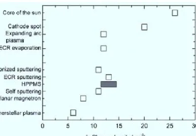
By some estimates, 99% of the observable universe is in the plasma state, also known as the fourth state of matter [39]. The use of the term "plasma" for an ionized gas started in 1927 with Irving Langmuir (1881-1957). Plasma is defined as a quasi-neutral gas of charged and quasi-neutral particles that exhibit a collective behavior [40]. The charged particles must satisfy two important criteria for the gas to qualify as a plasma; first, sufficiently high charge density, so that the long range Coulomb forces are a significant factor in determining the statistical properties of the particles. Second, low enough density, so that the Coulomb force of a near neighbor particle is much less than the cumulative long range Coulomb force exerted by the many distant particles. These criteria are satisfied in a range of plasmas as shown in Fig.1 (a), where the densities corresponding to the different plasmas are presented. The HPPMS plasma covers a large electron density range, with consequences related to the ionization fraction in the plasma and to the film deposition and structure.
Fig. 1. Different plasmas and their corresponding electron densities are shown. The box representing the HPPMS plasma is shaded.
Plasmas, unlike ordinary gases, support a wide variety of wave modes because of the charged particles [40,41]. One example of these wavelike disturbances is the ion acoustic wave, which is an electrostatic wave occurring in a field free plasma. Another is the magnetosonic wave, which is an acoustic wave in which the compressions and rarefactions are produced not by motions along the electric field E but by E×B drifts across E [40], where B represents the magnetic field. Finally, the electromagnetic wave, which can typically be described by an electric field of the form E(x,t)=E0exp
[
i(
kx−ωt)]
, where k is called the wave vector, x is thepropagation direction, and ω is the oscillation frequency. E0 and ω are in general
complex valued. If the wave is electromagnetic in nature then the magnetic field obeys a similar relation also. The (angular) frequency (ω) and the wave vector (k) are functionally related to one another, at least in linear theories of plasma waves, through a dispersion relationship ω(k). Knowledge of the dispersion characteristics of the propagating waves is certainly necessary for an understanding of the plasma state. Plasma, however, is a non-linear medium and unless the waves are truly of small amplitude, non-linear effects must be taken into account. Such non-linear effects conspire to produce wave-like disturbances that do not conform to the forms given above, and interesting phenomena such as solitary waves (solitons) are observed as a result in plasmas. This will be discussed in more detail in section 2.5.
2. 2. Plasma generation
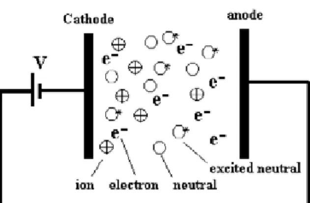
In order to start a discharge, some charged particles are needed to start the process of ionization and excitation of gas particles. This is provided to a large extent by weak background radiation from radioactive minerals where some ionized molecules and free electrons exist, and to a less extent from cosmic rays. By applying an electric field these electrons gain enough energy for further ionization and generation of secondary electrons, which in turn contributes to creating a so-called Townsend discharge with a relatively low, but avalanching, current (Fig. 2).
Fig. 2. A schematic of the Townsend discharge created by applying an electric field between two plates.
If the current increases enough, exceeding the breakdown field for the gas, a glow discharge is obtained. Figure 3 shows a photo of an Ar discharge for a sputtering system with a Ta sputter source (target). In order to sustain the plasma, regeneration of electrons is necessary [42]. This is often the most important means of maintaining the plasma as electrons lose their energy to the gas in ionizing processes, and are eventually lost to the surrounding chamber walls. Electrons also lose energy by elastic collisions, transforming energy to neutral species thereby putting them in an excited state, i.e., not all collisions result in ionization and secondary electrons generation [41]. As a result, a range of kinetic energies is obtained for the electron population, often described mathematically by a Maxwell-Boltzmann, a Bi-Maxwellian, or a Druyvesteyn distribution. More on distributions is presented in section 2.3.
Fig. 3. A photograph showing the HPPMS discharge in a magnetron sputtering system with a cylindrical target.
In many magnetron-sputtering applications, low-pressure gas discharges are used with only about 1% or less of the gas ionized. Nevertheless, the excited neutral atoms, molecules, and radicals gain energy by collisions, and have typically a temperature close to room temperature (~1 meV), compared with the warmer ions (tens of meV) and hotter electrons (a few eV). The electron density in such plasmas is in the range 1014-1017 m-3, the lower values being for dc glow and dc magnetron
discharge, and the higher values more typical for electron cyclotron resonance and inductively coupled discharges.
2.3. Plasma modeling and the distribution function
Plasmas are commonly modeled as fluids, and each species is described with its fluid equation [41]. The fluid approximation is sufficiently accurate to describe the majority of observed phenomena. When this is not viable, the accumulated behavior of an ensemble of charged particles is described using a statistical approach, in the form of the velocity distribution function. The distribution function is given as
f(r,v,t)d3rd3v, i.e., the number of particles inside a six-dimensional phase space volume d3rd3v at (r,v), at time t [41]. Knowing the distribution function, fundamental features of a large ensemble can be quantified. Knowing the cross sections for the relevant interactions in the plasma, measurements of the EEDF can be used to calculate all the rates [43]. By integrating over velocity, the average or (macroscopic) quantities associated with the plasma can be defined:
∫
= r u u
r t f t d
n( , ) ( , , ) plasma number density (1)
∫
= u ru u r u f t d n t) 1 ( , ,) ,( plasma bulk fluid velocity (2)
∫
− = u u r r f n m t ( ) ( 2 ) , ( 2ε ,u d,t) u plasma mean kinetic energy (3)
where m is the particle mass. This procedure of integrating over velocity space is referred to as taking velocity moments, with each one yielding a physically significant quantity. Knowledge of the electron energy distribution function, thus, permits us to determine important parameters. In modeling work, the electron distribution is often assumed to be Maxwellian. However, the electron energy distribution in a dc magnetron discharge has been shown to be bi-Maxwellian like [44] or Druyvesteyn like [41,45]. A Druyvesteyn like electron energy distribution has significantly fewer high-energy electrons than the Maxwellian for the same overall mean energy; resulting in less excitation and ionization.
2. 3. 1. The distribution function and the plasma parameters
As we know at this stage, the electron energy (speed) distribution function (EEDF) should be determined in order to study the plasma energetics. Fortunately,
one can measure the EEDF using a Langmuir probe. The use of a Langmuir probe to determine the EEDF was shown by Druyvesteyn in 1930, but it is not until the introduction of digital filtering in the 1990’s that it was put in practice in a significant way. Progress in analog and digital electronics has made it feasible to perform EEDF measurements in any modern plasma experiment where a classical Langmuir probe can be used. The EEDF, ge(V) can be calculated if the current-voltage relation in the
plasma is determined, in which case it is given by the Druyvesteyn formula as [41]:
2 2 2 1 2 2 2 ) ( dV I d m eV A e m V g e pr e = (4)
where Apr is the probe area, Ie is the electron current and V=Φp-Vb is the potential difference between the plasma potential, defined as the potential at which 2 0
2 =
dV I d e ,
and the probe potential. With ε=eV, the electron density ne is determined as:
∫
∞ = 0 ) (ε dε g ne e (5)If the plasma is in equilibrium, the EEDF is Maxwellian it simplifies the task, as the Maxwellian distribution can be described by a single electron temperature, which makes all the calculations simpler. For non-equilibrium plasmas, such as low-pressure discharge plasmas, the electrons are not in equilibrium with other plasma components, nor with the electron ensemble itself [46]. This means that the EEDF is non-Maxwellian, and as a result, the plasma modelling is much more complicated [45]. Consequently, a single electron temperature cannot fully describe the EEDF and it is more appropriate to describe the energetics of the electrons by a mean electron energy. At low pressures, the EEDF is generally non-Maxwellian and the electron temperature is thought of as an effective electron temperature, Teff, representing the mean electron energy determined from the EEDF [41] according to:
( )
ε ε ε ε g d n e e∫
∞ = 0 1 and k ε 3 2 = eff T (6)2. 4. HPPMS discharge diagnostics
2. 4. 1. Measurement setup
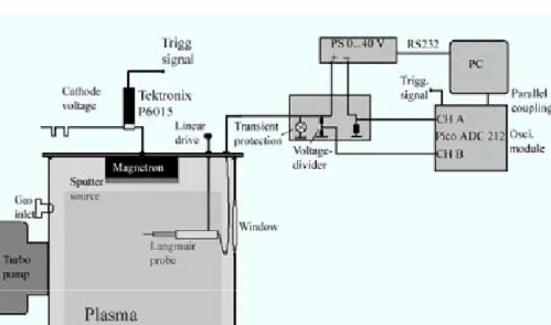
Figure 4 shows a schematic of the plasma measurement setup used in this work. A standard planar weakly unbalanced magnetron is operated with a metal target of 15 cm in diameter. The sputtering target is located inside a stainless steel chamber of a radius of 22 cm and a height of 75 cm. The chamber wall serves as an anode for the discharge. The cathode is driven by a pulsed power supply from Chemfilt R&D AB that delivers voltages of up to 2500 V and currents of up to 1200 A, at a repetition frequency of 50 Hz. The pulse length is in the range of 50 – 100 µs. Measurements are made with average powers comparable with those used in dcMS, i.e., the thermal load of the target is kept low. The target current and voltage are monitored by a Tektronix TDS 520 C (500 MHz, 1 Gs/s) oscilloscope using a Tektronix P6015 high voltage probe (1000 × attenuation), a Tektronix CT-04 high current transformer (20 kA peak current and bandwidth 20 MHz), and a TCP202 current probe (15A ac/dc and bandwidth 50 MHz).
Fig. 4. Experimental setup: A Langmuir probe is placed at different positions in the space in front of the target surface. The probe voltage and current are measured using an oscilloscope module and a PC. The data is stored in the PC, which functions also as the probe’s power-supply control-unit, for later analysis.
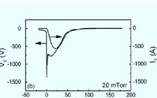
Figure 5 shows typical voltage and current time-traces in a HPPMS system, run with a Ta target and an Ar gas pressure held at 20 mTorr, as reported in paper III. For the applied target negative voltage VT, a decrease of a few hundred volts is seen
as the plasma ignites, and a maximum current of hundreds of Amperes is achieved 20-40 µs after the pulse initiation. This corresponds to peak powers of hundreds of kW, and peak power densities of a few kWcm-2, calculated over the target surface.
Fig. 5. Time evolution of the target voltage (VT) and current (IT) for a Ta target and
an Ar gas pressure of 20 mTorr.
2. 4. 2. Plasma measurements using a Langmuir cylindrical probe
Electrostatic probe diagnostic is the primary technique for obtaining information concerning the plasma state in low-pressure gas discharges. In its classical application, known as the Langmuir technique, the probe method allows us to find the basic plasma parameters such as the electron temperature, the plasma and floating potentials, the plasma density, and the electron energy distribution function (EEDF) [47]. The floating potential is defined as the potential at which the current drawn by the probe is nil, i.e., equal electron and ion currents. The plasma potential is defined as the potential at which ions cease to be attracted to the probe [41].
A Langmuir probe in its most basic form is simply a thin wire, typically made of tungsten, that is inserted into a plasma. The probe is biased at a variable voltage, and the collected current provides information on various plasma properties. Fig. 6 shows a schematic of a simple Langmuir probe.
Fig. 5. A schematic of the Langmuir cylindrical probe
2. 4. 2. 1. Measuring the I-V curve
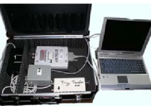
The relation between the applied Langmuir probe voltage and the collected current can be presented by a curve commonly known as probe I-V characteristic. This is quite a straightforward measurement in equilibrium plasmas, where plasma conditions do not change with time. However, in time dependent plasmas, as is the case of the HPPMS plasma, time-dependent electron currents are measured for a number of voltages, spanning over the voltage range of interest. In order to make the measurements more efficient and less time consuming, we built a probe station specific for the HPPMS discharge. The Langmuir probe is coupled via a coaxial cable to part 2 of the station. The different parts of the probe station that are presented in Fig. 6 are:
Part 1: A power supply. It supplies the probe with a voltage of ± 20 V
Part 2: It contains voltage-dividers for measurement of the probe voltage and the probe current; it also contains a transformer for powering the oscilloscope module (part 3).
Part 3: The oscilloscope module with a 12-bit resolution.
Part 4: A trigger signal transformer. It transforms the target voltage trigger signal into a suitable trig signal to the oscilloscope (± 5V).
The probe station is controlled using PScope, which is a software that we developed and tested successfully at Linköping University. PScope saves the measured data in the computer, in a form that is suitable for further analysis in the Matlab software package.
A typical measurement setup starts by measuring the probe current, as the voltage drop across a resistance Rs=10 Ω. The time resolved probe current is recorded for a fixed probe bias voltage, for 1000 microseconds from the pulse initiation. This is repeated in the voltage range -10 V to +15 V with a step voltage of 0.1 V. Thus, each
data set is a 1000 × 500 matrix with the 1st column showing time, the 2nd, 4th, 6th,…
columns representing the probe voltages, and the 3rd, 5th, 7th,… representing the probe currents. For each time value the I-V curve is then constructed and analyzed using a software specially developed for the purpose [48] and revised for analyzing the HPPMS discharge. The measured data is noisy, and the first step in the analysis is therefore filtering and smoothing of the data.
Fig. 6. A photo of the probe station with the different parts numbered according to the text.
4. 2. 2. Filtering and smoothing
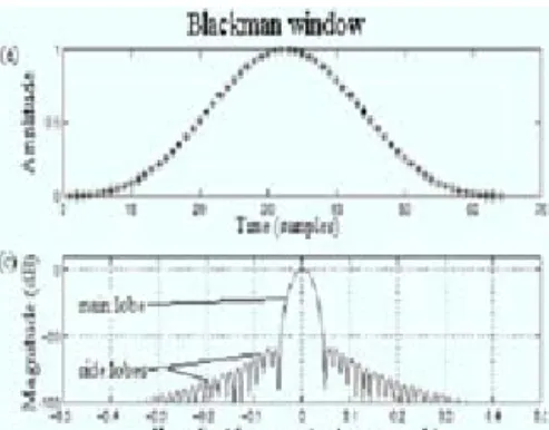
The plasma is inherently noisy. In order to calculate the EEDF, the second derivative of the measured curve is needed, which amplifies the noise. Therefore the data is filtered before proceeding further. This can be done by using a Blackman filter. The Blackman filter window is given by [49]:
M n M n n wB π π 4 cos 08 . 0 2 cos 5 . 0 42 . 0 ) ( = − + , n= 0, 1, 2, …, M (7)
where M is the size of the window and controls the degree of smoothing. The Blackman window minimizes the side-lobe level while having a steep roll-off on the side lobes (Fig. 7). This is a standard moving average window and the filtering is easy to implement in the Matlab software package [48].
Fig. 7. The Blackman window.
After filtering (Fig 8 (a), Fig. 8(b)), the derivative is found by applying the central difference approximation (Fig. 8 (c), Fig. 8(d)). It should be noted that there is always a compromise between accurately producing the EEDF maximum, and the high electron energy tail. Larger windows flatten the maximum while achieving good smoothing for higher energies, while small windows retain the shape of the maximum with less smoothing for higher energies. The effect of these distortions can be corrected for however, by fitting the measured EEDF to the function
) exp( x
R a b
g = ε − ε where a, b and x are constants. The Maxwellian and the Druyvesteyn electron energy distributions are special cases with x=1 and x=2 respectively. The value x is determined by performing a least-squares analysis of
) / ) (
ln(gR ε ε versus ε
x for various x. To find the best fit, the maximum of the EEDF and the corresponding energy are found. The equation is fitted to the measured EEDF (Fig. 8 (e)) from the electron energy at which the EEDF has the maximum until it has fallen two orders of magnitude. The best fit to the equation is then interpolated to zero electron energy, before the electron density (Fig. 8 (f)) and effective electron temperature are calculated.
Fig. 8. The I-V curve before (a) and after (b) filtering, measured 200 µs after the pulse start for a discharge with a Ta target and an Ar sputtering gas held at 20 mTorr. The corresponding first (c) and second (d) derivatives, and the calculated EEDF (e) and electron density (f) are shown.
2. 4. 3. Ion current determination
For ion current measurements, flat probes are commonly used. This is because by using this measurement setup, the collecting area is essentially independent of the sheath thickness [41]. A schematic of a flat probe is shown in Fig. 9, where a metal disk, in this case of 2 cm in diameter, is equipped with a guard ring that is kept at the
probe potential. However, only the current on the surface of the probe is collected. To keep the probe potential constant during operation of the cathode, a power supply with sufficient capacitance on the output circuit should be utilized. Principally, the theoretical approach for treating ion current measurements, using a flat probe, are the same as that of the wire probe described in the preceding section.
Fig. 9. A schematic of the flat probe with the probe and the guard ring kept at the same potential.
2. 4. 4. Direct display of plasma parameters using a triple probe setup
In some applications, measuring a time-dependent electron current that varies intensely in magnitude could cause problems related to the used measurement electronics. This is a typical problem in measuring the electron temperature in HPPMS, where the noise amplification resulting from the electron saturation current differentiation and the following integrations has been shown to give big uncertainties. To avoid this problem, the triple probe [50,51] with direct temperature display is utilized. The triple probe theory assumes a Maxwellian electron energy distribution. It consists of three Langmuir probes with identical geometrical configurations and dimensions. Figure 10 shows a schematic of such a probe.
Fig. 10. A schematic of the triple probe circuit with potentials and currents of each probe shown.
The three probes P1, P2 and P3 are placed 2 mm away from each other, forming an
equal-sided triangle. This is a large enough separation to adequately shield them from one another. Negative potentials Vd2 and Vd3 are applied to P2 and P3 with reference to
P1, so that the probes are individually biased to three different potentials. The current
density on each probe is the sum of the ion saturation current density and the electron current density so that:
-I1=-SJeexp(-φV1)+SJi(V1) (8)
I2=-SJeexp(-φV2)+SJi(V2) (9)
I3=-SJeexp(-φV3)+SJi(V3) (10)
φ=e/kTe (11)
Je=nee(kTe/2pme)1/2 (12)
Because the electron currents in all three probes are much higher that the corresponding ion currents, the assumption Ji(V1)= Ji(V2)= Ji(V3)=Ji is made. This gives: ) exp( 1 ) exp( 1 3 2 3 1 2 1 d d V V I I I I φ φ − − − − = + + (13)
This serves then to determine Te directly by drawing
curves of the probe-ratio (I1+I2)/(I1+I3) vs Te for a number of Vd3 and a fixed Vd2. It is also possible to have direct display of the electron temperature, using an oscilloscope for example. In that case one of the three probe is set to be floating (Fig. 11). With I2 =0, the currents flowing through P1 and P2 obey I1 =I3. Equation (13) will
therefore modify to give:
) exp( 1 ) exp( 1 2 1 3 2 3 1 2 1 d d V V I I I I φ φ − − − − = = + + (14)
This means that, having Vd3 fixed, the electron temperature can be determined solely
by knowing Vd2, using equation (11).
Fig. 11. A schematic of the triple probe circuit with one of the probes kept at the
2. 5. Solitary waves
As mentioned in section 2.1, a number of waves can exist in a plasma, ranging from electromagnetic to magnetosonic and ion acoustic waves. For a comprehensive theoretical description of these waves, I refer to the books in references 40 and 41. I will however provide in this section a short description of solitary waves that are also the subject of paper IV.
The discovery of solitary waves dates back to 1834, when John Scott Russell, a Scottish scientist was observing a boat being drawn along 'rapidly' by a pair of horses. When the boat suddenly stopped, Russell noticed that a bow wave continued forward "at great velocity, assuming the form of a large solitary elevation, a well-defined heap of water which continued its course along the channel apparently without change of form or diminution of speed". The photo in Fig. 12 shows a solitary wave generated in the Scott Russell Aqueduct on the Union Canal near Heriot-Watt University.
Fig. 12. Photo: courtesy to D B Duncan and J C Eilbeck, Heriot-Watt University, Edinburgh.
The observations of Russel appeared to contradict the nonlinear shallow-water wave theory of Airy (1845), which predicted that a wave with elevations of finite amplitude could not propagate without change of form, i.e., the top of the wave would move faster than the base resulting in a steepning of the wavefront (Fig 13) [52].
Fig. 13. A breaking wave.
This issue was resolved by Joseph Boussinesq (1871) and independently by Lord Reyleigh (1876), who showed that the steeping of the wave can be balanced by the dispersion (Fig. 14), where different frequency components of the wave travel with different speeds, leading to a wave of permanent form [52]. For a long time, however, the solitary wave was considered to be a rather unimportant curiosity in the mathematical structure of nonlinear wave theory. It was not until the 1950's that studies conducted by Fermi et al. [53] showed how important Russell's discovery had been [54,55]. Today's most advanced fiber-optic communications use stable pulses of light identical to Russell's waves, now called solitons (from solitary waves), to carry a large amount of information over thousands of kilometers of fibers.
Fig. 14. A dispersive wave.
Ion acoustic solitary waves can be described as constant-velocity waves where the group velocity is equal to the phase velocity. Thus ion-acoustic solitary waves are stationary pulses or wave packets that propagate in non-linear dispersive media. They were first observed in a double-plasma device by Ikezi et al. [56]. Since the initial
observation, these waves have been the subject of numerous experiments, including multiple soliton production in a double-plasma device [57,58] and excitation of cylindrical [59] and spherical [60] ion-acoustic solitons.
Hershkowitz and Romesser [59] summarized the distinguishing characteristics of the dimensional solitary wave. These characteristics are common to one-dimensional solitary waves in planar, cylindrical and spherical geometries and provide a reference for comparison with experimental data. They are:
1. Arbitrary positive (compressive) density perturbations evolve into a superposition of spatially separated solitons.
2. The number and amplitude of solitons is determined by the solutions of the time-independent Schrödinger equation.
3.The soliton velocity is given by:
) / ( 3 / 1 1 n n0 c u S δ + = (15)
where δn/n0 is the maximum density perturbation and cS is the ion acoustic velocity. 4. The square of the normalized spatial width, D2, of a soliton is proportional to
(δn/n0)-1, which implies D2δn/n0 = constant.
5. Solitons retain their identity upon collision with other solitons.
In addition to characteristic 2, it has been shown that to the continuous spectrum of unbound states corresponds a linear oscillatory tail [61]. The energy of a solitary wave is proportional to (δn/n0)2D. For non-planar expanding solitons the energy will decay due to geometry as 1/r and 1/r2, for cylindrical and spherical geometries, respectively. When combining this geometrical decay with characteristic 4, we find that the solitary wave’s amplitude will decay as:
η r nn ∝ ∂ ∂ 0 (16)
where r is the radius of the soliton and η=-2/3 for a cylindrical geometry but η=-4/3
for a spherical geometry [ 60,62].
3. Part II. Thin film growth and characterization
3. 1. Growth models
Thin films are formed on a substrate by a process of nucleation and growth. Nucleation depends on the interaction between the adatoms and the substrate and is affected by the surface mobility of the species, which depends on the substrate temperature, the deposition rate, and the energy of the arriving atoms. The shape of the nuclei depends on the relative interfacial free energies between the film and the substrate γfs, the film and the vapor γfv, and the substrate and the vapor γsv [63]. The
interfacial energies balance each other at the interface so that γsv= γfs+ γfvcosθ, where
θ is the contact angle, as seen in Fig. 15.
Fig. 15. Schematic of a nucleus and the corresponding free energies of the substrate, the film, and the vapor.
During the early stages of thin film growth, one of three main growth modes dominates [1]. If γsv≥ γfv+ γfs and θ=0, i.e., the atoms or molecules in the growing film
are more strongly bound to the substrate than to each other, the growth is two-dimensional Frank-van der Merwe (FM) or layer-by layer growth, where a continuous monolayer is formed prior to the deposition of a subsequent layer. If γsv< γfv+ γfs and
θ>0, the atoms or molecules in the growing film are more strongly bound to each other than to the substrate, and the film grows three-dimensionally. This is called Volmer-Weber (VW) or island growth, where the deposited atoms tend to aggregate into islands with thickness of several monolayers. Finally, the mixed Stranski-Krastanov (SK) mode, which starts in a monolayer growth mode for a few
monolayers before shifting to island growth. The intermediate SK mode is not completely understood, but factors such as the film-substrate, lattice mismatch, or the strain energy accumulated in the growing film, may be the cause of such growth [28]. For the three growth modes, similar mechanisms control depositions and agglomeration of the incoming atoms in the initial and stationary growth phases.
Fig. 16. Microstructure zone diagram for metals deposited by magnetron sputtering. T is the substrate temperature and Tm is the coating material melting point [64].
During deposition, the surface diffusion, the bulk diffusion, and the adsorption scale directly with the melting temperature of the deposit (metal). This can result in 4 different film structures (Fig. 16):
zone 1 exhibits amorphous or fine grained polycrystalline films, with a high density of crystallographic defects. The absence of surface diffusion at low Ts yields columnar structures and results in porous and rough films.
Zone 2 exhibits a dense columnar structure with smoother surfaces as a result of surface diffusion controlled growth. The columnar grains can have highly faceted surfaces due to surface energy anisotropy. The grain size generally increases with increasing Ts.
Zone T is the transition zone between zones 1 and 2 [64] that is observed when the gas pressure is decreased (Fig. 16). This is because a decrease of the gas pressure results in an increase in the energetic bombardment of the films.
The influence of ion bombardment on the film properties of metals can be reconciled with the accepted models of thin-film growth. However, energetic condensing particles or bombardment promote the zone T structure [64,65]. Evaporated atoms usually have energies in the vicinity of 0.1 eV, while sputtered atoms usually have energies of a few eV. Condensing atoms from these different sources will produce films of different densities.
3. 2. Deposition of thin films by magnetron sputtering
3. 2. 1. Magnetron sputtering
Figure 17 shows a typical magnetron set up where a permanent magnet is placed at the back of the cathode target, which generates magnetic field lines that enter and leave through the cathode plate, as shown in Fig. 18.
Fig. 17. A schematic of the magnetron with two concentric magnets with opposite polarities.
A simulation of the magnetic field is shown in Fig. 18. This was made for a magnetron sputtering cathode with a circular target (similar to the setup used in the work presented here) using the magnetic field shareware package Finite Element
Method Magnetics (FEMM) [66]. The simulation results show the magnetic field lines
that link the outer ring-magnet and the center pole. At a distance d ~ 6 cm from the target, the magnetic field strength is nil along the target axis, and at d > 6 cm, the magnetic field lines lead away from the target. The simulation also shows the magnetic field strength, B, in the form of shaded gray zones.
Fig. 18. Magnetic filed lines and flux simulated using FEMM.
The high B value near the surface of the target results in a trapping of the electrons in this area. This is because of the modification of the electron trajectory, as electrons within the dual (electric + magnetic) field environment experience the well-known Lorentz force. This is given in the equation,
) (E v B F= =−q + × dt mdv (17)
where q, m, and v¸ are the electron charge, mass, and speed, respectively. The magnetic field, prolongs therewith the electron residence time in the plasma close to the target, which allows for operating the sputtering discharge at higher current densities, lower voltages, and lower pressures than can be obtained in a conventional glow discharge. and thus enhance the probability of ionizing (and exciting) collisions.
3. 2. 2. The ionization process
In conventional magnetron sputtering techniques, the extra energy needed in some process conditions is usually provided by heating the entire substrate. This leads to restrictions with respect to choice of substrate material. Instead of using thermal energy as enhancement energy, various techniques, such as ion beam deposition [28], have made use of the kinetic energy of the accelerated ions. In conventional dcMS, the ion content of the discharge is usually low (~1 %). The plasma electron density is low (~ 1×10¹⁶ m-3), and the main mechanism for ionization is penning ionization [41].
In order to increase the ionization fraction, the plasma density in the region between the source and the substrate must increase so that the probability for electron-impact
ionization increases [41]. The higher ionization fraction is beneficial for the film morphology as particle bombardment of the growing film transfers kinetic energy to the adatoms of the growing surface [67], which can be used to lower the process temperature and modify the microstructure of the film. In order to do this, the power applied to the cathode is increased. However, since most of the power applied to the cathode is transformed into heat, the technique is limited to relatively low power densities before the target cooling is insufficient. High plasma density, without overheating the target, can be achieved by HPPMS, where high power pulses are applied to the target with a low duty cycle.
3. 2. 2. 1. Self-sputtering in HPPMS
In the HPPMS process (section 2.4), the sputtering takes place during the pulse-on time, with a duty factor of less than 1 %. Fig. 5 is redrawn here for convenience as Fig. 19, where the target voltage and current time-traces are shown. It is observed that the negative target voltage VT drops from 100V to ~ 800 V as the
target current IT starts to build up, and that a peak power of 0.4 MW is achieved 20 µs
after the pulse start. This is followed by a plasma decay as the voltage and the current decrease and eventually die out ~ 80 µs after the pulse initiation. During the pulse-on time, the composition of the discharge is dominated by the sputtered metal ions, as has been shown by optical emission spectroscopy (OES) [68]. This has not been fully explained at this point, nevertheless, it was suggested that during the pulse-on time, self-sputtering contributes to the sputtering process, i.e., sputtering is performed by the target metal ions that are attracted back to the target by the potential drop between the plasma and the target. [69,70].
Fig. 19. Time evolution of the target voltage (VT) and current (IT) for a Ta target and
an Ar gas pressure of 20 mTorr.
The effect of self-sputtering is examined, using a simple model, by comparing the sputtering yields obtained with HPPMS and dcMS, for the same average power (Fig. 20). The ratio between the HPPMS and the dcMS sputter yields (R=SHPPMS/SDC)
could be estimated by modeling the yields as follows:
SHPPMS = IAr×γAr + IMe×γMe – IMe (18) SDC = I’
Ar×γAr (19)
Where IAr and IMe are the Ar and metal ion currents in the HPPMS system and I’Ar is the Ar ion current in the dcMS system. γAr and γMe, are the sputter yield for Ar and metal respectively. If we write the total ion currents as:
IAr+IMe=IiH (20) I’ Ar≈Ii D (21)
Where IiH and IiD are the total ion currents in HPPMS and dcMS respectively, then,
(
)
(
)
Ar H i Me Me Ar Ar D i H i Ar D i Me Me Ar Ar DC HPPMS γ I 1 γ I γ I I I γ I 1 γ I γ I S S × − + × × = × − + × = = R − + − = Ar Me H i Me H i Me D i H i γ 1 γ I I I I 1 I I (22)
The current ratios D i H i I I and H i Me I I
could be determined by measuring the currents in the
system. However, it is seen from equation 22 that R not only depends on the applied average power, i.e., current ratios, but also on the target material and the sputter gas.
Fig. 20. A schematic of the sputtering process during HPPMS.
Weight gain differences between a floating and a positively biased substrate showed that, for a number of target materials, including Al, Zr, Ti, Ta, and Cr, the degree of ionization of the sputtered metal was high. Figure 21 shows the results obtained using a Ta target. Other works showed that the ionization fraction was 70 % using a Cu target [36], roughly 40 % using a TiAl-target [68] and 4.5 % using a C target [38].
Fig. 21. Ion deposition measurements. Experimental setup (a), and the ion fraction in the deposited film for Ta as a function of the pulse energy, measured as the weight gain difference between a floating and a biased substrate (b).
In order to put this in the context of self-sputtering, Fig. 22 shows measured relative deposition rate (HPPMS with respect to dcMS) for some metals as a function of the self-sputtering yield, SS, divided by the Ar yield, SAr. It is seen that materials
with high self-sputtering yields resulted in increased deposition rate ratios.
Fig. 22. Deposition rate ratio, of HPPMS by dcMS, versus the self-sputtering yield ratio SS/SAr [70].
3. 3. Effects of ionized deposition on film structure
3. 3. 1. Film densification using HPPMS
In general, ion bombardment is accompanied with surface defects such as surface vacancies [28,67]. The resulting film growth strongly depends on the individual film-substrate combination and the process parameters such as the substrate temperature, the ion energy, and the ion-to-neutral ratio [71]. It is therefore beneficial to acquire means to control the bombarding energy of individual ions in order to enhance the deposited film’s properties. In this regard, the advantages of using HPPMS are many. Dekoven et al. [67] found that ultra thin carbon films grown by HPPMS had significantly higher densities (2.7 g/cm3) than films grown by conventional dcMS (< 2.0 g/cm3), i.e., the film density is closely related to the ion bombardment and ion deposition during the process. This is also supported by paper VII where Ti-Si-C films were deposited by HPPMS. As is seen in the SEM micrographs in Fig. 23, the HPPMS film exhibited an enhanced morphology compared to the dcMS grown film.
Fig. 23. Thin Ti-Si-C films grown by dcMS (a), and HPPMS (b) at 14 cm from the target surface, using an average target power of 150 W, a substrate bias voltage of -10 V, and an Ar gas pressure of 5 mTorr.
Film densification was also investigated for deposition on tilted substrates. In general, deposition on complex-shaped substrates entails new challenges to the deposition process. When the substrate is tilted with respect to the deposition flux (oblique incidence), interesting effects such as shadowing can take place, which can
result in changes in the optical, magnetic and/or electrical properties of the deposited films [72,73]. The oblique incidence effect was discovered in 1959 and many papers have been written on the subject [72,73]. We investigate the oblique incidence effect in the HPPMS system by growing thin films on non-flat surfaces, mimicked by substrates at different angles with respect to the target surface, and compare their morphologies and properties with dcMS grown films. Scanning electron micrographs of Ta films deposited using a bias voltage of –50 V are shown in Fig. 24. They show that in the dcMS case, films deposited at a 90o inclination angle with respect to the
incidence flux exhibit a columnar morphology, with the columns tilted from the substrate normal toward the deposition flux incidence direction. The films are porous and rough. The HPPMS films, on the other hand, show a dense and smooth surface, and the columns are along the substrate normal. This indicates that a large part of the depositing species are ions, as is discussed in more detail in paper V. The ion bombardment is known to influence the morphology and the density of the growing film. However, the use of magnetron sputtering deposition for the purpose of depositing dense flat films on inclined surfaces is a feature that is unique to HPPMS at this point.
Fig. 24. SEM micrographs of Ta films deposited by dcMS (a) and HPPMS (b) at 10 cm from the target surface, with an average power of 300 W, a bias voltage of –50 V, and an Ar gas pressure of 5 mTorr.
Using HPPMS, densification of films grown facing away from the target (at a 180º angle with respect to the target surface) was possible by applying a bias to the substrate. Fig. 25 shows TEM micrographs of Ta films deposited by HPPMS, facing away from the target, at floating potential and at a –30 V bias voltage.
Fig. 25. TEM micrographs showing Ta films grown at a 180o angle with respect to the
target surface by HPPMS with the substrate floating (a) and biased with a voltage of -30 V (b). The average target power was 150 W and the Ar gas pressure 5 mTorr.
3. 3. 2. Phase formation
The formation of high compressive stress might promote the formation of dense phases of a material [28,74]. For example, at sufficiently high deposition energy, diamond can be grown as the thermodynamically stable phase rather than graphite. [28]. McKenzie et al. [75] showed that at a high pressure, corresponding to compressive stresses, sp3 bonding could be promoted in a growing C film by ion
bombardment. More clearly, the influence of the process parameters on the phase formation of coatings becomes evident for films with different crystalline phases. As an example, Ta thin films usually have a metastable tetragonal structure (β-Ta), when they are deposited on Si substrates. The formation of Ta metastable phases has been reported in a large number of sputtering experiments [76-82]. The β-phase is sometimes formed alongside the stable α-phase (bcc-Ta). β-Ta is harder, more brittle, and less ductile than the bcc-Ta. It is known to contain a high concentration of impurities and defects, and to have a resistivity of ~170–210 µΩcm, much higher than
the ~15–60 µΩcm of the stable bcc phase. [79,80,82]. Using HPPMS, β-Ta and bcc-Ta films could be grown for different bias voltages. This is shown in paper VI to be directly related to the ion energy and the ion-to-neutral ratio in the discharge.
3. 3. 3. Enhanced adhesion by HPPMS
Of fundamental importance to the performance of a thin film is its adherence to the substrate. Adhesion is determined by two main factors, one being the strength of the interface between film and substrate, the other one being the stress state of the film. Hirsh and Varga [83] found that ion bombardment increased the adhesion of IBAD (ion-beam assisted deposition) Ge films to glass substrates, as a result of reduced stress and formation of an intermediate layer between the film and the substrate, which increases the bonding forces [28]. Münz et al. [84] utilized ionized metal from an arc source for etching and implantation of a substrate with the aim to increase coating adhesion. The technique is named arc bond sputtering (ABS). An arc source gives a very high degree of metal ionization, but also produces macroscopic particles [85]. HPPMS is therefore an attractive alternative to ABS. HPPMS was also used to improve the adhesion of thin CrN films [86-88]. Its main advantage compared to arc deposition is that macroscopic droplets can be avoided. It was showen that the microstructure of the coatings grown by HPPMS was dense with, neither voids that are usually associated with conventional magnetron sputtered films, nor large-scale growth defects, usually observed in arc evaporated films, as shown in Fig. 26. Furthermore, the HPPMS-based sample pretreatment stage contributed to the strong adhesion of CrN coatings, with high sliding wear resistance. The films also showed better corrosion protection behavior than the corresponding ABS coatings due to the lack of macro-particles and the voids created close to them.
(a))
( axcdsaaa a)a
3. 4. Sputtering of a MAX-phase compound target by HPPMS
Mn+1AXn-phases (n= 1-3) are materials with exceptional structural, electrical,and mechanical properties [89]. They are ternary material-systems, where M is a transition metal, A is a group-A element, and X is C and/or N.
I II III IV V VI VII VIII
H
M
Early Transition Metal HeLi Be
A
A-Group Element III-V B C N O F NeNa Mg
X
Carbon and/or Nitrogen Al Si P S Cl ArK Ca Sc Ti V Cr Mn Fe Co Ni Cu Zn Ga Ge As Se Br Kr Rb Sr Y Zr Nb Mo Tc Ru Rh Pd Ag Cd In Sn Sb Te I Xe Cs Ba La- Hf Ta W Re Os Ir Pt Au Hg Tl Pb Bi Po At Rn Fr Ra Ac- Unq Unp Unh Uns Uno Une Uun Uuu Uub Uut Uuq Uup Uuh Uus Uuo
211 M2A1X1: Ti2AlN, Zr2SnC,…, ~ 50 Compounds
312 M3A1X2: Ti3SiC2, Ti3AlC2, Ti3GeC2
413 M4A1X3: Ti4AlN3, Ti4SiC3, Ti4GeC3
Fig. 27. The periodic table of elements showing the different groups that form the MAX-phase materials, with different shades. The box under shows the synthesized MAX-phase materials
Although they were discovered in the 1960’s [90,91], the interest in the Max-phase materials was only awakened in the later years [92], due mainly to the difficulty in synthesis. The MAX-phase materials combine metallic electrical properties and ceramic mechanical properties. Their crystal structure consists of a hexagonal unit cell belonging to the space group D4
6h-P63 / mmc. Fig. 28 illustrates the unit cells for Ti2SiC, Ti3SiC2, and Ti4SiC3 stoichiometric MAX-phases, where the edge-sharing
M6X (Ti6C or Ti6N) octahedra is interleaved with layers of pure A-elements, thus
forming a nanolaminated material [90]. Ti-Si-C is one of the materials system that has been studied the most. Films were grown by different methods [93-96] and their properties were studied. In this work Ti-Si-C thin film growth by HPPMS was pioneered. This is presented in paper VII, where the films were deposited on MgO
substrates at different substrate inclination angles with respect to the sputter source from a compound Ti3SiC2 target, for a range of deposition conditions. This resulted in
different film compositions as studied by XRD and XPS, indicating the possibility to tailor the film composition simply by inclining the substrate.
Fig. 28. Unit cells for Ti2SiC1, Ti3SiC2, and Ti4SiC3.
Results from these experiments (paper VII) and Ta deposition experiments (papers V and VI) show that in HPPMS three parameters related to ions in the plasma could be controlled, allowing for better control and potentially tailoring of the film structure. These are: the ion energy, which could be determined by choosing the power to the target and the bias voltage to the substrate, the ion to neutral flux, which was showed to depend on the deposition angle with respect to the target surface, and the metal flux in the plasma. Thus, work on the Ti-Si-C system shows that HPPMS can be used in more complex materials system, but a better understanding of the deposition dynamics and the gas phase scattering in the system is needed.
3. 5. Thin film characterization
In this chapter, the main tools used for characterization of the deposited films in the present work are shortly described. Different textbook references are given where appropriate for detailed descriptions of these techniques.
3. 5. 1. Transmission electron microscopy (TEM)
This is a powerful tool with an optimal image resolution of ~1 Å for an accelerating voltage of 200 kV. Figure 29 presents a comparison between a number of microscopic tools. Materials for TEM must be specially prepared to thicknesses that allow electrons to transmit through the sample. This is a time consuming task, especially for cross-sectional images. TEM images provide information on the sample microstructure and exhibit contrasts caused by defects and grain boundaries, for example. There are two mechanisms responsible for the contrasts observed in a TEM image, a diffraction contrast used for the formation of bright field and dark field images, which reveal areas that are diffracting the electron beam differently due to changes in orientation or presence of crystallographic defects, and a phase contrast that uses the phase difference of the transmitted electrons to form the image [99,100].
Fig. 29. The range of spatial resolutions for the eye and some microscopic techniques.
3. 5. 2. Scanning electron microscopy (SEM)
SEM is a versatile and widely used tool as it allows the study of both morphology and composition of materials. A monochromatic electron beam is focused onto a fine probe that is scanned over a rectangular area of the sample. As the electrons collide with the surface, a number of interactions occur resulting in the
emission of electrons and photons from the surface. The intensity of the secondary electrons is point-to-point mapped onto a screen. The electron beam in the cathode ray tube illuminating the screen is synchronized with the electron beam scanning the sample. High-resolution images of the morphology or topography of a specimen at very high magnifications can be obtained. Compositional analysis of a material may also be obtained by monitoring secondary X-rays produced by the electron-specimen interaction. Thus detailed maps of elemental distribution can be produced from multi-phase materials. Characterization of fine particulate matter in terms of size, shape, and distribution as well as statistical analyses of these parameters, may be performed. More details are found in reference [100, 101,102]
3. 5. 3. Atomic force microscopy (AFM)
AFM, also known as scanning force microscopy, is a technique with minimal sample preparation requirements. It is based on detection of force interaction between a sample surface and a sharp tip mounted on a flexible cantilever, and produces, in such a way, topographic images of a surface with atomic resolution in all three dimensions. When the tip is brought to a close proximity (a few Å) of a sample’s surface, repulsive Van der Waals forces between the atoms of the tip and those of the sample cause the cantilever to deflect. The magnitude of the deflection depends on the distance between the tip and the sample. Normaly the lateral AFM resolution of an AFM is about 1 nm. However, with a highly sharp tip and a flat sample, even atomic resolution could be obtained [100].
3. 5. 4. X-ray diffraction (XRD)
XRD is a powerful non-destructive method that is used to measure structural properties, such as residual stress, grain size, epitaxial relations, texture, defect structure, and crystal structure. The X-rays have wavelengths in the order of a few angstroms, the same as typical interatomic distances in crystalline solids. When certain geometric requirements are met, X-rays scattered from a crystalline solid can constructively interfere, producing a diffracted beam. The condition for constructive interference is given by the Bragg’s law:
where n is an integer, λ is the wavelength, d is the interatomic spacing, and θ is the diffraction angle. The diffracted beam intensity will depend on several factors such as the chemical composition of the film and the local arrangement of the atoms. Detailed descriptions of the technique are found in reference. [89, 103]
3. 5. 5. X-ray photoelectron spectroscopy (XPS)
XPS provides elemental analysis and information about chemical bonding properties. An incoming photon of known energy hω causes ejection of a core
electron, thus leaving the atom in an excited state. The kinetic energy EK of this core electron is equal to the excitation energy minus the binding energy and the work function φ. This is described by the following equation:
EB=hω-EK-φ.
XPS provides chemical bond information through the chemical shifts, i.e. an element experiences a slight net negative charge when bonded to an electropositive element. The opposite happens when the bond is to an electronegative element [100].
3. 5. 6. The four-point electrical probe
The four-point probe measurement setup is shown in Fig. 30. Four wires (or probes) are attached to a sample surface. A constant current is made to flow the length of the sample through probes labeled 1 and 4 in the figure. This can be done using a current source or a power supply as shown. The sample’s resistance to the flow of electrical current causes a potential drop as the current flows along the sample, for example between the two wires (or probes) labeled 2 and 3 in the figure. The resistance of the sample between probes 2 and 3 is the ratio of the voltage drop to the value of the output current of the power supply [1].
