1 2 3 4 5 6 7 8 9 10 11 12 13 14 15 16 17 18 19 20 21 22 23 24 25 26 27 28 29 30 31 32 33 34 35 36 37 38 39 40 41 42 43 44 45 46 47 48 49 50 51 52 53 54 55 56 57
Very Important Paper
Multivalent ions and biomolecules: Attempting a
comprehensive perspective
Olga Matsarskaia,*
[a]Felix Roosen-Runge,*
[b, c]and Frank Schreiber
[d] Ions are ubiquitous in nature. They play a key role for manybiological processes on the molecular scale, from molecular interactions, to mechanical properties, to folding, to self-organisation and assembly, to reaction equilibria, to signalling, to energy and material transport, to recognition etc. Going beyond monovalent ions to multivalent ions, the effects of the ions are frequently not only stronger (due to the obviously higher charge), but qualitatively different. A typical example is the process of binding of multivalent ions, such as Ca2 +
, to a macromolecule and the consequences of this ion binding such as compaction, collapse, potential charge inversion and precip-itation of the macromolecule. Here we review these effects and phenomena induced by multivalent ions for biological (macro) molecules, from the “atomistic/molecular” local picture of (potentially specific) interactions to the more global picture of phase behaviour including, e. g., crystallisation, phase
separa-tion, oligomerisation etc. Rather than attempting an encyclope-dic list of systems, we rather aim for an embracing discussion using typical case studies. We try to cover predominantly three main classes: proteins, nucleic acids, and amphiphilic molecules including interface effects. We do not cover in detail, but make some comparisons to, ion channels, colloidal systems, and synthetic polymers.
While there are obvious differences in the behaviour of, and the relevance of multivalent ions for, the three main classes of systems, we also point out analogies. Our attempt of a comprehensive discussion is guided by the idea that there are not only important differences and specific phenomena with regard to the effects of multivalent ions on the main systems, but also important similarities. We hope to bridge physico-chemical mechanisms, concepts of soft matter, and biological observations and connect the different communities further.
1. Introduction, motivation and scope
In this review, we will focus on effects induced in biological and chemical systems by multivalent ions. While general overviews of the influences of electrostatics on soft matter are given, e. g., in Refs. [1–3], it is generally accepted that the effects of multivalent ions go beyond such considerations.[4–6]
We will include different types of ions into our discussion: mono-atomic ions, such as Na+
and Mg2 +
; multi-atom ions (e. g., NHþ 4 or spermidine); nano-ions (such as polyoxometalates), and larger ions, e. g., oligo-arginine. We note that the classification of ions does not only depend on their net charge, but also on their other characteristics such as their (in)organic nature. The main aspects of this review are the charge-mediated effects of these
ions whereas their other properties play a less important role. We note that specific interactions of monovalent ions with biomolecules, such as that between Ag+
and DNA,[7]
have also been shown, implying that the role of monovalent ions can go beyond that of purely inert electrolytes. However, such interactions are not the main focus of this review.
Generally, ions are present ubiquitously and are therefore of fundamental interest for a large variety of topics and research areas. Starting from biology and physiology, the importance of ions becomes apparent immediately. A typical animal or human cell contains approximately 130 mM K+
and 10–20 mM Na+ cations and relies on an active ion exchange with its surroundings to maintain its electrochemical potential.[8]
Sim-ilarly, the signal transduction activity of neurons depends on charge exchange mechanisms.[9]
Inside the nucleus, the highly negative charge of nucleic acids (DNA and RNA) implies that ions – most frequently, Mg2 + – are required to screen their charges, thus enabling, e.g., nucleic acid-protein interactions.[9] Several other biological aspects depend on ions. For example, many enzymes host metal cations such as Ca2 + and Zn2 + in their catalytic centers; muscle contraction is made possible via myosin-Ca2 + interactions; and oxygen transport by hemoglobin is facilitated through Fe2 +/Fe3 + ions.[10] Generally, iron metabolism, storage and transport in mammals is a complex issue and involves, apart from hemoglobin, the proteins myoglobin, ferritin[10]and lactoferrin.[10,11]Divalent ions such as Ca2 + and Mg2 + have furthermore been shown to be present at the interfaces between virus particle subunits (see Ref. [12] and refs. therein), presumably fulfilling also structurally important roles in viruses. Investigations of silk feedstock [a] Dr. O. Matsarskaia
Institut Laue-Langevin, Grenoble, France E-mail: matsarskaia@ill.eu
[b] Asst. Prof. Dr. F. Roosen-Runge
Department of Biomedical Sciences and Biofilms-Research Center for Biointerfaces (BRCB), Faculty of Health and Society, Malmö University, Sweden
E-mail: felix.roosen-runge@mau.se [c] Asst. Prof. Dr. F. Roosen-Runge
Division of Physical Chemistry, Lund University, Sweden [d] Prof. Dr. F. Schreiber
Institute of Applied Physics, University of Tübingen, Germany E-mail: frank.schreiber@uni-tuebingen.de
© 2020 The Authors. Published by Wiley-VCH Verlag GmbH & Co. KGaA. This is an open access article under the terms of the Creative Commons Attribution Non-Commercial License, which permits use, distribution and reproduction in any medium, provided the original work is properly cited and is not used for commercial purposes Open access funding enabled and organized by Projekt DEAL.
1 2 3 4 5 6 7 8 9 10 11 12 13 14 15 16 17 18 19 20 21 22 23 24 25 26 27 28 29 30 31 32 33 34 35 36 37 38 39 40 41 42 43 44 45 46 47 48 49 50 51 52 53 54 55 56 57
indicated that the viscosity of the latter is strongly influenced by the type and valency of the cation present. Whereas the divalent Ca2 +
increases the viscosity by bridging acidic amino acids, the monovalent K+
reduces viscosity due to competition for binding to these types of residues.[13]
Given the prominent role of ions in physiology, it is obvious that biological and biotechnological experiments need to consider the native environment of the biomolecules they investigate. Thus, the success of ex vivo and in vitro experiments with, e. g. enzymes, strongly depends on the (ionic) composition of buffer solutions used.
The strong dependence of biological processes on ions can lead to peculiar evolution processes. As an example, due to low Zn2 +
levels in the deep sea, a certain diatom species relies on Cd2 +
instead[14]
– a heavy metal cation known to be toxic for many land-borne creatures. Another dependence on seemingly less physiologically relevant ions has been observed in M.
fumariolicum SolV, an extremophilic microbe native to Italian
volcanic mudpots. This bacterium utilises lanthanide cations to catalyse methane-based metabolic pathways.[15]
From ecologic and ecotoxicologic points of view, ions play significant roles as fertilisers. A large amount of fertilisers is phosphate-based,[16]
but lanthanides are also known to be used in agriculture.[17]
In industrial settings, polyanions such as polyacrylate can be used to prevent CaCO3precipitation (i. e., as scale inhibitors) due to their specific interactions with Ca2 +
in pipelines.[18]
At the same time, ions can be successfully employed to manipulate macromolecules in biotechnology, e. g. as crystal-lisation agents. This can be demonstrated in the case of negatively charged proteins where multivalent cations have been shown to promote crystallisation.[19–27]
In addition, a purification protocol using Zn2 +
ions has recently been established for recombinant antibodies.[28]
Here, we first introduce the theoretical aspects of charge-mediated interactions. We then summarise the current knowl-edge regarding the role of charges in (bio)(macro)molecules including DNA, surfactants, interfaces and proteins. We focus especially on charge-mediated modifications of static, dynamic or thermodynamic properties of the macromolecules in ques-tions, including the intriguing possibilities to rationally tune (bio)molecular interactions. Finally, we briefly cover other systems such as ion channels and synthetic polymers. We mention experimental methods where necessary, but do not consider them the main subject of this review.
Through this review, we aim to provide a comprehensive overview of the manifold ways in which charges can influence the behaviour of macromolecules. By emphasising how physical concepts can be used to understand biological and soft matter systems, our goal is to enhance mutual understanding and communication between different scientific communities tack-ling manifold questions. We hope to stimulate further dis-cussion and inspire both experimental and theoretical inves-tigations of these complex aspects. We will mostly focus on the static/equilibrium behaviour, but we note that there are of course also interesting multivalent ion-mediated effects on the dynamics, kinetics, and viscosity of (biological) soft matter.[29,30]
2. Background and theoretical concepts
In this section (partly based on Ref. [31] and Ref. 39), we provide an overview of the theoretical concepts behind ion-related interactions of soft matter. In particular, we focus on charge effects as accounted for in mean-field, Poisson-Boltzmann and Derjaguin-Landau, Verwey-Overbeek (DLVO)[32,33]
theories, out-lining their strengths and shortcomings especially in the context of ion-specific effects and multivalent ions. The two latter aspects are then outlined in more detail using the
Dr. Olga Matsarskaia obtained her PhD on the topic of tuning protein interactions using multivalent ions in the group of Prof. Frank Schreiber (University of Tübingen, Germany) in 2018. After postdoctoral stays at the Institut Laue-Langevin (ILL, Grenoble, France) and at the Université Grenoble-Alpes, she is, as of February 2020, co-responsible of the small-angle scattering instrument D22 at the ILL. Her primary research interests consist in using physical concepts in order to understand the mechanisms underlying the functionality of soft and biological matter.
Associate Senior Lecturer Dr. Felix Roosen-Runge obtained his PhD at University of Tübingen (Germany). After postdoctoral stays at the Institut Laue-Langevin (Grenoble, France) and Lund University (Sweden), he started as an Associate Senior Lecturer at Malmö University (Sweden) in 2019. His research interests are centered around dy-namics and structure in soft and biological
matter, with a special focus on the physical chemistry of proteins. He aims for an integra-tive picture combining in particular dynamic and static scattering techniques with coarse-grained simulations and modeling.
Professor Dr. Frank Schreiber obtained his PhD at Bochum University (Germany). He spent his postdoctoral research at Princeton University (USA) and gained his habilitation at Stuttgart University (Germany). After a three-year period as a Lecturer at Oxford University (UK), he has been a full professor at the University of Tübingen since 2004. His re-search interests include the physics and physical chemistry of molecular and biological matter, including equilibrium as well as non-equilibrium aspects. His group makes strong use of X-ray and neutron scattering techni-ques.
1 2 3 4 5 6 7 8 9 10 11 12 13 14 15 16 17 18 19 20 21 22 23 24 25 26 27 28 29 30 31 32 33 34 35 36 37 38 39 40 41 42 43 44 45 46 47 48 49 50 51 52 53 54 55 56 57
Hofmeister series as an example. We note that for a detailed review on polyelectrolytes and, inter alia, their interactions with counterions, the interested reader is referred to Ref. [2]. In the following, we will describe mean-field and beyond-mean-field approaches to ion condensation. As a very general principle, we shall briefly mention here that the two main parameters governing the interaction of ions and (macro)molecules are the enthalpic contributions of their electrostatic interactions and the loss of the ions’ conformational entropy upon binding to the molecules, the former compensating the latter.[3,34]
2.1. Mean-field theory of charged matter: Poisson-Boltzmann theory
Ion Distribution and Charge Screening: Poisson-Boltzmann and DLVO Theory
Considering a charged object in a solution with ions, the Poisson-Boltzmann (PB) theory provides a basic mean-field approach to describe the ion distribution. This approach combines the exact Poisson equation with a mean-field relation between the electrostatic potential and the potential of mean force on the ions.[1,35,36]
The resulting ion distribution around charged objects forms the so-called electrostatic double-layer that causes a screening of charges in electrolyte solutions, over the Debye screening length
lD¼ e0erkBT 2000e2I � � 1=2 ¼ 8000pIl1 B � � 1=2 (1) where ɛ0is the vacuum permittivity, ɛris the relative permittivity
of the sample, kB is the Boltzmann constant, and T the
temperature. The Bjerrum length, λB, quantifies the distance on
which the interaction between two elementary charges equals
kBT:[1,3,37]
lB¼ e2
4pe0erkBT (2)
which, in water at room temperature, is approximately equal to 0.72 nm.[37] Given a solution of several ions with molar concentration niand valency Zi, the ionic strength
I ¼12 P
i
niZi2 (3)
provides a valency-squared-weighted concentration of ions, implying that even in the mean-field approach multivalent ions have a stronger effect compared to monovalent ions.
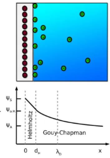
The decay of the double-layer potential described by the Poisson equation has been historically rationalised by different approaches. The Helmholtz theory disregards thermal motion of the counterions, assuming an unrealistic rigidity of the counterion layer.[38] This drawback is addressed in the Gouy-Chapman model which pictures the counterion layer as diffuse, but has the shortcoming of assuming that the charges in question are point-like. Rigid and diffuse models are combined
in the Stern model (Figure 1), resulting in a more comprehen-sive and realistic description of the interactions between charged surfaces and counterions.[38]
The PB theory and its linearised version, the Debye-Hückel theory,[40]
provide a very useful and important framework for the understanding of electrostatic phenomena in soft matter. PB theory has been fairly successful in describing, e. g., distributions of mono- and divalent ions around DNA,[41–43] although it is well-known that PB theory cannot fully describe the effects of multivalent ions.
One very important consequence of the PB approach is the DLVO theory. In the DLVO potential, screened Coulomb interaction and van der Waals attraction are combined to explain the charge stabilisation of solutions with charged solutes (Figure 2). With increasing ionic strength, the charge
Figure 1. Stern model combining the rigid (Helmholtz) and diffuse (Gouy-Chapman) double layer models. The grey shaded area represents a surface immersed into bulk liquid (blue continuum). The red circles on the shaded area represent negatively charged particles, the green circles illustrate positively charged ones. The potential ψ decays linearly between the surface (ψS) and the outer Helmholtz layer (ψo.H.at a distance dH). At dH, the diffuse
double-layer begins and ψ decays exponentially, asymptotically approaching a value ψAat long distances from the charged surface. The thickness of the
diffuse double-layer corresponds to the Debye screening length (eqn. 1). Figure reproduced and adapted from Refs. [38] and [39].
Figure 2. DLVO potential for varying salt concentration cs. With increasing cs,
the potential changes from repulsive to attractive. The aggregation barrier reflects the charge stabilisation behaviour that becomes weaker due to charge screening.
1 2 3 4 5 6 7 8 9 10 11 12 13 14 15 16 17 18 19 20 21 22 23 24 25 26 27 28 29 30 31 32 33 34 35 36 37 38 39 40 41 42 43 44 45 46 47 48 49 50 51 52 53 54 55 56 57
screening decreases the electrostatic repulsion more efficiently and on shorter ranges, and finally the van der Waals attraction causes aggregation and precipitation.
Van der Waals forces (for a detailed description, the reader is referred to Ref. [44]) account for attractive interactions arising from interactions between permanent and induced dipoles, and their azimuthal orientation and range depend on the macro-molecular structure. An essential aspect is the fact that the attraction decays on shorter length scales than electrostatics.[44] While not a part of the initial DLVO theory, we remark that in practice an attraction induced, e. g., by depletion or hydration can produce qualitatively similar effects, so that experimental interpretations using DLVO theory should not solely be attributed to van der Waals interactions .
The PB theory is based on strong assumptions. In particular it ignores ion–ion correlations, arising e. g. due to excluded volume and electrostatics. Furthermore, other ion properties such as polarisability and hydration effects are not included, but can actually cause significant effects. On the one hand, it is interesting that PB and DLVO theories perform so well in many cases, providing approximate theoretical predictions when a full description of the system is out of reach even with elaborate and costly computer simulations. On the other hand, it is not surprising that various ion effects have been observed that cannot be explained by PB DLVO theories.[1,45,46]
2.2. Charge effects beyond mean field: counterion condensation and ion-ion correlations
Very importantly, while in principle PB theory allows for Z > 1, there are effects not contained in the PB picture. The most obvious of these is probably the possibility of charge inversion and like-charge attraction. We will briefly elaborate on these phenomena in the following.
If the charge density in the system is strong, significantly modified ion distributions are obtained compared to the mean-field PB approach. Manning et al.[47]
found a condensation of counterions on surfaces as long as the surface charge density is higher than a critical value which depends on the surface geometry and counterion valence. Generally, the Manning condensation model was introduced to obtain an estimate of the number of counterions condensing onto polyelectrolytes. The model assumes an idealised polyelectrolyte via an infinitely long, charged line. For simplification, interactions between these idealised polyelectrolytes are ignored and the dielectric constant of the surrounding medium is assumed to be that of the bulk solvent.[3,47]In addition, Olvera de la Cruz[48]observed a precipitation of polyelectrolytes induced by tri- and tetravalent salts in a computational study, accounting for the probability of the binding of a condensed ion layer by PB theory with cylindrical geometry.
A general overview on ion-ion correlations has been given by Jönsson and Wennerström.[4] More recently, statistically advanced approaches accounting for ion–ion correlations due to strong Coulomb coupling have found counterion condensa-tion at strongly charged surfaces as well as ion distribucondensa-tions
that depend on the valence and size of the counterions.[1,49–52] In this case, ion–ion correlations cause an inversion of the surface charge.[51,53–55]
Theoretical approaches even predict a so-called “giant overcharging”[56]
due to increasing monovalent salt content, while simulations suggest a lower reversed charge for these conditions.[57]
The effects of ion–ion correlations are, in general, expected to be small compared to specific interactions between ions and surfaces.[54]
While ion–ion correlations might add a finite contribution to the protein–ion interaction, other more specific effects appear to be more relevant.[58]
An interesting point concerns competing-ion and co-ion effects, both for different multivalent ions as well as for a given species of multivalent ions[59]
in the presence of a monovalent ion.[60]
We shall mention that ion effects and in particular multivalent effects are also connected with the pH of the system, but unless explicitly stated otherwise, the effects we are discussing are dominated by the charge itself, and the pH is a secondary (although quantitatively important) effect. The effects of both ionic strength and pH (pD) has been studied for lysozyme by Kundu et al.[61]
For information on the quantitative modelling of the coupling of charge state and pH in the context of multivalent ions we refer to Ref. [62].
2.3. Ion-specific effects: hydration, Hofmeister, coordinative binding
While DLVO theory performs well for dilute monovalent ionic systems, a classical DLVO approach to biological systems fails due to the fact that it is no longer applicable at physiologically relevant ionic strengths above 0.1 M, as elaborated by Boström et al.[46]
In addition, Boström et al. point out that in order to allow for an appropriate comparison between theory and experiments, dispersion forces strongly depending on ion-specific effects need to be taken into account.[46]
Such effects include ion size, electronic structure, charge density and the resulting polarisability. Moreover, given that an overwhelming majority of biological and physiological processes take place in aqueous environments, the hydration properties of ions are of particular importance.
Systematic reviews of ion-specific effects and properties have been published, e. g., by Kunz.[5,6]Detailed theoretical and simulation studies have been described, e. g., by Lund et al., Jungwirth et al., Kalcher et al., Moreira et al., Schwierz et al., Lenz et al., Kunz et al., Smiatek et al., Lesch et al. and Kalayan et al.[49,57, 63–70] We will not review the results here, but instead refer the interested reader to the corresponding publications.
In the following, we will briefly indicate the current state of rationalisation of the Hofmeister series. We will then focus our discussion on ion-specific effects and those mediated by multivalent ions in protein systems.
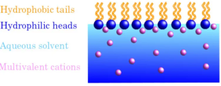
The study of salt-induced phase behaviour in protein solutions ranges back to the Hofmeister series on protein solubility in the presence of different salts[71] and the related salting-in and salting-out behaviour.[72] Figure 3 shows part of the Hofmeister series for anions and cations. The combination
1 2 3 4 5 6 7 8 9 10 11 12 13 14 15 16 17 18 19 20 21 22 23 24 25 26 27 28 29 30 31 32 33 34 35 36 37 38 39 40 41 42 43 44 45 46 47 48 49 50 51 52 53 54 55 56 57
of cations and anions strongly affects the ability of the salt to precipitate (“salt out”) or stabilise (“salt in”) colloidal solutions.[73] These variations of the phase behaviour cannot be explained by the DLVO theory and imply that the protein–ion interactions vary considerably beyond Poisson-Boltzmann theory due to ion-specific effects.
A comprehensive molecular understanding of ion-specific effects is a challenge for theory,[5]
although it is clear that water-mediated effects are a key ingredient. Baldwin[74]
argues that the Hofmeister effect can be understood considering the two competing abilities of ions to “salt out” nonpolar functional groups and “salt in” the polar peptide group.
A prominent theme for the molecular origin of the Hofmeister effect is the propensity of ions to change the water structure, i. e. the ion hydration.[75–77]
If certain ions – so-called “kosmotropes” – interact with water strongly, the surrounding water is aligned relative to the ion, and thus additional water structure is formed. Ions with weak interaction and inappro-priate size – so-called “chaotropes” – are not able to induce any water structure and even distort the bulk structure. Kosmo-tropic cations are also referred to as “soft” and large with a low charge density and weak hydration while the opposite is true for kosmotropic anions which are considered “hard”, strongly hydrated and assumed to have a high charge density. In turn, chaotropic cations are considered “hard” and are strongly hydrated and chaotropic anions are “soft” and weakly hydrated.[5,6]
This concept of “hard” and “soft” ions has important implications for the formation of ion pairs in aqueous solutions. One possible interpretation of this phenomenon has been given by Collins[76,78]
who formulated the “law of matching water affinities”. This law approximates ions as spheres with point charges. In the case of small, “hard” ions, their hydration shell is strongly bound; the hydration shells of large, “soft” ions, however, are only loosely associated to the ions. Collins assumes that two “hard” ions of opposite charge experience a strong mutual attraction and, upon approaching each other, their strong attraction will allow them to shed their hydration shells and form an ion pair. Two “soft” ions with opposite charges will experience a much weaker mutual attraction than two “hard” ions, but their weakly associated hydration shells are readily shed, allowing them to pair up with each other easily.[5,78] Another theme involves the change of the dielectric constant at the protein–water interface, which allows non-localised adsorp-tion of polarisable ions at non-polar, hydrophobic areas of the protein surface,[64,79,80]representing another possible mechanism for the Hofmeister effect via dispersion forces.[81–83]
Finally, the interfacial tension of the protein–water interface has been linked to the Hofmeister series.[84]
Interestingly, Okur et al.[85]
emphasise that Hofmeister cations and anions may follow different trends depending on the part of the protein they interact with. Potentially, the typical Hofmeister trend can even be reversed, an observation corroborated by Schwierz et al.[66,82]
Thus, although of practical importance and known for over a century, the Hofmeister effect, and salt effects on protein solutions in general, remain an interesting and challenging field of research. While the multivalent ion effects discussed in the remainder of this review go beyond the Hofmeister effects, some of the ingredients at least for ion-specific effects (ionic radius, polarisability etc.) are similar.
In Sec. 2.4, we will provide a brief summary of selected physiochemical aspects of ions in solution before focusing on specific types of (biological) (macro)molecules and describing their interactions with multivalent cations in Sec. 3.
2.4. Physicochemical aspects of ions in solution
In the following, we will summarise selected physico-chemical properties of some ions of particular relevance in biological and soft matter systems. Table 1 provides an overview on the ionic radii, hydration numbers and electron configurations. We note that in the case of the binding of transition metals (especially, but not only lanthanides) to biomolecules, their electronic configurations and particularly the presence of f-orbitals, is likely to play a highly complex role at the quantum chemical level. Non-trivial trends in the protein binding behaviour of lanthanide and yttrium cations have been observed, e. g., by Gomez et al.,[86]
Mulqueen et al.[87]
and the authors of this review.[59]
These effects can be tentatively attributed to the particular electron configurations of these ions and other highly complex properties of transition elements (polarisability, rela-tivistic effects, anisotropic binding to biomolecules). A detailed discussion of these effects is beyond the scope of this review. In the context of the hydration numbers given in Table 1, it is of interest to briefly mention the general effect that solutes Figure 3. Part of the Hofmeister series for anions and cations. Ions on the left
hand side of the series destabilise solutions and “salt out” solutes, whereas ions on the right stabilise (“salt in”) solutions.
Table 1. Selected ion properties. Unless indicated otherwise, the ionic radii are taken from Table 2.2 in Ref. 88. aindicates an approximate value,[88] bfrom Ref. 89;cfrom Ref. 90;dfrom Ref. 91efrom Ref. 92.
Ion Radius in solution (nm) Hydration number Electron configuration Na+ 0.102 3.5 [Ne] 3s0 K+ 0.138 2.6 [Ar] 4s0 Mg2 + 0.072 10.0 [Ne] 3s0 Ca2 + 0.100 7.2 [Ar] 4s0 Fe3 + 0.065 6.0e [Ar] 3d5 Y3 + 0.102b 8.0d [Kr] 4p05s0 La3 + 0.125c 10.3 [Xe] 4f0 (CH3)4N + 0.280 1.3 – Cl 0.181 2.0 [Ne] 3s23p6 F 0.133 2.7 [He] 2s22p6 CO2 3 0.178 a 4.0 SO2 4 0.230 3.1 PO3 4 0.238 4.5
1 2 3 4 5 6 7 8 9 10 11 12 13 14 15 16 17 18 19 20 21 22 23 24 25 26 27 28 29 30 31 32 33 34 35 36 37 38 39 40 41 42 43 44 45 46 47 48 49 50 51 52 53 54 55 56 57
such as ions can have on water structure. According to Marcus,[88]
a highly suitable strategy to quantify ion-mediated effects on water structure is to determine the change of the average number of hydrogen bonds of the ion-water structures. This parameter can be quantified by exploiting the fact that hydrogen bonds in heavy water (D2O) are stronger than those in light water (H2O). The experimental determination of D2 O-and H2O-specific parameters thus helps quantify the effect of solute molecules such as ions on water.[88,93]
When considering the binding of (multivalent) ions to macromolecules, an obvious question concerns the influence of the co-ions associated to the ions in question. While we note that a detailed discussion of this question is beyond the scope of this review, it is important to keep in mind that co-ions can have dramatic effects on ion adsorption to macromolecules. This has been shown for colloidal solutions, e. g., by Alfridsson et al.[94]
and Karaman et al.[95]
and for protein phase behaviour.[96] Importantly, in addition to unexpectedly specific co-ion adsorp-tion, Karaman et al. found that gas dissolved in the solutions played an important role in the context of emulsion stabilities, with implications even for highly complex phenomena such as enzymatic catalysis.[95]
It is furthermore important to mention that, in solution, different ion hydroxide and oxide complexes can be formed. This is especially well-known in the case of iron[97]
and implies that the ions can no longer be considered as single-atom ions. Iron is generally known to induce strong water protolysis, thereby acidifying aqueous iron solutions. In biological systems where pH regulation is essential for the functionality of biomolecules, the intricate interplay between the formation of such hydroxide complexes and pH effects[62]
needs to be considered on a case-to-case basis for different ions.
3. Proteins
The interior of living cells features a high ionic strength with typical intracellular concentrations of Na+
and K+
being 12 and 140 mM, respectively.[9]
These ions are of vital importance for,
inter alia, maintaining a physiological osmotic pressure in living
tissues and signal transduction in neurons. In addition to the ubiquitously present monovalent ions mentioned above, multi-valent ions play equally crucial roles in ensuring the stability and functionality of different proteins. In this section, we focus on the interactions of multivalent cations with proteins as a specific example of biological soft matter. We describe the molecular mechanisms by which cations bind to proteins as well as the physiological effects these cation-protein interac-tions have. Finally, we give an overview on the physico-chemical effects of cation-protein associations by describing phase diagrams of protein-multivalent cation systems. Remarks on ion channels, a very specific subtype of proteins interacting with cations, will be provided in Section 3.2.2. Parts of this section are based on Refs. [31] and [39].
As an example, calcium (Ca2 +) can be mentioned as a multivalent cation responsible for several phenomena related to the cytoskeleton[98] and muscle cells,[99] including the
contraction of heart muscle cells.[100]
In addition, Ca2 + is involved in the formation of protein aggregates of relevance for the food industry.[101–103]
Crucially, the (dis)assembly of viruses features a pronounced dependence on Ca2 +
.[104–108]
Further multivalent ions of physiological relevance for proteins include Zn2 +
, which is a cofactor of several enzymes[9,109]
, as well as iron (Fe2 +
and Fe3 +
) being an integral part of a variety of so-called heme proteins.[110]
These and other multivalent ions playing important roles in the context of physiology are discussed in more detail in Sec. 3.1.
There are some interesting aspects of physiological rele-vance of elements not occuring as natural constituents of living cells. Artificially introducing such elementary metals or their ions or their complexes into the human body is common practice in the field of medical imaging. For example, tumours and abscesses can be imaged by intravenously administering gallium 67
Ga-citrate.[111]
The gamma radiation energies emitted by 67
Ga and suitable for imaging are 93, 184, 296 and 388 keV.[112]
Animal studies showed 67
Ga-citrate to bind exclu-sively to transferrin and to be transported inside the body by the latter.[111]
Another radioactive element, 166
Ho, has been shown to efficiently label chelate-conjugated antibodies,[113] which offers a valuable method to trace the uptake and distribution of antibody-based therapeutics.
Barium (Ba) and gadolinium (Gd) are often used as contrast agents for X-ray scans and for magnetic resonance imaging (MRI). The respective advantageous properties for the techni-ques in techni-question are a high atomic number and therefore X-ray contrast enhancement (Ba) as well as an increase of the local longitudinal and transverse water proton relaxation rates.[114] Similarly, neodymium cations (Nd3 +
) have been used for an investigation of histidine in aqueous solution[115]
using nuclear magnetic resonance (NMR). Further information on lanthanides in structural biology can be found, e. g., in Refs. [116–118]. Metals and metal cations can also find applications in, e.g., cancer therapy. For example, attempts have been made to selectively target cancerous liver tissue by microspheres con-taining90
Y.[119,120]
Apart from these obviously beneficial, albeit not side effect-or risk-free, interactions between proteins and ions, attention needs to be drawn to the potential toxicity of ions. Amongst other pathways, the latter can be mediated by ion-protein interactions. Poisoning due to an ingestion of, or exposure to, pathologic concentrations or levels of mercury (Hg), lead (Pb) or cadmium (Cd) are a well-known danger. Further metals with potentially toxic properties are copper (Cu) and aluminium (Al). Copper poisoning primarily affects the liver[121] with possible damage being inflicted to the kidneys and the brain as well.[122] Aluminium (Al) concentrations > 0.1 mg/ml in drinking water have been associated with a potentially elevated risk of developing dementia and Alzheimer’s disease.[123,124]In addition, Mn2 + has recently been proposed to be involved in neurotoxic effects in the context of parkinsonianism.[125]
1 2 3 4 5 6 7 8 9 10 11 12 13 14 15 16 17 18 19 20 21 22 23 24 25 26 27 28 29 30 31 32 33 34 35 36 37 38 39 40 41 42 43 44 45 46 47 48 49 50 51 52 53 54 55 56 57
3.1. Local picture of cation binding sites of proteins
Given the obvious importance of protein-ion interactions under both physiological and pathological conditions in medical and biological contexts, much research effort has been invested into studying these interactions from the chemical and physical points of view. A necessary step to investigate proteins the functionality of which depends on ions was to determine and characterise their ion binding sites.
On the protein surface, numerous side chains with different physico-chemical properties are exposed to the solvent. As a first consequence, in aqueous solutions charge regulation of the protein surface occurs. Functional surface groups – basic (Lys, Arg) and acidic (Glu, Asp, His) amino acid side chains and the carboxy and amino termini of the protein – are (de)protonated depending on the pH and the charges in the environment,[126,127]
thus coupling pH to protein surface charge. As a second and indeed key effect in the context of this review, also ions other than the hydronium ion, in particular multivalent ones, interact with functional groups. Numerous studies report equilibrium constants for the binding of multi-valent counterions to proteins specialised in metal storage and transport.[128–132]
Moreover, models for ion binding have been developed in order to understand the interactions of proteins with ions and ligands.[133–136]
On the molecular level, amino acids with carboxylate, hydroxyl, thiol, thioether, and imidazole side chain groups bind transition metal ions coordinatively.[19,137–143]
In fact, the binding of the potentially toxic heavy metal ions Cd2 +
and Cr3 +
to the cysteine-rich protein Cry has been suggested to be an environmentally friendly method of eliminating said cations from water.[144]
The binding of ions is enhanced at hydrophilic sites surrounded by hydrophobic surface areas.[145]
The overall ubiquity of these surface groups suggests that the association of salt counterions with side chains of the opposite charge at the protein surface is at the heart of the model for the interaction of ions and proteins.[64]
This notion has been explicitly supported for a study on the oligopeptide tetra-aspartate. Kubíčková et al.[146]
observed a charge inversion both experimentally and by molecular dynamics simulations for tetra-aspartate with trivalent cations. Mono- and divalent ions also decreased the overall charge, but did not overcome the initial negative protein charge. As the basic mechanism, the ion binding to carboxylic acids is evidenced by radial distribution functions that also show the different behaviour of multi- and monovalent cations.[146]
As will be discussed in the following, this type of multi-dentate coordination of multivalent cations by negatively charged or polar residues is observed in many proteins. Nevertheless, other cation-binding mechanisms shall also be briefly mentioned here. As an example, the side chains of aromatic amino acids such as tyrosine, phenylalanine or tryptophan feature π electron systems which have been shown to undergo so-called cation-π interactions.[147,148] According to Dougherty,[148] all kinds of cations can be part of this type of interaction. Given the hydrophobic nature of π-electron
systems, however, they may be more likely to occur between π systems and hydrophobic cations such as quaternary ammonium ions or even the protonated guanidino group of the amino acid arginine.[148]
We shall briefly note here that binding of (monovalent) anions to nonpolar surface patches has been observed in molecular dynamics simulations.[149]
This phenomenon has been traced back to solvent-assisted attraction of the ion to the protein surface.
Having outlined general characteristics of cation binding sites on the protein surface, we will discuss specific examples of protein-cation systems in the following.
Calcium. In the human body, calcium is involved in a variety
of processes in living cells, including cytoskeleton mobility, muscle contraction, bone formation, blood coagulation and hormone-mediated metabolism regulation[9,10, 109]
(for a detailed description, see also the review by Kretsinger[99]
). In fact, Ca2 + is often referred to as a so-called “second messenger” due to its ubiquity in physiological processes.[10,99]
Thus, it is of particular physiological relevance to consider different Ca2 +
-binding proteins. Amongst these, a specific helix-loop-helix motif referred to as the “EF-hand” is a commonly shared feature.[150–153] Examples of proteins containing an EF-hand motif are calbindin, myosin, troponin, calmodulin and parvalbumin (see Ref. [150] and refs. therein). The Ca2 +
ion is usually coordinated by aspartic acid, asparagine or serine[150]
(see visualisation in Figure 4). Experimental studies have shown that the affinity of the EF-hand motif for Mg2 +
can be increased while decreasing that for Ca2 +
by residue-specific mutations,[154]
implying that subtle effects are important in determining the cation specificity of EF-hands.
In addition to the EF-hand protein family, Ca2 +
interacts with actin (see, e. g., the review by Janmey[155]
) and gelsolin[156,157]
or both actin and gelsolin simultaneously.[157] These Ca2 +
-protein interactions are involved in the regulation of cytoskeletal motility. According to Robinson et al.,[157]
both actin and gelsolin bind Ca2 +
ions via aspartate and glutamate residues. Osteopontin, a protein abundantly present in the bone and teeth matrices, binds calcium in an inorganic form (hydroxyapatite) through phosphorylated serine and threonine residues as well as polyaspartate sequences.[158]The physiolog-ical role of osteopontin is briefly discussed in Sec. 3.2.1. Interestingly, in osteocalcin (another important protein in bone tissue), Ca2 + cations are bound by γ-carboxyglutamic acid residues,[159]
a rarely occuring version of glutamic acid.[9] The tripeptide peptide Tyr-Asp-Thr with a very high Ca2 + chelating propensity has been isolated from whey protein,[160]evidencing the cooperative effect of several amino acids for binding of Ca2 +.
For more detailed and elaborate discussions on the binding of calcium ions to proteins, we refer the reader to Ref. [99]. Protein self-assembly in the presence of Ca2 + is discussed in Sec. 3.2.1.
Iron. In mammal physiology, iron is known particularly well
for its role in protein-mediated oxygen homeostasis and is often found in a coordination complex with porphyrin structures. This iron-porphyrin complex is referred to as a “heme group” and is
1 2 3 4 5 6 7 8 9 10 11 12 13 14 15 16 17 18 19 20 21 22 23 24 25 26 27 28 29 30 31 32 33 34 35 36 37 38 39 40 41 42 43 44 45 46 47 48 49 50 51 52 53 54 55 56 57
present as a prosthetic group in a variety of proteins such as myoglobin, hemoglobin, plant leghemoglobin and cytochromes (proteins involved in electron transport processes of the cellular respiratory chain)[109] as well as catalases, peroxidases, and mono- and dioxygenases[110] (enzymes catalysing redox reac-tions). For an overview of iron binding, the reader is also referred to Ref. [161].
The structure of myoglobin has been extensively studied, providing detailed insights into the local environment of the Fe2 + cation bound to the heme. The latter is located in a pocket-like structure of myoglobin, thus being protected from the surrounding solvent. Importantly, this steric protection
prevents the Fe2 +
ions from being oxidised to Fe3 +
, which is not able to bind oxygen.[9]
Iron metabolism in mammals furthermore involves the non-heme proteins ferritin,[10]
lactoferrin[10,11]
and transferrin with its corresponding receptor protein.[162]
As an example, one of the two iron-binding sites of transferrin consists of two tyrosines, a histidine and an aspartate residue and involves a carbonate anion[162]
(Figure 4). Several so-called iron-sulfur proteins can furthermore bind Fe ions via cysteine side chains.[9]
Moreover, while the ionic form of iron is clearly an important factor in the physiological context, it can also interact with proteins in other forms. Prominent examples are other types of iron-sulfur proteins, namely those hosting inorganic iron-sulfur (FeS) clusters. Those are, for example, found in the protein ferredoxin of the bacterium Anabaena. A particularly curious use of inorganic iron structures are so-called magnetosomes (inor-ganic, iron-containing crystals such as Fe3O4crystals) providing bacteria with the ability to orient themselves along magnetic fields (for an overview on this interesting phenomenon, see Ref. [163] and refs. therein).
The iron- and calcium-binding sites mentioned above share a common feature – the multivalent cations bound to the respective proteins are complexed by charged and/or polar amino acid residues such as aspartate and tyrosine. Some examples of such binding sites are shown in Figure 4.
Magnesium. Mg2 +
cations are known to play an important role in enzymatic reactions catalysing the cleavage of phosphate bonds. This can be especially relevant in sugar metabolism and nucleic acid (RNA and DNA) degradation. The latter is often catalysed by so-called nucleases such as ribonuclease H. Generally, magnesium ions are coordinated octahedrally.[165]
In the active centre of the ribonuclease RNase H, Mg2+
ions are also surrounded by hydration shells (see Ref. [166] and refs. therein as well as Ref. [167]). As opposed to other alkaline metals, Mg2 +
appears to have a particularly strong affinity to water molecules in its inner coordination shell,[166]
making water an important constituent of catalytically active Mg2+
-enzyme complexes. In addition to these
in vivo roles of Mg2 +
, a very interesting magnesium-mediated transition from binary to unitary protein structures has been demonstrated by Künzle et al.[168]
Zinc. A crucial physiological role of zinc is its stabilisation of
insulin microcrystals in the pancreas,[169,170]
with obvious implications for diabetes. Zinc is furthermore known to be involved as a cofactor in several enzymes. Examples include carboanhydrase,[109]
several proteases[10]
and alcohol dehydrogenase.[9]In numerous proteins, Zn2 + ions are coordi-nated by a so-called zinc finger motif consisting of cysteine and histidine residues (“Cys2His2”).[171]
Copper. In physiology, the most prominent role of copper is
the electron transport in the respiratory chain. Here, it is bound to sulfhydryl groups of the protein cytochrome oxidase.[9]It is important to note that the oxidation state of the copper ions involved in the reduction of oxygen to water changes throughout the catalysis.[172] Interestingly, the enzyme copper-zinc superoxide dismutase – which catalyses the reaction of superoxide radicals to hydrogen peroxide and molecular oxy-gen – uses both copper and zinc for said catalysis.[173]Structural Figure 4. Binding sites of multivalent ions in proteins (see text for details).
The image illustrates the pivotal roles of negatively charged amino acid residues in coordinating the respective ions. As opposed to the Y3 +cations
bound by BLG (b) and the Ca2 +ions bound by calbindin (c), binding of Fe3 +
requires carbonate ions in addition to the protein residues coordinating the ion (seen on the right side of the orange sphere representing Fe3 +in (a)).
The structures were visualised using UCSF Chimera[164]based on PDB IDs
1 2 3 4 5 6 7 8 9 10 11 12 13 14 15 16 17 18 19 20 21 22 23 24 25 26 27 28 29 30 31 32 33 34 35 36 37 38 39 40 41 42 43 44 45 46 47 48 49 50 51 52 53 54 55 56 57
studies reveal that the ions are coordinated by histidine and aspartate (Zn) and histidine and arginine residues (Cu).[173]
Molybdenum and vanadium. In plant metabolism,
molyb-denum (Mo), together with iron, plays an important role as a cofactor in the enzyme dinitrogenase. In addition, Mo is part of the active centre of the enzyme nitrogenase from Azotobacter
vinelandii.[174]
Interestingly, some dinitrogenase versions contain vanadium (V) instead of Mo.[9]
Furthermore, vanadium is of great importance for the biosynthesis of halogenated products by marine organisms.[175]
Cobalt. A Co3 +
ion is complexed by the corrin ring of coenzyme B12, a slightly modified form of vitamin B12. The latter is, inter alia, a cofactor of the enzyme methyl malonyl-CoA mutase which catalyses a step of a complex metabolic process referred to as β-oxidation of fatty acids. Importantly, the cobalt ion allows the reaction to proceed via an extremely unusual intermediate step involving a hydrogen radical[9]
by undergoing a change in oxidation state from + 2 to + 3.
Lanthanides. Complexes of lanthanide ions and some organic
ligands possess several favourable fluorescence properties such as long fluorescence lifetimes, strong Stokes shifts and distinct emission peaks;[176]
depending on the choice of the ligand, the fluorescence can be enhanced.[177]
Thus, lanthanides proved valuable structural probes to tackle questions related to cation binding sites of proteins. As an example, Harris and co-workers[178–182]
examined the binding of various non-ferrous cations to human transferrin and lactoferrin, notably including the lanthanides Lu3 + , Er3 + , Ho3+ , Tb3 + , Gd3+ , Sm3+ , Nd3 + and Pr3 + . The authors report two lanthanide binding sites involving tyrosine residues[183]
and a decrease in the number of cations bound with increasing cation radius.[180]
Here, we remind the reader of the phenomenon of lanthanide contraction,[184]
another important property of lanthanides in addition to those mentioned above. Lanthanide contraction describes the continuous decrease of the ionic radii from lanthanum (La) to hafnium (Hf) due to the successive increase in the occupation of the 4f orbitals and the simultaneous increase of the nuclear charge.[184]
Apart from purely structural studies, the influence of lanthanides on biological protein activities has been investi-gated as well. Smolka et al.[185]
analysed the consequences of replacing calcium by trivalent lanthanides and Y3 + in the calcium-dependent enzyme α-amylase. This study suggests an inverse linear proportionality of the enzyme efficiency on cation radius, underlining cation-specific effects. No strong structural changes of the protein were observed. A similar study replacing Ca2 + by lanthanides in trypsinogen and evaluating the respective efficiencies of the cations in catalysing the con-version to trypsin was conducted by Gomez et al.[86] The dependence of the conversion rate efficiency is non-linear, but also inversely proportional to the cation radius. Interestingly, Nd3 + and Pr3 + were shown to be even better trypsinogen-trypsin conversion activators than Ca2 +, which was ascribed to their higher charge.[86]
The study conducted by Gomez et al. highlights an important property of lanthanides. With their radii being similar to that of Ca2 +, they can replace Ca2 + not only in vitro, but also in vivo, being of toxicological relevance.[128,186]
Lanthanides as well as yttrium are usually found in the form of trivalent cations that strongly interact with binding sites formed by carboxylic residues. This coordinative binding of Y3 + is apparent from protein crystals, where the cations bridge different protein molecules.[19]
Importantly, the driving force for this binding is not enthalpy alone, but hydration entropy. In particular, a physicochemical characterisation of the binding reveals a lower critical solution temperature,[187]
and the water coordination around Y3 +
is reduced upon binding to the protein,[188]
both of which evidence the release of hydration water molecules with substantial related entropy gains. Finally, we refer the interested reader to a recent overview on the roles of lanthanides in biochemistry by Daumann.[189]
Other cations. There are, of course, many other cations that
could be mentioned here, but a full list would be beyond the scope of this review. We therefore refer the reader to the detailed works by Lipfert,[190]
Permyakov,[128]
Frausto da Silva and Williams[191]
and Evans.[192]
The above should suffice to indicate the main phenomena and concepts.
3.2. Physico-chemical and global effects of protein-cation interactions
In this section, we will discuss the global, physico-chemical behaviour of several systems composed of proteins and multi-valent cations. We will explain the phase behaviour of several selected systems and discuss their respective origins. A particular focus will be on the role of the multivalent cations and their interactions with the proteins in question. We remark that we intentionally limit ourselves to a few examples of systems, and related implications, as a complete review of protein-cation interactions would be too voluminous for this review. In this context, we aim for a balanced account of basic references and new studies evidencing ongoing work.
3.2.1. Calcium-induced effects on protein assembly
Calcium represents one of the most common multivalent cations, which is why we dedicate an extra paragraph to it. It has effects on protein systems exploited both in nature (e. g. in blood coagulation, for viral assembly, and bone formation), as well as in nano- and biotechnological contexts such as food engineering. In the following paragraphs, we will outline a few examples to show the various functions and structures that are controlled by calcium ions.
Milk proteins. As one well-studied example in food science,
milk proteins represent calcium-controlled molecules which sig-nificantly contribute to the calcium intake into the human body. The presence of calcium strongly affects the aggregation of whey protein,[193] and the resulting gel structure.[194,195] As a particular example, calcium has a dramatic effect on the speed of the gelation of whey aggregates, and mildly strengthens the resulting gels.[196,197] These structural variations have been shown e. g., to regulate the release of drugs from whey hydrogels.[198]
1 2 3 4 5 6 7 8 9 10 11 12 13 14 15 16 17 18 19 20 21 22 23 24 25 26 27 28 29 30 31 32 33 34 35 36 37 38 39 40 41 42 43 44 45 46 47 48 49 50 51 52 53 54 55 56 57
The second protein source in milk, casein, is also strongly affected by Ca2 +
and calcium-phosphate clusters. The calcium effects range from changing the micellar structure of casein,[199–201]
over varied aggregates after thermal denaturation,[202]
to macroscopic effects such as the texture of milk-derived products, e. g. yoghurt.[203,204]
For a detailed over-view of the functionality, association and aggregation of caseins, we refer to Ref. [205].
Fibrin clot formation. Calcium is an essential cofactor in the
initial step of blood coagulation, i. e., fibrin clot formation. Fibrin clots are the organism’s immediate response to injury in order to prevent excessive blood loss, and fibrin assembly thus has fundamental as well as applied relevance e. g. for drug carriers and fibrin sealants. Ca2 +
controls the cross-linking of fibrin protofibrils into fibers and hydrogel structures.[206]
In particular, Ca2 +
tunes the fibrin cross-linking rate,[207]
resulting in values ranging from seconds to tens of minutes depending on the overall conditions of the fibrin solutions.[208]
In this context, Ca2 + also affects the resulting gel structure, thereby generally enhancing the elasticity[209]
and non-monotonically adapting the gel permeability.[210]
Consequently, calcium is a very common additive in fibrin sealants, as well as in applications for drug delivery and bone tissue engineering.[211–213]
Inhibition of calcium crystal growth by osteopontin.
Osteopontin is known to be an important constituent of body fluids with a high calcium content, such as milk and urine. It is therefore assumed that it is involved in the prevention of calcium salt precipitation (reviewed in Ref. [158]). Indeed, amongst its other roles (reviewed in Ref. [214]), e. g., in bone tissue homeostasis, osteopontin has been shown to inhibit the nucleation and growth of calcium oxalate.[215–217]
A study combining molecular modelling and atomic force microscopy (AFM)[218]
revealed that osteopontin strongly changed the morphology and growth of calcium oxalate crystals. Interest-ingly, the strength of these effects were pronounced to different degrees for different crystal faces of calcium oxalate crystals, indicating a strong interaction specificity between osteopontin and oxalate.[218]
Virus assembly. A crucial role of Ca2 +
has been found for a large range of viruses. Early findings reported already that most plant viruses rely on correct Ca2 + binding for controlled structural assembly.[219]
Similar indications were found for the bacteriophage PM2 and papillomaviruses, where Ca2 + was found to be essential for viral reassembly in vitro and during infection.[106,220]
In the case of PM2, Ca2 +
was hypothesised to stabilise the lipid bilayer of the virus before the protein outer layer is deposited on top of the lipid structure.[106] For bovine papillomavirus, the role of Ca2 + appeared to be to stabilise the protein capsid.[220] A recent study reports the requirement of Ca2 + for Rubella virus infections, as well as viral fusion and liposome insertion,[221] evidencing that Ca2 + enables virus function via structural adaption of the virus.
For simian virus 40 (SV40), Ca2 +, along with pH effects,[222]was found to be important for the accuracy of the assembled structure, and appropriate affinities of the viral protein capsid to Ca2+ regulate assembly and disassembly of the virus.[104]The presence of Ca2 + also proved relevant for the cell and nuclear entry during
infection with simian virus 40, and Ca2 +
was proposed to not only change the assembly state, but also the flexibility of the capsid.[105] Related to this, SAXS investigations showed that chelating Ca2 + caused a uniform swelling of SV40,[223]
stressing the role of Ca2 + in regulating the virus structure.
A comparable picture is found for the hepatitis B virus (HBV), where calcium signalling plays an important role for DNA replication.[224]
Again, Ca2 +
was found to be important for the HBV core assembly.[108]
Importantly, the knowledge on the Ca2 + effects for virus assembly even translates into nanotechnology. As an example besides the more general establishment of purification schemes,[219]
an encapsulation system based on the hepatitis B virus allows to adapt the affinity to the cargo molecule via the Ca2 +
concentration.[225]
Lipoprotein metabolism. Calcium has been found to be
effective in regulating the low-density lipoprotein receptor (LDLR), which controls the body’s cholesterol homeostasis. Indeed, a relation between calcium intake and the lipoprotein metabolism has been suggested.[226]
On a molecular level, a recent study suggests that LDLR senses Ca2 +
and unfolds partially,[227]
thereby providing an alternative route for trigger-ing of LDL release apart from the acidic-induced release.[228] Similar strong binding affinity of Ca2 +
is found in a LDLR related protein abundant in the liver.[229]
Furthermore, calcium also acts on the lipoprotein metabolism by assembling the lipoprotein lipase into its functional dimeric structure.[230]
3.2.2. Ion Channels
While ion channels are a specific type of proteins, they represent a slightly different topic in the context of this review, one particular characteristic being the fact that they are transmembrane proteins. We shall therefore limit ourselves to a few comments here.
The main functions of ion channels include the maintenance of physiological ionic strengths inside cells and the transduction of electrochemical signals along neurons. Prominent examples of ion channels include the Na+
K+
ATPase antiporter and the SERCA pumps (sarco/endoplasmic reticulum Ca2 +
ATPase).[231,232] The function of the latter is to transport Ca2 + ions across the membrane of the sarco/endoplasmic reticulum. Thereby, SERCA pumps maintain an intracellular Ca2 + storage and also termi-nate Ca2 +-mediated signalling. Just as is the case for the EF-hand motif in proteins, SERCA proteins coordinate the Ca2 + ions via glutamate residues. In addition, glutamine and asparagine residues are involved (reviewed in Ref. [233]).
The functionality of ion channels relies on their selective permittivity with respect to different ion types. Indeed, the uptake of the “wrong” type of ions such as La3 +instead of Ca2 + can have drastic toxicological consequences.[128,186] Similar effects have been demonstrated for Gd3 +.[234]
On the other hand, ion selectivity can also be used to prevent cell death caused by toxic ion channels. This has been shown by Menestrina[235]in a study of the α-toxin of S. aureus. This toxin binds to the cell membrane, inserts itself into the membrane and forms ion channels, thus causing K+
1 2 3 4 5 6 7 8 9 10 11 12 13 14 15 16 17 18 19 20 21 22 23 24 25 26 27 28 29 30 31 32 33 34 35 36 37 38 39 40 41 42 43 44 45 46 47 48 49 50 51 52 53 54 55 56 57
which results in an osmotic shock and, ultimately, cell death.[236] Menestrina demonstrated that the channels formed by S. aureus
α-toxin, which are open in a KCl solution, can be closed - and
their function thus inhibited - by multivalent cations. The inhibition efficiency was shown to be
Zn2þ>Tb3þ>Ca2þ>Mg2þ>Ba2þ:
(4) Menestrina provides a mathematical model to quantify the multivalent cation-mediated inhibition of the channel in which it is assumed that one multivalent cation binds to the channel in its open and one in its closed state. In addition, Menestrina suggests that a carboxyl group is involved in the binding of the cations,[235]
which is consistent with the mechanisms described in Sec. 3.1. Similarly, Döbereiner et al.[237]
observed an inhibition of the conductance of ion channels formed by α-hemolysin (HlyA) from E. coli[238]
upon addition of multivalent cations. Here, the divalent cations Sr2 +
and Ba2 +
were able to induce HlyA-mediated erythrocyte hemolysis, albeit less efficiently than Ca2 + . Mg2 + , Cu2 + , Mn2 + , Zn2 + and Pb2 +
did not lead to hemolysis; neither did the trivalent cations Fe3 +
and La3 + . In addition, Mg2 + , Ca2 + , Sr2 + and Ba2 +
inhibited HlyA conductance; Fe3 +
and La3 +
did so with greater efficiency. Döbereiner et al.[237]
suggest that the cation radius plays an important role in cation recognition by HlyA.[237]
In order to better understand the selectivity of ion channels, Kumpf and Dougherty[239]
performed computational studies on the affinity of Li+
, Na+ , K+
and Rb+
to benzene. The latter was chosen as a model of the hydrophobic core of a specific type of K+
channel. Their results demonstrate a preference of benzene for K+
and indicate that so-called cation-π interactions – that is, interactions of cations with delocalised π electron ring systems, of which benzene is representative – appear to occur in hydrophobic regions of ion channels. In particular, these interactions could give a hint towards the ion selectivity of ion channels. More information on this intriguing subject is found in Ref. [240].
3.2.3. Lanthanide-induced phase behaviour in protein solutions
Apart from their roles as structural probes and in medical imaging, lanthanide ions can be used to tune the phase behaviour of protein solutions, including the rational induction of protein crystal growth.
The local interactions of multivalent ions with proteins have profound consequences for the global behaviour, qualitatively different from, say, Na+
. Here, we discuss the phase behaviour and related collective phenomena of protein systems. Special attention will be paid to those types of phase behaviour induced by multivalent ions in negatively charged proteins.
Generally, a rich phase behaviour has been found in protein solutions, including liquid liquid phase separation (LLPS), the formation of protein clusters, and crystallisation as well as other aggregates such as fibers. The nucleation kinetics differ considerably for different phases, which allows for metastable phases such as LLPS or clusters as precursor structures during
crystallisation, as well as arrested phases such as gels to occur.[241,242]
In this section, we provide an overview of the different phenomena that also play an important role in the present context.
Protein Surface Charge and Ion-Induced Charge Inversion.
Charges on the protein surface are an important feature ensuring stability and functionality of proteins.[243–245]
Charge patterns lead to anisotropic interaction patches that affect the phase behaviour of protein solutions[246–249]
as well as pathways for aggregation and crystallisation.[250–252]
Protein–protein interactions are linked to charge regulation, which is, in turn, a complex process depending on system geometry and ion specific effects like binding or condensation. A comprehensive understanding of charge regulation at the protein surface is also needed in order to account for ion-specific effects such as binding and condensation as well as for the system geometry, e. g. the proximity of a wall.[245,253]
A special case is charge inversion, i. e., overcompensation, of surfaces in the presence of counterions. A comprehensive understanding of charge inversion has to account for both local ion binding and non-local contributions such as ion–ion correlations and hydrophobic effects.[254,255]
Charge inversion has been observed for a broad range of systems such as silica spheres,[54]
insoluble oxides[254]
and also biological systems such as DNA.[56]
The latter is discussed in more detail in Sec. 4. In particular, charge inversion has been observed in solutions of globular, negatively charged proteins with multi-valent cations.[58,256, 257]
The lower charge density of the protein surface speak against ion–ion correlations being the main cause of charge inversion. Instead, Zhang et al.[19]
support the notion of a charge inversion due to ion binding to acidic residues on the protein surface, based on information from crystal struc-tures. Note that not only cations, but also negatively charged molecular complexes have been shown to interact specifically with net negatively charged proteins, as demonstrated for human and bovine serum albumin.[258]
Remarkably, a protein crystallisation strategy similar to the one demonstrated by Zhang et al.[19]
has been pursued using negative multivalent ion complexes.[24–26, 259]
Reentrant Condensation. The inversion of the surface
charge is related to a specific phase behaviour called reentrant condensation known from polyelectrolytes (see, e. g., Ref.[260]
), which has been observed in aqueous solutions of negatively charged proteins with trivalent[58,256,261] and tetravalent cations,[262]
as illustrated schematically in Figure 5. At a given protein concentration cp and a low salt concentration cs, the
system is a homogeneous liquid (Regime I), charge-stabilised by the initially net protein charge. A continuous increase in cs,
while keeping cp constant, decreases the negative surface
charge of the protein and eventually condenses the protein molecules in solution into cluster-like structures. This con-densed state is referred to as Regime II, the entrance into which is marked by a critical salt concentration, c*. A further increase of salt concentration leads to overcharging of the protein, and the clusters redissolve upon surpassing a second critical salt concentration, c** (Regime III), stabilised by the reversed charge of the protein-cation complex. Computer simulations confirmed
![Figure was rendered using UCSF chimera [164] and Avogadro. [349]](https://thumb-eu.123doks.com/thumbv2/5dokorg/4098694.86316/14.892.455.829.97.425/figure-rendered-using-ucsf-chimera-avogadro.webp)