ACTA UNIVERSITATIS
Digital Comprehensive Summaries of Uppsala Dissertations
from the Faculty of Medicine
1029
Regulation of Gene Expression
in Multiple Myeloma Cells and
Normal Fibroblasts
Integrative Bioinformatic and Experimental
Approaches
Dissertation presented at Uppsala University to be publicly examined in Rudbeck salen, Rudbecklaboratoriet, Dag Hammarskjolds väg 20, Uppsala, Thursday, 13 November 2014 at 09:15 for the degree of Doctor of Philosophy (Faculty of Medicine). The examination will be conducted in English. Faculty examiner: Håkan Axelson (Lund University).
Abstract
Agarwal, P. 2014. Regulation of Gene Expression in Multiple Myeloma Cells and Normal Fibroblasts. Integrative Bioinformatic and Experimental Approaches. Digital Comprehensive Summaries of Uppsala Dissertations from the Faculty of Medicine 1029. 71 pp. Uppsala: Acta Universitatis Upsaliensis. ISBN 978-91-554-9045-4.
The work presented in this thesis applies integrative genomic and experimental approaches to investigate mechanisms involved in regulation of gene expression in the context of disease and normal cell biology.
In papers I and II, we have explored the role of epigenetic regulation of gene expression in multiple myeloma (MM). By using a bioinformatic approach we identified the Polycomb repressive complex 2 (PRC2) to be a common denominator for the underexpressed gene signature in MM. By using inhibitors of the PRC2 we showed an activation of the genes silenced by H3K27me3 and a reduction in the tumor load and increased overall survival in the in vivo 5TMM model. Using ChIP-sequencing we defined the distribution of H3K27me3 and H3K4me3 marks in MM patients cells. In an integrated bioinformatic approach, the H3K27me3-associated genes significantly correlated to under-expression in patients with less favorable survival. Thus, our data indicates the presence of a common under-expressed gene profile and provides a rationale for implementing new therapies focusing on epigenetic alterations in MM. In paper III we address the existence of a small cell population in MM presenting with differential tumorigenic properties in the 5T33MM murine model. We report that the predominant population of CD138+ cells had higher engraftment potential, higher clonogenic growth, whereas the CD138- MM cells presented with less mature phenotype and higher drug resistance. Our findings suggest that while designing treatment regimes for MM, both the cell populations must be targeted.
In paper IV we have studied the general mechanism of differential gene expression regulation by CGGBP1 in response to growth signals in normal human fibroblasts. We found that CGGBP1 binding affects global gene expression by RNA Polymerase II. This is mediated by Alu RNA-dependent inhibition of RNA Polymerase II. In presence of growth signals CGGBP1 is retained in the nuclei and exhibits enhanced Alu binding thus inhibiting RNA Polymerase III binding on Alus. Hence we suggest a mechanism by which CGGBP1 orchestrates Alu RNA-mediated regulation of RNA Polymerase II.
This thesis provides new insights for using integrative bioinformatic approaches to decipher gene expression regulation mechanisms in MM and in normal cells.
Keywords: Integrative Bioinformatics, Epigenetics, Multiple myeloma, RNA polymerase, CGGBP1, gene expression
Prasoon Agarwal, Department of Immunology, Genetics and Pathology, Hematology and Immunology, Rudbecklaboratoriet, Uppsala University, SE-751 85 Uppsala, Sweden. © Prasoon Agarwal 2014
ISSN 1651-6206 ISBN 978-91-554-9045-4
List of Papers
This thesis is based on the following papers, which are referred to in the text by their Roman numerals.
I. Antonia Kalushkova*, Mårten Fryknäs*,Miguel Lemaire,Charlotte Fristedt,Prasoon Agarwal,Maria Eriksson,Sarah Deleu,Peter Atad-ja,Anders Österborg, Kenneth Nilsson, Karin Vanderkerken,Fredrik Öberg, Helena Jernberg-Wiklund(2010) Polycomb target genes are silenced in multiple myeloma. PLoS ONE 2010 Jul; 5(7):e11483 II. Prasoon Agarwal*, Antonia Kalushkova*, Stefan Enroth, Kenneth
Nilsson, Fredrik Öberg, Helena Jernberg-Wiklund. The epigenomic map of multiple myeloma reveals the importance of Polycomb gene silencing for the malignancy (Manuscript)
III. E Van Valckenborgh, W Matsui, P Agarwal, S Lub, X Dehui, E De Bruyne, E Menu, C Empsen, L van Grunsven, J Agarwal, Q Wang, H Jernberg-Wiklundand K Vanderkerken. (2012) Tumor-initiating capacity of CD138- and CD138+ tumor cells in the 5T33 multiple myeloma model. Leukemia 2012 Jun; 26(6): 1436-9
IV.
Prasoon Agarwal, Stefan Enroth, Martin Teichmann, Helena Jern-berg Wiklund, Arian Smit, Bengt Westermark, Umashankar Singh. Growth signals employ CGGBP1 to suppress transcription of Alu-SINEs.Cell Cycle (in press).
All the published articles in this thesis have been reproduced by due permission from the concerned publishers
Paper I (Public Library of Science (PLOS)), Paper III (Macmillan Publishers Lim-ited), Paper IV Landes biosciences (Taylor & Francis)
Contents
Introduction ... 11
Studying mechanisms of gene expression regulation in the post-genomic era ... 11
Integrative Bioinformatics ... 12
Significance of bioinformatics and present challenges ... 13
Project 1 ... 14
Multiple myeloma: a background ... 14
Genetics of multiple myeloma ... 15
Classification of multiple myeloma ... 16
Tissue microenvironment in multiple myeloma ... 17
Heterogeneity of multiple myeloma ... 19
Experimental models of multiple myeloma ... 19
Epigenetic regulation in multiple myeloma ... 21
DNA methylation ... 21
Histone modifications ... 22
Polycomb group proteins ... 24
Epigenetic regulation of transcription in multiple myeloma ... 25
Project 2 ... 27
Major signal transduction pathways in normal fibroblasts ... 27
Brief overview of transcription ... 27
Structure and function of gene promoter ... 28
Retroelements and mammalian genome ... 29
CGGBP1: a regulator of normal and cancer cell biology ... 30
Summary of Paper I ... 33
Title: Polycomb target genes are silenced in Multiple Myeloma. ... 33
Summary ... 33
Aim ... 33
Results ... 33
Discussions and conclusions ... 34
Summary of Paper II ... 35
Title: An epigenomic map of multiple myeloma reveals the importance of Polycomb gene silencing for the malignancy. ... 35
Summary ... 35
Results ... 36
Discussion and conclusions ... 37
Summary of Paper III ... 38
Title: Tumor-initiating capacity of CD138- and CD138+ tumor cells in the 5T33 multiple myeloma model ... 38
Summary ... 38
Aim ... 39
Results ... 39
Discussion and conclusions ... 40
Summary of Paper IV ... 41
Title: Growth signals employ CGGBP1 to suppress transcription of Alu-SINEs ... 41
Summary ... 41
Aim ... 41
Results ... 41
Discussion and conclusions ... 43
Acknowledgements ... 44
Abbreviations
BAFF
B cell activating factor
BM
Bone marrow
Bmi-1
B lymphoma Mo-MLV insertion region 1
CBX Chromobox
CCND Cyclin
D
CDKN
Cyclin dependent kinase inhibitor
CGGBP1 CGG
triplet
repeat-binding protein 1
ChIP-seq Chromatin
Immunoprecipitation-Sequencing
CpG
5´ Cytosine phospodiester Guanine 3´
DAP
Death associated protein
DNMT DNA
methyltransferase
DS Durie/Salmon
ECM Extracellular
Matrix
EED
Embryonic ectoderm development
ESC Embryonic
Stem
Cells
EZH
Enhancer of zeste
FISH
Fluorescence in situ hybridization
GEP
Gene Expression Profiling
HAT Histone
acetyltransferases
HDAC Histone
deacetylases
HMT Histone
methyltransferases
IGF-1 Insulin-like
growth factor 1
IGF-1R Insulin-like
growth factor receptor
IgH Immunoglobulin
heavy
chain
IL-6 Interleukin
6
IRF8
Interferon regulatory factor 8
ISS
International Staging System
JAK Janus
Kinase
MAPK Mitogen-activated protein kinase
MBD
Methyl CpG binding domain
MGUS Monoclonal
gammopathy
of undetermined signif-
-icance
MIP1α
Macrophage inflammatory protein-α
miRNA Micro-RNA
MM Multiple
Myeloma
MMSET
Multiple Myeloma SET domain
mRNA Messenger
RNA
NGS Next-generation
sequencing
PC Plasma
Cell
PcG Polycomb
group
PCL
Plasma cell leukemia
PHC Polyhomeotic
PI3K Phosphoinositol-3
kinase
PRC
Polycomb Repressive Complex
RANK
Receptor Activator of Nuclear Factor κ B
RING1
Really interesting new gene 1
SCID
Severe combined immunodeficiency
SMM Smoldering
multiple
myeloma
SOCS1
Suppressor of cytokine signaling 1
STAT3
Signal transducer and activator of transcription 3
SUZ Suppressor
of
zeste
TGFBR2
TGF beta receptor 2
TrxG Trithorax
group
Introduction
Studying mechanisms of gene expression regulation in the
post-genomic era
Gene expression is the fundamental and primary step in functional manifes-tation of the genome. In addition to the genomic sequence, the chromatin also influences this step dramatically. Major advances in our understanding of regulation of gene expression were limited to targeted- and candidate-gene approaches in the pre-genomic era. The availability of whole-genome sequence along with advanced computational analysis tools has drastically changed the way gene expression regulatory mechanisms are being studied in the post-genomic era. The advent and development of sequencing tech-nology has helped in deciphering the human genome sequence which (a haploid genome) comprises of approximately 3 billion nucleotide base pairs (Lander et al., 2001; Venter et al., 2001); the sequence of these bases encod-ing the blueprint of our life. A major paradigm shift was the development of next-generation sequencing (NGS), which adapted the core philosophy of massive parallel sequencing initially used by shotgun sequencing (Fleischmann et al., 1995). The collective approaches used by NGS is either DNA synthesis or ligation for reading through many different DNA tem-plates in parallel (Fuller et al., 2009). Several NGS methods have recently evolved for large-scale sequencing and are being employed for studying transcriptomes (Mortazavi et al., 2008), alternative splicing (Sultan et al., 2008), DNA methylation (Taylor et al., 2007), protein DNA interactions (Barski et al., 2007), and somatic mutations (Govindan et al., 2012).
The NGS approaches have been applied extensively for studying genetic, epigenomic and genomic alterations in several types of cancers (Banerji et al., 2012; Daniels et al., 2012; Ojesina et al., 2014; Zang et al., 2012). The most widely used NGS methods are whole genome, exome and transcriptome se-quencings that help in obtaining a global or high-resolution targeted map of cancer genomes. These studies also elucidate novel genetic alterations that can contribute to oncogenesis, metastasis and progression of cancer (Agrawal et al., 2011; Berger et al., 2012), and in studying tumor complexity, heteroge-neity (Gerlinger et al., 2012) and clonal evolution (Ding et al., 2012).
Another area of widespread application of NGS is the deciphering of DNA-protein complexes that help in studying the structure and function of chromatin. DNA-protein interactions have regulatory influences on gene
expression. In mammals the regulation of gene expression is extremely com-plex and contains multiple layers of regulators. NGS technology has been applied for understanding the mechanism of mammalian gene expression regulation (Blecher-Gonen et al., 2013; Johnson et al., 2007). Techniques like MNase-, DNaseI- and ChIP-sequencing have been used as main tools to investigate open chromatin, binding of a transcription factor and epigenet-ically marked histone proteins respectively. These technologies are robust, generate huge volumes of data, yet prone to generate artifacts due to large-scale nature of the protocols involved. To analyze, interpret and produce biological relevance of the data from these experiments, advanced bioinfor-matics and innovative computational analysis is required.
Integrative Bioinformatics
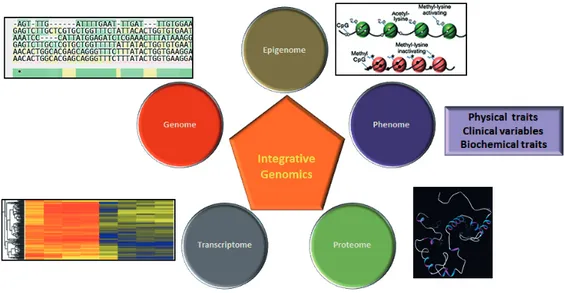
Integrative bioinformatic analysis is a powerful approach to determine infor-mation across multiple high-throughput data generating platforms (Marconett et al., 2013). For example, figure 1 depicts a schematic workflow of integrat-ed analysis of genomic and transcriptomic data. This type of analysis helps in deducing connotations that are not obvious when analyzing dataset represent-ing a srepresent-ingle aspect, such as copy number variation or gene expression pro-files, both of mRNA and miRNA. The fundamental principle of integrative genomic analysis is that, all living organism are made of different layers of molecular mechanisms through which the genome manifests its function, and only by understanding the interactions between these layers one can under-stand the phenotypic characters of the organism (Joyce and Palsson, 2006; Kristensen et al., 2014). One of the early prospects for integrative genomics ascended from the development of different dataset spawned by screening of NCI60 panel of cancer cell lines for both drug sensitivity and genomic con-tours (Weinstein, 2004). As an attempt to analyze different molecular profiles generated from different platforms a tool called ‘CellMiner’ (Shankavaram et al., 2009) was built. This tool is a relational database, which contains differ-ent molecular profiles generated for NCI60 cell-lines panel. Thus the idea of analyzing the data across different diverse experimental platforms has led to the development of several data-mining platforms or databases such as ‘On-comine’ (Rhodes et al., 2004), ‘Gene Aging Nexus’ (Pan et al., 2007), ‘TCGA’ (Cancer Genome Atlas Research et al., 2013), ‘G-DOC’ (Madhavan et al., 2011). The different pipelines used for the integrative analysis utilizes a combination of graph theory, correlation algorithms and statistical methods for finding connotations between different categories of transcriptomic or genomic aberrations and clinical or other phenotypic outcomes from the bio-logical systems being studied. The work presented in this thesis has employed novel integrative bioinformatic and experimental approaches to study DNA-protein interactions and associated transcriptomic changes in (i) MM and (ii) cellular response to growth signals.
Figure 1. Integration of data from different platforms to understand the mech-anism of complex biological system. By studying the gene expression using mi-croarray alone will answer question about the mechanisms underlying regula-tion of expression levels of mRNA or miRNA. However, if the microarray data is integrated with the epigenomic or genomic data, it gives more insight of the possible mechanism of differential gene expression. Further if this data is ana-lyzed together with the information about proteins in the same samples, it un-covers more information about the translational variation occurring in the system and its correlation with transcription and translation of a protein. (Part of figure modified from original source (Gudjonsson and Krueger, 2012)).
Significance of bioinformatics and present challenges
The reduction in the cost and time of sequencing has led to the generation of unprecedented volumes of sequencing data. Bioinformatics plays a central role in managing, handling and analyzing such high-throughput data. A vari-ety of softwares are available for NGS data analysis and many more are be-ing developed as the needs and experience in NGS advance. Some major areas where bioinformatics plays a crucial role includes (i) alignment of reads to a reference genome (Langmead et al., 2009; Li and Durbin, 2010), (ii) de novo assembly (Simpson et al., 2009), (iii) assembly viewers (Li et al., 2009), (iv) base calling and genetic variation detection (Goya et al., 2010; Li et al., 2009), (v) genome annotation (Stone and Sidow, 2005). There are several bioinformatic impediments when handling or analyzing the high-throughput data like alignment, assembly, annotation, quality control, short read mapping and data storage (Xuan et al., 2013). Thus, it becomes necessary to broaden our knowledge and understand the principles, benefits or limitations of bioinformatics tools and further design appropriate pipe-lines, which are capable of generating good and reliable analytical results.
Due to the large amount of information available about DNA-binding pat-terns of different protein being churned out, it is necessary to establish their effects on gene expression to elucidate the effects of DNA-protein interac-tions. An experimental design that addresses DNA-protein interaction and gene expression in a certain context forms the core of such an approach. The parallel and corresponding analysis of such massively parallel sequencing and expression datasets requires intuitive and context-dependent usage of bioinformatic tools. The work presented in the papers included in this thesis employ a bioinformatic approach that on one hand analyzes the DNA-binding pattern of transcription-regulatory factors and on the other hand establishes its correlation with expression data. Where expression and DNA-binding pattern data are unavailable from the same sources, extensive com-putational usage of information available from relevant samples in different databases have been utilized to derive putative functional effects of DNA-protein interaction patterns on gene expression. In addition, many tools have been utilized in customized ways to test the correspondence between DNA-protein interaction and expression datasets.
Project 1
Multiple myeloma: a background
The term “Multiple Myeloma” was suggested by von Rustizky in 1873. MM, also known as plasma cell myeloma is characterized by malignant plasma cells classically residing in the bone marrow. According to the currently held view, MM cells derive from a post germinal center, long lived, somatic hy-permutated and isotype switched B-cell. The primary genetic events that are suggested to be involved in the tumor pathogenesis include translocation during IgH switch recombination, whereas secondary or late events involved in progression include activating mutations in RAS and inactivating muta-tions in tumor protein 53 (TP53) (Bergsagel and Kuehl, 2003). The malig-nant plasma cells interact with the bone marrow microenvironment compris-ing of extracellular matrix, lymphoid, myeloid cells, stromal cells, osteo-clasts, osteoblasts, and blood vessels. Both genetic aberrations and bone marrow microenvironmental interactions contribute to MM pathogenesis. MM is the second most common hematological cancer (Avet-Loiseau et al., 2007) with a worldwide annunal occurrence estimate of 0.8% of all new cancer cases and 0.9% of all cancer deaths (Becker, 2011). The median age at diagnosis is around 70 years (Palumbo and Anderson, 2011). Some of its clinical hallmarks are the occurrence of bone damage, or osteolytic lesions, renal failure, hypercalcemia and immunodeficiency. MM can either occur in
de novo or it can advance from its benign stage known as monoclonal
into an advanced premalignant and asymptomatic stage known as smolder-ing multiple myeloma (SMM). MM has a further advanced stage known as plasma cell leukemia (PCL) where the malignant plasma cells disseminate into the peripheral blood. Decades of research have led to several treatment modalities that have resulted in increasing the response to treatment and survival of MM patients. However, MM still remains incurable as a majority of patients eventually get a relapse of the disease and death occurs due to the emergence of therapy-refractory disease. A possible reason could be the presence of tumor-initiating populations that are resistant to current regimes of therapies and possess the capability to recapitulate tumors (Reya et al., 2001). These tumor-initiating cells are expected to carry the etiologically important genetic and epigenetic lesions. Several attempts have been made to characterize the genetic aberrations that are likely to play a key etiological role in development and progression of MM.
Genetics of multiple myeloma
Chromosomal abnormalities play an important role in disease evolution of several hematological malignancies MM has several incidences of complex chromosomal abnormalities that are commonly present and are early events in the clonal development of the tumor. In spite of genetic complexities MM patients based on considerably better diagnosis can be divided into two ma-jor classes: the high-risk non-hyperdiploid and the low-risk hyperdiploid (Weh et al., 1995). Abnormal karyotype with several trisomies of odd num-bered chromosomes is the main characteristic of hyperdiploidy. In case of non-hyperdiploid MM, translocations occur in immunoglobulin heavy-chain (IgH). The translocations detected are likely caused by errors in VDJ recom-bination and IgH switch recomrecom-bination augmented by somatic hypermuta-tion (Bergsagel and Kuehl, 2003).
The five recurring IgH translocations (figure 2) described as early events of tumor development are: (i) t(11;14) involved in deregulation of Cyclin D1 (CCND1) and Cyclin D2 (CCND2), (ii) t(6;14) in deregulation of Cyclin D3 (CCND3), (iii) t(4;14) involving fibroblast growth factor receptor 3 (FGFR3) and Wolf-Hirschhorn Syndrome candidate 1 (WHSC1) (also called multiple myeloma SET domain containing protein (MMSET)) (iv) t(14;16) and (v) t(14;20) involved in deregulation of musculoaponeurotic fibrosar-coma (MAF) and musculoaponeurotic fibrosarfibrosar-coma homolog B (MAFB) respectively (Chesi et al., 1998a; Hanamura et al., 2001). The t(4;14) and t(14;16), but not t(11;14) have been associated with poor prognosis (Fonseca et al., 2002; Moreau et al., 2002). IgH translocations in MGUS are as preva-lent as in MM and could be referred to as early genetic events in MM trans-formation (Avet-Loiseau et al., 1999). TP53 mutations and rearrangements of MYC (Shou et al., 2000) are considered to be secondary events in the tumor progression in multiple myeloma (Hallek et al., 1998; Hanamura et
al., 2006). Around 35-50% of MM has RAS-activating mutations (Fonseca et al., 2004; Liu et al., 1996). RAS mutations are rarely found in MGUS, suggesting that it is a molecular marker rather being causative of progression from MGUS to MM (Bezieau et al., 2002; Corradini et al., 1993; Fonseca et al., 2004).
Figure 2. Genetic modification occurring in MM. MGUS is an indolent, asymp-tomatic condition, which may develop into MM. Smouldering MM does not have clinical features as compared to symptomatic MM. In MM the clinical features are collectively referred to as (CRAB) calcium, renal, anemia and bone abnormality. The two main classes of genetic changes in MM are classified as ‘hyperdiploid’ and the ‘non-hyperdiploid’. The five primary translocations affect Cyclins, FGFR3, MAF, and MAFB. (Modified from original source (Morgan et al., 2012)).
Classification of multiple myeloma
Based upon the clinical parameters, Durie/Salmon (DS) in 1975 introduced a staging system for MM (Durie and Salmon, 1975). This system included parameters like levels of calcium and hemoglobin, number of bone lesions, levels and types of monoclonal proteins. In 2005 an International Staging System (ISS) was developed based on the survival tree model (Greipp et al., 2005). According to this system, which is currently widely used, MM is
divided into three risk groups based on the serum levels of β2-microglobulin and albumin (Greipp et al., 2005; Palumbo and Anderson, 2011).
Based on conventional cytogenetics and fluorescence in situ hybridization (FISH), MM is classified into two prognostic groups: high risk and standard risk. High risk and poor prognosis group is defined by presence of hypodip-loidy, t(4;14) or t(14;16), while presence of hyperdiploidy or t(11;14) was defined as standard risk or better prognostic group (Palumbo and Anderson, 2011; Sawyer, 2011).
The advent of high-throughput approaches has made our understanding even better in molecular classification of multiple myeloma. One such classi-fication was performed by using transcriptome-wide gene expression profil-ing (GEP) where normal plasma cells (PCs) were compared with MM PCs and MM PCs were classified into four distinct groups (Zhan et al., 2002). In another classification, known as TC classification, MM is classified based on IGH translocations and overexpression of cyclin D genes (Bergsagel et al., 2005). According to this system eight TC groups were proposed. Later, MM was classified based on the presence of translocations or hyperdiploidy into seven different classes (Zhan et al., 2006). With the help of expression-based multivariate discriminant analysis, a ‘17 upregulated genes’ signature was developed that has been suggested to be predictive of poor prognosis (Shaughnessy et al., 2007). In another study a ‘15 gene signature’ was fined in newly diagnosed and relapsed myeloma patients. This model de-fined high and low-risk subpopulations, and suggested that gene expression profiles can be used to understand the molecular mechanism underlying re-sistance to drug treatments (Decaux et al., 2008). Notably, the overlap of single genes constituting these signatures of high-risk myeloma is small and the biological rational of these genes remains to be elucidated
Tissue microenvironment in multiple myeloma
The bone marrow microenvironment plays a crucial role in the pathogenesis of the MM (Chauhan et al., 2004; Mitsiades et al., 2006). The stroma con-sists of several types of myeloid cells, lymphoid cells, extracellular matrix (ECM) proteins e.g. osteopontin, fibronectin, laminin and various cell types including erythrocytes, bone marrow endothelial cells, osteoclasts, and oste-oblasts (Hideshima et al., 2007). The interaction of the myeloma cells with the ECM proteins and accessory cells (figure 3) has an established and piv-otal role in the pathogenesis of MM (Chauhan et al., 2004; Mitsiades et al., 2006), and results in the secretion of different cytokines and growth factors such as interlukin-6 (IL-6), vascular endothelial growth factor (VEGF), B-cell activating factor (BAFF), and insulin-like growth factor-1 (IGF-1) (Jernberg-Wiklund and Nilsson, 2007; Kawano et al., 1988; Moreaux et al., 2004; Podar et al., 2001).
Figure 3. Interaction between MM cells and bone marrow microenvironment. MM microenvironment plays a crucial role in the growth and survival of the bone marrow resident MM cells. MM cells helps in the osteoclastogenesis by secreting) C-C chemokines such as MIP-1A (CCL3), which help in stimulating the expression of receptor activator of NF-kappa B ligand (RANKL) in stromal cells. MM cells constitutively express VEGF, which helps in osteoclast for-mation by RANKL. Osteoclasts secrete anti-apoptotic chemokines like IL6, APRIL and BAFF for the myeloma cells. Osteoclasts secret matrix metallopro-teinase (MMP9) and play an important role in enhanced angiogenesis. Interac-tion of stromal cells with myeloma cells helps in the proliferaInterac-tion and survival of the MM cells via elaborating factors such as IL6 and IGF1. The terminally differentiated mature osteoblasts inhibit the growth and survival of MM cells. MM cells block the osteoblastic differentiation by secreting Dikkopf1 (DKK1), which is a WNT signal inhibitor. (Modified from original source (Abe, 2011)). IL-6 is one of the most important cytokines that promote growth and surviv-al in MM in a paracrine and autocrine fashion (Spets et surviv-al., 1997; Zhang et al., 1989). IL-6 signaling mediated via the gp-130 transducing receptor sub-unit activates the following pathways: RAS/mitogen-activated protein kinase (MAPK), Phosphoinositol-3-kinase (PI3K)/AKT, and Janus kinase (JAK)/signal transducer and activator of transcription 3 (STAT3) which are related to growth and survival of MM cells (Brocke-Heidrich et al., 2004; Ogata et al., 1997; Tu et al., 2000).
The imbalance in function of osteoclasts and osteoblasts leads to the for-mation of bone lesions in patients detected with MM. The osteoblasts are suppressed on the inhibition of Wnt pathway, while receptor activator of nuclear factor-kappa B (RANK) pathway amplification and action of macro-phage inflammatory protein-α (MIP-1α; also called CCL3) activates osteo-clasts (Roodman, 2009). VEGF leads to the abnormal growth of myeloma tumor vessels (Hideshima et al., 2007). Myeloma cell growth and survival is also affected by IGFs (Kimata and Yoshida, 1994). IGF1 is produced by
different cells e.g hepatocytes, osteoclasts, and fibroblasts of which hepato-cytes produce it largely, and it promotes proliferation and survival of MM cells (Ferlin et al., 2000; Georgii-Hemming et al., 1996; Jelinek et al., 1997). IGF1 binds to its receptor Insulin-like growth factor receptor (IGF1R) and stimulates proliferation and survival of MM cells by activating signaling pathways like MAPK and PI3K pathways (Qiang et al., 2002). It is likely that the microenvironment in MM provides the bioavailability of the ligand to the tumor cells. It may increase the survival and growth of MM cells by autocrine or paracrine stimulation of IGF1R by IGF1.
Heterogeneity of multiple myeloma
MM still remains a severe disease due to the fact that treatment eventually leads to fatal relapse. Such observations indicate clearly that there are some MM cells that are not targeted and eradicated completely by current thera-pies. This notion has given rise to several hypotheses on the existence of small populations of cells that are drug resistant, persistent and may have tumor-initiating and self-renewing potential (Matsui et al., 2004; Matsui et al., 2008). According to other suggestions a clonogenic population exists in the dominant population of human CD138+ plasma cells (Chiron et al., 2012; Kim et al., 2012). These contrasting findings underline the importance of studies investigating the functional properties like engraftment potential, clonogenic growth and drug resistance in CD138+ and CD138- cell popula-tions.
Experimental models of multiple myeloma
MM progression depends largely on the interaction of the MM cells with the bone marrow environment, including stromal cells, osteoblasts, osteoclasts and normal hematopoiesis cells. To study these aspects of MM, several in
vitro and in vivo models have been described.
In vitro models
Several established MM cell lines are dependent on growth factors for their maintenance in vitro. Most of them were established from patients with ad-vanced, often terminal disease. It has been challenging to establish MM cell lines as MM cells have slow proliferative activity in culture, and in some cases, require anchorage-dependence from a feeder layer of stromal cells. There are at present more than 80 MM cell lines established for studying the MM disease (Jernberg-Wiklund and Nilsson, 2007; Nilsson, 1977; Nilsson et al., 1970; Nilsson and Ponten, 1975; Pellat-Deceunynck et al., 1995; Ralph, 1985).
In vivo models
A preferred in vivo model is one, which reflects the disease both biologically and genetically. In MM, the malignant plasma cells home to the bone mar-row, where they proliferate and contemporaneously interact with the bone microenvironment. To study these interactions, MM in vivo models reflect-ing this process have been established. There are several in vivo MM mouse models like 5T, severe combined immune-deficient (SCID), MOPC315, non-obese diabetic (NOD), SCID/NOD, and SCID/beige and Vk*Myc (Chesi et al., 2008; Cheung et al., 2004).
5TMM models
The 5TMM is a syngeneic mouse model obtained from elderly mice of the C57BL/KalwRij strain where MM developed spontaneously (Radl et al., 1979). There are two variants of this model; 5T2MM and 5T33MM (Asosingh et al., 2000). There are two different mechanisms that are cardinal for MM growth (i) MM developing from the preexisting MGUS (1/3 of the MM occurrences) and (ii) de novo development of MM (2/3 of the MM oc-currences). The above two models represent de novo myelomas (Radl, 1990). The clinical symptoms of these models are similar to the human mye-loma e.g. presence of serum M-component, selective localization of MM cells in the bone marrow, increased angiogenesis in the bone marrow, and induction of osteolytic bone disease (Radl et al., 1979; Van Valckenborgh et al., 2002; Vanderkerken et al., 1997).
SCID models
SCID mice are generated by a point mutation in chromosome 16 in the CB17 inbred mouse strain. This mutation leads to an interruption of lymphocyte maturation resulting in a deficiency of T and B-cells. On the other hand the innate immunity of these mice is intact which involves cells like monocytes, natural killer cells and granulocytes.
To understand the biology of MM several experimental and biological SCID models have been developed. In SCID xenotransplant models MM cell lines are injected intravenously in mice (Huang et al., 1993; Tsunenari et al., 1997). The injected cells home to the bone marrow where the microenvi-ronment feeds the malignant plasma cells (Asosingh et al., 2000). Non-obese and diabetes free (NOD/SCID) mice are immunodeficient SCID variant with defects in both innate and adaptive immunologic functions (Shultz et al., 1995). These mice provide a suitable non-human environment for supporting engraftment of human cells. In SCID-hu mice, a piece of human bone is grafted to compartmentalize tumor growth (Yaccoby et al., 1998). This mod-el may be instrumental in studying the MM pathophysiology in presence of human BM microenvironment (McCune et al., 1988).
Vk*MYC model
This model is made by introducing a stop codon (TAG) in the third codon of V-kappa exon. The transcription is similar to the formerly generated model Vk-MYC (Robbiani et al., 2005) but in this model the translation gets prem-aturely aborted thus hampering the production of MYC protein. In this mod-el activation of MYC is AID-dependent occurring in the germinal centers i.e. in the late stages of B-cell differentiation. This model is helpful in under-standing the role of MYC in development of MM in the mice (Chesi et al., 2008).
Epigenetic regulation in multiple myeloma
Epigenetic changes are heritable chemical modifications that occur in the genome without any alteration in the DNA sequence itself (Bird, 2002). These chemical alterations orchestrate the development and maintenance of the organism by switching parts of genome ‘on’ and ‘off’ at strategic times and locations during development and differentiation. These changes are inherited stably through the cell division and are potentially reversible. Epi-genetic regulation plays vital roles in several diseases including cancer. There are different regulatory mechanisms by which these changes can be exerted are DNA methylation, expression of non-coding RNAs and histone modifications. Among these, DNA methylation is more stable whereas his-tone modifications are relatively flexible marks. DNA methylation, hishis-tone modifications and mechanisms affecting them, mentioned in subsequent sections, have been extensively studied in context of several diseases, exclu-sively cancer.
DNA methylation
DNA methylation is one of the most studied epigenetic mechanisms and occurs through the addition of a methyl group on the 5th carbon of cytosine
preceded by a guanine residue (Bird, 1986). The genomic regions longer than 200bp with G+C content >50% and at least 0.60 as CpG ratio of ob-served/expected are known as CpG islands (Ioshikhes and Zhang, 2000). In humans these islands are linked with approximately 50% of gene promoters. The reaction of methylation is catalyzed by DNA methyltransferases (DNMTs) and these methylation marks are read by methyl CpG binding proteins (MBDs). In the case of mammals there are three major types of DNMTs: DNA (cytosine-5-)-methyltransferases 1 (DNMT1), DNA (cyto-sine-5-)-methyltransferase 3 alpha (DNMT3A) and DNA (cytosine-5-)-methyltransferase 3 beta (DNMT3B). DNMT1 is required for retaining CpG methylation and DNA mismatch repair (Robert et al., 2003; Wang and James Shen, 2004). DNMT3A and 3B play significant roles in development and
disease as they carry out de novo methylation in early embryonic stages (Okano et al., 1999). In normal cells, bulks of gene promoters bearing CpG islands are usually unmethylated and are associated with a euchromatic state. These genes are generally transcriptionally active. However, there are some exceptions such as the genes subjected to genetic imprinting and X-chromosome inactivation where methylation silences genes in an allele-specific manner (Bird, 1999; Singal and Ginder, 1999). On the other hand, in cancer cells these CpG islands can be abnormally methylated in a tumor specific manner resulting in gene silencing. Hypermethylation of these CpG islands of tumor suppressor genes results in the loss or decrease of gene ex-pression, which leads to absence of tumor suppression function (Esteller, 2008; Herman and Baylin, 2003; Jones and Baylin, 2002). However, apart from hypermethylation, a reduction of methylation, which occurring in the repetitive DNA sequences and demethylation of coding regions and introns (Feinberg and Tycko, 2004) also contributes to the development of cancer. The three different mechanisms: generation of chromosomal instability (Eden et al., 2003), reactivation of transposable elements (Bestor, 2005), and loss of imprinting (Cui et al., 2003; Kaneda and Feinberg, 2005) have been proposed to explain the role of hypomethylation in cancer development. There is interdependence between locus-specific patterns of DNA methyla-tion and histone modificamethyla-tion profiles, latter being a major focus of work presented in paper III. Both these epigenetic mechanisms coordinate to achieve a certain epigenetic structure of the genome and thereby affect the transcriptome.
Histone modifications
DNA together with the histone proteins makes up the chromatin, which is packed in the nucleus of the cell. The DNA is wrapped around the histone octamer core, which consists of two subunits of each of the four histones H2A, H2B, H3, and H4. The fifth histone H1 acts as a linker protein thus making the nucleosome the basic subunit of DNA packaging in eukaryotes. (Felsenfeld and Groudine, 2003; Shukla et al., 2008). Different covalent modifications occurring in these proteins can alter the chromatin structure, and thus prevent the accessibility to transcriptional factors and other regula-tory proteins controlling gene expression (Yoo and Jones, 2006). The differ-ent modifications including ubiquitinylation, methylation, acetylation, phos-phorylation, or SUMOylation occur at different residues of lysine, arginine, and serine amino acids at the exposed C- and N-terminal tails of histones.
Figure 4. A list of histone modifications, their location with reference to associ-ated genes and their effects on transcription is shown. (Modified from original sources (Li et al., 2007) and (Young et al., 2011))
These modifications lead to either activation or silencing of gene expression. The different enzymes that catalyze these reactions involve histone deacety-lases (HDACs), histone methyltransferases (HMTs), and histone acetyltrans-ferases (HATs) (Jenuwein and Allis, 2001; Strahl and Allis, 2000). Among, the several histone modifications listed above acetylation is associated with transcriptional activation, while the function of histone methylation depends on the type of amino acid and its position in the histone tail (Bernstein et al., 2007; Mack, 2006). For example tri-methylation on H3K27 is associated with gene silencing while tri-methylation of H3K4 is associated with active-ly transcribed genes. While these two histone modifications constitute the main focus of work presented in this thesis, many other histone modifica-tions have been reported with diverse effects on chromatin structure and function (Li et al., 2007).The outcome of histone modifications on gene
ex-pression greatly depends on the distribution patterns of modified histones along the target genes (Li et al., 2007) (Figure 4).
In addition to histones, there are other proteins, which by being a part of the chromatin shape the epigenetic state and fate of target genes. Prominent among these are the Polycomb group proteins, a major focus of experiments in paper I.
Polycomb group proteins
The Polycomb Group (PcG) and trithorax group (trxG) proteins are required for maintenance of correct expression levels of several key developmental regulators like the homeobox (HOX) genes (Mahmoudi and Verrijzer, 2001). Several PcG protein domains are conserved from files to humans (Levine et al., 2002). These proteins are believed to function in distinct multimeric complexes that control transcription by changing the structure of chromatin (Otte and Kwaks, 2003). The deregulation of expression and function of PcG proteins has been linked to altered state of cellular proliferation and cancer (Jacobs and van Lohuizen, 2002; Valk-Lingbeek et al., 2004). Polycomb repressive complex 1 and 2 (PRC1 and PRC2) are the two well-characterized PcG complexes (figure 5) (Jacobs and van Lohuizen, 2002; Otte and Kwaks, 2003). In mammals PRC1 is composed of chromobox homolog (CBX), real-ly interesting new gene 1 (RING1), poreal-lyhomeotic (PHC), and B-cell real- lym-phoma Mo-MLV insertion region 1 (BMI1). In mammals PRC2 complex consists of four core proteins: suppressor of zeste 12 (SUZ12), retinoblasto-ma binding protein (RBBP7/4), embryonic ectoderm development (EED) and enhancer of zeste (EZH2) or its close homolog EZH1 (Di Croce and Helin, 2013; Schuettengruber and Cavalli, 2009). EZH2 contains the SET domain and is the main catalytic subunit, which has methyltransferase activi-ty. It catalyzes the mono-, di- and tri-methylation of lysine 27 on histone H3. Previously, biochemical studies suggested that the chromodomain of Poly-comb, which is a subunit of PRC1, is specifically recognized by the product of EZH2 i.e. H3K27me3 (Fischle et al., 2003; Min et al., 2003). However, recent findings propose that additional mechanisms may be required for PRC1 recruitment and alone H3K27me3 may not suffice (Tavares et al., 2012; Yu et al., 2012).
PRC2 plays an important role in several biological processes like cellular differentiation, proliferation, maintenance of cellular identity and stem cell plasticity (Margueron and Reinberg, 2011). PRC2 components have been reported to be over-expressed in several cancers like melanoma, lymphoma, prostate cancer, and breast cancer (Margueron and Reinberg, 2011). Studies in breast and prostate cancers have shown EZH2 to be a marker of aggres-sive stages (Kleer et al., 2003; Varambally et al., 2002), and EZH2 has also been indicated as an oncogene that promotes the neoplastic transformation of a benign prostatic epithelial cell line (Karanikolas et al., 2009). EZH2
ex-pression is controlled by several microRNAs, for example hsa-mir-101 (Cao et al., 2010), hsa-mir-26a (Dang et al., 2012) and hsa-mir-214 (Juan et al., 2009). A deregulation of microRNAs could result in the overexpression of EZH2 in cancer (Margueron and Reinberg, 2011). PRC1 and PRC2 com-plexes seems to help in the acquisition of pluripotency, as embryonic stem cells (ESC) lacking either of the two complexes were unable to successfully reprogram B-cells towards pluripotency (Pereira et al., 2010), thus, suggest-ing that PcG proteins are required for establishsuggest-ing the pluripotent state of the cell.
Figure 5. PRC1 and PRC2 complex proteins and how they are involved in the transcriptional repression. EZH1 or 2 contain the SET domain and are respon-sible for the enzymatic activity of PRC2 complex. EZH2 trimethylates the his-tone H3 at lysine 27, yielding an H3K27me3 mark; this is followed by recruit-ment of PRC1 complex to this mark. The CBX recognizes the H3K27me3 mark. The enzymatic subunit of PRC1 complex is RING1a or RING1b, which acts as an E3 ubiquitin ligase and monoubiquitylates histoneH2A at lysine 119. Mode of action of both the complexes leads to transcriptional repression. PRC2 complex recruits the DNMTs. (Modified from original source (Choi and Friso, 2010)).
Epigenetic regulation of transcription in multiple myeloma
In MM several tumor suppressor genes regulating the cell cycle are found to be silenced by DNA hypermethylation, like cyclin dependent kinase inhibi-tor 2A (CDKN2A) (Chim et al., 2007; Guillerm et al., 2001), cyclin depend-ent kinase inhibitor 2B (CDKN2B), death associated protein kinase (DAP kinase) (Ng et al., 2001) suppressor of cytokine signaling 1 (SOCS1) (Galm
et al., 2003), interferon regulatory factor 8 (IRF8) (Tshuikina et al., 2008) and the secreted frizzled- related protein (SFRP) family (Jost et al., 2009). Hypermethylation of some genes have been correlated with poor outcomes in patients e.g. TGF beta-receptor 2 (TGFBR2) (de Carvalho et al., 2009) and O-6-methylguanine-DNA methyltransferase (MGMT) to extramedullary disease (Yuregir et al., 2010) although a cause-effect relation in remains unclear. In contrast to DNA hypermethylation, some recent studies have identified that DNA hypomethylation of the repetitive sequences like Alu, LINE-1, and satellite alpha (SAT-alpha) are linked to genomic instability and associated with tumor progression in multiple myeloma patients (Bollati et al., 2009).
In addition to DNA methylation, histone modifications also play an im-portant role in controlling the gene expression. MMSET was the first chro-matin modifying gene identified in MM in the t(4;14) translocation carrying subpopulations (Chesi et al., 1998b). EZH2 was reported to have oncogenic activity in MM and its induction is correlated with tumor burden (Croonquist and Van Ness, 2005). The other PcG protein family member BMI1 was found to be important in myelomagenesis through suppression of proapop-totic protein BIM (Jagani et al., 2010). Independently, it has been shown that IGF1 treatment results in epigenetic silencing of BIM by increasing H3K9 dimethylation (De Bruyne et al., 2010). Furthermore, inactivating somatic mutations in the histone H3 lysine 27 demethylase UTX was reported in several tumor types with highest prevalence in MM (Chapman et al., 2011; van Haaften et al., 2009). Recently it has been reported that in t(4;14) mye-loma cells MMSET controls histone methylation. The overexpression of MMSET leads to globally high anomalous levels of H3K36me2 convoyed by decreased levels of H3K27me3 (Martinez-Garcia et al., 2011). Thus we have investigated MM through a combination of experimental as well as integrative bioinformatic approaches in the first three papers.
Integration of experimental approaches with customized bioinformatic analysis can also be used to understand mechanisms of normal cell biology. Such a combined application of bioinformatics and experiments empowers us to understand how DNA-protein interactions translate into gene expres-sion patterns with effects on cellular phenotypes. In project 2, we applied such an integrative bioinformatic approach to understand the correlations between DNA-protein interactions and their effects on transcription in nor-mal cells. The results expose how DNA-protein interactions could affect the RNA profile of a cell without necessarily altering the histone code or chro-matin structure of target genes in cis. The work in this project touches upon areas of transcription, RNA Polymerases, interspersed repeats and the human protein CGGBP1.
Project 2
Major signal transduction pathways in normal fibroblasts
The stimulation of mammalian cell replication is executed by growth factors such as EGF, PDGF or FGF, which bind to and activate their cognate tyro-sine-kinase receptors. The activated receptors phosphorylate downstream targets and activate various signaling pathways. Canonically, as exemplified by the EGF-EGFR axis, the growth-promoting signal is transduced through the “GRB2-SOS RAS RAF ERK”, “PLC DAG/IP3 PKC/CaM-kinase” and “PI3K PDK1 AKT mTOR” pathways. The extent and importance of crosstalk and inter-regulation between these RTK-downstream events and other signaling pathways such as G-protein signal-ing, cytokine signaling can only be understated. All these extrinsic stimula-tions ultimately are signaled down to regulate specific transcription fac-tors/co-factors. Some well-known examples are E2F, MYC, MAX, FOS, JUN downstream of RTKs, CTNNB-TCF downstream of WNT signaling, HES family factors downstream of NOTCH signaling, GLI transcription factors downstream of SHH signaling, STAT downstream of JAK signaling and CREB downstream of G-protein signaling. Some factors like the serum-response factor (SRF) respond to the components of serum, a widely used source of growth and survival factors in culture. It is well known that the RTK-activated transcriptional program mediated by E2F, MYC, MAX, and FOS etc. increases the expression of genes required for cell cycle (Cyclin E, ribosomal RNA and proteins) and at the same time inhibits the activity and expression of cell cycle inhibitory proteins such as p21 and p53. Growth factor signals are thus transduced to orchestrate a transcriptional program to achieve cell growth and proliferation.
Brief overview of transcription
Transcription is a biological process in which a complementary RNA is syn-thesized from the DNA by enzymes called RNA Polymerases. While tRNA and rRNA thus produced contribute in translation, the mRNA thus produced is used as a template for the synthesis of protein. In eukaryotes the main RNA Polymerases involved in transcription are RNA Polymerase I, II and III (Roeder and Rutter, 1969). RNA Polymerase I initiates ribosomal bio-genesis and helps in regulating eukaryotic cell growth (Engel et al., 2013), RNA Polymerase II is involved in the transcription of messenger RNA (mRNA) and non-coding RNA including small nuclear RNA (snRNA) and microRNAs (miRNAs) (Koleske and Young, 1995; White, 2011) and RNA Polymerase III is involved in synthesis of tRNA, 7SL RNA and 5S rRNA (Dumay-Odelot et al., 2010; White, 2011). Retrotransposons interspersed in our genomes also use RNA Polymerases to synthesize their RNA. For
ex-ample, the long interspersed nuclear elements (LINE-1) require RNA Poly-merase II for transcription whereas short interspersed nuclear elements (SINEs) require RNA Polymerase III.
Structure and function of gene promoter
Each transcribed unit has an upstream or embedded element that acts as a transcriptional promoter. In eukaryotes transcription of protein-coding genes is regulated by numerous events like nucleosome remodeling, histone modi-fication, decondensation or condensation of the transcribed locus, binding of transcriptional activators and co-activators to regulatory sequence regions (promoters and enhancers) and binding of the basal transcription factors to the core promoters (Smale and Kadonaga, 2003). The core promoter consists of a TATA box, mostly present 25 to 30 bp upstream of the transcription site of about a quarter of all known human genes (Yang et al., 2007). It is strictly conserved and where present is essential for transcription initiation (Suzuki et al., 2001). TATA binding protein (TBP) binds to the TATA sequence (Kim et al., 1993). Data from promoters of most of the protein-coding genes reveal that in an initiator region (Inr), an adenosine is present at transcription start site (+1), and a cytosine at (-1) position with a few pyrimidines sur-rounding around the nucleotides (Corden et al., 1980). Downstream core promoter (DPE) sequence is another downstream core promoter sequence located at +28 to +32 relative to Inr (Kutach and Kadonaga, 2000). Another core promoter sequence TFIIB recognition element (BRE) is present in pro-tein-coding gene promoters, which is present upstream of TATA box (Hain et al., 1992).
In the eukaryotic core promoter of small nuclear RNA (snRNA) a highly conserved sequence called the proximal sequence element (PSE), it is pre-sent between -45 to -60 bps relative to transcription start site (Schramm and Hernandez, 2002). PSE sequence can act differently within a single organ-ism; it can initiate transcription for some snRNA by RNA Polymerase II and by RNA Polymerase III for different subsets of snRNA (Schramm and Hernandez, 2002).
RNA Polymerase I promoter contains two regulatory elements an up-stream control element (UCE) from -156 to -107 from the start site and a core promoter element from -45 to +18 relative to transcription start site (Goodfellow and Zomerdijk, 2013; Russell and Zomerdijk, 2005).
RNA Polymerase III promoters are typical in that they employ down-stream regulatory sequences contained within the transcribed unit. In case of eukaryotes RNA Polymerase III promoters are of three different types de-pendent on the kind of regulatory sequences they contain and the factors that regulate them (Orioli et al., 2012). The type 1 promotes have an intermediate element and a C box instead of B box while the type 3 promoters lack both the A and the B boxes and transcription for these promoters rely on external
upstream elements like TATA box, PSE elements (Schramm et al., 2000; Teichmann et al., 2000).
In the type 2 RNA Polymerase III promoters the transcription units con-tain within the transcribed region regulatory elements known as A and B boxes. The A box is present approximately 12-20 bp downstream of TSS and B box lies 30-60 bp downstream of the A box. This promoter organization is particularly present in tRNA, SINEs and some non-coding RNA genes (Dieci et al., 2007). The A-box sequence binds to the RNA Polymerase III components BRF1 (a subunit of TFIIIB) and TFIIIC (of which GTF3C1 is a major component) whereas B box binds to GTF3C1 without BRF1. Binding of BRF1 at A box serves as a positioning factor required for position-specific recruitment of other RNA Polymerase components. The assembly of BRF1 and TFIIIC facilitates the recruitment of the catalytic units of RNA Polymerase III, including POLR3F required for transcription. The A box thus acts as a positing as well as enhancing element for RNA Polymerase III-driven transcription, whereas the B box functions only as an enhancing se-quence. Thus far, only transcription-enhancing effects of A- and B-box-binding proteins have been reported. The function of the intervening se-quence between the two elements and possible transcription factors that might bind them and regulate RNA Polymerase III activity is not known. The only known regulator of RNA Polymerase III activity is MAF1 (Boguta, 2013; Li et al., 2007; Rollins et al., 2007; Upadhya et al., 2002) that acts in a tyrosine phosphorylation-dependent manner (Michels et al., 2010).
Retroelements and mammalian genome
The high-throughput DNA sequencing has helped in deciphering the ge-nomic constitution of different organisms. Human genome can be divided into coding and non-coding regions. The coding part constitutes of all the protein coding genes while the non-coding genome consists of intergenic regions, introns (although a part of the protein-coding gene), pseudogenes, non-coding RNAs, regulatory DNA sequences, simple and satellite repeat sequences and sequences related to mobile genetic elements including the retrotransposons Alu-SINEs and LINE-1 elements. The full-length LINEs, though classified as belonging to the non-coding part of our genome, actual-ly do code for proteins that aid their transposition. More than 50% of the human genome is made of repetitive elements of which one third are the interspersed repeats (Cordaux and Batzer, 2009; Smit, 1996). LINEs occupy 20% of the genome while SINEs are 13%. Full-length L1-LINE elements are 6-8kb long and Alu-SINEs are less than 300 bps long and both constitute the majority of LINEs and SINEs respectively (Cordaux and Batzer, 2009; Singer, 1982; Ullu and Tschudi, 1984). Alu constitute 11% of the human genome (Lander et al., 2001). They have a wide range of influences on gene expression and mRNA polyadenylation (Chen et al., 2009), and splicing
(Shen et al., 2011). Alus have originated from head-to-tail fusion of 7SL RNA (figure 6). An internal RNA Polymerase III promoter is present in the Alu sequence that helps in the transcription of Alus. An Alu element does not contain any well-defined transcription termination sequence and uses either an Alu-specific or any gene-associated downstream TTTT sequence for termination (Deininger, 2011). Alu insertion can disrupt the coding re-gions or splice sites thus contributing to mutations and diseases (Belancio et al., 2008; Deininger and Batzer, 1999). Alus can also affect gene expression by integrating close to promoter elements and inactivating them because Alus are prone to silencing by DNA methylation. Alu elements contain CpG residues that are subject to methylation and thus are accountable for approx-imately 25% of genome methylation (Xie et al., 2009).
Figure 6. Structure of an Alu element. Alu elements, derived from a head-to-tail fusion of copies of 7SL gene have the fused 7SL monomers represented as Alu-left arm and Alu-right arm. The left arm consists of RNA Polymerase III-binding sites (A-box and B-box), which act as transcription enhancing as well as polymerase positioning elements respectively. A short poly-A region sepa-rates the two arms. The regions overlapping with A-box, depicted as ATE re-gion has been described in paper IV.
CGGBP1: a regulator of normal and cancer cell biology
CGGBP1 was discovered as a CGG triplet repeat-binding protein of 20 KDa (Deissler et al., 1997). It is expressed ubiquitously in different tissues and acts as a transcription regulator (Muller-Hartmann et al., 2000; Naumann et al., 2004). It binds to CGG repeats in the promoter of FMR1 gene (Muller-Hartmann et al., 2000). Its binding is proposed to keep the CGGxn element-controlled promoter in a transcriptionally suppressed state. From this, it has been inferred that CGGBP1 is a transcription repressor. In situ and in vitro experiments have shown that CGGBP1 localizes to the ribosomal gene clus-ters in the short arms of acrocentric human chromosomes (Muller-Hartmann et al., 2000). Its expression is detectable at RNA level in multiple tissues (Naumann et al., 2004). The phylogeny of CGGBP1 shows that it has evolved in the reptilian lineage and is highly conserved (NCBI Homolo-Gene). The human amino acid sequence of CGGBP1 is 88% similar to birds (G. Gallus) and 98% similar to mammals (mouse and human CGGBP1 dif-fer only by two amino acids). Reports of functional studies on CGGBP1 have been sparse. Attempts to create CGGBP1-null mice have been
incon-clusive (Naumann, 2003). Presence of Polymorphisms/mutations in cancer cell lines (CBioPortal) show that only silent mutations in CGGBP1 are se-lected (Singh and Westermark, unpublished observations), making it argua-ble that any deviations from wild-type sequence is not conducive for cellular growth and.
A reinvigoration of interest in the functions of CGGBP1 took place with the screening of genes involved in glioblastoma. In a murine model of retro-virally overexpressed PDGFB-induced glioblastoma (Johansson et al., 2004), Nuclear factor 1-X (NFIX) was one of the strongest candidate brain tumor loci found. Mechanistic studies on NFIX that included yeast-2-hybrid screen, microarray-based expression analyses and targeted ChIP experiments demonstrated that heat shock-sensitive interactions between CGGBP1 and NFIX regulate cellular stress response (Singh et al., 2009). CGGBP1 could thus influence cancer development through modulation of stress response.
Roles of CGGBP1 in multiple cellular functions were elucidated through studies on normal human cells. Acute depletion of CGGBP1 induced tetra-ploidy due to failure of cytokinesis (Singh and Westermark, 2011). CGGBP1 localizes to the nuclei in interphase, to the metaphase chromatin during met-aphase, spindle midzone during anaphase and on midbodies during telophase and cytokinesis (Singh and Westermark, 2011). Knockdown of CGGBP1 lead to mitotic slippage or regression of cleavage furrow leading to tetra-ploidy. While in normal cells this is manifested as S and G2/M arrest, in cancer cells the cell cycle block caused by CGGBP1 knockdown results in a G0/G1 arrest (Singh et al., 2011). These studies were performed in cancer cells of various backgrounds (TP53-deficient, CDKN1A-deficient, INK4A/ARF-deficient, RB-deficient or advanced cancer cells with unknown profiles of major cancer-relevant genes (Singh et al., 2011).
The intolerance of cells to changes in CGGBP1 peptide sequence is demonstrated by dominant negative effects of point mutant CGGBP1. Serine 164 on CGGBP1, a part of an SQ motif and a target site for PI3 kinase-like kinases (ATR /ATM kinases) when mutated to Alanine results in reduction of serine phosphorylation on immunoprecipitated CGGBP1 (Singh et al., 2014). Overexpression of S164A mutant CGGBP1 in presence of endoge-nous WT CGGBP1 lead to endogeendoge-nous DNA damage response focused mainly at the telomeres (Singh et al., 2014). This telomeric damage was associated with telomere shortening, telomeric fusions, failure of resolution of lagging anaphase chromatin in dividing cells leading to chromatin bridges between telophase and post-telophase nuclei. These experiments demon-strated that CGGBP1 mediates the preventive effects of ATR on endogenous telomere damage and normalcy of midbodies. Interestingly, all these delete-rious effects of loss of S164 phosphorylation of CGGBP1 on cells were res-cued by S164E mutant CGGBP1 overexpression wherein E acts as a phos-phoserine-mimicking residue (Singh et al., 2014).
While the molecular mechanisms underlying these effects of CGGBP1 remain elusive, there are two major lines of evidence. First, the effect of CGGBP1 on telomeric DNA damage could be due to direct interaction be-tween CGGBP1 and telomere protector POT1. Evidence suggests that CGGBP1 regulates POT1 DNA binding (Singh et al., 2014). Second, CGGBP1 being a transcription regulator, could affect a variety of cellular processes by controlling the expression of its target genes (Singh et al., 2011). For example, it regulates expression of genes like FMR1, HSF1, CDKN1A and GAS1 (Singh et al., 2011), as well as transcription of rRNA (Muller-Hartmann et al., 2000), telomeric protection (Singh et al., 2014) and cell cycle progression (Singh et al., 2011; Singh and Westermark, 2011).
The amino acid sequence of CGGBP1 shows that it has a C2H2 Zn finger DNA-binding domain that overlaps with the nuclear localization domain Ensembl database). Bioinformatic prediction of functional domains on CGGBP1 reveals several residues that could potentially be involved in signal transduction as well as interaction with transcription-regulatory factors. Some known interacting partners of CGGBP1 are SUV39H2, UBC, NFIX, HMGN1, ATR and POT1 (Abdelmohsen et al., 2009; Danielsen et al., 2011; Singh et al., 2009; Singh et al., 2014; Stelzl et al., 2005; Stes et al., 2014).
With this knowledge it seems that the signal transduction and DNA-binding-related activities of CGGBP1 hold key to its functions and thereby its effects on cell cycle progression. The work on CGGBP1 presented in this thesis is based on experimental and integrated bioinformatics analyses of DNA-binding profile of CGGBP1 vis-à-vis its role in growth factor signal transduction.
Summary of Paper I
Title: Polycomb target genes are silenced in Multiple Myeloma.
Summary
MM is a genetically heterogeneous incurable malignancy of mature plasma cells. To elucidate a common mechanism for initiation and progression is still a challenge Here we have applied an integrative bioinformatic approach using gene expression data from MM patients to identify a common denomi-nator for the under-expressed genes in MM. The under-expressed gene sig-nature was found to overlap with previously reported Polycomb group pro-tein targets. Five randomly selected genes from our list were used for con-firmation of the presence of tri-methylation marks on histone H3 lysine 27 (H3K27me3) in two MM cell lines (RPMI 8226 and U-266-1984) and pri-mary cells from four patients. By using the previously reported inhibitors of HMTs and HDACs (DZNep and LBH589 respectively) we could reactivate these signature set of under-expressed genes. The drug treatment also result-ed in the down regulation of EZH2 expression. These drugs further rresult-educresult-ed tumor load, induced apoptosis and prolonged survival both in vitro models and in vivo using the 5T33MM murine model.
Aim
To elucidate a common denominator behind the under-expressed genes in MM, to validate that the under-expressed genes are H3K27me3-bearing Pol-ycomb protein targets and to test if expression of these genes could be re-verted by the use of chemical inhibitors of histone methylation and deacety-lation.
Results
First the publically available patient expression datasets (Agnelli et al., 2007; Zhan et al., 2007; Zhan et al., 2002) were analyzed through integration of different bioinformatic approaches and a common set of under-expressed genes was defined in MM. This gene set correlated to the previously defined Polycomb targets in human embryonic fibroblasts (Bracken et al., 2006),
which bear the H3K27me3 mark incorporated by the Polycomb catalytic subunit EZH2. We further confirmed this in underexpressed gene dataset by randomly selecting five genes (CIITA, GATA2, CDH6, CXCL12 and, ICSBP/IRF8) in four MM patients and two MM cell lines. These results suggested an involvement of Polycomb proteins in deregulation of gene ex-pression in MM.
Further we investigated if these H3K27me3 marks could be reactivated by the use of two previously reported small molecule inhibitors LBH589 (a broad spectrum HDAC inhibitor) and DZNep (a broad spectrum HMT inhib-itor) (Fiskus et al., 2006; Tan et al., 2007). Treatments with both the inhibi-tors lead to an increase of mRNA levels of under-expressed genes and simul-taneously reduced the EZH2 levels and the number of viable MM cells and increased the fraction of apoptotic cells. We further extended our investiga-tion in vivo using the 5T33MM murine model, where on treatment with LBH589, the candidate set of under-expressed genes was reactivated, tumor load reduced and an increment in survival was seen.
Discussions and conclusions
Gene-silencing mediated by the Polycomb is an important contributing fac-tor in cancer. In several cancers its role has been associated with a poorly differentiated tumor phenotype (Ben-Porath et al., 2008; Mills, 2010). Our findings for the first time define a common under-expressed gene expression profile in MM compared to normal controls mediated by Polycomb repres-sive complex. Interestingly, the silenced profile was more prominent with the advancement of the disease. Taken together our results from the MM cell lines and MM patient primary cells suggest that the gene silencing mediated by Polycomb plays a crucial role in MM and can possibly be a novel target of therapeutic interventions.
Summary of Paper II
Title: An epigenomic map of multiple myeloma reveals the
importance of Polycomb gene silencing for the malignancy.
Summary
A hallmark of MM is the accumulation and expansion of neoplastic plasma cells in bone marrow. These cells produce monoclonal immunoglobulin (Ig). Genetically, MM is very heterogeneous and therefore it has been difficult to identify a common underlying mechanism for the disease. Recently (paper I), we suggested a possible role of Polycomb-mediated gene silencing in the development of MM. Here for the first time we have defined an epigenomic map of MM. We have isolated plasma cells from four patients and se-quenced the genomic DNA associated with the histone marks H3K27me3 and H3K4me3, extracted by ChIP. Through a parallel transcriptome-wide expression analysis, we found 1205 genes associated with H3K27me3 mark to be commonly under-expressed in all the four patients. Similarly for the H3K4me3 mark, 5269 genes were commonly overexpressed and for bivalent set (bearing both marks) 281 genes in all the patients. On performing the hierarchical clustering for the functional annotations for all the patients based on the p-values we found that bivalent genes clustered together with the H3K27me3 targets thus indicating that they are more similar in functions as compared to the H3K4me3 targets. Furthermore, we compared common H3K27me3 genes to several publically available MM patient gene expres-sion datasets and found that they were enriched for genes important for prognosis and survival in MM. Additionally; these genes were significantly enriched for already defined Polycomb complex protein target datasets.
Aim
To define the genome wide enrichment profile for two histone marks H3K27me3 and H3K4me3 in the multiple myeloma patients’ plasma cells, and to find the biological relevance of these marks in the disease context.