R E S E A R C H
Open Access
β-TCP/HA with or without enamel matrix
proteins for maxillary sinus floor
augmentation: a histomorphometric
analysis of human biopsies
James Carlos Nery
1,6*, Luís Antônio Violin Dias Pereira
2, George Furtado Guimarães
1, Cassio Rocha Scardueli
3,4,
Fabiana Mantovani Gomes França
1, Rubens Spin-Neto
4and Andreas Stavropoulos
5Abstract
Background: It is still unclear whether enamel matrix proteins (EMD) as adjunct to bone grafting enhance bone healing. This study compared histomorphometrically maxillary sinus floor augmentation (MSFA) withβ-TCP/HA in combination with or without EMD in humans.
Methods: In ten systemically healthy patients needing bilateral MSFA, one side was randomly treated usingβ-TCP/
HA mixed with EMD (BC + EMD) and the other side using onlyβ-TCP/HA (BC). After 6 months, biopsies were
harvested from grafted areas during implant installation, being histologically and histomorphometrically analyzed. Differences between the groups considering new bone formation, soft tissues, and remaining BC were statistically evaluated.
Results: All patients showed uneventful healing after MSFA, and dental implant installation was possible in all patients after 6 months. Histological analysis showed newly formed bone that was primarily woven in nature; it was organized in thin trabeculae, and it was occasionally in contact with residual bone substitute particles, which appeared in various forms and sizes and in advanced stage of degradation. Mean bone area was 43.4% (CI95 38.9; 47.8) for the BC group and 43.0% (CI95 36.6; 49.5) for the BC + EMD group. Mean soft tissue area was 21.3% (CI95 16.5; 26.2) for BC group and 21.5% (CI95 17.7; 25.3) for BC + EMD group, while the remaining biomaterial was 35.3% (CI95 36.6; 49.5) and 35.5% (CI95 29.6; 41.3) for BC and BC + EMD group, respectively.
Conclusions: MSFA with BC resulted in adequate amounts of new bone formation allowing successful implant installation; adding EMD did not have a significant effect.
Keywords: Bone substitute, Maxillary sinus floor, Enamel matrix proteins, Histomorphometry, Human Background
Reconstruction of the edentulous and severely atrophied posterior maxilla is often performed by means of maxil-lary sinus floor augmentation in combination with dental implants [1, 2]. Various bone graft materials are typically used for enhancing bone formation within the sinus cavity; autogenous bone (AB) is considered as the gold
standard due to its osteogenic, osteoinductive, and osteoconductive properties [3–5]. However, harvesting AB from intraoral sites is associated with a number of pitfalls such as donor site morbidity, surgical complica-tions, and extra time, while in some occasions there is limited availability in intraoral bone [6]. Furthermore, the available scientific evidence neither supports nor refutes the superiority of AB over other graft materials for maxillary sinus augmentation with regard to implant survival or complications at the recipient site [7].
Various bone substitute materials, that attempt to incorporate several features of AB, have been evaluated * Correspondence:jamescnery@gmail.com
1
Department of Implantology, São Leopoldo Mandic Research Center, Brasília, DF, Brazil
6Implant Center, SEPS 710/910, Lotes CD, Office 226, CEP: 70390-108 Brasília,
DF, Brazil
Full list of author information is available at the end of the article
© The Author(s). 2017 Open Access This article is distributed under the terms of the Creative Commons Attribution 4.0 International License (http://creativecommons.org/licenses/by/4.0/), which permits unrestricted use, distribution, and reproduction in any medium, provided you give appropriate credit to the original author(s) and the source, provide a link to the Creative Commons license, and indicate if changes were made.
with the aim to replace AB grafting [1]. Biphasic calcium phosphate has been widely used as a bone substitute in orthopedics, periodontology, and maxillofacial and oral surgery. It has been shown to be a safe biocompatible scaffold supporting new bone formation, used either alone or in combination with growth factors [8, 9]. Bone Ceramic® (BC; Straumann, Basel, Switzerland) is among the biphasic calcium phosphates currently available in the market. It is a fully synthetic bone graft substitute of medical grade purity in particulate form (particle size 500–1000 μm), consisting of 60% hydroxyapatite (HA) and 40% beta tri-calcium phosphate. Studies have shown that BC acts as osteoconductive material when used for maxillary sinus floor augmentation [4, 10].
An enamel matrix protein derivative (EMD; Emdogain, Straumann, Basel, Switzerland) has been used in peri-odontal regenerative procedures for over 20 years, and it has been shown to efficiently enhance the outcome of healing [11, 12]. Although the few available preclinical studies have not shown any clear benefit when EMD was used for bone regeneration, emerging evidence shows that EMD upregulates the expression of several chemokines and growth factors relevant for bone wound healing [13]. In this context, clinical testing on the pos-sible potential of EMD to enhance bone formation in other types of bone defects (i.e., non-periodontal) is sparse and the results are unclear [14].
The aim of the present study was to compare histo-morphometrically the outcome of maxillary sinus floor
augmentation with β-TCP/HA with or without enamel
matrix proteins (BC + EMD and EMD, respectively) in humans.
Methods
This research project was approved by the Ethics Com-mittee of the School of Dentistry and Dental Research Center São Leopoldo Mandic, Brazil, under the protocol 2010/0360.
Sample definition
Ten consecutive patients (age range 35–75 years) with the need of bilateral maxillary sinus floor augmentation prior to the placement of four dental implants (two in each side of posterior maxilla) were selected for the study. The main inclusion criterion was a vertical di-mension of the residual alveolar bone between 3 and 5 mm in the sites selected for implant placement in the posterior maxilla, as assessed on a cone beam CT. Only patients with no need for additional bone augmentation (i.e., lateral or vertical) were included. The patients did not suffer from any systemic disease that might interfere with bone healing (e.g., uncontrollable diabetes; osteo-porosis) and did not smoke more than 10 cigarettes per day. Sample size calculation was based on the statistical
mean and standard deviation of percent new bone for-mation within the augmented maxillary sinus, reported previously in a similar study including histomorpho-metric evaluation [15].
Maxillary sinus floor augmentation, biopsy harvesting, and dental implant placement
All patients received systemic antibiotics (amoxicillin 500 mg, every 8 h for 7 days) and anti-inflammatory drugs (nimesulide 100 mg twice daily for 5 days), start-ing all the medication 1 h before surgery. Patients were also prescribed analgesics (paracetamol 750 mg, max. four times a day) if there was pain. Chlorexidine digluco-nate 0.12% mouth rinses, four times daily, were also pre-scribed for 14 days post-operatively.
Surgery was planned using cone beam CT images (i-CAT, Image Sciences International, USA) with 0.25 mm voxel size, in 1-mm-thick sections, generated every 1 mm in the region of interest (posterior maxilla). After extra and intraoral disinfection of the operating field, local anesthesia was administered using lidocaine hydro-chloride 2% with epinephrine 1:100.000 (DFL Industry and Trade, Rio de Janeiro, Brazil). Maxillary sinus floor augmentation with a lateral window technique was per-formed, and each of the sinuses received either β-TCP/ HA (Straumann® BoneCeramic, Basel, Switzerland – BC group) or β-TCP/HA manually mixed using a periosteal elevator with EMD (Straumann® Emdogain, Basel, Switzerland), in a proportion of 1 g of BC for 0.3 ml of EMD (BC + EMD group), in a random fashion (by tos-sing a coin) and utos-sing a split-mouth design. In both groups, a very limited amount of sterile ;physiological sa-line solution (NaCl 0.9%) was added to the graft material mixture, insufficient amount to provide the consistency needed to ease handling and transferring into the sinus. No membrane or other material was used for closing the lateral window. After flap repositioning, closure was per-formed using simple interrupted nylon sutures (4-0, Ethicon, Johnson & Johnson). No radiographic examin-ation immediately after sinus augmentexamin-ation procedure was undertaken.
Six months after grafting, another CBCT examination was carried out for implant planning. In the sequence,
following the previously described antiseptic and
anesthetic procedures, two implants with a sand-blasted and acid etching surface were installed in each of the grafted sinuses, i.e., 40 implants in total (32—Neopor-ous, Neodent, Curitiba, Paraná, Brazil; 8—SLA, Strau-mann, Basel, Switzerland). A 10-mm-long cylindrical bone biopsy was harvested using a 2-mm internal diam-eter trephine bur during preparation for the most anter-ior implant site (i.e., two biopsies were retrieved from each patient). Six months later, the prosthetic rehabilita-tion of the patient was performed.
Biopsy handling and evaluation
Immediately after retrieval, the apical aspect of the har-vested biopsies was marked using India ink, to be used as a guide during histological evaluation. The biopsies were rou-tinely processed (maintained in formaldehyde during 2 days, washed, and decalcified using EDTA solution, under con-tinuous shaking, for 2 months) and embedded in paraffin. Six 6-μm-thick sections representing the central aspect of the cylindrical biopsy were obtained from each biopsy. These sections were stained using hematoxylin-eosin and were used for histological and histomorphometric analyses. Images were acquired using a DIASTAR light microscope (Leica Reichert & Jung products, Germany) connected to a Leica Microsystems DFC-300-FX digital camera (Leica Microsystems, Germany). Additional sections were stained using picrosirius-hematoxylin for microscopic examination under polarized light.
From the entire biopsy, only the 6 mm towards the apical aspect was considered as the region of interest (ROI), in order to allow visualization of approximately 80% of grafted bone and 20% of resident bone. Histo-logical evaluation assessed morphoHisto-logical characteristics of the newly formed bone, remaining grafted material, integration of the grafted material with the newly formed bone, soft tissues, and local inflammation. Also, the newly formed bone was assessed regarding the aggrega-tion and organizaaggrega-tion of the collagen bundles, reflected in the variation in birefringence intensity. The relative amounts (%) of bone, soft tissues, and “other material” (i.e., remaining grafting material or empty spaces due removal of the grafting material during histological processing, artifacts, and debris), within the ROI were planimetrically estimated using ImageJ (NIH, Bethesda, MD, USA) (Fig. 1).
Data analysis
The data for each tissue component from the three histological sections were averaged to represent the bi-opsy. Commercially available software (GraphPad Prism 5.0 for Windows, GraphPad Software Inc., USA) was uti-lized for statistical comparisons between groups and for drawing the graphics. The assumption of normality was checked using D’Agostino & Pearson omnibus test. The data for each evaluated tissue, for BC and BC + EMD groups were analyzed as two paired samples from nor-mal distributions based on a pairedt test. Estimates were given with 95% confidence intervals, and statistical sig-nificance was set at 5% (p < 0.05).
Results
Clinical evaluation
All ten patients showed uneventful healing after the sinus floor augmentation procedure as well as after den-tal implant placement, with no overt postoperative
inflammation or infection. Consistently, in all ten patients, no significant jiggling of the drill was noticed during biopsy harvesting, while subjective drilling resist-ance during implant placement was similar in both groups and all implants had appropriate primary stability as judged clinically. Further, even though bone substitute particles could still be recognized in the retrieved biopsy, all particles appeared well integrated in the biopsy tissue.
Histological evaluation
The histological evaluation showed various amounts of newly formed bone, soft tissue, and remaining grafted ma-terial particles in all biopsies, with no apparent difference between groups (Figs. 2 and 3). In all samples, most part of the grafted material was removed due to decalcification during the histological processing. From the ghost images of the grafted material, the particles appeared in various forms and sizes, and in advanced stage of degradation. Evaluation under polarized light showed both areas of high birefringence in the newly formed bone, indicative of the high aggregation and organization of collagen bundles of mature lamellar bone, as well as areas of low birefrin-gence, indicative of the disorganized collagen bundles of immature bone. No apparent differences in bone matur-ation were observed between the groups (Figs. 4 and 5). The new bone was in contact with the remaining graft particles at a variable extend within each biopsy, but again there were no apparent differences between the two groups. In all samples, only few inflammatory cells, mostly macrophages, were observed.
Histomorphometric analysis
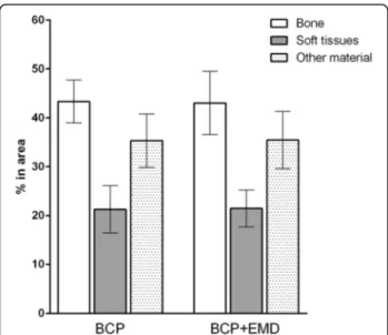
Within the ROI, mean bone area was 43.4% (SD 6.1; CI95 38.9–47.8) for the BC group and 43.0% (SD 9.0;
Fig. 1 Histomicrograph illustrating the various tissue areas measured on the sections: newly formed bone (green mask), soft tissues (purple mask), and“others”, including residual bone substitute particles and empty spaces either due to removal of the bone substitute particles during to the decalcification processing or due to artifacts (white mask)
CI95 36.6–49.5) for the BC + EMD group. The mean soft tissue area was 21.3% (SD 6.8; CI95 16.5–26.2) for BC group and 21.5% (SD 5.3; CI95 17.7–25.3) for BC +
EMD group. The mean area of “other material” was
35.3% (SD 9.0; CI95 36.6–49.5) for BC group, and 35.5% (SD 8.2; CI95 29.6–41.3) for BC + EMD group. The data is graphically presented in Fig. 6. No differences between the groups were found for any of the three parameters
evaluated (p value was 0.94 for bone, 0.96 for soft tissue, and 0.97 for other materials).
Discussion
The present study compared the histological and histo-morphometrical outcome of healing after maxillary sinus floor augmentation with BC with or without EMD, based on human biopsies. The results showed that
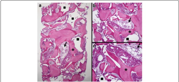
Fig. 2 Histomicrograph of a biopsy from the BC group. a Overview—×25 magnification; b ×30 magnification; c ×60 magnification. Areas corresponding to BC removed during histological processing (square) in direct contact with newly formed bone (asterisk), containing a large number of osteocytes, and with soft tissue (arrow) can be observed (hematoxylin-eosin stain)
Fig. 3 Histomicrograph of a biopsy from the BC + EMD group. Overview—×25 magnification; b ×30 magnification; c ×60 magnification. Areas corresponding to BC + EMD removed during histological processing (square) surrounded by newly formed bone (asterisk), with large numbers of osteocytes and soft tissue (arrow) can be observed. There is direct contact between the BC reminiscent, soft tissues, and vital bone (hematoxylin-eosin stain)
addition of EMD did not enhance the outcome of heal-ing, neither in terms of quality nor quantity of new bone. Nevertheless, the amount of bone generated after maxillary sinus floor augmentation with BC or BC + EMD was adequate to support successful implant place-ment and osseointegration of implants.
EMD is used for almost 20 years for enhancing tissue regeneration in periodontal defects, and it has been shown to exert anabolic action on several types of cells and factors relevant for bone regeneration [11, 12]. Nevertheless, there is still only sparse information from the clinic on the possible beneficial effect of adding EMD on a bone substitute material in terms of enhan-cing bone tissue regeneration in non-periodontal sites. In particular, a single study has previously evaluated the BC + EMD combination vs. BC in sinus lift, but due to the fact that only radiographic analysis was performed, the results were unclear [14]; thus, the present study, including histological evaluation, was performed. After 6 months of healing, about 43% of the evaluated part of
the biopsy consisted of newly formed mineralized bone and about 35% consisted of grafting material; no differ-ences between the groups were observed also in regard to bone tissue organization and maturation, as revealed by analysis of birefringence. Herein, only the 6 mm to-wards the apical aspect of the 10-mm-long biopsies was considered as the region of interest (ROI), in order to minimize any influence on the results from counting aspects of the alveolar ridge present before surgery.
Aiming to enhance bone formation and bone quality when bone substitute materials such as BC are used, biologics have often been added and positive results have occasionally been observed [16, 17]. The possibility that absence of any beneficial effect of EMD on bone regen-eration herein was due to the sterile physiological saline solution added to the graft material mixture to facilitate its handling and transferring into the sinus, cannot be excluded. Indeed, the saline solution may have either diluted the concentration of EMD necessary to exert a beneficial effect or it may have interfered with adequate
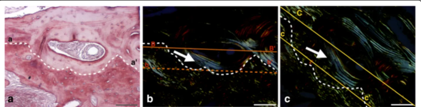
Fig. 4 Histomicrograph of a biopsy from the BC group, showing an aspect of newly formed bone. Section stained with picrosirius-hematoxylin and digitalized with bright-field (a) and linearly polarized light (b and c). b, c Results of near 45° section rotation (between axes B–B’ and C–C’) to compensate some of the orientation-related effects associated with linearly polarized light. In a, typical Haversian systems are showed (area observed above dotted line, a to a’). In b and c, the arrows indicate thin birefringent collagen bundle (appearing as bright lines) arranged around Haversian canals, suggestive of mature lamellar bone. The area observed below the dotted line is suggestive of immature (non-lamellar) bone, where collagen fibers undulations can be observed. The dark area corresponds to complete disorganization of the collagen fibers. Bar = 100μm
Fig. 5 Histomicrograph of a biopsy from the BC + EMD group, showing an aspect of newly formed bone. Section stained with picrosirius-hematoxylin and digitalized with bright-field (d) and linearly polarized light (e and f). e, f Results of near 45° section rotation (between axes B–B’ and C–C’) to compensate some of the orientation-related effects associated with linearly polarized light. In d, typical Haversian systems are showed (area observed above dotted line, a to a’). In e and f, the arrows indicate thin birefringents (appears visually as brilliance) collagen arrangement around Haversian canals suggestive of lamellar mature bone. Areas observed below dotted lines are suggestive of immature (non-lamellar) bone where collagen fiber undulations can be observed. The dark area corresponds to complete disorganization of the collagen fibers. Bar = 100μm
adsorption of EMD on the BC particles, resulting in altered (reduced) presence of EMD on the site during healing. In fact, in an in vitro study, published after the clinical procedures of the present study were concluded, it was shown that best adsorption of EMD on bone substi-tute particles is achieved when particles are dry and EMD is allowed to adsorb for at least 5 min. Further, in that study it was shown that inadequate adsorption of EMD on the bone substitute particles had negative influences in osteoblast proliferation and differentiation [14, 15, 18].
Nevertheless, the amount of bone generated with BC or BC + EMD herein was adequate to support successful im-plant placement and osseointegration of imim-plants. In fact, more or less similar amounts of bone formation have been reported in studies evaluating human sinus biopsies after grafting with a variety of biomaterials (bone formation ranging approximately from 30 to 50%) [19]. On the other hand, an ideal situation would be that BC becomes grad-ually resorbed and completely substituted by vital bone tissue [8]. A few studies have indeed showed that biologics accelerate the degradation of biomaterials and conse-quently lead to larger bone formation at the grafted region [20, 21]. However, in the present study, EMD did not seem to influence graft remodeling in that manner. The possible biological and biomechanical long-term chal-lenges of a loaded implant inserted in largely non-vital BC-grafted bone sites remain unknown. Recent studies, in fact, indicate high failure rates of implants inserted in sites augmented laterally and/or vertically with fresh-frozen allogeneic bone blocks [22], a material that remains largely necrotic for several months, despite good clinical graft incorporation [22–24]. In perspective, high long-term im-plant survival rates are reported after sinus augmentation with a variety of bone substitute materials [25].
Conclusions
The present study showed that maxillary sinus floor aug-mentation with BC resulted in adequate amounts of new bone formation allowing successful implant installation, while adding EMD did not have a significant effect.
Authors’ contributions
JCN, LAVDP, GFG, and RSN performed the experiments and data analysis. CRS, FMGF, RSN, and AS conceived and designed the study, performed the experiments, and wrote the manuscript. CRS, RSN, and AS participated in the manuscript preparation. All authors read and approved the final version of the manuscript.
Competing interests
James Carlos Nery, Luís Antônio Violin Dias Pereira, George Furtado Guimarães, Cassio Rocha Scardueli, Fabiana Mantovani Gomes França, Rubens Spin-Neto, and Andreas Stavropoulos declare that they have no competing interests.
Publisher’s Note
Springer Nature remains neutral with regard to jurisdictional claims in published maps and institutional affiliations.
Author details
1Department of Implantology, São Leopoldo Mandic Research Center,
Brasília, DF, Brazil.2Department of Biochemistry and Tissue Biology, UNICAMP
– State University of Campinas, Institute of Biology, Campinas, São Paulo, Brazil.3Department of Periodontology, UNESP– Univ. Estadual Paulista,
Araraquara Dental School, Araraquara, São Paulo, Brazil.4Department of
Dentistry and Oral Health– Oral Radiology, Aarhus University, Aarhus, Denmark.5Department Periodontology– Faculty of Odontology, Malmö
University, Malmö, Sweden.6Implant Center, SEPS 710/910, Lotes CD, Office
226, CEP: 70390-108 Brasília, DF, Brazil.
Received: 8 December 2016 Accepted: 22 April 2017
References
1. Esposito M, Felice P, Worthington HV. Interventions for replacing missing teeth: augmentation procedures of the maxillary sinus. Cochrane Database Syst Rev. 2014;17:CD008397.
2. Jungner M, Cricchio G, Salata LA, Sennerby L, Lundqvist C, Hultcrantz M, et al. On the early mechanisms of bone formation after maxillary sinus membrane elevation: an experimental histological and
immunohistochemical study. Clin Implant Dent Relat Res. 2015;17:1092–102. 3. Boyne PJ, James RA. Grafting of the maxillary sinus floor with autogenous
marrow and bone. J Oral Surg. 1980;38:613–6.
4. Frenken JW, Bouwman WF, Bravenboer N, Zijderveld SA, Schulten EA, ten Bruggenkate CM. The use of Straumann bone ceramic in a maxillary sinus floor elevation procedure: a clinical, radiological, histological and histomorphometric evaluation with a 6-month healing period. Clin Oral Implants Res. 2010;21:201–8.
5. Wallace SS, Froum SJ. Effect of maxillary sinus augmentation on the survival of endosseous dental implants. A systematic review. Ann Periodontol. 2003;8:328–43.
6. Galindo-Moreno P, Padial-Molina M, Fernandez-Barbero JE, Mesa F, Rodriguez-Martinez D, O’Valle F. Optimal microvessel density from composite graft of autogenous maxillary cortical bone and anorganic bovine bone in sinus augmentation: influence of clinical variables. Clin Oral Implants Res. 2010;21:221–7.
7. Nkenke E, Stelzle F. Clinical outcomes of sinus floor augmentation for implant placement using autogenous bone or bone substitutes: a systematic review. Clin Oral Implants Res. 2009;20 Suppl 4:124–33. 8. Stavropoulos A, Becker J, Capsius B, Acil Y, Wagner W, Terheyden H.
Histological evaluation of maxillary sinus floor augmentation with recombinant human growth and differentiation factor-5-coated beta-tricalcium phosphate: results of a multicenter randomized clinical trial. J Clin Periodontol. 2011;38:966–74.
Fig. 6 Histomorphometric evaluation results (considering six sections for each biopsy), for newly formed bone, soft tissues, and others
9. Stavropoulos A, Windisch P, Gera I, Capsius B, Sculean A, Wikesjo UM. A phase IIa randomized controlled clinical and histological pilot study evaluating rhGDF-5/beta-TCP for periodontal regeneration. J Clin Periodontol. 2011;38:1044–54.
10. Schmitt CM, Doering H, Schmidt T, Lutz R, Neukam FW, Schlegel KA. Histological results after maxillary sinus augmentation with Straumann(R) BoneCeramic, Bio-Oss(R), Puros(R), and autologous bone. A randomized controlled clinical trial. Clin Oral Implants Res. 2013;24:576–85.
11. Miron RJ, Sculean A, Cochran DL, Froum S, Zucchelli G, Nemcovsky C, et al. Twenty years of enamel matrix derivative: the past, the present and the future. J Clin Periodontol. 2016;43:668–83.
12. Sculean A, Nikolidakis D, Nikou G, Ivanovic A, Chapple IL, Stavropoulos A. Biomaterials for promoting periodontal regeneration in human intrabony defects: a systematic review. Periodontol 2000. 2015;68:182–216. 13. Wyganowska-Swiatkowska M, Urbaniak P, Nohawica MM, Kotwicka M,
Jankun J. Enamel matrix proteins exhibit growth factor activity: a review of evidence at the cellular and molecular levels. Exp Ther Med. 2015;9:2025–33. 14. Favato MN, Vidigal BC, Cosso MG, Manzi FR, Shibli JA, Zenobio EG. Impact
of human maxillary sinus volume on grafts dimensional changes used in maxillary sinus augmentation: a multislice tomographic study. Clin Oral Implants Res. 2015;26:1450–5.
15. Boeck-Neto RJ, Gabrielli M, Lia R, Marcantonio E, Shibli JA, Marcantonio Jr E. Histomorphometrical analysis of bone formed after maxillary sinus floor augmentation by grafting with a combination of autogenous bone and demineralized freeze-dried bone allograft or hydroxyapatite. J Periodontol. 2002;73:266–70.
16. Alam I, Asahina I, Ohmamiuda K, Enomoto S. Comparative study of biphasic calcium phosphate ceramics impregnated with rhBMP-2 as bone substitutes. J Biomed Mater Res. 2001;54:129–38.
17. Alam MI, Asahina I, Ohmamiuda K, Takahashi K, Yokota S, Enomoto S. Evaluation of ceramics composed of different hydroxyapatite to tricalcium phosphate ratios as carriers for rhBMP-2. Biomaterials. 2001;22:1643–51. 18. Miron RJ, Bosshardt DD, Hedbom E, Zhang Y, Haenni B, Buser D, et al.
Adsorption of enamel matrix proteins to a bovine-derived bone grafting material and its regulation of cell adhesion, proliferation, and differentiation. J Periodontol. 2012;83:936–47.
19. Friedmann A, Dard M, Kleber BM, Bernimoulin JP, Bosshardt DD. Ridge augmentation and maxillary sinus grafting with a biphasic calcium phosphate: histologic and histomorphometric observations. Clin Oral Implants Res. 2009;20:708–14.
20. Koo KT, Susin C, Wikesjo UM, Choi SH, Kim CK. Transforming growth factor-beta1 accelerates resorption of a calcium carbonate biomaterial in periodontal defects. J Periodontol. 2007;78:723–9.
21. Wikesjo UM, Sorensen RG, Kinoshita A, Wozney JM. RhBMP-2/alphaBSM induces significant vertical alveolar ridge augmentation and dental implant osseointegration. Clin Implant Dent Relat Res. 2002;4:174–82.
22. Carinci F, Brunelli G, Franco M, Viscioni A, Rigo L, Guidi R, et al. A retrospective study on 287 implants installed in resorbed maxillae grafted with fresh frozen allogenous bone. Clin Implant Dent Relat Res. 2010;12:91–8.
23. Spin-Neto R, Stavropoulos A, Coletti FL, Faeda RS, Pereira LA, Marcantonio Jr E. Graft incorporation and implant osseointegration following the use of autologous and fresh-frozen allogeneic block bone grafts for lateral ridge augmentation. Clin Oral Implants Res. 2014;25:226–33.
24. Spin-Neto R, Stavropoulos A, Coletti FL, Pereira LA, Marcantonio Jr E, Wenzel A. Remodeling of cortical and corticocancellous fresh-frozen allogeneic block bone grafts–a radiographic and histomorphometric comparison to autologous bone grafts. Clin Oral Implants Res. 2015;26:747–52. 25. Corbella S, Taschieri S, Del Fabbro M. Long-term outcomes for the
treatment of atrophic posterior maxilla: a systematic review of literature. Clin Implant Dent Relat Res. 2015;17:120–32.
Submit your manuscript to a
journal and benefi t from:
7 Convenient online submission 7 Rigorous peer review
7 Immediate publication on acceptance 7 Open access: articles freely available online 7 High visibility within the fi eld
7 Retaining the copyright to your article