Interobserver variability of
peak systolic velocity
measurements in
the common carotid artery
PAPER WITHIN Biomedical science, clinical physiology AUTHORS: Ewelina Antonijev & Frida Åstrand TUTOR: Rachel de Basso, PhD at Jönköping University
EXAMINER: Magnus Karlsson, PhD at Jönköping University JÖNKÖPING May 2016
Summary
Stroke is ranked as the number one cause of death in Vietnam and the second leading cause of death in the world. Stenosis in the carotid arteries is one of the major risk factors for stroke, making it extremely important to confirm the diagnosis. Duplex ultrasound including measurements of the peak systolic velocity is considered the most accurate method when it comes to screening of carotid stenosis. However, the survey method is user dependent and differences in the results related to the scanning technique of the examiner may occur. The aim of this study was to determine the diagnostic variability for measuring peak systolic velocity in the common carotid artery in order to investigate whether the obtained results differ depending on the examiner. 30 healthy volunteers underwent carotid duplex ultrasound performed by two different examiners. Peak systolic velocity was measured with pulsed waved Doppler caudally of the carotid bifurcation. A T-test and a Bland-Altman plot were later applied in order to determine interobserver variability between the examiners. Results showed no significant difference in the measurements, with a mean difference of -0.01 m/s. Despite low interobserver variability, reliable conclusions cannot be drawn from the results since both examiners lacked adequate experience.
Table of contents
Background ... 1
Cerebrovascular disease ... 1
Anatomy of the carotid arteries ... 2
Duplex ultrasound ... 2
Doppler Techniques ... 3
Duplex examination of the carotid arteries ... 4
Importance of accurate diagnosis ... 5
The significance of implementing this study in Vietnam ... 5
Study aim ... 6
Material and method ... 7
Design ... 7 Equipment requirements ... 7 Procedure ... 8 Statistical analysis ... 10 Ethical considerations ... 11
Results ... 12
Discussion ... 15
Cardiovascular disease and hypertension ... 17
Cardiovascular disease and BMI ... 18
Ergonomics ... 20
Conclusions ... 21
Acknowledgements ... 21
References ... 22
Appendix 1 -
Questionnaire/Phiếu thăm dòAppendix 2 -
Jönköping University Ethical Consideration for Student's ThesisAppendix 3 -
Letter of consent to participants1
Background
Cerebrovascular disease
Cerebrovascular disease can rapidly cause disruptions of brain function caused by vascular abnormalities. Stroke is caused by either cerebral infarction (arterial occlusion) or hemorrhage in the brain. This causes a blocked blood flow to the brain, resulting in hypoxia and damaged brain tissue (Ericson & Ericson, 2012).
Stroke is the second leading cause of death in the world and it is expected to continue keeping this ranking in 2030 (Mathers & Loncar, 2006). Stroke is also the most common cause of severe disability (Lännergren, Westerblad, Ulfendahl, & Lundeberg, 2012).
In several Asian countries, Vietnam amongst them, stroke is ranked as the number one cause of death. In the year of 2009, the number of deaths caused by stroke constituted 15.9% of the total amount of deaths in Vietnam (Hoy, Rao, Hoa, Suhardi & Lwin, 2013). Vietnam is undergoing an epidemiological transition, which means that developmental changes happen due to increased economic status, population growth and innovation in medical treatments. Deaths due to non-contagious diseases have increased rapidly during the last 20 years, due to this transition. In the year of 2010, morbidity from non-contagious diseases was three times higher than the morbidity of contagious diseases. This changing of the Vietnam disease profile can be explained by the increase in life expectancy. When an individual lives longer, the risk of cardiovascular disease increases due to prolonged exposure of risk factors (Anh Ha, Goldberg, Allison, Hong Chu & Nguyen, 2013).
Atherosclerosis, i.e. calcification of arteries, is the main cause of stroke and other
cardiovascular diseases. Pieces of calcified plaque can come loose from the carotid arteries, travel with the blood flow and get stuck as embolisms in small arteries in the brain. This causes restricted blood flow in the affected part of the brain, which leads to hypoxia and stroke. Stenosis in a major vessel supplying the brain with blood causes no symptoms until the diameter is reduced by about 75%, which is the reason why atherosclerosis in large
arteries seldom is the direct cause of stroke. However, pieces of plaque from these arteries can come loose and clog smaller vessels leading to brain ischemia. Stroke mostly affects
individuals with calcified vessels, since both arterial occlusion and bleeding often occur in vessels with atherosclerotic changes (Lännergren et al., 2012).
2 Anatomy of the carotid arteries
The brachiocephalic trunk originates from the aortic arch and divides into the right subclavian artery and the right common carotid artery (CCA). The right CCA then divides into the right internal carotid artery (ICA) and the right external carotid artery (ECA). On the left side, the left subclavian artery and the left CCA originates directly from the aortic arch. The left CCA then divides into the left ICA and the left ECA. The vein that normally closely follows the CCA is the internal jugular vein, which drains deoxygenated blood from the head, via the superior vena cava and back to the heart (Jogestrand & Rosfors, 2002).
Typically in the middle of the neck, the CCA widens and forms the carotid bulb bilaterally, after which it divides into the ICA and the ECA. Generally, the ICA is located postero-laterally and the ECA is located antero-medially [Fig. 1], although variances is common. The ICA usually does not branch out, unlike the ECA that divides into smaller vessels which extracranially supply blood to the face and the scalp. The ICA continues intracranially via the oval foramen. The opthalmic artery originates from the ICA, after which the ICA divides into the middle and anterior cerebral arteries, which are parts of the circle of Willis. The circle of Willis supplies blood to the brain and its surrounding structures (Jogestrand & Rosfors, 2002).
In case of severe obstruction in the arteries of the neck, several collateral blood flows may form. Possible variances include for example collaterals from the opthalmic artery originating from the ICA or collaterals from the vertebral artery, which originates bilaterally from the subclavian arteries (Jogestrand & Rosfors, 2002).
Duplex ultrasound
Duplex ultrasound involves an ultrasound with simultaneous Doppler imaging, and is a frequently used survey method to investigate stenosis in the carotid arteries leading to the brain. Duplex ultrasound combines ultrasound that provides a visual two dimensional image of the carotid arteries morphology, with Doppler imaging that provides information about the current blood flow (Jogestrand & Rosfors, 2002).
When the ultrasonic wave emitted during a duplex examination encounters a blood flow or any other movable structure in the vessel, the ultrasound will be reflected and the reflected ultrasound will have a frequency different from the emitted ultrasonic frequency. The
3
difference in frequency that occurs is called the Doppler shift and it is the registration of this Doppler shift that allows detection of a blood flow. Information about blood flow direction may also be obtained, since the reflected ultrasound from blood flowing in the direction towards the ultrasound transducer has a higher frequency than the reflected ultrasound from blood flowing in the opposite direction of the ultrasound transducer (Jonson & Wollmer, 2011).
In addition to demonstrating blood flow and yield information about the blood flow direction, blood flow velocity can also be determined using Doppler imaging (Jonson & Wollmer, 2011). The blood flow velocity is calculated from the recorded difference in frequency and the angle between blood flow and the ultrasonic signal (Jogestrand & Rosfors, 2002). Modern appliances used in duplex examinations automatically calculates the measured flow velocity in meters per second from the values obtained in the two dimensional image (Jonson & Wollmer, 2011).
Doppler Techniques
There are two different Doppler techniques, the continuous wave (CW) Doppler and the pulsed wave (PW) Doppler. The CW Doppler sends and receives the reflected ultrasound signals continuously, providing registrations from all measurement depths in the direction of the signal. However, this type of Doppler does not provide any knowledge of the specific measurement depth for a single reflected signal. The CW Doppler can measure high blood flow velocities at great depths and the technique is often used for flow velocity measurements in the heart (Jonson & Wollmer, 2011).
The PW Doppler sends out short, individual signals and allows us to select the measurement depth from where registration will take place, since the ultrasonic velocity in the tissue is known [Jonson & Wollmer, 2011]. The PW Doppler is generally the one used for duplex examinations of the carotid arteries, since measurements of blood flow velocities from defined measuring depths are needed (Jogestrand & Rosfors, 2002). Unlike the CW Doppler, the PW Doppler lacks ability to measure high velocities at great depths. This however, does not cause any trouble when performing ultrasound examinations of the carotid arteries since these arteries are located superficially (Jonson & Wollmer, 2011).
4 Duplex examination of the carotid arteries
The information of the blood flow supplied by Doppler at the duplex examination allows assessment of the blood flow velocity in the main carotid branch (the CCA) and its cranial branches (the ICA and the ECA). Provided that the blood flow is unaffected, severe stenosis in these arteries will yield increased blood flow velocity in conjunction with decreasing cross-sectional area. Grading of stenosis by measuring the peak systolic flow velocity has the
highest diagnostic value in the ICA, as the ICA is the artery that supplies the brain (Jogestrand & Rosfors, 2002).
At the duplex examination, the CCA is visualized on level with the clavicle and is then followed cranially to the carotid bifurcation. The ICA and the ECA originating from the carotid bifurcation are then visualized as far up the neck as possible. Distinguishing the ICA and the ECA is of major importance before performing blood flow velocity measurements, because of the fact that changes in the vascular walls of the ECA lack diagnostic value (Jogestrand & Rosfors, 2002).
Differences in the appearance of the Doppler signal is used for the distinguishing of the ICA and the ECA, since the peripheral resistance of their vascular supply areas vary. The ICA supplies the brain which is a low resistance area, which gives a less pulsatile flow and a smaller difference in velocity between the systolic and diastolic flow. The ECA supplies the scalp and the face, which are regions with high peripheral resistance. This results in a more pulsatile flow and a distinct difference in velocity between systolic and diastolic flow (Jogestrand & Rosfors, 2002). In addition to the variations detected in the Doppler signals, there are other typical differences between the two arteries that facilitate the identification. For instance the ECA has branches unlike the ICA, while the ICA usually is the wider of the two arteries (Jogestrand & Rosfors, 2002).
Blood flow velocity measurements with PW Doppler is always performed in longitudinal section with combined color Doppler when an increased velocity causes distortion of the color Doppler signal, which means that the peak systolic velocity (PSV) can be derived with the Doppler sample volume. Although measurements of the peak systolic flow velocity have the highest diagnostic value in the ICA, measurements are also performed in the CCA since the ratio between the PSV in the CCA and the ICA is an important parameter. A ratio between
5
systolic blood flow velocity in the CCA and the ICA exceeding 4 indicates severe stenosis with >80 % vessel diameter reduction (Jogestrand & Rosfors, 2002).
Importance of accurate diagnosis
The increase of PSV is a parameter of great value for diagnosis, making it extremely important to get accurate and correct readings. A PSV exceeding 1.25 m/s suggests that the diameter of the vessel is reduced by at least 50% due to stenosis, and a PSV higher than 4 m/s suggests severe stenosis. Severe stenosis of the carotid arteries can reduce blood flow to the brain, which then can cause symptoms from lack of oxygen in the brain (Jonson & Wollmer, 2011).
Blood flow velocity is calculated based on the registered Doppler shift and the angle between blood flow and the ultrasonic signal direction [Jogestrand & Rosfors, 2002]. The values obtained are dependent of the individual performer's scanning technique and management of the examination. The positioning of the Doppler marker, the selection of measuring area and the incident angle, are all important factors for the velocity measured. If scanning technique is handled differently between observers there is a risk that the values obtained will differ, which can then lead to misdiagnosis (Thomson, Woods, Lannos & Sage, 2001).
How the results vary in relation to various performers is described as interobserver variability. Equivalent results regardless of the performer provides a low interobserver variability and thus a reliable method. If the collected data cannot be reproduced, they should be discarded (Corrivaeu & Johnston, 2004; de Oliviera, Moreira, Velarde, Monterio & Netto, 2011).
The significance of implementing this study in Vietnam
Today, the development of technology in medical diagnostics goes rapidly forward. Apparatus and survey methodology is constantly improving and becoming more
sophisticated. There are roughly 91 million inhabitants in Vietnam. The high number of inhabitants and the increase in life expectancy means that large diagnostic needs will need to be fulfilled as modern and more advanced technology becomes available (World Health Organization, 2015). With rapid development follows improved diagnostics, though it may also mean that quality assurance such as controls in relation to performance may be sidelined, making the performing of this study interesting.
6
Since Da Nang University of Medical Technology and Pharmacy in Da Nang, Vietnam, was recently appointed a university (in 2013), performing this study there will also serve an academic purpose. The students at the university will hopefully get a chance to get inspired and learn more of how to perform a methodological study, knowledge which they will be able to use for their own bachelor theses in the future. The teachers at the university will have to supervise these students when they perform studies of their own and so they might also benefit and learn from the experience of the performing of this study at their university.
Study aim
The aim of this study was to determine the diagnostic variability for measuring PSV in the CCA using the ultrasound technique and to investigate whether obtained results differ depending on the examiner.
The null hypothesis of this study was that there is no significant difference in ultrasound measurements of PSV between the two examiners.
7
Material and method
Design
This quantitative observational study was made in order to investigate interobserver variability of PSV measurements in the CCA.
The study sample consisted of 30 students at Da Nang University of Medical Technology and Pharmacy, Da Nang, Vietnam. The participants were young and healthy subjects without known cardiovascular disease and no ongoing medication or other treatment. All subjects were participating on a voluntary basis. In the event that pathological abnormalities would be detected during any of the examinations, a supervising physician was available during every examination.
Participants with arrhythmia or established hypertension were excluded from the study. Inability with one or both examiners of acquiring a measurable view of the CCA was also considered an exclusion criterion.
Equipment requirements
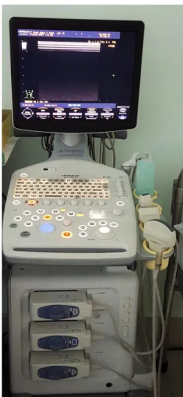
Blood pressure was measured using Omron M3 automatic blood pressure monitor, manufactured by Omron Healthcare.The ultrasound equipment that was used during all examinations was a machine manufactured by Hitachi Aloka, model F37, made in Japan [Fig. 1]. A gel improving the acoustic impedance by minimizing the amount of air between the transducer and the patient's skin, was used at each examination. A high frequency, linear array ultrasonic transducer (model UST-5413) with the frequency range of 5-13 MHz was used at every ultrasound examination.
8
Fig. 1. Ultrasound machine (Hitachi Aloka model F37) that was used.
Procedure
Upon arriving at the Medical Diagnostics Centre of the University in Da Nang, Vietnam, the participants received a questionnaire containing questions about age, sex, smoking habits, health status and current medications [Appendix 1]. They answered the questionnaire on their own and had their height and weight measured by a nurse working for the Medical
Diagnostics Centre. Upon entering the examination room, the participant got to rest lying down,quietly, for 10 minutes. A timer was used to ensure correct resting duration. After resting, heart rate and blood pressure were measured on the right side in supine position, using a validated oscillometric sphygmomanometer.
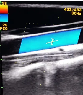
The ultrasound examinations were then performed in a separate room. The ultrasound measurements were made in the CCA. The CCA was visualized in cross section view and then followed all the way up to the carotid bifurcation. Once the bifurcation was visible, the probe was turned in order to show the carotid arteries in longitudinal section view. Color Doppler was used in order to acquire visualization of the blood flow. The color box beam steer was set to 15°in order for it to follow the direction of the blood flow in the vessel. PSV was then measured with PW Doppler in the CCA, approximately two to four centimeters caudally of the carotid bifurcation. PW Doppler angle of 60° was implemented and the PW
9
Doppler sample volume was set to two millimeters. The cursor was placed in the center of the vessel, with the angle cursor as parallel as possible to the lumen of the vessel [Fig. 2].
Fig. 2. Ultrasound picture showing the CCA with color Doppler and cursor angle of 60°.
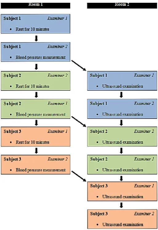
The blood flow measurements were at all ultrasound examinations performed on the right side of the neck and the participants were lying down during the procedure. Each participant was examined by two separate examiners. The first examiner was to find the measurement point and use the PW Doppler to measure PSV in the CCA. A total of three separate PSV values were measuredand thereafter a mean value of these three measurements was obtained [Fig. 3]. The ultrasound procedure was then repeated by the second examiner [Fig. 4]. Maximum duration of each separate ultrasound examination was set to be 10 minutes and a timer was used to ensure that the time frame was kept.
10
Fig. 4. Flow chart of the course of procedure.
Statistical analysis
For statistical analysis and calculations, the IBM software programme Statistical Package for the Social Sciences (SPSS) was used. Microsoft Office Excel 2007 was used for the making of tables. Descriptive statistics calculations were performed in SPSS for the obtaining of demographic data of the study population [Table 1; Table 2]. Three scatter plots were made in order to investigate whether or not this study showed a correlation between PSV and
11
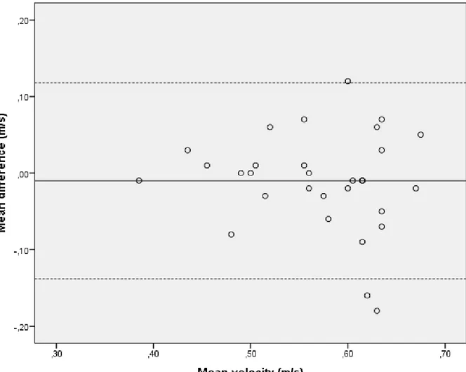
was normally distributed, after which a paired sample T-test (significance interval 95%, α = 0.05) was performed to determine whether or not to reject the null hypothesis. A Bland-Altman plot was applied to determine interobserver variability between the two examiners in measuring PSV in the CCA [Fig. 6].
Ethical considerations
In this study, the "Jönköping University Ethical Consideration for Student's Thesis" was followed [Appendix 2].
Prior to the data collection, all participants received clear information about the purpose of the study and how the examinations were to be performed. The survey methodology was
noninvasive and completely painless. This information was communicated through a form, through which the participants also left their written, informed consent. On this form, the participants were given the opportunity to submit their e-mail address for receiving a personal copy of the finished report of the study. The information was beforehand translated into the participants' native language, Vietnamese, to ensure avoidance of misunderstandings [Appendix 3; Appendix 4].
The measured variables and collected information about each participant was treated
confidentially. Coding of identities, using numbers instead of names, ensured that the results could not be linked to specific individuals. Because of language barriers, the participants may have felt some level of discomfort from uncertainty about what they were supposed to do. Difficulties understanding the instructions, particularly those regarding that they were supposed to rest alone for 10 minutes, may possibly have caused some participants to feel uneasy.
All participation was voluntary and the participants could choose to terminate their
12
Results
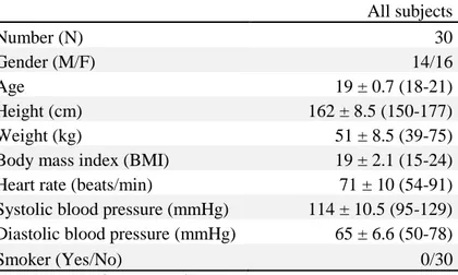
The study sample consisted of a total of 30 participants (14 male, 16 female), ages 18-21 years. All 30 participants completed the questionnaire and the ultrasound examinations, thereby not causing any loss of sample. All of the participants were students at Da Nang University of Medical Technology and Pharmacy, Da Nang, Vietnam. Complete
demographics including gender, age, height, weight, BMI, heart rate (HR), systolic blood pressure (SBP), diastolic blood pressure (DBP) and cigarette smoking are presented in Table 1.
Table 1. Descriptive demographics of the subjects participating in this study.
All subjects Number (N) 30 Gender (M/F) 14/16 Age 19 ± 0.7 (18-21) Height (cm) 162 ± 8.5 (150-177) Weight (kg) 51 ± 8.5 (39-75) Body mass index (BMI) 19 ± 2.1 (15-24) Heart rate (beats/min) 71 ± 10 (54-91) Systolic blood pressure (mmHg) 114 ± 10.5 (95-129) Diastolic blood pressure (mmHg) 65 ± 6.6 (50-78)
Smoker (Yes/No) 0/30
Data is presented as mean ± SD (range)
Table 2 presents mean, maximum and minimum PSV values measured by the two examiners. A mean difference in measurements between the two examiners is also shown in Table 2. Since none of the participants had high blood pressure [Table 1], ≥140/90 mmHg (World Health Organization, 2013), no correlation between high blood pressure and PSV could be detected. All of the participants had normal heart rates (beats/min). A scatter plot showed that the heart rates of the participants close to the limits of bradycardia, ≤50 beats/min, and
13
Table 2. Paired sample overview of measured common carotid artery peak systolic velocities (m/s) between the two examiners.
Number (N) Minimum Maximum Mean SD
Examiner 1 30 0.38 0.7 0.56 0.8
Examiner 2 30 0.39 0.72 0.58 0.8
Mean difference* 30 0.0 -0.18 -0.01 0.06
*Mean difference = negative value meaning examiner 1 obtained the lower measurement value compared to examiner 2.
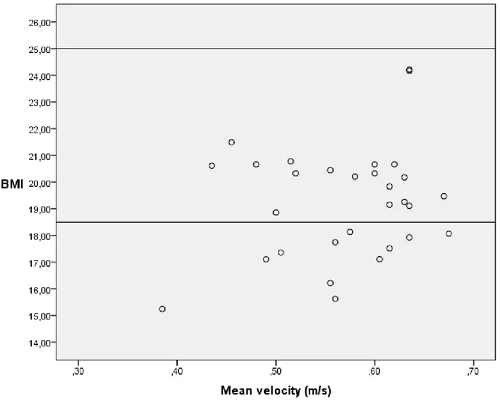
This study showed no correlation between decreased or increased PSV and low body mass index (BMI). The scatter plot representing this comparison showed similar distribution of mean PSV values in all individuals, including those with low BMI and normal BMI [Fig. 5].
Fig. 5. Scatter plot showing comparison between BMI and mean PSV in the CCA. The reference lines respectively represent low BMI (<18.5) and high BMI (≥25). N = 30.
14
A paired sample T-test did not show a significant difference between the two examiners (P-score = 0.355 > α = 0.05). Thereby, the null hypothesis claiming that there was no significant difference in PSV measurement values between the two examiners was not rejected. The mean difference between the two examiners was -0.01 m/s [Table 2; Fig. 6]. None of the two examiners measured PSV systematically lower or higher, the differences were random. In 53% and 47% of the cases respectively, examiner one and examiner two obtained the lower PSV measurement value [Table 2].
Fig. 6. Bland Altman-plot showing interobserver variability in PSV measurements in the CCA between the two examiners. The middle line shows the mean difference (-0.01 m/s) and the two dotted lines show the limits of agreement, in between which 95% of the study population is located. N = 30.
15
Discussion
Performing this methodological study has helped improving the authors' sense of critical thinking. When working with advanced technology it is of outmost importance to know not to blindly trust all obtained measurements, but to assess them with certain caution. Even though the results in this particular study did not show significant differences between the two examiners, despite the fact that they were both inexperienced, the authors' have become more aware of the importance of critical thinking when it comes to this kind of examinations. Cautious assessments are necessary for the assurance of diagnostic confidence and patient safety. Therefore, development of one's critical thinking skills is of great value when leading a professional career as a biomedical scientist (Institutet för biomedicinsk laboratorievetenskap, 2016).
The aim of this study was to determine the interobserver variability for measuring PSV in the CCA with ultrasound. The results of this study showed no significant difference in the
measurements between the two examiners [Fig. 6]. However, there are limitations to this study making it important to use caution when drawing conclusions from these results.
Duplex scanning of the carotid arteries, including measurements of the PSV, is considered the most accurate imaging method when it comes to screening, follow up and preoperative
assessment of carotid stenosis (Corrivaeu & Johnston, 2004). Yet, it is important to know that ultrasound duplex of the carotid arteries is a complex type of examination that demands a certain amount of skill in the examiner. The greatest disadvantage with this type of examination is that it is user dependent and that it requires extensive practice and skill to acquire satisfying results (Jogestrand & Rosfors, 2002). Since this study was not conducted in order to investigate pathological conditions, measurements of the PSV were not performed in the ICA, but only in the more easily localized CCA, to minimize error sources. Corrivaeu and Johnston (2004), claims that the interobserver variability between different examiners needs to be low to ensure reliable measurements.
There are several factors that can cause errors in the PSV measurements, not least errors related to the technique of the examiner such as experience, selected size of the sample volume and angle of insonation (Corrivaeu & Johnston, 2004). Even though the results in this
16
study only varied minimally between the two examiners, it is not proof that the values
obtained were actually correct since the two examiners did not have the extensive experience that is pointed out to be necessary to ensure reliable measurements when performing
ultrasound duplex examinations of the carotid arteries. An option for reducing this uncertainty concerning the measured PSV values could have been to let an experienced examiner make measurements for the study in addition to the two inexperienced examiners, thereby reducing the concerns about whether or not the obtained results were in fact accurate.
The angle of insonation between the flow direction and the direction of the Doppler beam is an important parameter to obtain precise measurements of the velocity. An angle greater than 60° is a source of error that leads to unacceptably large measurement errors and should not be used. Depending on the physical properties of Doppler, measurements made with an angle greater than 60° will result in inaccurate readings even when the appropriate angle adjustment is used (Tola & Yurdakul, 2006; Jogestrand & Rosfors, 2002).
In the present study, all measurements were performed with a fixed 60° angle of insonation. The angle was automatically calculated by the machine. To ensure the correct angle, the cursor was positioned parallel to the vessel wall in each examination. However, the blood flow is not always necessarily parallel to the vessel wall, and the angle of insonation is a factor that is related to the scanning technique of the separate examiner. This meaning that even if the cursor is parallel to the vessel wall, it might not be parallel to the blood flow (Corrivaeu & Johnston, 2004). There are different opinions regarding what angle that should be used in Doppler velocity measurements. Some consider that measurements should be performed at an angle of exactly 60°, while others believe that measurements can be
performed with an angle less than or equal to 60°. According to Tola & Yurdakul (2006) there are significant differences in results from Doppler velocity measurements made in the ICA at different fixed insonation angles. They maintain that standardized angle-specific thresholds should be applied in grading of stenosis.
Corrivaeu & Johnston (2004) believes that standardized duplex scanning machine settings, such as transducer aperture size, gain and sample volume is required in order to isolate error sources. In the present study, all examinations were performed with the same equipment and with the same standardized machine settings. Furthermore, the two examiners had the same amount of experience and all the examinations were performed at the same anatomical
17
location on the right side of the neck. These factors mentioned enhance the reliability of this study.
Cardiovascular disease and hypertension
It is generally known that hypertension increases the risk of stroke since high blood pressure causes increased arterial wall stress and atherosclerosis. Agunloye & Owolabi (2014) argues that it is desirable to investigate additional risk factors for stroke apart from hypertension itself and highlights the link between hypertension and carotid sonographic parameters. For example, hypertension has been found to correlate positively with a larger diameter of the CCA lumen (Agunloye & Owolabi, 2014) but is also believed to be one of several
contributing factors to an increased carotid intima media thickness (Zhang, Feng, Chen, Geng & Xu, 2014).
Whether the carotid blood flow velocity is affected by blood pressure has also been
investigated. Agunloye & Owolabi (2014) found a reduction in both PSV and end diastolic velocity (EDV) in the CCA and the proximal ICA among hypertensive patients, however the authors claim that only the reduction seen in EDV is significantly associated with an
increased stroke risk.
Correlation regarding blood pressure and PSV in the CCA cannot be seen in the present study, since blood pressure outside the normal limits could not be detected in any of the examined subjects. The fact that only the PSV was measured also makes it difficult to draw any
conclusions regarding a correlation between blood pressure and CCA blood flow velocities.
Another limitation to this study was the fact that the participants were supposed to rest quietly, lying down for 10 minutes before having their blood pressure measured. Due to misunderstandings that emerged, some of the participants did not rest consistently during the time that was deposited, which may have affected the measurement results. However, despite this error source, none of the participants exhibited hypertension [Table 1] although some of them did fit in the category of prehypertension.
Hypertension is defined as 140/90 mmHg and prehypertension is defined as 120/80 mmHg (World Health Organization, 2013). If the systolic and diastolic pressures happen to belong in different groups, the categorization is always decided by the highest blood pressure value
18
(Lindgärde, Thulin & Östergren, 2009). In our study, none of the participants had diastolic pressure exceeding 80 mmHg but 12 out of 30 participants had systolic pressure exceeding 120 mmHg. According to Lindgärde, Thulin & Östergren (2009), this puts these individuals in the prehypertension category.
In 2014 another bachelors' thesis conducted at the Da Nang University of Medical
Technology and Pharmacy, Da Nang, Vietnam, showed that 4 % of students had hypertension and 25 % had prehypertension (Shafie & Tran, 2014). The present study showed no
participants with hypertension and 40 % (n: 12) with prehypertension. Since both studies used the same population for the sample, these differences may be explained by several other factors. First of all, the study by Shafie & Tran (2014) used a larger study sample than this study (a total of 121 and 30 subjects respectively). Since the study sample in this study was small, no conclusions can be drawn regarding the larger population. It is likely that the larger sample of 121 subjects reflects the larger population more accurately. Shafie & Tran (2014) performed the blood pressure measurements on subjects in a seated position, whereas in this study blood pressure was measured in supine position. According to Cicolini et al., (2011), mean systolic pressure is generally higher in supine compared to seated position, which may also explain the higher percentage of prehypertensive participants in this study.
Cardiovascular disease and BMI
In the early stages of the atherosclerosis process, the arterial luminal diameter increases in order to compensate for the decreased vascular lumen that occurs because of plaque
formation. One study has shown a correlation between increased carotid arterial diameter and increased body mass index (BMI), in both men and women. Large arterial diameter can lead to disrupted blood flow regulation because of the fact that larger vessels have less ability to adjust to an increased blood flow. This is what makes enlargement of the arterial diameter a risk factor for continuous development of atherosclerosis (Özdemir, Artas, Serhatlioglu & Ogur, 2006).
Özdemir et al. (2006) claims that overweight, or increased BMI, increases the luminal
diameter in the arteries. In their study, the diameter was measured in the CCA and was higher in those individuals whose BMI was high compared to those with normal BMI. Another finding was that the individuals with high BMI also had lower blood flow velocity in the CCA
19
and the ICA than those with normal BMI. Özdemir et al. (2006) concludes that decreased blood flow velocity associated with increased arterial diameter can be a sign of early atherosclerosis.
The endothelium is the single cell layer that separates the blood from the rest of the body. This layer secretes vasoactive substances which regulates vascular tone and the most potent vasodilating substance is the gas nitric oxide (NO). Blockade of NO-synthase leads to vasoconstriction, which tells us that humans normally have continuous secretion of NO. Besides contributing to vasodilation, NO also contributes to the mitigation of thrombus formation and atherosclerosis (Lindgärde, Thulin & Östergren, 2009).
According to Higashi et al. (2003) this kind of endothelium-dependent vasodilatation is defective in obese individuals because of decreased NO-bioavailability. Since endothelial dysfunction is a finding in the early atherosclerosis process, this reduction of NO in the body increases the risk for cardiovascular disease (CVD). The cause of this reduction in NO bioavailability is increased oxidative stress. Indexes of oxidative stress have shown to be significantly increased in individuals with high BMI, ≥25(World Health Organization, 2000), compared to those with normal BMI. An interesting finding in the study by Higashi et al. (2003) was that not only those with high BMI but individuals with low BMI, <18.5 (World Health Organization, 2000), also had increased indexes of oxidative stress, showing that low BMI may also be a risk factor for CVD.
In the present study, none of the participants had high BMI, but 11 out of 30 participants had low BMI. This study showed no correlation between low BMI and increased/decreased PSV [Fig. 5].The participant's low BMI may however possibly be putting them at risk for
developing CVD in the future because of the likelihood of them having increased indexes of oxidative stress and therefore decreased bioavailability of NO. Although the correlation between cardiovascular complications and low BMI is controversial in today's society, studies have shown a connection between increased risk of cardiovascular morbidity and low BMI (Higashi et al., 2003).
A major risk for developing atherosclerosis, and thereby CVD, is cigarette smoking. Smoking is estimated to increase the risk of stroke by two to four times in comparison to non-smokers and to quit smoking is the most important lifestyle change to adapt in order to prevent CVD
20
(Halldén et al., 2013; Lindgärde, Thulin & Östergren, 2009). Of all deaths related to CVD, about 10 % is due to cigarette smoking (Halldén et al., 2013). One of the negative effects of smoking is increased intima-media thickness (IMT). Thickening of the intima-media complex in the carotid artery has been proven to be an independent predictor for the future
development of CVD (Langham et al., 2005; Lindgärde, Thulin & Östergren, 2009). In one study, it was shown that young smokers had increased IMT in comparison to young non-smokers. This points to the fact that cigarette smoking is a risk factor for developing a thickened intima-media complex, independently of the factor of increased age (Langham et al., 2015). All of the participants in the present study claimed to be non-smokers and are therefore not at risk for developing any cardiovascular complications due to cigarette smoking [Table 1].
Ergonomics
During the ultrasound examinations, difficulties related to the work environment emerged since it was not possible to adjust the height of the examination bed. Due to the nonadjustable bed that was too low in relation to the examiners' heights, measurements were carried out under poor ergonomic conditions with too great a distance between the examiner and the individual being examined. According to Roll, Selhorst & Evans (2012), increased height of the sonographer and inappropriate bed height are both factors associated with poor
positioning while performing scans.
It is unclear whether the ergonomic conditions had any impact on the measurements obtained in the present study, however ideal ergonomic conditions are of major importance. Work-related discomfort and musculoskeletal disorders are common in the sonography profession. Evans, Roll & Baker (2009), found that 90 % of diagnostic medical sonographers and
vascular technologists felt pain during examinations. On average, sonographers hold their arm statically for 73 % of the time during an examination, and the shoulder also tends to work statically for a longer time in carotid scans compared with other scan types (Roll, Selhorst & Evans, 2012).
21
Conclusions
The null hypothesis of this study was confirmed since no significant difference in PSV measurement values between the two examiners was found. Because of the fact that the two examiners did not have the profound experience that is pointed out to be necessary for this type of ultrasound examination, the results achieved in this study cannot be used to draw any general, reliable conclusions. An option for reducing the uncertainty about the values obtained would be to let an examiner with longer experience perform measurements for the study in addition to the two inexperienced examiners, which aside from more reliable results also could lead to interesting conclusions regarding the amount of experience needed for this kind of examination.
Acknowledgements
What made it possible for us to perform this study in Da Nang, Vietnam, was the funding by Minor Field Studies (MFS). We are grateful to Thomas Ehn for helping us in the process of writing our application for the MFS-scholarship. Thanks to School of Health and Welfare and the International Office at Jönköping University for granting us this scholarship.
A special thanks to our Vietnamese supervisor Dr. Võ Hồ Quỳnh Như at Da Nang University of Medical Technology and Pharmacy in Da Nang, Vietnam, for all the help provided during our data collection. We would also like to give our thanks to our Swedish supervisor Rachel De Basso, PhD at Jönköping University, for guiding us through the process of performing this study.
22
References
Agunloye, M. A., & Owolabi, M. O. Exploring Carotid Sonographic Parameters Associated With Stroke Risk Among Hypertensive Stroke Patients Compared to Hypertensive Control. Journal of
ultrasound in medicine. 2014; 33(6). 975-83. DOI: 10.7863/ultra.33.6.975
Anh Ha, D., Goldberg, R-J., Allison, J-J., Hong Chu, T., & Nguyen, H-L. Prevalence, Awareness, Treatment, and Control of High Blood Pressure: A Population-Based Survey in Thai Nguyen, Vietnam. PLoS ONE. 2013;8(6):e66792. DOI: 10.1371/journal.pone.0066792
Cicolini, G., Pizzi, C., Palma, E., Bucci, M., Schioppa, F., Mezzetti, A., & Manzoli, L. Differences in blood pressure by body position (supine, Fowler's, and sitting) in hypertensive subjects. American
Journal of Hypertension. 2011;24(10):1073-1079. DOI: 10.1038/ajh.2011.106
Corrivaeu, M., & Johnston, W. Interobserver variability of Doppler peak velocity measurements among technologists in an ICAVL – accredited vascular laboratory. Journal of Vascular surgery. 2004;39(4).735-741. DOI: 10.1016/j.jvs.2003.12.017
Ericson E., & Ericson T. Medicinska sjukdomar. 4:e upplagan. Lund: Studentlitteratur 2012.
Evans, K., Roll, S., & Baker, J. Work- Related Musculoskeletal disorders (WRMSD) Among Registered Diagnostic Medical Sonographers and Vascular Technologists – A representative Sample.
Journal of Diagnostic Medical Sonongraphy. 2009; 25(6): 287-299.
DOI: 10.1177/8756479309351748
Halldén, S., Sjögren, M., Hedblad, B., Engström, G., Narkiewicz, K., Hoffmann, M., ... Melander, O. Smoking and obesity associated BDNF gene variance predicts total and cardiovascular mortality in smokers. Heart. 2013;99:949-953. DOI: 10.1136/heartjnl-2013-303634
Higashi, Y., Sasaki, S., Nakagawa, K., Kimura, M., Noma, K., Sasaki, S., ... Yoshizumi, M. Low Body Mass Index Is a Risk Factor for Impaired Endothelium-Dependent Vasodilation in Humans: Role of Nitric Oxide and Oxidative Stress. Journal of the American College of Cardiology. 2003;42(2):256-263. DOI: 10.1016/S0735-1097(03)00630-2
Hoy, D-G., Rao, C., Hoa, N. P., Suhardi, S., & Lwin, A. M. M. Stroke mortality variations in South-East Asia: empirical evidence from the field. International Journal of Stroke. 2013;8:21-27.
23
Jogestrand, B., & Rosfors, S. (2002). Klinisk fysiologisk kärldiagnostik. Lund: Studentlitteratur.
Jonson, B., & Wollmer, P. (2011). Klinisk fysiologi. Stockholm: Liber.
Langham, M., Zhou, Y., Chirico, E., Magland, J., Sehgal, C., Englund, E., ... Wehrli, F. Effects of age and smoking on endothelial function assessed by quantitative cardiovascular magnetic resonance in the peripheral and central vasculature. Journal of Cardiovascular Magnetic Resonance.
2015;17(1):19-32. DOI: 10.1186/s12968-015-0110-8
Lindgärde, F., Thulin, T., & Östergren, J. (2009). Kärlsjukdom (3rd edition). Lund: Studentlitteratur AB.
Lindskog, B. (2014). Medicinsk miniordbok (8th edition). Lund: Studentlitteratur AB.
Lännergren, J., Westerblad, H., Ulfendahl, M., & Lundeberg, T. (2012). Fysiologi. Lund: Studentlitteratur.
Mathers, C-D., & Loncar, D. Projections of Global Mortality and Burden of Disease from 2002 to 2030. PLoS Medicine. 2006;3(11):2011-2030.
de Oliveira, C., Moreira de Sá, R., Velarde, L., Nascimento Pereira Monteiro, V., & Netto H. Doppler Velocimetry of the Ophthalmic Artery. Journal of Ultrasound in Medicine. 2012;31(6):879-884.
Roll, S., Selhorst, L., & Evans, K. Contribution of positioning to work- related musculoskeletal discomfort in diagnostic medical sonographers. Work. 2012; 47(2):253-260. DOI: 10.3233/WOR-121579
Shafie, G., & Tran, A. Blood pressure in young adults in Vietnam - a quantitative exploratory study.
Jönköping University: School of Health Sciences (Bachelor thesis in the Department of Natural Science and Biomedicine). 2014.
Thomson, H., Woods, A., Lannos, J., & Sage, M. The inter‐sonographer reliability of carotid duplex ultrasound. Australasian Radiology. 2001;45(1):19-24.
Tola, M., & Yurdakul, M. Effect of Doppler Angle in Diagnosis of Internal Carotid Artery Stenosis.
24
World Health Organization. (2000). Obesity: preventing and managing the global epidemic. [Retrieved 2016-04-16] Available at:
http://www.who.int/nutrition/publications/obesity/WHO_TRS_894/en/
World Health Organization. 2013. High blood pressure - a public health problem. [Retrieved 2016-04-18] Available at:
http://apps.who.int/iris/bitstream/10665/113242/1/Fact_Sheet_WHD_2013_EN_14870.pdf?ua=1
World Health Organization. 2015. Viet Nam: WHO statistical profile. [Retrieved 2016-04-11]. Available at: http://www.who.int/gho/countries/vnm.pdf?ua=1
Yrkesetisk kod för biomedicinska analytiker. Institutet för biomedicinsk laboratorievetenskap.
[Retrieved 2016-05-03] Available at:
http://ibl-inst.se/wp-content/uploads/2011/09/Yrkesetisk_biomed_20111.pdf
Zhang, F., Feng. L., Chen, Y., Geng, Z., & Xu, X. Relationship between carotid artery intima media thickness and cardiovascular risk factors in Chinese Uygur population. International Journal of
clinical and experimental medicine. 2014; 7(12).5412-20.
Özdemir, H., Artas H., Serhatlioglu, S., & Ogur, E. Effects of overweight on luminal diameter, flow velocity and intima-media thickness of carotid arteries. Diagnostic and interventional radiology. 2006;12:142-146.
1
Appendix 1 - Questionnaire/Phi
ếu thăm dòParticipant number: _____
Số của người tham gia:
Age: ______
Tuổi
Sex: Male Female
Giới tính: Nam Nữ
Do you smoke? Yes/Có No/Không Bạn có hút thuốc không?
Do you consider yourself healthy? Yes/Có No/Không Bạn có cho là mình khoẻ mạnh không?
Do you take any medications? Yes/Có No/Không Bạn có uống thuốc gì không?
If yes, specify which medications you are currently taking: ______________________
Nếu có, ghi tên các loại thuốc bạn hiện đang dùng:
Values below will be measured by the examiners:
Các giá trị dưới đây sẽ được đo bởi các người khám:
Height: _____ cm Weight: _____ kg
Chiều cao Cân nặng
Blood pressure: _______ mmHg Heart rate: ______ beats/min
Huyết áp Nhịp tim
Examiner 1/ người khám 1
Peak systolic velocity a. carotis communis: 1. ______ m/s 2. ______ m/s 3. ______ m/s
Tốc độ dòng máu tại động mạch cảnh chung: Mean value: ______ m/s
Examiner 2/ người khám 2
Peak systolic velocity a. carotis communis: 1. ______ m/s 2. ______ m/s 3. ______ m/s
2
3
Appendix 3 - Letter of consent to participants
We are two students at the Biomedical Science programme, clinical physiology, at Jönköping University in Sweden. Our purpose here in Vietnam is to collect data for our bachelor thesis. The aim of our study is to determine the diagnostic variability for measuring blood flow velocity in the common carotid artery using the ultrasound technique, to investigate whether the obtained results differ depending on the examiner.
First, we will measure your height and length, after which you will get to lie down and rest for 10 minutes. After those 10 minutes, we will measure your heart rate and blood pressure. We will then individually perform an ultrasound examination of the common carotid artery and later analyze and compare our results in order to determine interobserver reliability for measuring blood flow velocity in the common carotid artery.
This means that you as a participant will undergo two separate, but identical, ultrasound examinations performed by two different examiners.
The procedure is noninvasive, painless and will take up to 30 minutes. Participation is voluntary and you are free to withdraw from this study at any time if desired.
To be included in this study you need to be in good health with no established hypertension or arrhythmia and no ongoing medications or other treatments.
A copy of the report of this study will be made available to you via personal email. Please provide your email address below if you wish to receive a personal copy.
Investigators
Ewelina Antonijev
ewelinaantonijev@hotmail.com Frida Åstrand
frida.astrand@hotmail.com
Supervisor in Sweden Supervisor in Vietnam
Rachel De Basso, PhD, Senior Lecturer. Võ Hồ Quỳnh Như
rachel.de-basso@ju.se vhqnhu@gmail.com
--- I have received the information and understand the content of the study, and I choose to participate on a voluntary basis.
Name: ______________________________________ Date: ____________________
Signature: ___________________________________
4
Appendix 4 - Letter of consent (Vietnamese translation)
Chúng tôi là hai sinh viên của chương trình khoa học y sinh, thuộc khoa sinh lý học lâm sàng, trường Đại học Jönköping, Thuỵ Điển. Chúng tôi đang trong quá trình làm luận văn cử nhân và có mục đích thu thập số liệu cho đề tài này tại Việt Nam, chúng tôi mong muốn các bạn sẽ là người tham gia cho nghiên cứu này. Mục tiêu của nghiên cứu là xác định độ tin cậy chẩn đoán trong đo vận tốc dòng máu tại động mạch cảnh chung bằng kỹ thuật siêu âm, để xem xét sự khác nhau trong kết quả nhận được giữa những người khám siêu âm.
Chúng tôi sẽ thực hiện các bước sau:
Trước tiên, người tham gia sẽ được đo chiều cao và cân nặng, sau đó nằm nghỉ tại giường trong 10 phút. Bước kế tiếp, người tham gia sẽ được đo nhịp tim và huyết áp.
Cuối cùng, mỗi người tham gia sẽ được siêu âm đo vận tốc động mạch cảnh chung bởi từng người khám siêu âm chúng tôi. Điều này có nghĩa là mỗi người khám siêu âm chúng tôi sẽ có kết quả siêu âm riêng biệt cho mỗi người tham gia.
Đây là một kỹ thuật không xâm lấn, không gây đau và mất khoảng 30 phút cho mỗi người tham gia. Sự tham gia của các bạn là hoàn toàn tự nguyện và bạn có quyền từ chối tham gia tại bất kỳ thời điểm nào trong quá trình. Người tham gia phải là người khoẻ mạnh hoàn toàn với huyết áp bình thường và hiện không dùng loại thuốc nào hay đang có điều trị gì khác.
Nếu bạn muốn một bản sao tường trình kết quả của mình, vui lòng điền địa chỉ email của bạn vào mục bên dưới. Người thực hiện EwelinaAntonijev ewelinaantonijev@hotmail.com Frida Åstrand frida.astrand@hotmail.com
Người hướng dẫn tại Thuỵ Điển Người hướng dẫn tại Việt Nam Rachel De Basso, PhD, Senior Lecturer. Võ Hồ Quỳnh Như
rachel.de-basso@ju.se vhqnhu@gmail.com
--- Tôi đã nhận được đầy đủ thông tin, hiểu nội dung của nghiên cứu và tự nguyện đồng ý tham gia:
Họ tên: ______________________________________ Ngày: ____________________ Chữ ký: ___________________________________