Processing of the amyloid precursor
protein and its paralogues
amyloid precursor-like proteins 1 and 2
Linda Adlerz
Doctorial dissertation, 2007 Department of Neurochemistry
Arrhenius Laboratories for Natural Sciences Stockholm University
SE-106 91 Stockholm Sweden
© Linda Adlerz, Stockholm 2007 ISBN (978-91-7155-417-8)
Printed in Sweden by Universitetsservice US-AB, Stockholm 2007 Distributor: Department of Neurochemistry, Stockholm University
Proteins – the poem Like food, water and sleep You are essential to me. My body whither and weep when You are absent to me. You are complexed as few, with 20 faces, quite a lot. Some are useless that is true, other eight is definitely not. I share my love for Your being with people all over the earth. Some have yet not started seeing,
they wait to experience their knowledge birth. The more I learn about your inside,
the bigger gets my screaming whole. Soon You will have me as a much to young bride,
tell me Your secrets, whisper my soul.
List of publications
This paper is based on the following publications, which are referred to in the text by their indicated Roman numerals:
Paper I. Adlerz L, Beckman M, Holback S, Tehranian R, Cortés Toro V, Iverfeldt K.
Accumulation of the amyloid precursor-like protein APLP2 and reduction of APLP1 in retinoic
acid-differentiated human neuroblastoma cells upon curcumin-induced neurite retraction.
Brain Res Mol Brain Res. 2003, 119(1):62-72. Paper II. Holback S*, Adlerz L*, Iverfeldt K.
Increased processing of APLP2 and APP with concomi-tant formation of APP intracellular domains in BDNF and retinoic acid-differentiated human neuroblastoma cells. J Neurochem. 2005, 95(4):1059-68.
Paper III. Holback S, Adlerz L, Gatsinzi T, Iverfeldt K.
The APP processing enzyme ADAM10 is up-regulated by retinoic acid in a PI3-kinase-dependent manner
Submitted manuscript.
Paper IV. Adlerz L, Holback S, Multhaup G, Iverfeldt K.
IGF-1-induced processing of APP family proteins is medi-ated by different signalling pathways.
Additional publication
Adlerz L, Soomets U, Holmlund L, Viirlaid S, Langel Ü, Iverfeldt K.
Down-regulation of amyloid precursor protein by peptide nucleic acid oligomer in cultured rat primary neurons and astrocytes.
Abstract
Alzheimer’s disease (AD) is today the most common form of dementia. It is a neurodegenerative disorder which is histopathologically characterised by amyloid plaques and neurofibrillary tangles. Amyloid plaques consist of the amyloid β-peptide (Aβ) that can form aggregates in the brain. Aβ is gener-ated from the amyloid precursor protein (APP) through proteolytic cleavage. APP belongs to a conserved protein family that also includes the two paralogues, APP-like proteins 1 and 2 (APLP1 and APLP2). Despite the immense amount of research on APP, motivated by its implication in AD, the function of this protein family has not yet been determined. In this thesis, we have studied the expression and proteolytic processing of the APP pro-tein family. Our results are consistent with previous findings that suggest a role for APP during neuronal development. Treatment of cells with retinoic acid (RA) resulted in increased synthesis. In addition, we observed that RA treatment shifted the processing of APP from the amyloidogenic to the non-amyloidogenic pathway. The proteins in the APP family have been hard to distinguish both with respect to function and proteolytic processing. How-ever, for development of new drugs with APP processing enzymes as targets this is of great importance. Our studies suggest similarities, but also differ-ences in the mechanism regulating the processing of the different paralogues. We found that brain-derived neurotrophic factor (BDNF) had different im-pact on the members of the APP family. Most interestingly, we also found that the mechanism behind the increased processing in response to IGF-1 was not identical between the homologous proteins. In summary, our results indicate that in terms of regulation APLP1 and APLP2 differ more from each other than from APP. Our studies open up the possibility of finding means to selectively block Aβ production without interfering with the proc-essing and function of the paralogous proteins.
Table of contents
INTRODUCTION...13
1.1. Alzheimer’s disease ...13
1.1.1. Histopathological hallmarks of Alzheimer’s disease...14
1.1.1.1. Neurofibrillary tangles...14
1.1.1.2. Amyloid plaques ...14
1.1.2. The amyloid cascade hypothesis...14
1.1.3. Alzheimer’s disease-linked mutations...15
1.1.3.1. APP mutations...15
1.1.3.2. PS1 and PS2 mutations ...16
1.1.3.3. ApoE and other risk factors ...16
1.2. APP and APP-like proteins...17
1.2.1. Structure...17
1.2.2. Biosynthesis and localisation...18
1.2.3. Functions...19
1.2.4. Processing and proteolytic fragments ...22
1.2.5. APP processing enzymes...25
1.2.5.1. α-secretases...25
1.2.5.2. β-secretase...27
1.2.5.3. γ-secretase ...28
1.3. Therapeutic targets in AD...30
1.3.1. Available therapies...30
1.3.2. Aβ immunisation ...30
1.3.3. Prevention of Aβ formation ...31
1.3.3.1. γ-secretase inhibition...31
1.3.3.2. β-secretase inhibition ...31
1.3.3.3. Stimulation of α-secretase activity ...32
1.4. Neurotrophic factors, their receptors and signalling pathways ...32
1.4.1. RA ...32
1.4.2. BDNF ...32
1.4.3. Insulin/IGF-1 ...35
2. AIMS OF THE STUDY ...36
3. METHODOLOGICAL CONSIDERATIONS...37
3.1. Cell cultures...37
3.1.2. Treatments...37
3.1.3. Cell viability and neurite outgrowth ...39
3.2. Western blot analysis ...40
3.3. ELISA ...41
3.4. Metabolic labelling and immunoprecipitation ...42
3.5. Northern blot analysis...42
3.6. Statistical analysis ...43
4. RESULTS AND DISCUSSION ...44
4.1. Neuronal differentiation ...44
4.1.1. RA induces neuronal differentiation of SH-SY5Y cells (papers I and II) ...44
4.1.2. Curcumin counteracts the effect of RA on neurite outgrowth (paper I) ...45
4.2. Regulation of the synthesis of the APP protein family ...46
4.2.1. RA induces expression of the APP protein family (paper I, II and III) ...46
4.2.1.1. mRNA levels are up-regulated in response to RA ...46
4.2.1.2. Protein levels are up-regulated in response to RA ...46
4.2.1.3. Possible signals involved in the effects of RA on the expression levels ...47
4.2.2. BDNF together with RA selectively increases the protein levels of APP (paper II) ...48
4.3. Processing of the APP protein family...49
4.3.1. RA increases proteolytic processing (papers II and III)...49
4.3.1.1. RA increases the levels of secreted APP and APLP2 ectodomains as well as the levels of the α-secretases ADAM10 and TACE...50
4.3.1.2. RA affects the production of C-terminal fragments from APP and APLP1 as well as the levels of BACE1...52
4.3.2. BDNF increases the proteolytic processing of APP and APLP2 in RA-differentiated cells (paper II and III) ...53
4.3.3. RA and BDNF affect the electrophoretic mobilities (paper I and II) ...54
4.3.4. Insulin and IGF-1 increase proteolytic processing (paper IV) ...55
4.3.4.1. Signalling cascades involved in IGF-1-induced secretion of the APP family ...56
4.3.5. Effects of curcumin on the protein levels (paper I) ...57
5. CONCLUSIONS...58
6. ACKNOWLEDGEMENTS ...61
Abbreviations
DNA deoxyribonucleic acid Aβ β-amyloid peptide
AD Alzheimer's disease
ADAM a disintergrin and metalloprotease AICD APP intracellular domain ALID APP-like intracellular domain ANOVA analysis of variance AP-1 dimeric transcription factor
activating protein 1 APH-1 anterior pharynx defective 1 APLP1 APP-like protein 1 APLP2 APP-like protein 2 ApoE apolipoprotein E
APP amyloid precursor protein Asp2 novel aspartic protease 2 BACE β-site APP cleaving enzyme BDNF brain-derived neurotrophic factor bFGF basic fibroblast growth factor C50 50 amino acids long APP CTF C57 57 amino acids long APP CTF C59 59 amino acids long APP CTF C83 83 amino acids long APP CTF C99 99 amino acids long APP CTF cdk5 cyclin-dependent kinase 5 cDNA complementary DNA CNS central nervous system CS GAG chondroitin sulphate
glycosaminoglycan CSF cerebrospinal fluid CTF C-terminal fragment DAG diacylglycerol
DIG digoxigenin
ds DNA double stranded DNA EGF epidermal growth factor ELISA enzyme-linked immunosorbent
assay EOAD early-onset AD
ER endoplasmatic reticulum FAD familiar AD
Gab1 Grb2 associated binder GAP-43 growth-associated protein 43 GAPDH glyceraldehyd 3-phosphate
dehydrogenase
GDP guanosine diphosphate Grb-2 growth factor receptor-bound
protein 2
GTP guanosine triphosphate HIV human immunodeficiency virus HRP horseradish peroxidase IGF-1 insulin-like growth factor-1 IGF-1R IGF-1 receptor IL interleukin IP3 inositol tri-phosphate IR insulin receptor IRS insulin receptor substrate JNK c-Jun N-terminal kinase KPI Kunitz protease inhibitor LDL low-density lipoprotein LOAD late-onset AD
LTP long term potentiation M1 muscarinic acetylcholine
MAPK mitogen-activated protein kinase
MEK MAPK kinase
me-mapsin2
membrane aspartic protease/pepsin 2 mRNA messenger RNA NFκB nuclear factor κB NFTs neurofibrillary tangles NGF nerve growth factor NICD Notch intracellular domain NMDA N-methyl-D-aspartate NSAIDs nonsteroidal anti-inflammatory
drugs
NT neurotrophin NTF N-terminal fragment NTR neurotrophin receptor PC proprotein convertase PDGF platelet-derived growth factor PDK 3’-phosphoinositide-dependent
kinases
PEN-2 presenilin enhancer 2 PHFs paired helical filaments
PI3-K phosphatidylinositol 3-kinase PIP2 phosphatidylinositol bisphosphate PIP3 phosphatitylinositol triphosphate PKB protein kinase B (or Akt)
PKC
RNA ribonucleic acid RXR retinoid X receptor sAPLP1 secreted APLP1 sAPLP2 secreted APLP2 sAPP secreted APP SDS-PAGE sodium dodecyl sulfate
polyacrylamide gel electrophoresis SH2 Src homology region 2
Shc Src homology/collagen siRNA short interfering RNA
SOS son of sevenless SPP signal peptide peptidase ssDNA single stranded DNA TACE TNFα convertase
TM transmembrane domain TNFα tumour necrosis factor-α Trk tropomyosin related kinases
protein kinase C PLC phospholipase C PNS peripheral nervous system PS presenilin
PTB phosphotyrosine binding PVDF polyvinylidene difluoride RA retinoic acid
RAC related to protein kinase A and C Raf rapidly growing fibrosarcoma RARE RA-responsive elements Ras p21src protein of sarcoma virus
INTRODUCTION
1.1. Alzheimer’s disease
Alzheimer’s disease (AD) is a disorder of the brain which mainly affects the elderly and it is today the most common form of dementia. This devastating disease was documented already by the ancient Greeks. Those affected by AD are deprived of human cognitive functions such as memory, speech and the ability to reason and to orient oneself in time and space. About 10% of the population over the age of 65 suffer from AD and the prevalence in-creases almost exponentially with age and by the age of 80 nearly half the population is affected (www.alzforum.org or www.alzheimerforeningen.se). By the year of 2010 over 100 000 patients in Sweden are expected to be diagnosed with AD. From the onset of symptoms the disease progresses for 2-15 years, with an average extent of 7 years, before ending in death usually caused by pneumonia or lack of nutrition. In Sweden, the total treatment costs for one AD patient is over two million SEK. With increased longevity among the population this cost is bound to further burden the health care budget. Clearly not only the patients and their relatives, but also the society craves for a cure in the nearby future.
The disease was given the eponym of Alois Alzheimer by his senior col-league in 1910. Alois Alzheimer described both the clinical and pathological features of one of his demented patients suffering from AD. The lecture took place during a meeting of the Psychiatrists of South West Germany on No-vember 3, 1906 (Alzheimer et al. 1995; Hardy 2006; Alzheimer 1907). His findings were published a year later.
“Pre-senile” dementia, with an age of onset <65 sometimes as early as in the age of 20 (also known as early-onset, EOAD, or familiar AD, FAD) and “senile” dementia with an age of onset >65 (also known as late-onset AD, LOAD or sporadic AD) were originally considered to be two distinctive diseases. It was not until 1976 that these two types of dementia were ac-cepted as sharing a common histopathological profile and was subsequently both referred to as AD (Katzman 1976).
1.1.1. Histopathological hallmarks of Alzheimer’s disease
Alois Alzheimer’s patient, Auguste D, died at the age of 55 after some years of progressive dementia. Post mortem dissection of her brain revealed the presence of plaques and tangles. These histopathological hallmarks of AD are protein aggregates, which build up in the brain and are believed to be involved in the process which leads to progressive neuronal degeneration and subsequently death. Other characteristics of the disease are inflamma-tion, synapse loss and neurodegeneration eventually resulting in massive atrophy of the brain.
1.1.1.1. Neurofibrillary tangles
Neurofibrillary tangles (NFTs) (Alzheimer 1907) are intraneuronal lesions made up of paired helical filaments (PHFs) (Kidd 1963). PHFs in turn, are composed of fibrillar polymers of the abnormally phosphorylated tau protein (Grundke-Iqbal et al. 1986a; Grundke-Iqbal et al. 1986b; Goedert et al. 1988; Wischik et al. 1988a; Wischik et al. 1988b). Tau is a microtubule-associated protein. Hyperphosphorylation of tau is believed to destabilise microtubule assembly and thereby impair the axonal transport in neurons (recently reviewed in Mi and Johnson 2006).
1.1.1.2. Amyloid plaques
In 1906, Alois Alzheimer described the amyloid plaques that he observed in the post mortem brain of his demented patient. However, it was not until almost 80 years later that the amyloid β-peptide (Aβ) was isolated as the main constituent of these plaques (Glenner and Wong 1984; Masters et al. 1985). Amyloid plaques (also known as senile or neuritic plaques) are ex-tracellular aggregates of Aβ that are surrounded by dystrophic neurites, reac-tivated astrocytes and acreac-tivated microglia (Glenner et al. 1984). These le-sions predominantly affect the neocortex and hippocampus, which are areas of the brain important for cognitive functions like association and memory formation.
1.1.2. The amyloid cascade hypothesis
Although there is still an ongoing debate over whether the senile plaques are the cause or a consequence of AD, the amyloid cascade hypothesis has dominated the last decades of AD research (Glenner and Wong 1984; Mas-ters et al. 1985; Selkoe 1991; Hardy and Higgins 1992). After isolation of Aβ from senile plaques, the amyloid precursor protein (APP) was cloned and identified as the precursor of Aβ (Goldgaber et al. 1987; Kang et al. 1987). The APP gene was localised to chromosome 21 and it is known that patients with Down’s syndrome (having an extra copy of this gene) will invariably develop AD-like pathology (Wisniewski et al. 1978; Mann et al. 1984;
Goldgaber et al. 1987). In addition, most known mutations in APP (section 1.1.3.1) and presenilin-1 (PS1, section 1.1.3.2) causing FAD, set off abnor-malities in the processing of APP leading to more Aβ accumulation. This fact constitutes the strongest evidence for the amyloid cascade hypothesis. When the amyloid cascade hypothesis was postulated it was the general be-lief that it was Aβ in its aggregated form that was neurotoxic and that initi-ated AD pathology (NFTs, inflammation, neurodegeneration) and ultimately dementia. However, a more recent version of the hypothesis takes into ac-count the possibility that other forms of Aβ (monomers, oligomers or proto-fibrils) may instead be the most neurotoxic species (reviewed in Walsh and Selkoe 2007). It should be pointed out, however, that there is still much de-bate over which form of Aβ, that is the most toxic one. Nevertheless, if it turns out to be a soluble form, this fact would obliterate the foremost accept-able argument against the amyloid cascade, namely that the amyloid burden does not correlate with the degree of dementia in AD. It could also explain why APP transgenic mice possess cognitive abnormalities even prior to the detection of plaque formation (Westerman et al. 2002).
In opposition to the amyloid cascade hypothesis, is the proposal that tau pathology is more important and that NFTs or PHFs are the real neurotoxic specimen driving the neurodegenerative cascade (Jellinger et al. 1991; Hutton et al. 1998). However, there are other dementias, such as frontotem-poral dementia with parkinsonism that are also linked to tau pathology but lack amyloid plaques. This suggests that the presence of tangles do not nec-essarily result in the formation of amyloid aggregates (Spillantini et al. 1996; Hardy et al. 1998; Spillantini et al. 1998). In addition, there are also studies supporting the notion that amyloid deposition precedes tangle formation (Lemere et al. 1996; Lewis et al. 2001; Smith et al. 2001; Oddo et al. 2003; Oddo et al. 2004). Therefore, the amyloid cascade hypothesis still stands, albeit in a somewhat modified form.
1.1.3. Alzheimer’s disease-linked mutations
Mutations in three known genes – APP, PS1 and PS2 – cause autosomal dominant inherited FAD. FAD and sporadic AD accounts for 5% and 95% of all AD cases, respectively. Although sporadic AD accounts for the major-ity of AD cases, the discovery of FAD mutations has enabled a better under-standing of the biochemical processes that is common to both types of the disease.
1.1.3.1. APP mutations
The first mutation to cause FAD was discovered, as late as in 1991, in the
APP gene (Goate et al. 1991). Today, there are 27 known mutations in APP
All mutations in APP are within the proximity of the different cleavage sites (see section 1.2.4 and 1.2.5 for cleavage sites and secretases) and subse-quently either affect the quantity and length of Aβ that is formed or alterna-tively the properties of Aβ. As an example, the Swedish mutation Lys595-Met596→Asn595-Leu596 (Mullan et al. 1992), which is located N-terminally of the secretase site, makes APP a better substrate for β-secretase and consequently results in a higher production of Aβ (Citron et al. 1992). The London mutation Val642ÆIle642 (Goate et al. 1991), on the other hand, which is located C-terminally of the γ-secretase cleavage, affects the γ-secretase in such a way that more Aβ42 is produced (Suzuki et al. 1994a). This longer form of Aβ is more prone to aggregate into fibrils (Hil-bich et al. 1991; Burdick et al. 1992), promotes the aggregation of Aβ40 and thus drives Aβ deposition and formation of plaques (Jarrett et al. 1993). The Arctic mutation Glu618ÆGly618 (Kamino et al. 1992), results in lower levels of Aβ in plasma. However, this is due to the ability of Aβ with the arctic mutation to more rapidly form protofibrils (Nilsberth et al. 2001). As the authors propose, the increased formation of protofibrils as a driving force in the development of AD should also be considered when designing thera-peutic therapies for sporadic AD.
1.1.3.2. PS1 and PS2 mutations
The majority of FAD families do not have APP mutations and research in-stead suggested linkage to a different chromosome (Schellenberg et al. 1992). In 1995, PSEN1 (coding for PS1, for ‘pre-senile’, function discussed in section 1.2.5.3) was identified as the major locus on chromosome 14 (Sherrington et al. 1995). Furthermore, mutations linked to FAD were also found in the homologue PS2 (encoded by PSEN2 on chromosome 1) (Levy-Lahad et al. 1995b; Levy-(Levy-Lahad et al. 1995a; Rogaev et al. 1995). As of the time of writing, 161 PS1 mutations found in 355 FAD families and 11 PS2 mutations found in 19 FAD families, have been submitted to the Alzheimer Disease & Frontotemporal Dementia Mutation Database (see link above). The location of the mutations in PS1 and PS2 are not as straightforward as those in APP. In general PS mutations affect APP processing in such a way that the longer and more amyloidogenic Aβ42 is generated to a higher de-gree (reviewed in Selkoe 2002). PS mutations are highly penetrant and in general, PS1 mutations result in a faster disease progression and cause FAD with symptoms presented earlier in life as compared to PS2 mutations (mean familiar age of onset 44.1 versus 57.1 years) (Bertram and Tanzi 2004). 1.1.3.3. ApoE and other risk factors
In addition to the autosomal dominant pathogenic mutations in APP and the two presenilins, there are also polymorphisms in several different genes that have been linked to altered risk or age of onset of AD (Palotas and Kalman 2006). The most important gene in this respect is ApoE (apolipoprotein E).
The ApoE ε4 allele has been linked as a strong risk factor for both sporadic and FAD (Pericak-Vance et al. 1991; Corder et al. 1993; Poirier et al. 1993; Saunders et al. 1993; Strittmatter et al. 1993). ApoE is a lipoprotein which is involved in the LDL (low-density lipoprotein) receptor family mediated uptake of cholesterol and phospholipids (reviewed in Poirier 2000; Vance et al. 2005). There are three different isoforms of apoE (4, 3 and 2) that give rise to the homozygous phenotypes ε4/4, ε3/3 and ε2/2 and the heterozygous phenotypes ε4/3, ε4/2 and ε3/2. No clear consensus has yet been reached regarding the mechanism behind ApoEs effect in AD. However, the ApoE ε4 allele seems to decrease the age of onset in a dose-dependent manner, since homozygous ε4/4 phenotypes display lower age of onset than het-erozygous or ε3/3 or ε2/2 phenotypes (Blacker et al. 1997). Intriguingly, ApoE ε4 was associated with lower Aβ levels in CSF (cerebrospinal fluid) whereas the deposition of plaques in the brain was increased (Beffert et al. 1999; Prince et al. 2004). Furthermore, ApoE-/- knock-out mice display con-siderably reduced deposition of Aβ (Bales et al. 1997).
As already mentioned, FAD accounts for only a small proportion of AD cases. Thus, the main risk factor for developing the disease is considered to be high age. Other non-genetic risk factors are head injury, cardiovascular diseases, smoking, female gender and poor education (reviewed in Mayeux 2003). Also, although somewhat speculative, protective factors might be physical and mental activities in mid-life, moderate alcohol consumption (although heavier drinking does increase the risk for dementia) and the use of anti-inflammatory agents and lipid-lowering drugs (discussed in section 1.3.3.3).
1.2. APP and APP-like proteins
Proteolytic processing of APP (1.2.4 and 1.2.5) can give rise to the amyloi-dogenic Aβ peptides. However, APP certainly has a physiological role other than to cause AD. APP (Goldgaber et al. 1987; Kang et al. 1987; Tanzi et al. 1987) belongs to an evolutionary highly conserved gene family and in mammals, two paralogues, APLP1 (Wasco et al. 1992; Paliga et al. 1997) and APLP2 (Sprecher et al. 1993; Wasco et al. 1993) (APP-like protein 1 and 2) have been identified. Homologues in other species include APPL (Rosen et al. 1989) in Drosophila Melanogaster and APL-1 (Daigle and Li 1993) in Caenorhabditis elegans.
1.2.1. Structure
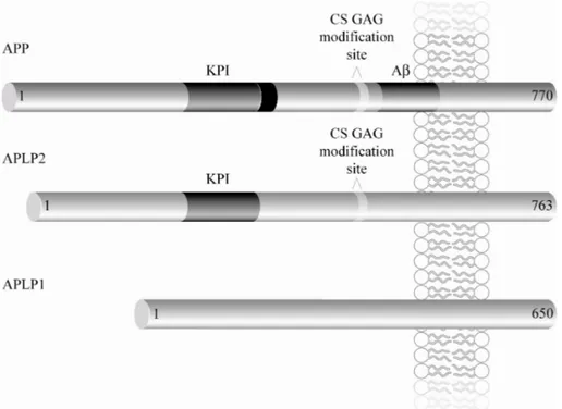
All members of the APP family are single transmembrane glycoproteins, with a large exoplasmic domain and a short cytoplasmic domain (Fig. 1 and table 1) (cf. Kang et al. 1987; Dyrks et al. 1988). Alternative splicing gives rise to different isoforms of APP and APLP2 (but not APLP1), which are
named according to the number of amino acids (reviewed in Sandbrink et al. 1996). The major existing isoforms of APP and APLP2 are APP695, APP751, APP770, APLP2763 and APLP2707. The longest isoforms, APP751, APP770 and APLP2763 (Ponte et al. 1988) contain a KPI (Kunitz protease inhibitor) do-main, which has been shown to exhibit protease inhibitory activity (Kitagu-chi et al. 1988). In addition, alternative splicing can give rise to APP and APLP2 isoforms that lack 12 amino acids (APP677, APP733, APP752, APLP2751 and APLP2695). These isoforms can be CS GAG (chondroitin sul-phate glycosaminoglycan) modified (Thinakaran and Sisodia 1994; Pangalos et al. 1995b; Pangalos et al. 1995a; Thinakaran et al. 1995a). APLP1 has been found to consist of 650 amino acids and no additional isoforms have yet been detected (Wasco et al. 1992; Paliga et al. 1997).
Figure 1. Schematic representation of the APP protein family. Both APP and APLP2 isoforms can contain a KPI domain and a CS GAG modification site. In addition, APP contains Aβ. An extra insert C-terminal of the KPI-domain is present in the longest isoform.
1.2.2. Biosynthesis and localisation
APP and APLP2 are ubiquitously expressed throughout the organism while the expression of APLP1 seems to be more restricted to the peripheral (PNS) and central nervous system (CNS) (Slunt et al. 1994; Lorent et al. 1995). Moreover, isoform specific expression has been reported suggesting that APP695 is preferentially expressed in cells with neuronal origin (Ponte et al.
1988), whereas KPI-containing (LeBlanc et al. 1991) and APP CS GAG modified (Sandbrink et al. 1994) forms are primarily expressed by non-neuronal cells in the brain.
APP and its paralogues are synthesised in the endoplasmatic reticulum (ER). Maturation (i.e. N- and O-glycosylation) occurs on the way through the sec-retory pathway (Dyrks et al. 1988; Weidemann et al. 1989; Lyckman et al. 1998). In addition, other posttranslational modifications, such as CS GAG modification, sulfation, sialyation and phosphorylation can take place (Hung and Selkoe 1994; Suzuki et al. 1994b; Thinakaran and Sisodia 1994; Panga-los et al. 1995b; PangaPanga-los et al. 1995a; Thinakaran et al. 1995a; Suzuki et al. 1997; Walter et al. 1997). Proteolytic cleavage of APP by secretases (section 1.2.4 and 1.2.5) mainly occurs at the plasma membrane and in the en-dosomes (Sambamurti et al. 1992; Parvathy et al. 1999). APP also undergoes anterograde transport in vesicles along the axons (Koo et al. 1990) and can be detected in presynaptic terminals (Lyckman et al. 1998). APP can be lo-calised to several membrane compartments and the protein can be retrogra-dally and transcytotically transported (Yamazaki et al. 1995). APLP1 local-isation has been reported to be restricted to postsynaptic terminals (Kim et al. 1995; Lyckman et al. 1998), in contrast to APLP2 which has been de-tected both pre- and postsynaptically (Lo et al. 1995; Thinakaran et al. 1995b; Lyckman et al. 1998).
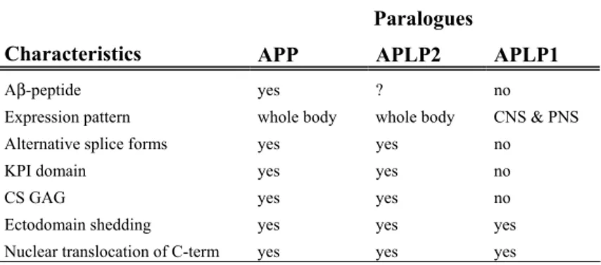
Table 1. A summary of characteristics displayed by the different paralogues.
Paralogues
Characteristics APP APLP2 APLP1
Aβ-peptide yes ? no
Expression pattern whole body whole body CNS & PNS Alternative splice forms yes yes no KPI domain yes yes no CS GAG yes yes no Ectodomain shedding yes yes yes Nuclear translocation of C-term yes yes yes
1.2.3. Functions
The general consensus is that APP is the only member of the protein family generating Aβ-peptides, although one study claims that APLP2 also give rise to Aβ-like peptides (Eggert et al. 2004). However, all paralogues release their large extracellular domain as a result of proteolytic processing (section 1.2.4 and 1.2.5). These secreted forms are denoted sAPP, sAPLP1 and sAPLP2 (Webster et al. 1995; Paliga et al. 1997; Walsh et al. 2003; Eggert et al. 2004; Li and Südhof 2004; Endres et al. 2005). Both sAPP and sAPLP2
was reported to stimulate neurite outgrowth (Araki et al. 1991; Cappai et al. 1999). When APP was first cloned, it was according to the primary structure suggested to resemble a cell-surface receptor (Kang et al. 1987). Since then, APP has been shown to interact with numerous proteins (reviewed in Turner et al. 2003; King and Scott Turner 2004) indicating that this initial notion might be correct. A most intriguing feature of APP in this context is the sug-gestion that the APP family might work as a receptor, similar to Notch. Pio-neering studies in 2001 demonstrated that the intracellular domain of the APP family was released into the cytoplasm and translocated into the nu-cleus where it was implicated in transcriptional regulation (Cao and Südhof 2001; Cupers et al. 2001; Kimberly et al. 2001; Scheinfeld et al. 2002; Walsh et al. 2003). Intriguingly, the intracellular domain has been reported to regulate the expression of its own precursor (i.e. APP) (von Rotz et al. 2004). It is not yet clear whether the most important functions of the APP family are mediated by the full length proteins or by their proteolytic frag-ments.
Even though APP has been extensively studied, due to the pivotal role of Aβ in the pathogenesis of AD, the exact biological function of this protein and its paralogues is still unknown. However, numerous in vitro studies have proposed several different functions for APP, such as involvement in cell adhesion, stimulation of neurite outgrowth and synaptogenesis, modulation of synaptic plasticity (affecting learning and memory) and neuroprotection (reviewed in Mattson 1997; Reinhard et al. 2005; Zheng and Koo 2006). In
vivo studies have further reinforced these proposed functions of APP. An
increase in APP expression at the time of axon elongation and synaptogene-sis was observed in a study of the hamster retinofugal pathway (Moya et al. 1994). Further evidence for the involvement of the APP family in develop-ment comes from studies demonstrating that the mRNA expression from whole mouse embryos increases during embryogenesis (Lorent et al. 1995). APLP1 synthesis, in particular, paralleled the timeline of the most prominent development of the nervous system. The Drosophila homologue APPL, has the highest homology to human APLP1 (Kim et al. 1995). In Drosophila, several studies have been undertaken that show important roles for APPL (or APLP1) in the nervous system. In a recent study, human APP was shown to rescue the lack of post-developmental axonal arborisation observed in Dro-sophila due to deletion of APPL (Leyssen et al. 2005). This demonstrates not only a function of APP and APP-like proteins, but also that there is func-tional redundancy between homologues. Moreover, the same study showed up-regulated expression of APPL in Drosophila in regions exposed to trau-matic brain damage, supporting a role in axonal outgrowth in response to injury. In mice, sAPP and sAPLP2 (but not sAPLP1) induced proliferation of cells in the subventricular zone of the lateral ventricles, where neurogene-sis occur in the adult brain (Caille et al. 2004). These results, on the other
hand, suggest different functions of the paralogues during post-developmental neurogenesis. Short interfering RNA (siRNA)-mediated re-duction of APP and APLP2 expression resulted in decreased synaptic activ-ity in rat retinal terminal in response to visual stimuli (Hérard et al. 2006). Impaired memory was observed after intraventricular infusion of antibodies against the N-terminal (but not against the C-terminal) of APP in rats (Doyle et al. 1990; Huber et al. 1993). Furthermore, rats displayed increased mem-ory retention, in parallel with an increased number of presynaptic terminals in the frontoparietal cortex, in response to intraventricular infusion of a syn-thetic peptide derived from sAPP (Roch et al. 1994). In turn, the extracellu-lar levels of sAPP were increased in a N-methyl-D-aspartate (NMDA)-dependent manner after induction of long term potentiation (LTP) in the dentate gyrus (a part of the hippocampus, involved in memory formation) of rats (Fazeli et al. 1994). Neuronal overexpression of APP in mice could de-crease the loss of neuronal dendrites and presynaptic terminals in response to injury (induced by the HIV-glycoprotein and kainate, respectively) (Masliah et al. 1997). This implicates APP as a mediator of protection against chronic as well as acute neurotoxicity.
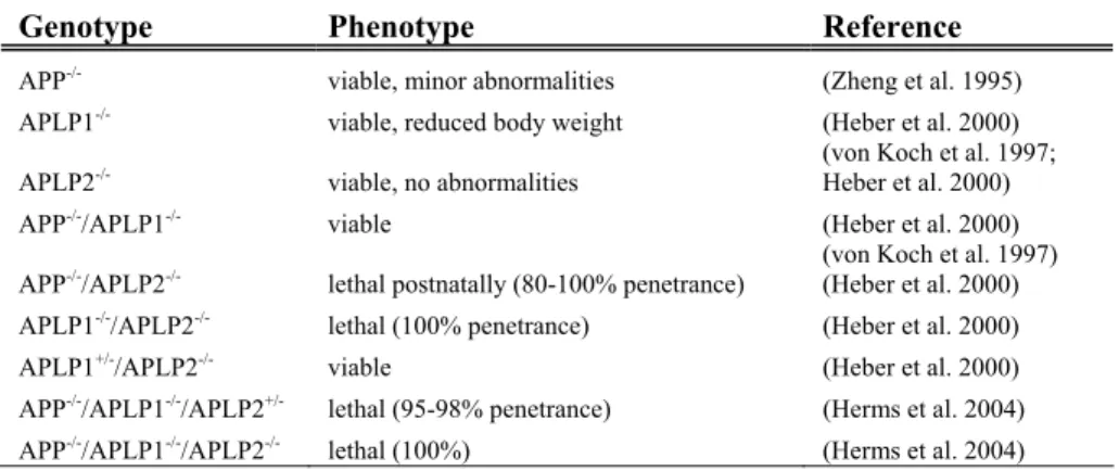
The fact that APP is highly conserved across species and ubiquitously ex-pressed suggests that it fulfils important physiological functions. However, although APP-/- mice were shown to have reduced body weight, grip strength and locomotor activity as well as reactive gliosis in the brain, they were ap-parently viable and fertile with no obvious abnormalities or significant neu-roanatomical differences (Zheng et al. 1995). APLP2-/- (von Koch et al. 1997) and APLP1-/- (Heber et al. 2000) mice were found to be normal. Vi-able and apparently normal single knock-out mice would argue against es-sential functions of the protein. Even though no compensatory upregulation of the homologous family members in single knock-out mice could be ob-served (Zheng et al. 1995; von Koch et al. 1997), functional redundancy between the APP family members might explain the subtle phenotypes ob-served. Perhaps the best tools when investigating the function of these pro-teins are double and triple knock-out mice (reviewed in Anliker and Müller 2006). Indeed, APP-/-/APLP2-/- and APLP1-/-/APLP2-/- mice die within the first week after birth (von Koch et al. 1997; Heber et al. 2000). Partial re-dundancy was suggested since, conversely, APP-/-/APLP1-/- and APLP1 +/-/APLP2-/- do survive (Heber et al. 2000). Table 2 summarises the results that APLP2 was crucial for survival, although APP and APLP1, in combination (but not alone) could compensate for the essential APLP2 function and res-cue the lethal phenotype. It should be added that this did not seem to be a consequence of compensatory expression of either APP or APLP1. Studies in N2a cell cultures using gene silencing have suggested that APLP1 might be more important for neurite outgrowth and survival at least in neuroblas-toma cells (Sakai and Hohjoh 2006). Results that further corroborate the
involvement of the APP family in synapse formation and function come from studies on the neuromuscular junction in APP-/-/APLP2-/- mice. These mice exhibited defective synapses, displaying reduced number of synapses, reduced number of vesicles and active zones and an aberrant neuronal sprouting (Wang et al. 2005; Yang et al. 2005). Embryonic triple APP family knock-out mice displayed abnormal positioning of cortical neurons, sug-gested to be a result of a decreased number of cortical Cajal Retzius cells (Herms et al. 2004). In addition to the functional redundancy, a direct inter-action between paralogues can occur. A recent study elegantly demonstrated that the APP family was able to form both homo- (APP/APP, APLP1/APLP1 and APLP2/APLP2) and heterotypic (APP/APLP1, APP/APLP2, APLP1/APLP2) complexes in a trans-cellular fashion and that this interaction promoted cell adhesion (Soba et al. 2005).
Table 2. APP family knock-out mice. APLP2 is crucial for survival unless both APP and APLP1 are present to compensate for the essential function of APLP2.
Genotype Phenotype Reference
APP-/- viable, minor abnormalities (Zheng et al. 1995)
APLP1-/- viable, reduced body weight (Heber et al. 2000)
(von Koch et al. 1997; Heber et al. 2000) APLP2-/- viable, no abnormalities
APP-/-/APLP1-/- viable (Heber et al. 2000)
(von Koch et al. 1997) APP-/-/APLP2-/- lethal postnatally (80-100% penetrance) (Heber et al. 2000)
APLP1-/-/APLP2-/- lethal (100% penetrance) (Heber et al. 2000)
APLP1+/-/APLP2-/- viable (Heber et al. 2000)
APP-/-/APLP1-/-/APLP2+/- lethal (95-98% penetrance) (Herms et al. 2004)
APP-/-/APLP1-/-/APLP2-/- lethal (100%) (Herms et al. 2004)
Clearly the APP family of proteins do perform essential functions. Further studies are needed to fully demonstrate the elusive postnatal physiological role of the different members of this family.
1.2.4. Processing and proteolytic fragments
The processing of APP can be divided into two different pathways, the non-amyloidogenic and the non-amyloidogenic (Figs. 2 and 3). As indicated by the name, the amyloidogenic pathway ultimately leads to the generation of Aβ after cleavage of APP by β- and γ-secretase (section 1.2.5). Conversely, Aβ formation is precluded in the non-amyloidogenic pathways, since α-secretase (section 1.2.5) cleaves in the middle of the Aβ sequence at the Lys16-Leu17 bond (Esch et al. 1990; Sisodia et al. 1990). Not only does α-secretase cleavage preclude the formation of Aβ, but sAPPα has been
Figure 2. Schematic illustration of the proteolytic processing of APP and resulting fragments.
shown to have neuroprotective properties (Araki et al. 1991; Mattson et al. 1993; Schubert and Behl 1993; Furukawa et al. 1996a; Furukawa et al. 1996b). sAPPα is constitutively released (basal shedding) (Esch et al. 1990), but can also be secreted as a result of a stimulus (regulated shedding) (cf. table 3 in section 4.3.1). After cleavage by α-secretase, a C-terminal stub of 83 amino acids (C83) is left in the membrane. This fragment can be further processed by the γ-secretase complex resulting in secretion of the small pep-tide p3.
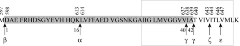
The amyloidogenic processing of APP starts with the β-secretase cleavage N-terminal of Asp1 of Aβ. This step also leads to shedding of a large ecto-domain, sAPPβ, subsequently leaving a membrane-bound C-terminal stub of 99 amino acids (C99). Aβ formation is complete after cleavage of C99 by γ-secretase, either at the Val40-Ile41 bond or after Ala42 generating Aβ40or Aβ42, respectively (reviewed in Esler and Wolfe 2001).
Figure 3. Proteolytic cleavage sites in APP. The Aβ sequence is marked in grey. Numbers above the sequence correspond to amino acid residue of the APP695 iso-form. Greek letters below graph represent the different cleavage sites. Numbering below the sequence is in relation to the Aβ sequence. The grey rectangle corre-sponds to the putative transmembrane region.
γ-secretase cleavage of APP to liberate Aβ40 or 42 would leave a C-terminal fragment (CTF) of 59 or 57 amino acids (C59 or C57) in the membrane. However, the remaining CTF of APP that is released into the cytoplasm has been shown to be equivalent to the 50 most C-terminal amino acids (C50) (Sastre et al. 2001). The elegant analogy to cleavage of Notch and the subse-quent release of NICD (Notch intracellular domain) led the authors to pro-pose the name AICD (APP intracellular domain) for C50. Both the amyloi-dogenic and the non-amyloiamyloi-dogenic pathway result in AICD formation. This cleavage between Leu646-Val647 of APP695 (Fig. 3) is, like the γ-site cleav-age, dependent on PS1 (section 1.2.5.3) and is also known as the ε-cleavage site (Yu et al. 2001; Weidemann et al. 2002). Cleavage at the corresponding sites in APLP1 and APLP2 has also been demonstrated (Gu et al. 2001). The remaining CTFs of APLP1 and APLP2 are, in accordance with AICD for APP, denoted ALID1 and 2 (APP-like intracellular domain 1 and 2) (Naruse et al. 1998; Scheinfeld et al. 2002; Walsh et al. 2003; Eggert et al. 2004). An additional γ-secretase dependent cleavage site of APP (ζ-cleavage site
be-tween Val643-Ile644) has also been suggested (Zhao et al. 2004). However, there is still debate over whether the γ-, ε- and ζ-cleavages are dependent or independent catalytic events.
1.2.5. APP processing enzymes
1.2.5.1. α-secretases
The identity of α-secretase is controversial. There are several good candi-dates and most likely there are several different proteinases that are able to cleave APP at the α-secretase site. Protease inhibitor studies have revealed that α-secretase is a zinc metalloproteinase (Roberts et al. 1994), proposed to belong to the ADAM (a disintegrin and metalloprotease) family. ADAM9, ADAM10 and TACE (tumour necrosis factor-α convertase also known as ADAM17) fulfil criteria for α-secretase as discussed below. ADAMs are type I integral membrane proteins consisting of a signal peptide, a pro-domain followed by a cleavage site for proprotein convertases (PCs), a cata-lytic metalloprotease domain containing the HEXXH zinc-binding motif, a disintegrin/cystein-rich domain, a transmembrane domain (TM) and a short cytoplasmic domain (Howard et al. 1996; Black et al. 1997; Moss et al. 1997).
One of the first pieces of evidence for ADAM10 acting as an α-secretase was data showing that it cleaved a peptide spanning the α-secretase cleavage site between the Lys16-Leu17 bond of Aβ as expected (Lammich et al. 1999). Expression of ADAM10 mRNA and APP have been shown to over-lap in mouse brain and in human cortical cells (Marcinkiewicz and Seidah 2000). Furthermore, in transfected HEK293 cells, the majority of ADAM10 was found inside the cell as a proenzyme, whereas the proteolytically active form was localised in the plasma membrane (Lammich et al. 1999), where most of the α-secretase processing of APP takes place (Parvathy et al. 1999). Overexpression of ADAM10 in HEK293 cells led to several-fold increase of both basal and protein kinase C (PKC)-stimulated release of sAPPα and in addition, to increased levels of C83 (Lammich et al. 1999). In LoVo cells, the overexpression of ADAM10 also results in amplified secretion of sAPPα (Lopez-Perez et al. 2001). In addition, in brain samples from double-transgenic ADAM10xAPP[V717I] mice, augmented secretion of sAPPα and C83 in parallel with reduced secretion of sAPPβ as well as Aβ40 and Aβ42 was observed. Furthermore, these mice developed no plaques. Double trans-genic mice expressing a dominant negative mutant of ADAM10, on the other hand, developed more plaques, both faster and with larger size (Postina et al. 2004). ADAM10-/- mice die at day 9.5 during embryogenesis and this early lethality has prevented analysis of neuronal cell cultures. However, in fibroblasts from ADAM10-/- mice, the α-secretase activity is preserved.
Al-though no differences in ADAM9 and TACE expression could be detected, this result suggests that other proteases can compensate for the loss of ADAM10 (Hartmann et al. 2002). Both the constitutive and the regulated secretion of sAPPα can be substantially inhibited by a dominant negative form of ADAM10 or a hydroxamic acid-based zinc metalloprotease inhibitor (Lammich et al. 1999). In contrast, increased maturation of ADAM10 in-duced by overexpression of either PC7 or furin is paralleled by an increased secretion of sAPPα (Anders et al. 2001; Lopez-Perez et al. 2001). Indeed, α-secretase activity in brain from AD patients (Tyler et al. 2002), as well as ADAM10 protein levels in platelets were lower, analogously to the reduced levels of sAPPα in platelets as well as CSF in comparison to age-matched control patients (Colciaghi et al. 2002).
Another protease that may work as an α-secretase is TACE. Like ADAM10, TACE has benn shown to be able to cleave a synthetic peptide encompassing 10 amino acids at the α-secretase site within Aβ (Buxbaum et al. 1998). It was later shown that this cleavage was very slow, as demonstrated by the modest kcat/Km and almost 100-fold less efficient than the cleavage of the TNFα peptide (Mohan et al. 2002). However, this does not exclude a more efficient cleavage of full-length and membrane-bound APP, since additional factors may be required for efficient processing of APP by TACE. In pri-mary embryonic fibroblasts derived from TACE-deficient mice, the PKC-stimulated shedding of sAPPα was abolished (Buxbaum et al. 1998; Merlos-Suárez et al. 1998). Since the constitutive release of sAPPα was not affected, these studies suggest that TACE might be involved in the regulated, but not the constitutive, release of sAPPα. Additional support for this theory comes from studies in the furin-deficient LoVo cell line, where TACE overexpres-sion fails to increase the basal sAPPα recovery (Lopez-Perez et al. 2001). In contrast, TACE and APP co-transfection in HEK-293-M3 cells resulted in increased basal shedding of sAPPα in a dose-dependent manner in relation to TACE cDNA expression. On the other hand, no effect on the regulated shedding could be observed (Slack et al. 2001). This would implicate TACE in the constitutive release of sAPPα. However, the authors of this study also observed that the inhibition profiles of endogenous and TACE-induced basal sAPPα release were different. It was therefore suggested that another metal-loprotease might be responsible for the endogenous α-secretase activity in HEK-293-M3 cells. In HEK-293 as well as in SH-SY5Y cells, PMA treat-ment resulted in elevated sAPPα recovery, despite reduced levels of the mature and catalytically active form of TACE (Endres et al. 2003). Addi-tionally, in SH-SY5Y cells, the inhibition profile of basal and stimulated shedding of sAPPα could not be distinguished. Further, a hydroxamate-based TACE inhibitor did not block α-secretase activity, suggesting that TACE is not involved in the regulated secretion of sAPPα (Parkin et al.
2002). Another TACE inhibitor was reported to prevent regulated sAPPα release in primary human neurons (Blacker et al. 2002). However, although it was not commented on by the authors, a small inhibition of the basal se-cretion of sAPPα could also be detected, further illustrating the complexity of APP processing. In addition, the expression of TACE in mouse brain is low (Kärkkäinen et al. 2000) and in situ hybridisation revealed only a partial overlap of the expression of APP and TACE (Marcinkiewicz and Seidah 2000).
Yet another enzyme suggested to be involved in the processing of APP at the α-secretase site is ADAM9 (Koike et al. 1999; Roghani et al. 1999; Hotoda et al. 2002). The evidence pointing to an involvement of ADAM9 is less convincing than that for the involvement of ADAM10 or TACE. However, when comparing the α-secretase activity in ADAM9, ADAM10 or TACE transfected COS-7 cells, they all possessed the ability to release sAPPα through both constitutive and regulatory mechanisms (Asai et al. 2003). ADAM9 was also suggested to contribute to the increased sAPPα levels through activation of ADAM10 rather than through direct cleavage of APP (Cissé et al. 2005). However, only ADAM10 (and not ADAM9 or TACE) was required for green-tea polyphenol-induced α-secretase cleavage in APP-Swe transfected N2a cells as well as in primary neuronal cells from Tg2576 mice (Obregon et al. 2006).
To summarise, there is convincing evidence pointing to the involvement of ADAM10 acting as an α-secretase in constitutive and regulated shedding of sAPPα. There is a notion of multiple α-secretases working together possibly to a differing degree under different conditions and in different cell types. More studies are needed to establish the roles of TACE and ADAM9 as po-tential α-secretases.
1.2.5.2. β-secretase
For over a decade the enzyme responsible for cleavage of APP to generate the N-terminus of Aβ was unknown and was simply referred to as the β-secretase. Then in 1999, the enzyme BACE1 (β-site APP cleaving enzyme), also known as Asp2 (novel aspartic protease 2) or memapsin2 (membrane aspartic protease/pepsin 2), was simultaneously discovered by several inde-pendent groups and found to fulfil the criteria for β-secretase (Hussain et al. 1999; Sinha et al. 1999; Vassar et al. 1999; Yan et al. 1999; Lin et al. 2000). A year later BACE2, a homologue to BACE1, was found (Farzan et al. 2000). However, although BACE2 can cleave APP at the β-secretase site, it cleaves with higher efficiency within the Aβ region, C-terminally of Phe19 or Phe20 (cf. Fig. 3). Consequently, it may rather function as an alternative α-secretase (Farzan et al. 2000; Yan et al. 2001). In addition, the expression of BACE2 mRNA in the brain, is in contrast to the ubiquotous expression of
BACE1 mRNA, restricted to certain discrete nuclei (Vassar et al. 1999; Ben-nett et al. 2000a). Even more decisively, no Aβ could be detected in brain homogenate or in cultures from primary cortical neurons from BACE1 knock-out mice (Luo et al. 2001b; Roberds et al. 2001). Thus, BACE2 can be ruled out as a major contributor to Aβ formation in vivo, even though the Flemish FAD mutation at Ala21 causes elevated Aβ levels (Farzan et al. 2000). Furthermore, BACE1 expression and activation has been correlated to AD, both when it comes to levels in the affected brain regions as well as in platelets (reviewed in Johnston et al. 2005).
BACE1 is an aspartyl protease with a conserved active site including the amino acid residues Asp93 and Asp289. ProBACE1 is comprised of 501 amino acids, contains a single membrane spanning domain and is synthe-sised with a prodomain (Hussain et al. 1999; Vassar et al. 1999). This pro-domain is cleaved off by a PC in the Golgi apparatus to yield mature BACE1 (Bennett et al. 2000b; Benjannet et al. 2001). During maturation, BACE1 can also be phosphorylated (Walter et al. 2001), N-glycosylated (Vassar et al. 1999; Huse et al. 2000), palmitoylated and sulphated (Benjannet et al. 2001). It has been suggested that N-glycosylation may affect the protease activity of BACE1 (Charlwood et al. 2001). ProBACE1 exhibit some β-secretase activity. Thus, atypically, the pro region does not suppress the pro-tease activity but has instead been suggested to assist in the proper folding of the protein (Shi et al. 2001). Palmitoylation may influence the intracellular localisation and also inhibit ectodomain shedding of BACE1 (Benjannet et al. 2001). Phosphorylation of the C-terminal Ser498 affects the intracellular trafficking of BACE1 in such a way that BACE1 can be retrieved from early endosomes to late endosomes and Golgi and subsequently recycled into sec-retory vesicles (Walter et al. 2001). APP and BACE1 co-localise within the secretory pathway, and the majority of BACE1 immunostaining is found within the Golgi apparatus and endosomes. The optimal pH of BACE1 activ-ity is approx. 4.5, suggesting that cleavage of APP takes place on the luminal side in an acidic cellular compartment like the endosome (Vassar et al. 1999).
1.2.5.3. γ-secretase
Initial evidence for the involvement of PS1 and PS2 in γ-secretase activity came from genetic linkage analysis. Mutations in these genes could cause an autosomal and highly penetrant form of early-onset AD (section 1.1.3). Ad-ditional evidence for the role of PS1 in γ-secretase processing of APP comes from neuronal cultures devoid of PS1, where Aβ40 as well as Aβ42 de-creased dramatically in correspondence with accumulation of C-terminal stubs generated by the preceding α- and β-secretase cleavage (De Strooper et al. 1998). The presenilins were first proposed to be comprised of eight (Doan et al. 1996; Li and Greenwald 1996) and later of nine (Laudon et al. 2005;
Oh and Turner 2005) membrane spanning domains. Regulated endoproteoly-sis results in an N-terminal fragment (NTF) and a CTF, which remain asso-ciated in a stable complex (Thinakaran et al. 1996; Thinakaran et al. 1997). There are contradictory reports as to whether the endoproteolysis of PS1 and the γ-secretase activity are connected or distinct features. However, two con-served aspartate residues, one in TM6 and the other one in TM7, have been found to be essential for the endoproteolysis of PS as well as for the genera-tion of Aβ (Steiner et al. 1999; Wolfe et al. 1999). Furthermore, transition-state analogues of γ-secretase inhibitors were shown to bind directly to pre-senilin NTF and CTFs (Esler et al. 2000; Li et al. 2000; Seiffert et al. 2000). Finally, the key support for presenilin working as an aspartyl protease came from the identification of Gly384 in PS1 as a part of a highly conserved GxGD motif (including the aspartate residue in TM7) which is essential for γ-secretase function (i.e. endoproteolysis, Aβ generation from APP C-terminal stubs and Notch cleavage) (Steiner et al. 2000). With the finding of signal peptide peptidase (SPP) and its homologues (Weihofen et al. 2002), it became clear that presenilins belong to a novel family of polytopic aspartyl proteases (Haass and Steiner 2002). These findings strongly supported the theory that presenilin could be the true γ-secretase. However, it was also reported that PS1 existed in larger molecular complexes and thus could work as the catalytic component in concert with other interacting proteins (Seeger et al. 1997; Capell et al. 1998; Yu et al. 1998). Today, three additional and essential members of the γ-secretase complex are known; nicastrin, anterior pharynx defective 1 (APH-1) and presenilin enhancer 2 (PEN-2) (Goutte et al. 2000; Yu et al. 2000; Francis et al. 2002; Edbauer et al. 2003). Nicastrin is needed for APP and Notch cleavage and is believed to have a function in the correct assembly of the γ-secretase complex as well as in substrate inter-action. APH-1 appears to mediate stabilisation and assembly of PS1, whereas PEN-2 is implicated in endoproteolysis and γ-secretase activity (reviewed in Verdile et al. 2006; Wolfe 2006). Assembly of γ-secretase (re-viewed in Kaether et al. 2006a) takes place in the ER, where the interaction of APH-1 and Nicastrin is the first event, followed by the incorporation of presenilin into the complex. PEN-2 then enters the complex and endoprote-olysis of presenilin takes place. Subsequently the assembled complex is transported via the secretory pathway to the plasma membrane and the en-dosomes where interaction and cleavage of C83 and C99 take place (Esler et al. 2002; Kaether et al. 2006b).
Presenilins have been ascribed many different functions and the number of substrates are also constantly increasing (reviewed in Vetrivel et al. 2006). One of the most intriguing potential roles for PS1 is its implications in main-taining cholesterol through Aβ formation (Grimm et al. 2005). PS1 has also been suggested to be involved in the subcellular trafficking of APP, that
could also affect the subsequent processing of APP (Leem et al. 2002; Cai et al. 2003).
1.3. Therapeutic targets in AD
1.3.1. Available therapies
Over the last decades the understanding of the mechanism involved in the development of AD has increased dramatically. However, despite tremen-dous efforts, only two types of drugs are today available on the market for AD patients (www.alzforum.org). Donezepil and galantamine are choli-nesterase inhibitors and memantine is an NMDA receptor antagonist. Ace-tylcholine is an important neurotransmitter for cognitive function and an early pathogenic event in AD is loss of neurons in the basal forebrain that synthesise acetylcholine. Thus, cholinesterase inhibitors aim at stalling the enzyme that degrades acetylcholine after it has been released at the synapse, thereby prolonging the effects of the neurotransmitter. Glutamate signalling, on the other hand, may cause neurodegeneration as a result of excitotoxicity in AD and other neurodegenerative diseases. Memantine is believed to exert its beneficial effects through its low affinity for NMDA receptors preventing excessive glutamate transmission but allowing for normal transmission to take place. Neither of these drugs prevent the progression of the disease, but rather mitigate the symptoms and in the best scenario, delay the cognitive decline.
Massive research into different approaches to treat AD is currently being undertaken by scientists and pharmacological industry all over the world. Obvious therapeutic tactics would be to prevent the formation of Aβ, to pre-vent assembly of Aβ species, or increase the degradation of Aβ or increase the transport of Aβ out of the brain.
1.3.2. A
β immunisation
In my opinion, the most intriguing approach comes from studies involving immunisation against Aβ. Active immunisation of young transgenic mice prior to the occurrence of pathological changes prevented plaque formation (Schenk et al. 1999). In older mice, which already displayed plaques prior to immunisation, the plaque burden was decreased. Additionally, a report using passive immunisation in mice showed that plaque formation was reversed by peripheral administration of antibodies against Aβ (Bard et al. 2000). This finding is extraordinary, since it indicates that antibodies can cross the blood-brain barrier and induce microglial cells to phagocytise Aβ. Unfortu-nately, the first clinical trial on humans using active immunisation with Aβ was terminated, since 18 of 298 immunised patients developed meningoen-cephalitis (Orgogozo et al. 2003). However, post mortem examination of the
brains from three patients in this trial, corroborated previous findings in ani-mal models, demonstrating almost complete removal of amyloid plaques (Nicoll et al. 2003; Masliah et al. 2005). New approaches have been under-taken to circumvent the detrimental immune response leading to meningoen-cephalitis and there are currently two studies (one using passive and the other one using active immunisation) in Phase II clinical trials.
1.3.3. Prevention of A
β formation
Theoretical tactics to block the formation of Aβ would be to inhibit the en-zymes responsible for generating Aβ, namely β- and/or γ-secretase (section 1.2.5.2 and 1.2.5.3), or to increase the α-secretase (section 1.2.5.1) activity.
1.3.3.1. γ-secretase inhibition
The lethal phenotype of PS1-/- mice (De Strooper et al. 1998), as a result of disturbed Notch signalling, in addition to the rapidly increasing number of known secretase substrates, indicates serious problems with the use of γ-secretase inhibitors. However, epidemiological studies have shown that long-term use of NSAIDs (nonsteroidal anti-inflammatory drugs) reduces the risk of developing AD. This is probably not solely due to inhibition of the inflammatory responses in AD. Intriguingly, studies suggest that γ-secretase can be allosterically modulated by NSAIDs (reviewed in Evin et al. 2006) to produce less of the longer, more amyloidogenic, form of Aβ (Weg-gen et al. 2001). In fact, one NSAID, flurbiprofen, has been demonstrated to selectively lower Aβ42 levels and is currently being investigated in Phase III clinical trials. It should not be forgotten that γ-secretase cleavage of the APP family members results in nuclear translocation of the C-terminal domain. γ-secretase inhibitors would presumably affect the translocation of ALID1 and ALID2 as well. Possible side-affects could arise, since expression of the APP family is essential.
1.3.3.2. β-secretase inhibition
BACE1-/- mice are, in contrast to PS-/- mice, viable and fertile with no major abnormalities (Luo et al. 2001b; Roberds et al. 2001) thus turning BACE1 into a therapeutic key target. However, the design of small organic com-pounds inhibitors (that would be able to cross the blood-brain barrier and act in the brain) for BACE1 (reviewed in Citron 2004) is a challenge due to the large size of the active site in BACE1. Nevertheless, there are currently BACE1 inhibitors being investigated in preclinical trials (Thompson et al. 2005). Another shrewd preclinical approach to block β-secretase, is to ex-press intracellular single chain antibodies (intrabodies) directed to an epitope in APP next to the site where β-secretase cleaves. These intrabodies bind close to the β-secretase cleavage site and were shown to block proteolytic processing at that site (Paganetti et al. 2005).
1.3.3.3. Stimulation of α-secretase activity
An increase in α-secretase activity not only precludes the formation of Aβ, but instead favours formation of the neurotrophic sAPPα. Agonists for the muscarinic acetylcholine receptor M1, have been investigated in AD patients as a tool to increase the non-amyloidogenic pathway (Nitsch et al. 2000). Cholesterol lowering drugs such as statins, have been correlated in retrospec-tive studies to a reduced risk of developing AD. Besides their anti-inflammatory and antioxidant properties, cholesterol lowering drugs might exert their effect through an increase in the expression of ADAM10 (Kojro et al. 2001). Similarly, cholinesterase inhibitors (discussed above) have been proposed to increase ADAM10 activity by promoting its trafficking in neuroblastoma cell lines (Zimmermann et al. 2004). Also, cholesterol is speculated to increase the generation of Aβ as an effect of APP and BACE1 co-localisation within lipid rafts. A decrease in cholesterol would subse-quently increase the opportunity for α-secretase to encounter APP and hence the non-amyloidogenic pathway would be favoured (reviewed in Johnston et al. 2005; Cordy et al. 2006). Several cholesterol-lowering drugs are currently in Phase II clinical trials.
1.4. Neurotrophic factors, their receptors and signalling
pathways
Reduced levels of various neurotrophic factors have been implicated in AD and replacement therapies have also been considered. Below, the signalling pathways for important neurotrophic factors that we have focused on in our studies are described.
1.4.1. RA
RA is a derivative of retinol (vitamin A). In the CNS system, RA is involved in pattern formation during embryogenesis and has commonly been used for differentiation of neuroblastoma cells. Retinol enters the cell and is subse-quently converted to retinal and further to RA. RA then enters the nucleus where it can bind to two types of receptors, the RA receptors and the retinoid X receptors. There are two isoforms of RA, all-trans-RA and 9-cis-RA that bind the different receptors with different affinity. RA receptors are ligand-activated nuclear transcription factors that, after activation, bind to RARE (RA-responsive elements) and hence control gene expression.
1.4.2. BDNF
The mammalian neurotrophin family consists of NGF (nerve growth factor), BDNF (brain-derived neurotrophic factor) and the NT3 and NT4/5 (neuro-trophin-3 and 4/5). As the name neurotrophin suggests, they promote cellular
growth, differentiation and survival. Three major pathways; the PI3-K (phos-phatidylinositol 3-kinase), MAPK (mitogen-activated protein kinase) and PLC (phospholipase C) signalling cascades are involved in neurotrophin signalling. It should be stated that the following description of the signalling pathways is schematic and highly simplified. Alternative splicing of recep-tors, formation of hybrid receprecep-tors, expression of different isoforms of intra-cellular signalling proteins and cross-talk between the major pathways and additional signalling proteins promote a much more complicated network in the cell.
Effects of the neurotrophins are mediated by binding to their corresponding receptors, Trks (tropomyosin related kinases) or p75NTR (neurotrophin re-ceptor). p75NTR is activated by all neurotrophins. TrkA is primarily acti-vated by NGF, TrkB by BDNF (and NT4/5) and TrkC by NT3. Trks are receptor tyrosine kinases. Ligand binding causes two receptors to dimerise, which enables the cytoplasmic tyrosine kinase domains to interact and for subsequent autophosphorylation to occur, resulting in activation. This activa-tion creates a binding site for the adaptor protein Shc (Src homol-ogy/collagen) that contains a PTB (phosphotyrosine binding) domain. When
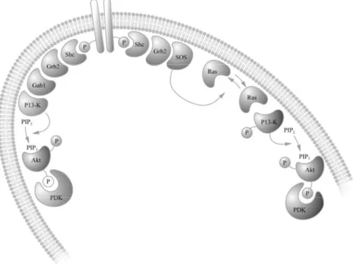
Figure 4. Ligand binding to a receptor tyrosine kinase can activate the PI3-K signal-ling cascade.
Shc in turn is phosphorylated, another small adaptor protein Grb2 (growth factor receptor-bound protein 2) is recruited, which contains both a SH2 and a SH3 domain (Src homology region 2 and 3). The SH2 domain recognises phosphorylated tyrosine residues (located in Grb2) and the SH3 domain as-sociates with the guanine nucleotide releasing protein SOS (son of sevenless). SOS stimulates the small GTPase Ras (p21src protein of sarcoma virus) to exchange its bound GDP for GTP and subsequently to become acti-vated. Activated Ras then recruits Raf (rapidly growing fibrosarcoma) to the membrane, promoting its activation through phosphorylation. Phosphoryla-tion of Ras initiates a series of phosphorylaPhosphoryla-tion events called the MAPK cascade.
Activation of PI3-K can be Ras-dependent (as described for MAPK activa-tion above) or independent. Ras-independent activaactiva-tion of PI3-K is enabled by the recruitment and binding of Gab1 (Grb2 associated binder) to Grb2. When active PI3-K converts the plasma membrane lipid PIP2 (phosphatidy-linositol bisphosphate) to PIP3 (phosphatidylinositol triphosphate), the ser-ine/threonine kinase Akt (also known as protein kinase B, PKB, or RAC, related to protein kinase A and C) is translocated to the plasma membrane. Akt contains a protein lipid domain and associates with PIP3, possibly pro-moting a conformational change of Akt. Constitutively active PDKs (3’-phosphoinositide-dependent kinases) can then activate Akt through phos-phorylation of exposed residues.
The third major pathway involved in Trk signalling is PLC. PLC is recruited to the C-terminal of the activated receptor and in turn become phosphory-lated. PLC thereafter hydrolyses PIP2 to generate IP3 (inositol tri-phosphate) and DAG (diacylglycerol). IP3 promotes release of internally stored Ca2+ after binding to IP3-gated channels located in the ER membrane. DAG, to-gether with the elevated intracellular Ca2+ levels activates enzymes like PKC and Ca2+-calmodulin-regulated protein kinases.
Signalling of neurotrophins through Trk receptors is regulated through inter-nalisation of Trk receptors together with its bound ligand in endocytotic vesicles. Internalisation serves at least two purposes. Firstly, transport of these vesicles enables the activated receptors to come into proximity to cell compartments and cell mediators where it is required for signalling. Sec-ondly, internalisation offers the possibility for receptor down-regulation and thereby desensitisation of the signal as a result of ligand binding. In addition, the receptor can then be recycled back to the membrane when needed.
Figure 5. Ligand binding to a receptor tyrosine kinase can activate MAPK and PLC signalling cascades.
1.4.3. Insulin/IGF-1
Insulin and IGF-1 are growth factors involved in survival as well as differen-tiation. The receptors for these growth factors (IR and IGF-1R) belong to a different subfamily than Trks, but are also receptor tyrosine kinases. Binding of insulin or IGF-1 to their receptors (or with lower affinity to each other’s receptors) result in activation of MAPK and PI3-K signalling cascades as described above but with some differences. Insulin and IGF-1R are tetrameric in structure (with two extracellular α-subunits and two transmem-brane intracellular β-subunits covalently linked by disulfide bridges). Hence, binding of ligand to the receptor results in conformational changes and auto-phosphorylation of the C-terminal domain rather than dimerisation. In addi-tion, activated receptors recruit the adaptor protein IRSs (insulin receptor substrates) with a PTB domain, which in turn associates with Shc, Grb2 and PI3-K. This result in activation of Akt and MAPK as described for neurotro-phin signalling above.
2. AIMS OF THE STUDY
For a complete understanding of APP processing and to enable the develop-ment of good therapeutic agents that efficiently block the formation of A β-peptide, devoid of undesirable side-effects, we need more knowledge about the functions and processing of the two paralogues APLP1 and APLP2. Thus, our studies were focused on comparing the regulation of expression and processing of the APP family. The specific aims of the work in this the-sis were as follows:
• To analyse the expression and processing of the APP family during neuronal differentiation (papers I and II)
• To investigate the mechanism behind the RA-induced expression and processing of the APP family (papers I-III)
• To examine the involvement of BDNF and TrkB in the synthesis and processing of the APP family (paper II)
• To determine the signalling pathways involved in IGF-1-induced processing of the APP family (paper IV)
3. METHODOLOGICAL CONSIDERATIONS
3.1. Cell cultures
Throughout this thesis, human SH-SY5Y neuroblastoma cells have been used to study the expression and proteolytic processing of the APP protein family. Immortalised cells are convenient to handle and experiments can be performed during continuous conditions in which biochemical processes easily can be studied. However, one should bear in mind that the nervous system include an enormous variety of cell types and cell contacts and that these factors are absent in an isolated cell line.
3.1.1. SH-SY5Y neuroblastoma cells
SH-SY5Y is a neuroblast-like subclone of the SK-N-SH neuroblastoma cell line, originally derived from a metastatic tumour in the bone marrow. This cell line was established already in 1970 and has, since then, been widely used to study neuronal differentiation and neurodegeneration (Biedler et al. 1973; Biedler et al. 1978). Thus, by now, SH-SY5Y cells constitute a well defined cellular system. An additional advantage is the human origin. The parental cell line of SH-SY5Y cells comprise of both neuroblastic and sub-strate adherent cell-types. However, SH-SY5Y cells can be differentiated into a more neuronal-like phenotype with extended neurites using different neurotrophic factors like, RA, BDNF and IGF-1 (Påhlman et al. 1984; Påhlman et al. 1991; Kaplan et al. 1993; Encinas et al. 2000). In addition, SH-SY5Y cells endogenously express all the members of the APP protein family (Beckman and Iverfeldt 1997).
3.1.2. Treatments
Treatments discussed below are illustrated in Fig. 6, to make the following section more comprehensible. In paper I and II, SH-SY5Y cells were differ-entiated with 10 μM RA during 6 days to achieve a fully differentiated neu-ronal-like population of cells. The effect of a PKC inhibitor, curcumin, on RA-differentiated cells was investigated. 10 μM curcumin was added during the last 24 h before harvesting or 2 μM curcumin was added concomitant with RA from the start. Two different protocols were used since 10 μM cur-cumin during longer periods was to harsh even for RA-differentiated cells. Metabolic labelling studies showed that the half life of the APP family in our
experiment during RA treatment was less than 90 minutes. Thus, using the two different protocols would have no impact on the expression levels of these proteins. When it comes to studying neurite outgrowth there is a differ-ence since RA treated cells are continuously extending their neurites. As a consequence adding 2 μM concomitantly with RA would resemble inhibi-tion of neurite outgrowth. Adding 10 μM curcumin during the last 24 h is rather equivalent to degeneration of neurites.
Figure 6. Treatments schematically illustrated. Arrows indicate change of medium.
In paper II, cells were first treated with RA during 3 days in order to induce expression of TrkB receptors and thereby responsiveness to BDNF (Kaplan et al. 1993). Subsequently two protocols were used, first BDNF were added concomitantly to RA for a 6 days period (to be compared with cells differen-tiated by RA alone for 6 days). However, even though differences in the mRNA expression of the APP family could be observed after 3 days, protein levels were significantly increased first after 6 days of RA treatment (in