http://www.diva-portal.org
Postprint
This is the accepted version of a paper presented at The Swedish AI Society (SAIS) Workshop SAIS, 14,
22-23 May 2014, Stockholm, Sweden.
Citation for the original published paper:
Barua, S., Begum, S. (2014)
A Review on Machine Learning Algorithms in Handling EEG Artifacts.
In: The Swedish AI Society (SAIS) Workshop SAIS, 14
N.B. When citing this work, cite the original published paper.
Permanent link to this version:
A Review on Machine Learning Algorithms in Handling EEG Artifacts
Shaibal Barua
1, Shahina Begum
2School of Innovation, Design and Engineering, Mälardalen University SE-721 23, Västerås, Sweden
1shaibal.barua@mdh.se 2shahina.begum@mdh.se
Abstract— Brain waves obtained by Electroencephalograms
(EEG) recording are an important research area in medical and health and brain computer interface (BCI). Due to the nature of EEG signal, noises and artifacts can contaminate it, which leads to a serious misinterpretation in EEG signal analysis. These contaminations are referred to as artifacts, which are signals of other than brain activity. Moreover, artifacts can cause significant miscalculation of the EEG measurements that reduces the clinical usefulness of EEG signals. Therefore, artifact handling is one of the cornerstones in EEG signal analysis. This paper provides a review of machine learning algorithms that have been applied in EEG artifacts handling such as artifacts identification and removal. In addition, an analysis of these methods has been reported based on their performance.
Keywords— Electroencephalograms(EEG), Artifacts,
Machine Learning
I. INTRODUCTION
Brain waves or neural signals obtained by Electroencephalograms (EEG) recordings is an important research area and plays a vital role in medical and health applications and in Brain Computer Interface (BCI). For instance, sleep study is one of the domains where EEG is used frequently. Several other medical and health-related research areas where EEG is extensively used are, but not limited to, epilepsy, neuroscience, cognitive science, and psychophysiological research. Like other biosignals, EEG is a non-stationary and nonlinear signal. Klonowski (2009) defined it as ‘3N’: nonstationary, nonlinear, and noisy. One of the crucial aspects of using EEG in medical applications and as well as BCI applications is to deal with noise and artifacts presented in the signals. Physiological signals other than brain activity, which contaminates the EEG signals, are referred to as artifacts. Artifacts make EEG signal uninterpretable and in EEG signal analysis it can also leads to serious misinterpretation. In addition, artifacts can cause significant miscalculation of measurements of the diagnosis that reduces the clinical usefulness of EEG signals. On the other hand, in BCI applications, problems can arise if the EEG signals that have been contaminated by subject-generated artifacts are used to control the BCI system. This is because the definition of BCI, which is a nonmuscular communication channel, will be violated. Therefore, in the EEG signal analysis artifacts and noise handling is one of the cornerstones, to identify whether the signals originated from the brain or other physiological source like heart, eye, muscles, or by electrical components from the equipments.
EEG measures the brain activity and it is a diagnosis method of the central nervous system. EEG artifacts have been defined as any undesired signals or potential difference due to an extra-cerebral source that interfere the recorded brain signal (Chadwick et al., 2011; Klass, 1995). In last decade, a large amount of studies have been carried out on EEG artifacts removal to analyse EEG signals in medical, psychophysiological and health, and BCI research. Several methods and algorithms have been proposed in different studies to identify and remove various artifacts from EEG signals. These methods include, but not limited to, adaptive filters, regression, blind source separation (BSS), and signal decompositions. Recently, machine learning algorithms (Jafarifarmand & Badamchizadeh, 2013; Nguyen et al., 2012; O’Regan & Marnane, 2013; Sheniha et al., 2013; Winkler et al., 2011) have been combined with existing methods to automate the artifacts handling techniques. In this paper, machine learning techniques are reviewed which have been proposed in handling artifacts from EEG signals. The main purpose of this review is to present state-of-the-art of machine learning and artificial intelligence algorithms in handling EEG artifacts. In this article, analyses of the different machine learning techniques that have been used in EEG artifacts handling are discussed. Comparison of the performance of these methods, and performance analysis methods are also presented in this article.
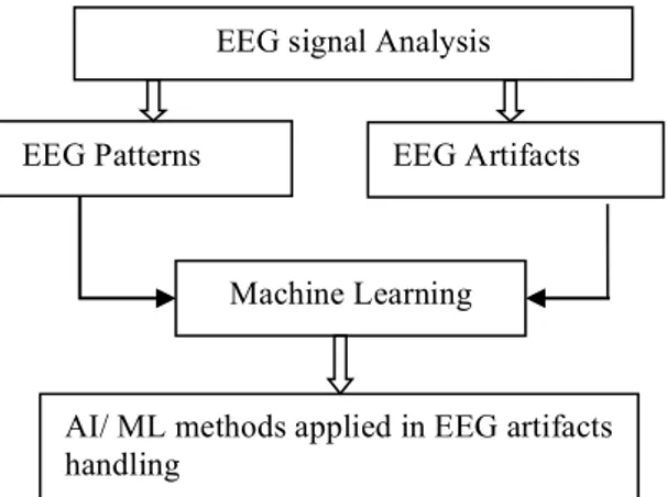
Study of EEG artifacts handling covers a variety of research area including computer science, neuroscience, health and medical science, and biomedical engineering, and various studies have been conducted over the years. In order to identify related literature, we used key words: EEG artifacts, machine learning technique, EEG signal analysis, automatic artifact handling, EEG pattern recognition. We refined the results in two categories 1) where machine learning and AI methods have been applied, and 2) other EEG artifacts handling methods. This paper focuses on the machine learning (ML) techniques use in EEG artifacts handling that are created or reported on between 2007 and 2014. A three-tier review methodology (Figure 1) has been applied to refine the machine learning methods applied in EEG artifacts removal.
Figure 1: Review methodology
In Section 2, we address various EEG artifacts, with central focus on physiological sources. In Section 3, existing methods for handling artifacts are addressed, with special focus on EOG and EMG artifacts. In Section 4, we present a review of artifact handling methods using machine learning. In Section 5, discussion of the machine learning methods are presented and finally, conclusions is presented in Section 6.
II. EEGARTIFACTS
EEG is inclined to various signal contaminations like other physiological signals. These contaminations are referred to as artifacts, which are signals of other than brain activity (Migotina et al., 2012; Talsma, 2008) and reduce the clinical usefulness of EEG (Kofronov & Biolek, 1996). The problems with artifacts in EEG signal are that they can make EEG uninterpretable, can change the brain signal and even mimic any cerebral activity that lead to serious misinterpretation (Chadwick et al., 2011; Klass, 1995). Moreover, in EEG signal processing e.g. power spectral analysis or topographic displays, artifacts can cause false conclusion unless artifactual data are handled or excluded from the processed EEG data. A. Sources of EEG Artifacts
EEG is the electric potential from the exposed surface of scalp and measured by the current flows when synaptic excitation of dendrites of many pyramidal neurons in the cerebral cortex. EEG signal is recorded from the scalp surface by electrodes and characterized by amplitude and frequency. The amplitude of the EEG signal is between 10-100 µV (Gratton, 1998; Senapati et al., 2010). Based on source, EEG artifacts can be divided into two categories a) Non-physiological and b) Physiological artifacts.
Non-physiological artifacts, also known as external artifacts are generally caused by device and recording equipment. These artifacts are occurred because of errors in the device, including interference from electric fields, poor electrode connection, electro-magnetic interference by nearby electronic devices etc. Daly I. et al. (2012) have mentioned power line noise at either 50 or 60 Hz, cable movement, sweating, electrode movement are the causes of non-physiological artifacts in EEG signal.
Physiological (or internal) artifacts in EEG signal are the main concern of this paper. The common causes of physiological artifacts are eye and head movements (Kiamini et al., 2009; O'Regan et al., 2010a; Sauter et al., 1990). Ma Junshui et al. (2012) have pointed out that the problem of EEG signals is often contaminated by skeletal or facial muscle activity i.e. muscle artifacts. Normal EEG signals are affected by high frequency signal components induced by Muscle artifacts and EEG power spectra at Beta and Gamma band are overpowered as a result (Ma et al., 2012). Cardiac activities can also cause artifacts in EEG signal. Therefore, based on the above description the sources of physiological artifacts can classify into three categories a) muscle activity (EMG) and b) ocular activity (EOG) and c) cardiac activity.
Positive cornea and negative retina of human eye generates electrical dipole and EOG signal is produced because of the change of the dipole by eye movement and blinks (Gratton, 1998; Shahbakhti et al., 2012). Ocular artifacts (OAs) are often dominant over other physiological artifacts i.e. head movement, muscle artifacts and most of the research articles are about dealing with ocular artifacts. EOG waveform depends on factors, for example, the direction of the eye movements. Eye blink artifacts are low frequency (<4 Hz) in nature and significant in amplitude. It can be located on frontal electrodes (FP1, FP2); which have symmetrical activity and low propagation. On the other hand eye movement artifacts are represented by low frequency (<4 Hz) but with higher propagation (Pourzare et al., 2012; Shahbakhti et al., 2012). Eye movements may occur in any direction and can be considered as combinations of rotations over two angles (a vertical angle and a horizontal angle). Different frequency ranges of EEG signal have been reported as neural information in several studies. Neural information can be obtained below 100 Hz from the EEG signal and in many applications information lies below 30 Hz (Akhtar et al., 2012). In (Kiamini et al., 2009) the range of EEG signal is 0 to 64 Hz and they mention that ocular artifacts occur within 0 and 16 Hz. A fraction of EOG contaminates the EEG signal and stronger peaks are introduced in the EEG signal because of the ocular artifacts (H. Ghandeharion & Ahmadi-Noubari, 2009; Kiamini et al., 2009; Ruijiang & Principe, 2006; Shahbakhti et al., 2012).
Another most common EEG artifacts are muscular artifacts. Both EOG and EMG artifacts overlap with neural brain activity and are recorded by sensors, which increase the difficulty to interpret the EEG signals. However, the hypothesis about these artifacts is that, they are independent from the brain activity, either normal or pathologic (Romo-Vazquez et al., 2007). Muscle artifacts are generally induced in the EEG because of head movements, jaw clenching, eyebrow raising etc. and several studies it is found that muscle artifacts mostly marked above frequency 13 Hz (Xinyi et al., 2008). Gasser et al.(2005) have pointed out that EMG artifacts occur in the frequency band 51-69 Hz which they have defined as muscle power. Cardiac activity such as heart-beats causes EEG artifacts (Jiang et al., 2007; Senapati et al., 2010).
III. EEGARTIFACTS HANDLING METHODS/ALGORITHMS
EEG Artifacts EEG signal Analysis EEG Patterns
Machine Learning
AI/ ML methods applied in EEG artifacts handling
A. Artifacts Identification
In EEG signal analysis a wide range of different features are considered to identify artifacts that characterise properties of artifacts, their spectral template, statistical properties of uni-variate of multivariate EEG. The question in artifact identification is, what clean/normal EEG looks like? Neurophysiology literature has defined some characteristics of clean EEG and based on that Daly (2012) has presented metrics of clean EEG, which are as follows:
Raw signal characteristics:
• Amplitudes should typically range between 10 and 100 uV (mostly below 50 uV).
• The signal should generally exhibit rounded or arc shaped sinusoidal morphology.
Alpha rhythm characteristics:
• The EEG between 8-12 Hz should exhibit a rounded or arc-shaped sinusoidal morphology. • b) Additionally, amplitudes typically take
values in the range of between 10 and 100 uV. • Alpha rhythms are typically larger over
paratial/occipital regions then frontal/central regions.
Beta rhythm characteristics:
• The EEG between 13-35 Hz should exhibit a rounded sinusoidal morphology.
• Amplitudes are typically lower than 30 uV. Power spectrum characteristics:
• Low frequencies typically exhibit high power while high frequencies exhibit low power.
The author has defined some metrics to check the range of values of the above quantities. These metrics are:
a) Maximum amplitude values.
b) Standard deviation of the amplitude values. c) Kurtosis of the amplitude values.
d) Skewness of the amplitude values
In (Daly et al., 2012; Hosna Ghandeharion & Erfanian, 2010), authors have also considered these metrics to identify artifacts in EEG signals.
B. Artifacts Handling
In the literature, there are several methods and algorithms for EEG artifacts detection and removal. However, most of the methods are to correct ocular artifacts in the EEG data. Many of these studies have discussed about muscle artifacts in the EEG signal. Several procedures or approaches that are used for EEG artifact correction are mostly based on 1) regression, 2) principal component analysis (PCA), 3) independent component analysis, 4) wavelet denoising, 5) filtering, 6) empirical mode decomposition.
1) Regression method: Regression-based method is the traditional approach of ocular artifacts corrections, which computes propagation factors or transmission coefficients to determine the correlation between one or more electrooculogram (EOG) channels and each EEG channel. In time or frequency domain it subtracts EOG portions that are contributing in EEG signal. The problem with regression
analysis is that it not only reduces ocular artifacts but also it may remove interesting cerebral activity. It also requires EOG reference channel for artifact removal and it requires a calibration trail to determine the transfer coefficients between EOG and EEG channels. Muscle noise removal is impractical by regression method since it requires multiple reference channels for multiple muscle groups. Also line noise removal in frequency domain is not feasible when 50 Hz to 60 Hz line frequency overlaps with the spectrum of EEG high frequencies (H. Ghandeharion & Ahmadi-Noubari, 2009; Hosna Ghandeharion & Erfanian, 2010; Hoffmann & Falkenstein, 2008; Kiamini et al., 2009; Senapati et al., 2010; Shahbakhti et al., 2012).
2) ICA: In many research disciplines, including neural network research the fundamental problem is to find a suitable representation of multivariate data. For computational simplicity the representation often required as a linear transformation of the original data. Independent component analysis (ICA) is a method which finds a linear representation of non-Gaussian data where data are statistically independent (Hyvärinen & Oja, 2000). Independent component analysis (ICA) is a statistical method, which can decompose observed signals into statistically independent components. ICA assumes a data model 𝑋 = 𝐴𝑆, where X is a queued column vectors of data recorded from individual EEG channels, A is a weight matrix for mixing independent components back to original signals, S is queued column vector of statistically independent components. The M observed EEG signals
𝑥 𝑡 = 𝑥! 𝑡 , 𝑥! 𝑡 , … … , 𝑥! 𝑡 ! are generated as a sum of the
N independent components 𝑡 = 𝑠!𝑡 , 𝑠!𝑡 , … … , 𝑠! 𝑡 ! ,
𝑋 = 𝐴𝑆 . The mixing matrix A is consisted of mixing coefficients 𝑎!,! 𝑖 = 1,2, … , 𝑁 𝑗 = 1,2, … , 𝑀 . In the ICA model,
number of sources N and the mixing matrix A are usually unknown. It is commonly supposed that M=N and the task of ICA method is to recover unknown source signals 𝑠 𝑡 by introducing unmixing matrix W; 𝑌 = 𝑊𝑋 , Where W is inverse matrix of the mixing matrix A. W obtained by considering the independence of the signal. Y represents the independent components that are estimates of sources S. Since there is no knowledge of matrix A, it is not possible to determine W exactly.
ICA is the most common method that has been used in EEG signal decomposition and it can be used to identify various artifacts from EEG signal. However, one problem with ICA is that it needs visual inspection of extracted components and manual classification of the interference components (Akhtar et al., 2012; H. Ghandeharion & Ahmadi-Noubari, 2009; Hosna Ghandeharion & Erfanian, 2010). This is time-consuming and undesirable for real-time artifact suppression. Therefore, methods e.g., wavelet denoising, mutual information have been combined with ICA to perform automatic artifacts identification and removing (Akhtar et al., 2012; H. Ghandeharion & Ahmadi-Noubari, 2009; Hosna Ghandeharion & Erfanian, 2010). Jiang et al. (2007) have developed an automatic method for detection and elimination of ECG artifacts from EEG signal using ensemble average subtraction (EAS) method, independent component
analysis (ICA), and adaptive noise cancelling theory. Wavelet based EEG signal decomposition has been performed to extract features and classify EEG data. Independents components can be separated by the kurtosis of their amplitude distribution over time, which could discriminate the EEG signals between strictly periodical signals, regularly occurring signals and irregularly occurring signals (Delorme et al., 2007; Vigário, 1997). In (Delorme et al., 2007) authors have compared five different methods including kurtosis to detect trails containing artifacts. Four other methods are: extreme values, linear trends, data improbability and spectral pattern. Daly et al (2013) have presented an ICA based method for reduction of head movement artifacts from EEG where authors have used an accelerometer to measure head movement. In addition, this method also removes the influence of head movement artifacts from EEG signal. Independent components of EEG signals, which are correlated with accelerometer above some threshold, are flagged as artifacts and later removed from the EEG signal. Thresholding method is another approach to identify artifactual components from independent components (Geetha & Geethalakshmi, 2012; Nolan et al., 2010).
3) PCA: Principal Component Analysis (PCA) decomposes signals into uncorrelated, but not necessarily independent components that are spatially orthogonal. PCA cannot completely separate eye artifacts from EEG signals, when they have comparable or similar voltage amplitudes. PCA extracts and sort out the principal components according to the influence on the overall data space. It is not likely that any or more principal components represent the artifacts. It requires some prior knowledge to identify the principal components as the artifact. A PCA based automatic artifact reduction algorithm is proposed in (Schachinger et al., 2007). The authors suggest that the proposed algorithm can be used to eliminate various kinds of artifacts from EEG signal with manual interaction. Different artifacts containing frequency bands are isolated from the decomposed EEG signal using the PCA based algorithm. Then the dominant artifacts activities are eliminated from the decomposed component using an adaptive threshold method.
4) EMD: Empirical Mode Decomposition (EMD) is a computational method for nonlinear and non-stationary signal analysis, proposed by N. E. Huang (Kiamini et al., 2009). EMD decomposes a signal without leaving time domain. Original signal is transformed to a complete and nearly orthogonal basis after functions are filtered out by the EMD. Completeness is based on the method of the EMD; the way it is decomposed implies completeness. Intrinsic Mode Function (IMF) is the output basis functions of EMD, are derived from the data and are susceptible to the Hilbert Transform for spectrum analysis. Unlike traditional signal analysis methods such as Fourier Transforms and wavelets, EMD does not require any priori known basis since it relies on fully data-driven mechanism and it is an intuitive, direct and adaptive method. The signal that is decomposed into functions, are all in time domain and of the same length as original signal. It is
important to obtain IMFs from the real world signals because generally natural processes have multiple causes and the causes may happen at specific time intervals. These data can be obtained by EMD analysis where this information is hidden in other analysis such as Fourier Transforms or wavelet coefficients. EMD generates a collection of IMFs, which are based on the direct extraction of the energy associated with various natural time scales. These IMFs are well-behaved Hilbert transforms from which the instantaneous frequencies can be calculated. The local energy and the instantaneous frequency derives from the IMF through the Hilbert transform can give us a full energy-frequency-time distribution of the data.
Most of the articles use EMD for removing EOG artifacts from EEG (Kiamini et al., 2009; Molla et al., 2012; Shahbakhti et al., 2012; Soomro et al., 2013; Zhang et al., 2008). In (Kiamini et al., 2009), EEG signal is classified into edges by correspond to eye blinks and those do not correspond to eye blinks, based on relative amplitude. Then they have applied their proposed EMD based method only to the detected ocular artifact zones. The advantage of this method is that it avoids the removal of background EEG information. In the paper (Shahbakhti et al., 2012), the authors suggest that eye blinks increase the power of the frequency at lower end of the EEG spectrum, therefore, artifacts component lie in the last end of several IMFs. Hence, until sifting process of EMD does not reach the artifact components, the entropy of two consecutive IMFs decreases towards decomposition level. So the authors first decompose the signal into IMFs then calculate the entropy between two consecutive IMFs. After that decomposition level M is identified, at which the entropy begins to go up and then clean signal is reconstructed by summing up the first M IMFs. Zhang et al (2008) proposed a novel and efficient algorithm based on EMD and they suggested that this algorithm can detect, separate and remove various artifacts from EEG signals. Authors have used this algorithm to remove power interference and EMG artifacts from EEG signal. Their experimental results suggest that proposed method is data-driven and adaptive and ideal for computations of nonlinear and non-stationary signal processing. EMD based approach has also been used to remove EMG artifacts from EEG signals (Safieddine et al., 2012).
5) Other methods: Apart from decomposition methods i.e. PCA, ICA, and EMD there are several methods that have been proposed in various studies including, but not limited to, different filter algorithms, autoregressive method and variety of signal transform algorithms. Several studies mentioned the drawback of PCA and ICA method (Djuwari et al., 2005) and therefore, different techniques have been proposed other than PCA and ICA. The major drawback of PCA is pointed out as, that; it cannot completely separate ocular artifacts when both EEG and EOG waveform have similar voltage amplitude. On the other hand ICA requires visual inspection to manually select and remove artifacts components from EEG signal (Senapati et al., 2010; Wallstrom et al., 2004).
Power spectrum analysis has been done in (Gasser et al., 2005) to correct muscle artifact in the EEG power spectrum. Authors have used Fast Fourier transform on 20s epoch or recording and spectral power was computed on delta, theta, alpha and beta band. They have defined muscle power in the frequency band 51.0–69.0 Hz as an indicator of EMG contamination in the EEG. To eliminate the influence of muscle artifacts a linear regression analysis has been performed with log power in the muscle band as explanatory variable and log EEG band power as outcome variable. Autoregressive (AR) method has been used in several studies to model EEG data and artifact correction (Cluitmans & Van de Velde, 2000; Lawhern et al., 2012; Lawhern et al., 2013b). Authors have also mentioned that AR model provides compact and computationally efficient representation of EEG signals. In addition, the advantage of AR model is that, the parameters of AR model are constant with respect to the scaling changes in the data, which can be occurred from inter-subject variations, for example, scalp and skull thickness. In the paper (Lawhern et al., 2013b) the authors have represented the statistical method for identifying the optimal AR features based on penalized multinomial regression. To determine the optimal features, elastic net penalization of the standard likelihood solution have been used since AR coefficients reveal a high degree of multivariable co-linearity. Their results are agreed with known brain physiological properties, like; eye movement artifacts are mostly strong at the frontal channels. In (Senapati et al., 2010), authors have proposed a new technique for ocular artifact removal from EEG signal using S-transform. The S-transform (ST) is a mathematical operation that produces frequency content at each time point within a time varying signal. It generates high amplitude S-coefficients for the artifactual instants in the signal. Authors used a statistical threshold function to filter out the artifacts in the S-domain. The main advantage of this method is that it is possible to remove artifact frequencies within a narrow time-frame while preserving the frequency information at all other time points. After removing the artifacts it also preserves the absolute referenced phase information of the signal. Wavelet based artifact detection is presented in the paper (Inuso et al., 2007) where Renyi’s entropy and kurtosis are used to identify artifacts from wavelet components. Four major EEG wave alpha, beta, theta and delta are extracted from the original EEG recording using wavelet transform and they measured the artifactuality rate of each wavelet component. The authors have also discussed the joint use of these three methods as a pre-processing step to optimize artifacts from EEG.
IV. MACHINE LEARNING ALGORITHMS IN EEGARTIFACTS
HANDLING
Different methods have been developed to identify and remove artifacts from EEG signals. However, expert’s observation is required to identify artifacts from the EEG signals. In the case of automatic artifacts handling, some threshold is required to classify artifactual components. Machine learning techniques are used to recognise the patterns in and improve the classification accuracy. Support vector
machine (SVM) and artificial neural network are the two most commonly used techniques in EEG artifacts handling.
Hybrid approach such as combination of ICA and SVM provides a promising method for automatic artifacts handling. Support vector machine is a supervised and one of the main statistical learning algorithms that can classify unseen data using decision boundaries, which is derived by some rule to separate data into discrete classes. One important property of SVM is its good generalization capacity independent of the input space dimension that makes SVM good candidate for the analysis of biomedical data e.g., multi-channel EEG recording.
Lawhern et al. (Lawhern et al., 2012; Lawhern et al., 2013a, 2013b) have developed a Matlab toolbox for EEG signal processing where SVM has been used for signal classification. They have used autoregressive model for feature extraction and characterisation of EEG signals containing various type of subject generated artifacts including jaw clenching, jaw movement, eye blinks, eye movements, raising and lowering eyebrows and rotation head side-top-side. To classify the artifacts conditions using the AR model parameter features, SVM is used and the results suggest that it is possible to discriminate artifactual data effectively using very low order AR model. O’Regan and his co-workers also worked with SVM to detect head movement artifacts in EEG signals (O'Regan et al., 2010a, 2010b; O’Regan et al., 2013; O’Regan & Marnane, 2013). In these articles the authors have discussed several feature extraction methods that can be used for head movement artifacts identification. Time, frequency and entropy features are extracted and evaluated in these papers. Mutual Information Evaluation Function and Linear Discriminant Analysis, Kolmogorov-Smirnov and Wilcoxon rank-sum non-parametric tests methods have been used to evaluate the features. To distinguish normal EEG and artifacts the authors have used Mutual Information as a measure of usefulness of individual features. Support Vector Machine and Linear Discriminant Analysis are the two methods that have been used in these papers to classify head movement related artifacts.
In (O’Regan et al., 2013) authors also applied fusion approach using EEG and gyroscope signals for classification of artifact and non-artifact data. In another study, based on the results, Shi-Yun (Shi-Yun et al., 2009; Shi-Yun et al., 2008) recommended that combination of ICA and Support vector machine (SVM) is a better approach for automatic artifact removal. They have compared the proposed SVM approach with other classification methods, namely, K-nearest neighbors (KNN), Gaussian mixture models (GMM), linear discriminant function (LDF) and standard SVM. And from the experimental results they found that the proposed method obtained better accuracy than the other methods they have compared with. SVM has also been considered as artifacts classification method in (Chin-Teng et al., 2012; Hsu et al., 2012; Tangermann et al., 2009; Winkler et al., 2011) and achieved good classification accuracy.
In several studies, another method that has been frequently used is Artificial Neural Network. In the paper (Hwa-Shan et
al., 2009), the authors suggest that machine learning tools can be effective to discriminate artifactual components from independent components (ICs) after applying ICA. They have used three machine learning methods multilayer perceptron architecture with hyperbolic tangent signal function and multilayer perceptron architecture with redial basis function signal function, and the radial basis function neural network and a simple majority voting algorithm fusion scheme to improve the system performance. An arm movement classification system from EEG signal is proposed by (Marquez L & Munoz G, 2013). The system also recognises the executed and imagined movements. The authors have used a multilayer perceptron neural network for the classification and feature extraction is performed using wavelet analysis. They have reported that the system identifies a great number of movements and real movements were associated with the imagined movements. An ANN multilayer perceptron is also proposed by (Sovierzoski et al., 2009) to classify eye blink in EEG signal. Nguyen et al., (2012) have proposed a wavelet neural network for removal of EOG artifacts from EEG. The advantage of their method is that it does not require EOG recording as reference for artifact removal.
Different clustering algorithms have been found in the literatures that are used for EEG artifacts identification. In (Yuan et al., 2012) a ICA based clustering method has been proposed to automatically extract event-related components and remove artifacts from EEG signals. The authors have used hierarchical clustering algorithm to group all independent components into several hierarchical clusters. Pre-determined template is constructed for the target event-related components and based on that they have selected suitable cluster, which is used later to refine the EEG signals through the inverse transform process of ICA. In the article (Patidar & Zouridakis, 2008) a hybrid algorithm using iterative ICA and fuzzy clustering has been proposed for artifacts rejection in EEG signals. They have first identified the ICs that represent artifacts from iICA and then clustered based on several spectral and temporal features. The combination of iICA and fuzzy clustering procedure has provided better results than the iICA alone. The authors quantify the results based on improvement of the signal-to-noise ratio after processing EEG signals. Nicolaou and Nasuto (2007) are suggested another clustering approach combined with temporal decorrelation source separation (TDSEP) method. They have used lag auto-mutual information as feature for clustering the estimated components into cerebral and non-cerebral activity. Later, the cluster that has contained ocular components is discarded and remaining components are used to reconstruct clean EEG signals. However, they have also used separate template for ocular activity that is obtained from another dataset. In (Pourzare et al., 2012) various facial muscle movement artifacts are classified using K-nearest neighbour algorithm. Features are extracted based on root mean square, polynomial fitting and Hjorth descriptors method. Authors mention that the simplicity of feature extractions is the good attribute of this proposed method. And, one of the uniqueness of this approach is the third order polynomial fitting method of
feature extraction. Results showed that k-NN algorithm performed good to classify the artifacts containing data set. Aydemir et al (2012) have proposed a k-NN based EMG and EOG artifacts classification approach. Here, they have extracted three features namely, root mean square (RMS), polynomial fitting, and Hjorth descriptors, and three classifier algorithms are used to classify artifacts. First, features are extracted by RMS and classified by first classifier. The results of the first classifier are temporarily recognised into five classes: class1, class2, class3, class4, and class5. If the classified result recognises as class1 or class2 then features are extracted using polynomial fitting method. Then the trained classifier2 algorithm is determined as exactly the trial class1 or class2. If the result recognises as class3 or class4 the features are extracted using Hjorth descriptors. Then the trained classifier3 algorithm is determined as exactly the trial class3 or class4. The last type class5 is determined directly as class5 from classifier1.
Junfeng et al (2009) introduces a learning method called manifold learning algorithm for dimensionality reduction of initial features and a classifier is used to identify artifact components from independent components of ICA. A decision tree classifier has been applied to classify artifacts in (Chadwick et al., 2011). The authors then use this classification to improve existing artifact removal methods. Mainly four types of classification are used in the experiment; artifacts occur by eye movement only, head movement only, joint eye and head movement and eye blink. Two classifiers, decision tree and Hidden Markov Model (HMM) are the methods that are chosen for artifact classification. The authors have selected decision tree because it has ability to select features. However, the drawback of decision tree is it cannot handle time-series data. Therefore, HMM has been chosen to model the data’s time component.
Other machine learning methods that are also found in the literatures are fuzzy inference system (Kezi Selva Vijilal et al., 2007; Sheniha et al., 2013), genetic algorithm (Fairley et al., 2010; Poli et al., 2011), and Bayesian model (Schetinin & Maple, 2007).
V. DISCUSSION
As it can be seen from the earlier chapters, a wide range of research studies has been done for EEG artifacts removal. Methods that have been proposed can be divided into manual, semi-automatic and automatic. Manual and semi-automatic methods require expert observations to identify artifacts in EEG signal. On the other hand, automatic methods require predefined threshold value. In the past few years, machine learning techniques have been advanced significantly and used in pattern identification and classification problems. Table 1 presents a summary of the papers based on the different machine learning algorithms presented earlier in this paper.
Table 1 shows that the SVM is the mostly used method and different approaches of SVM are applied to classify artifacts in EEG signal. Gaussian kernel and radial basis function (RBF) are found most appropriate approaches for EEG artifacts
classifications (Hsu et al., 2012; Lawhern et al., 2012; Phothisonothai et al., 2012; Winkler et al., 2011). “One-against-one” approach is used in these studies to solve multiclass classification problem. In separate paper Shi-Yun suggests two approaches, one is weighted SVM with error correction (Shi-Yun et al., 2009) and the other one is probabilistic multi-class SVM (Shi-Yun et al., 2008). On the other hand, multilayer perceptron neural network is the most common technique in EEG artifacts handling. The multilayer perceptron neural network is composed by sum of the
products of all the input signals including the bias, by respective synaptic weight of the connection and followed by activation function.
Cluster methods are also common to classify EEG artifacts. Yuan et al. (2012) have suggested that hierarchical clustering is better approach than k-mean and fuzzy c-mean clustering since in EEG artifacts handling, the number of clusters often unknown. K-mean and fuzzy c-means are both iterative methods and target number of clusters are required to terminate the clustering iterations.
TABLEI
SUMMARY OF PAPERS BASED ON DIFFERENT MACHINE LEARNING TECHNIQUES Machine Learning
Technique Associated Methods References
Support Vector Machine ICA, BSS, Autoregressive model
(Bartels et al., 2010; Chin-Teng et al., 2012; Gao, Yang, et al., 2010; Halder et al., 2007; Hsu et al., 2012; Lawhern et al., 2012; Lawhern et al., 2013a; O’Regan et al., 2013; O’Regan & Marnane, 2013; Phothisonothai et al., 2012; Shi-Yun et al., 2009; Shi-Shi-Yun et al., 2008; Singla et al., 2011; Tangermann et al., 2009; Winkler et al., 2011; Wu et al., 2009)
Artificial Neural Network ICA, Spectral analysis
(Chin-Teng et al., 2012; Jafarifarmand & Badamchizadeh, 2013; Junfeng et al., 2009; Marquez L & Munoz G, 2013; Nguyen et al., 2012; Singla et al., 2011; Sovierzoski et al., 2009)
Fuzzy Inference system Differential Evolution Adaptive Noise Cancellation (Kezi Selva Vijilal et al., 2007; Sheniha et al., 2013)
Clustering Kurtosis (Nicolaou & Nasuto, 2007; Patidar & Zouridakis, 2008; Yuan et al., 2012)
K-NN Polynomial fitting, Hjort descriptor (Aydemir et al., 2012; Gao, Lin, et al., 2010; Pourzare et al., 2012) Bayesian Model Spectral power (Schetinin & Maple, 2007)
Genetic programming Power spectral analysis, kurtosis (Fairley et al., 2010; Poli et al., 2011) Figure 2 shows the basic model of automatic EEG
artifacts handling using ICA and threshold method. Artifacts handling using machine learning method is shown in Figure 3. The main difference between these two approaches is that after extracting features some threshold
is applied to distinguish the artifactual and non-artifactual components in ICs, whereas, in machine learning approach components are classified based on the spectral and topographical characteristics.
Figure 2: Automatic EEG Artifacts Removal Raw EEG
Figure 3: EEG artifacts Removal using Machine Learning Algorithm Hybrid approaches are also found in few studies where
same machine learning technique or two different techniques are combined to classify different artifacts. Chin-Teng et al., (2012) have applied SVM and self-organizing map (SOM), where SVM is used to distinguish useful and artefact components and later SOM to identify different brain sources. Aydemir (2012) has used multilayer k-NN algorithm and in (Bartels et al., 2010) two SVM are applied to classify EOG and EMG artifacts.
Comparison of different machine learning methods has also been reported in several studies. In (Shi-Yun et al., 2009), authors compared their proposed method (weighted probabilistic SVM with error correction) with standard SVM, k-NN, linear discriminant function, Gaussian mixture model. From the experiment they have found that their proposed method provides better classification than others. In (Gao, Lin, et al., 2010) performance of SVM has been compared with Fisher discriminant analysis and back propagation neural network and SVM outperformed other two methods. In another studies (Gao, Yang, et al., 2010; Junfeng et al., 2009) four methods are compared, namely, Fisher discriminant analysis, neural network, PCA with neural network and manifold learing with k-NN. The results suggest that manifold learning with k-NN is better than other three approaches. Singla et al (2011) compared SVM and artificial neural network (ANN) to classify eye events and the experiment results have shown that SVM provided a maximum classification accuracy of 90.8% and ANN obtained 86.8%, which suggested that SVM has better performance than ANN classifier. Classification accuracy of the algorithms are measured based on Cross-validation, Mean Square Error (MSE), sensitivity and specificity, Receiver operator characteristics (Lawhern et al., 2012; Nguyen et al., 2012; O’Regan & Marnane, 2013; Sheniha et al., 2013; Sovierzoski et al., 2009).
VI. CONCLUSIONS
This paper presents a literature review of machine learning algorithms that are frequently used in EEG artifacts handling. This article provides an overview of how certain machine leaning techniques have been applied in handling different EEG artifacts. From the study, it is revealed that a large number of automatic and semi-automatic methods are
available for EEG artifacts removal. However, the usage of machine learning algorithms is limited. It is also found that machine learning algorithms provide better classification accuracy than other approaches. Most popular method of EEG signal classification is ICA but to identify artifacts from independent components of ICA it requires expert observation, where machine learning algorithms can be applied to ease the classification process. Moreover, comparison of different techniques is also studied and in several studies it is suggested that SVM is better classifier than other classification methods. Finally, the survey leaves us with focus on hybrid approaches i.e., using several machine learning algorithms.
ACKNOWLEDGMENT
The authors of this work are supported by the Vehicle Driver Monitoring,VDM project funded by Vinnova.
REFERENCES
[1] Akhtar, Muhammad Tahir, Mitsuhashi, Wataru, & James, Christopher
J. (2012). Employing spatially constrained ICA and wavelet denoising, for automatic removal of artifacts from multichannel EEG data. Signal Processing, 92(2), 401-416.
[2] Aydemir, O., Pourzare, S., & Kayikcioglu, T. (2012). Classifying Various EMG and EOG Artifacts in EEG Signals. PRZEGLĄD ELEKTROTECHNICZNY (Electrical Review), 88(11a), 218-222.
[3] Bartels, G., Shi, L. C., & Lu, B. L. (2010). Automatic artifact removal
from EEG - a mixed approach based on double blind source separation and support vector machine. Conference proceedings : Annual International Conference of the IEEE Engineering in Medicine and Biology Society. IEEE Engineering in Medicine and Biology Society. Conference, 2010, 5383-5386.
[4] Chadwick, N. A., McMeekin, D. A., & Tan, T. (2011, May 31
2011-June 3 2011). Classifying eye and head movement artifacts in EEG signals. Paper presented at the Digital Ecosystems and Technologies Conference (DEST), 2011 Proceedings of the 5th IEEE International Conference on.
[5] Chin-Teng, Lin, Yu-Kai, Wang, & Shi-An, Chen. (2012, 10-15 June 2012). A hierarchal classifier for identifying independent components. Paper presented at the Neural Networks (IJCNN), The 2012 International Joint Conference on.
[6] Cluitmans, P. J. M., & Van de Velde, M. (2000, 2000). Outlier detection to identify artefacts in EEG signals. Paper presented at the Engineering in Medicine and Biology Society, 2000. Proceedings of the 22nd Annual International Conference of the IEEE.
[7] Daly, I., Billinger, M., Scherer, R., & Muller-Putz, G. (2013). On the
Automated Removal of Artifacts Related to Head Movement From the EEG. Neural Systems and Rehabilitation Engineering, IEEE Transactions on, 21(3), 427-434.
Raw EEG
[8] Daly, I., Pichiorri, F., Faller, J., Kaiser, V., Kreilinger, A., Scherer, R., & Muller-Putz, G. (2012, Aug. 28 2012-Sept. 1 2012). What does clean EEG look like? Paper presented at the Engineering in Medicine and Biology Society (EMBC), 2012 Annual International Conference of the IEEE.
[9] Delorme, Arnaud, Sejnowski, Terrence, & Makeig, Scott. (2007).
Enhanced detection of artifacts in EEG data using higher-order statistics and independent component analysis. NeuroImage, 34(4), 1443-1449
[10] Djuwari, D., Kumar, D. K., & Palaniswami, M. (2005, 17-18 Jan. 2006). Limitations of ICA for Artefact Removal. Paper presented at the Engineering in Medicine and Biology Society, 2005. IEEE-EMBS 2005. 27th Annual International Conference of the. [11] Fairley, Jacqueline, Georgoulas, George, Stylios, Chrysostomos, &
Rye, David. (2010). A Hybrid Approach for Artifact Detection in EEG Data. In K. Diamantaras, W. Duch & L. Iliadis (Eds.), Artificial Neural Networks – ICANN 2010 (Vol. 6352, pp. 436-441): Springer Berlin Heidelberg.
[12] Gao, Junfeng, Lin, Pan, Yang, Yong, Wang, Pei, & Zheng, Chongxun. (2010). Real-time removal of ocular artifacts from EEG based on independent component analysis and manifold learning. Neural Computing and Applications, 19(8), 1217-1226.
[13] Gao, JunFeng, Yang, Yong, Lin, Pan, Wang, Pei, & Zheng, ChongXun. (2010). Automatic Removal of Eye-Movement and Blink Artifacts from EEG Signals. Brain Topography, 23(1), 105-114. doi: 10.1007/s10548-009-0131-4
[14] Gasser, Theo, Schuller, Jan C., & Gasser, Ursula Schreiter. (2005). Correction of muscle artefacts in the EEG power spectrum. Clinical Neurophysiology, 116(9), 2044-2050.
[15] Geetha, G., & Geethalakshmi, S. N. (2012). Artifact Removal from EEG using Spatially Constrained Independent Component Analysis and Wavelet Denoising with Otsu's Thresholding Technique. Procedia Engineering, 30(0), 1064-1071.
[16] Ghandeharion, H., & Ahmadi-Noubari, H. (2009, April 29 2009-May 2 2009). Detection and removal of ocular artifacts using Independent Component Analysis and wavelets. Paper presented at the Neural Engineering, 2009. NER '09. 4th International IEEE/EMBS Conference on.
[17] Ghandeharion, Hosna, & Erfanian, Abbas. (2010). A fully automatic ocular artifact suppression from EEG data using higher order statistics: Improved performance by wavelet analysis. Medical Engineering & Physics, 32(7), 720-729.
[18] Gratton, Gabriele. (1998). Dealing with artifacts: The EOG contamination of the event-related brain potential. Behavior Research Methods, Instruments, & Computers, 30(1), 44-53. [19] Halder, Sebastian, Bensch, Michael, Mellinger, Jurgen, Bogdan,
Martin, Kubler, Andrea, Birbaumer, Niels, & Rosenstiel, Wolfgang. (2007). Online Artifact Removal for Brain-Computer Interfaces Using Support Vector Machines and Blind Source Separation. Computational Intelligence and Neuroscience, 2007. [20] Hoffmann, Sven, & Falkenstein, Michael. (2008). The Correction of
Eye Blink Artefacts in the EEG: A Comparison of Two Prominent Methods. PLoS ONE, 3(8), e3004.
[21] Hsu, Wei-Yen, Lin, Chao-Hung, Hsu, Hsien-Jen, Chen, Po-Hsun, & Chen, I. Ru. (2012). Wavelet-based envelope features with automatic EOG artifact removal: Application to single-trial EEG data. Expert Systems with Applications, 39(3), 2743-2749. [22] Hwa-Shan, Huang, Pal, N. R., Li-Wei, Ko, & Chin-Teng, Lin. (2009,
14-19 June 2009). Automatic identification of useful independent components with a view to removing artifacts from eeg signal. Paper presented at the Neural Networks, 2009. IJCNN 2009. International Joint Conference on.
[23] Hyvärinen, A., & Oja, E. (2000). Independent component analysis: algorithms and applications. Neural Networks, 13(4–5), 411-430. [24] Inuso, G., La Foresta, F., Mammone, N., & Morabito, F. C. (2007, 8-11
July 2007). Brain Activity Investigation by EEG Processing: Wavelet Analysis, Kurtosis and Renyi's Entropy for Artifact Detection. Paper presented at the Information Acquisition, 2007. ICIA '07. International Conference on.
[25] Jafarifarmand, Aysa, & Badamchizadeh, Mohammad Ali. (2013). Artifacts removal in EEG signal using a new neural network enhanced adaptive filter. Neurocomputing, 103(0), 222-231.
[26] Jiang, Joe-Air, Chao, Chih-Feng, Chiu, Ming-Jang, Lee, Ren-Guey, Tseng, Chwan-Lu, & Lin, Robert. (2007). An automatic analysis method for detecting and eliminating ECG artifacts in EEG. Computers in Biology and Medicine, 37(11), 1660-1671.
[27] Junfeng, Gao, Chongxun, Zheng, & Pei, Wang. (2009, 17-19 Oct. 2009). Automatic Removal of Ocular Artifacts from EEG Signals. Paper presented at the Biomedical Engineering and Informatics, 2009. BMEI '09. 2nd International Conference on.
[28] Kezi Selva Vijilal, C., Kanagasabapathy, P., Johnson, S., & Ewards, V. (2007, 22-24 Feb. 2007). Artifacts Removal in EEG Signal using Adaptive Neuro Fuzzy Inference System. Paper presented at the Signal Processing, Communications and Networking, 2007. ICSCN '07. International Conference on.
[29] Kiamini, M., Alirezaee, S., Perseh, B., & Ahmadi, M. (2009, 6-9 May 2009). Elimination of Ocular Artifacts from EEG signals using the wavelet transform and empirical mode decomposition. Paper presented at the Electrical Engineering/Electronics, Computer, Telecommunications and Information Technology, 2009. ECTI-CON 2009. 6th International Conference on.
[30] Klass, D. W. (1995). The continuing challenge of artifacts in the EEG. American Journal of EEG Technology, 35(4), 239-269.
[31] Klonowski, Wlodzimierz. (2009). Everything you wanted to ask about EEG but were afraid to get the right answer. Nonlinear Biomedical Physics, 3(1), 1-5. doi: 10.1186/1753-4631-3-2
[32] Kofronov, M., & Biolek, D. (1996, 18-21 Aug 1996). Artefact filtering from human EEG. Paper presented at the Circuits and Systems, 1996., IEEE 39th Midwest symposium on.
[33] Lawhern, Vernon, Hairston, W. David, McDowell, Kaleb, Westerfield, Marissa, & Robbins, Kay. (2012). Detection and classification of subject-generated artifacts in EEG signals using autoregressive models. Journal of Neuroscience Methods, 208(2), 181-189. [34] Lawhern, Vernon, Hairston, W. David, & Robbins, Kay. (2013a).
DETECT: A MATLAB Toolbox for Event Detection and Identification in Time Series, with Applications to Artifact Detection in EEG Signals. PLoS ONE, 8(4), e62944. doi: 10.1371/journal.pone.0062944
[35] Lawhern, Vernon, Hairston, W. David, & Robbins, Kay. (2013b). Optimal Feature Selection for Artifact Classification in EEG Time Series. In D. Schmorrow & C. Fidopiastis (Eds.), Foundations of Augmented Cognition (Vol. 8027, pp. 326-334): Springer Berlin Heidelberg.
[36] Ma, Junshui, Tao, Peining, Bayram, Sevinç, & Svetnik, Vladimir. (2012). Muscle artifacts in multichannel EEG: Characteristics and reduction. Clinical Neurophysiology, 123(8), 1676-1686.
[37] Marquez L, A. P., & Munoz G, R. (2013, Sept. 30 2013-Oct. 4 2013). Analysis and classification of electroencephalographic signals (EEG) to identify arm movements. Paper presented at the Electrical Engineering, Computing Science and Automatic Control (CCE), 2013 10th International Conference on.
[38] Migotina, D., Calapez, A., & Rosa, A. (2012, 27-30 May 2012). Automatic Artifacts Detection and Classification in Sleep EEG Signals Using Descriptive Statistics and Histogram Analysis: Comparison of Two Detectors. Paper presented at the Engineering and Technology (S-CET), 2012 Spring Congress on.
[39] Molla, M. K. I., Tanaka, T., & Rutkowski, T. M. (2012, 25-30 March 2012). Multivariate EMD based approach to EOG artifacts separation from EEG. Paper presented at the Acoustics, Speech and Signal Processing (ICASSP), 2012 IEEE International Conference on.
[40] Nguyen, Hoang-Anh T., Musson, John, Li, Feng, Wang, Wei, Zhang, Guangfan, Xu, Roger, . . . Li, Jiang. (2012). EOG artifact removal using a wavelet neural network. Neurocomputing, 97(0), 374-389. [41] Nicolaou, N., & Nasuto, S. J. (2007). Automatic Artefact Removal
from Event-related Potentials via Clustering. The Journal of VLSI Signal Processing Systems for Signal, Image, and Video Technology, 48(1-2), 173-183. doi: 10.1007/s11265-006-0011-z [42] Nolan, H., Whelan, R., & Reilly, R. B. (2010). FASTER: Fully
Automated Statistical Thresholding for EEG artifact Rejection. Journal of Neuroscience Methods, 192(1), 152-162.
[43] O'Regan, S., Faul, S., & Marnane, W. (2010a, Aug. 31 2010-Sept. 4 2010). Automatic detection of EEG artefacts arising from head movements. Paper presented at the Engineering in Medicine and
Biology Society (EMBC), 2010 Annual International Conference of the IEEE.
[44] O'Regan, S., Faul, S., & Marnane, W. (2010b, 7-10 Nov. 2010). Automatic detection of EEG artefacts arising from head movements using gyroscopes. Paper presented at the Applied Sciences in Biomedical and Communication Technologies (ISABEL), 2010 3rd International Symposium on.
[45] O’Regan, Simon, Faul, Stephen, & Marnane, William. (2013). Automatic detection of EEG artefacts arising from head movements using EEG and gyroscope signals. Medical Engineering & Physics, 35(7), 867-874.
[46] O’Regan, Simon, & Marnane, William. (2013). Multimodal detection of head-movement artefacts in EEG. Journal of Neuroscience Methods, 218(1), 110-120.
[47] Patidar, U., & Zouridakis, G. (2008, 20-25 Aug. 2008). A hybrid algorithm for artifact rejection in EEG recordings based on iterative ICA and fuzzy clustering. Paper presented at the Engineering in Medicine and Biology Society, 2008. EMBS 2008. 30th Annual International Conference of the IEEE.
[48] Phothisonothai, M., Fang, Duan, Tsubomi, H., Kondo, A., Aihara, K.,
Yoshimura, Y.,Watanabe, K. (2012, 5-7 Dec. 2012). Artifactual
component classification from MEG data using support vector machine. Paper presented at the Biomedical Engineering International Conference (BMEiCON), 2012.
[49] Poli, R., Cinel, C., Citi, L., & Salvaris, M. (2011, April 27 2011-May 1 2011). A genetic programming approach to detecting artifact-generating eye movements from EEG in the absence of electro-oculogram. Paper presented at the Neural Engineering (NER), 2011 5th International IEEE/EMBS Conference on.
[50] Pourzare, S., Aydemir, O., & Kayikcioglu, T. (2012, 3-4 July 2012). Classification of various facial movement artifacts in EEG signals. Paper presented at the Telecommunications and Signal Processing (TSP), 2012 35th International Conference on.
[51] Romo-Vazquez, R., Ranta, R., Louis-Dorr, V., & Maquin, D. (2007, 22-26 Aug. 2007). EEG Ocular Artefacts and Noise Removal. Paper presented at the Engineering in Medicine and Biology Society, 2007. EMBS 2007. 29th Annual International Conference of the IEEE.
[52] Ruijiang, Li, & Principe, J. C. (2006, Aug. 30 2006-Sept. 3 2006). Blinking Artifact Removal in Cognitive EEG Data Using ICA. Paper presented at the Engineering in Medicine and Biology Society, 2006. EMBS '06. 28th Annual International Conference of the IEEE.
[53] Safieddine, Doha, Kachenoura, Amar, Albera, Laurent, Birot, Gwénaël, Karfoul, Ahmad, Pasnicu, Anca, . . . Merlet, Isabelle. (2012). Removal of muscle artifact from EEG data: comparison between stochastic (ICA and CCA) and deterministic (EMD and wavelet-based) approaches. EURASIP Journal on Advances in Signal Processing, 2012(1), 1-15.
[54] Sauter, D., Tomczak, M., Richard, A., Mouze-Amady, M., & Cail, F. (1990, 1-4 Nov 1990). Artefacts Detection And Pre-cleaning In Spectral EEG Analysis. Paper presented at the Engineering in Medicine and Biology Society, 1990., Proceedings of the Twelfth Annual International Conference of the IEEE.
[55] Schachinger, D., Schindler, K., & Kluge, T. (2007, 1-4 July 2007). Automatic Reduction of Artifacts in EEG-Signals. Paper presented at the Digital Signal Processing, 2007 15th International Conference on.
[56] Schetinin, V., & Maple, C. (2007, 1-4 July 2007). A Bayesian Model Averaging Methodology for Detecting EEG Artifacts. Paper presented at the Digital Signal Processing, 2007 15th International Conference on.
[57] Senapati, K., Kar, S., & Routray, A. (2010, 16-18 Dec. 2010). A new technique for removal of ocular artifacts from EEG signals using S-transform. Paper presented at the Systems in Medicine and Biology (ICSMB), 2010 International Conference on.
[58] Shahbakhti, M., Khalili, V., & Kamaee, G. (2012, 5-7 Dec. 2012). Removal of blink from EEG by Empirical Mode Decomposition (EMD). Paper presented at the Biomedical Engineering International Conference (BMEiCON), 2012.
[59] Sheniha, S. F., Priyadharsini, S. S., & Rajan, S. E. (2013, 3-5 April 2013). Removal of artifact from EEG signal using differential
evolution algorithm. Paper presented at the Communications and Signal Processing (ICCSP), 2013 International Conference on. [60] Shi-Yun, Shao, Kai-Quan, Shen, Chong-Jin, Ong, Wilder-Smith, E., &
Xiao-Ping, Li. (2009). Automatic EEG Artifact Removal: A Weighted Support Vector Machine Approach With Error Correction. Biomedical Engineering, IEEE Transactions on, 56(2), 336-344.
[61] Shi-Yun, Shao, Kai-Quan, Shen, Chong-Jin, Ong, Xiao-Ping, Li, & Wilder-Smith, E. (2008, 12-15 Oct. 2008). Automatic identification and removal of artifacts in EEG using a probabilistic multi-class SVM approach with error correction. Paper presented at the Systems, Man and Cybernetics, 2008. SMC 2008. IEEE International Conference on.
[62] Singla, R., Chambayil, B., Khosla, A., & Santosh, J. (2011). Comparison of SVM and ANN for classification of eye events in EEG. Journal of Biomedical Science and Engineering, 1(4), 62-69. [63] Soomro, M. H., Badruddin, N., Yusoff, M. Z., & Jatoi, M. A. (2013,
25-28 May 2013). Automatic eye-blink artifact removal method based on EMD-CCA. Paper presented at the Complex Medical Engineering (CME), 2013 ICME International Conference on. [64] Sovierzoski, M. A., Schwarz, L., & Azevedo, F. (2009, 14-16 Aug.
2009). Binary Neural Classifier of Raw EEG Data to Separate Spike and Sharp Wave of the Eye Blink Artifact. Paper presented at the Natural Computation, 2009. ICNC '09. Fifth International Conference on.
[65] Talsma, Durk. (2008). Auto-adaptive averaging: Detecting artifacts in event-related potential data using a fully automated procedure. Psychophysiology, 45(2), 216-228.
[66] Tangermann, M., Winkler, I., Haufe, S., & Blankertz, B. (2009). Classification of Artifactual ICA Components. International Journal of Bioelectromagnetism (IJBEM), 11(2), 110-114. [67] Vigário, Ricardo Nuno. (1997). Extraction of ocular artefacts from
EEG using independent component analysis.
Electroencephalography and Clinical Neurophysiology, 103(3), 395-404.
[68] Wallstrom, Garrick L., Kass, Robert E., Miller, Anita, Cohn, Jeffrey F., & Fox, Nathan A. (2004). Automatic correction of ocular artifacts in the EEG: a comparison of regression-based and component-based methods. International Journal of Psychophysiology, 53(2), 105-119.
[69] Winkler, Irene, Haufe, Stefan, & Tangermann, Michael. (2011). Automatic Classification of Artifactual ICA-Components for Artifact Removal in EEG Signals. Behavioral and Brain Functions, 7(1), 1-15.
[70] Wu, Jin, Zhang, Jiacai, & Yao, Li. (2009, 9-11 April 2009). An automated detection and correction method of EOG artifacts in EEG-based BCI. Paper presented at the Complex Medical Engineering, 2009. CME. ICME International Conference on. [71] Xinyi, Yong, Ward, R. K., & Birch, G. E. (2008, 12-14 March 2008).
Facial EMG contamination of EEG signals: Characteristics and effects of spatial filtering. Paper presented at the Communications, Control and Signal Processing, 2008. ISCCSP 2008. 3rd International Symposium on.
[72] Yuan, Zou, Hart, J., & Jafari, R. (2012, 25-30 March 2012). Automatic EEG artifact removal based on ICA and Hierarchical Clustering. Paper presented at the Acoustics, Speech and Signal Processing (ICASSP), 2012 IEEE International Conference on.
[73] Zhang, De-xiang, Wu, Xiao-pei, & Guo, Xiao-jing. (2008, 16-18 May 2008). The EEG Signal Preprocessing Based on Empirical Mode Decomposition. Paper presented at the Bioinformatics and Biomedical Engineering, 2008. ICBBE 2008. The 2nd International Conference on.