www.nature.com/scientificreports
Colony stimulating factor-1 in saliva
in relation to age, smoking, and
oral and systemic diseases
Ronaldo Lira-Junior
1,2, Sigvard Åkerman
3, Anders Gustafsson
1, Björn Klinge
1,4&
Elisabeth A. Boström
1Colony stimulating factor (CSF)-1 is a growth factor that stimulates the survival, proliferation and differentiation of mononuclear phagocytes, which has been implicated in several inflammatory diseases. This study evaluated the possible influence of age, sex, smoking, periodontitis, caries, and several systemic conditions on salivary levels of CSF-1. Four-hundred and forty-one individuals were enrolled in this study. All participants answered a health questionnaire and underwent a comprehensive oral examination. Stimulated saliva was collected and CSF-1 levels were analysed by enzyme-linked immunosorbent assay. Salivary levels of CSF-1 were significantly increased in participants over 64 years old and in non-smoking individuals, whereas no difference was observed between men and women. Individuals having periodontitis and manifest caries had significantly higher levels of CSF-1. Participants with muscle and joint disease exhibited increased CSF-1 levels as compared to those without. Age, smoking, percentage of pockets ≥4 mm, number of manifest caries lesions, and presence of tumor were associated with CSF-1 levels. Salivary levels of CSF-1 are associated with age, smoking, periodontitis, manifest caries, and the presence of muscle and joint diseases and tumors. CSF-1 might be a promising biomarker candidate in saliva of both local and systemic conditions that needs further investigation.
The search for biomarkers that can accurately reflect and monitor disease states is a goal of molecular diagnostics. In this regard, saliva is emerging as a desirable body fluid that is simple, noninvasive, and inexpensive to collect. Saliva is an oral fluid whose protein composition derives mainly from salivary acinar cells, with contributions from epithelial cells shed from mucosa, blood content and tissue fluid from gingivae, and oral microorganisms1. It
contains several biomolecules, such as DNA, RNA, proteins, metabolites, and microbiota, which can be applied to the early detection, risk assessment, diagnosis, prognosis, and monitoring of several oral and systemic infectious and immune diseases2. Approximately 27% of the proteins found in saliva are also found in blood3.
Indeed, several studies have pointed out the utility of saliva to evaluate biomarkers associated with both oral and systemic diseases4–8, such as higher interleukin-1β (IL-1β) levels in patients with periodontitis, and
inflam-matory bowel disease8, 9. However, most biomarker candidates so far are not solely expressed in diseased tissues,
and their levels are affected by genetic, physical constitution, lifestyle and medication10. In fact, genetic, clinical
and lifestyle factors have been shown to influence the levels of plasma biomarkers, in which as much as 56% of the biomarker variance can be attributed to non-disease factors10. To the best of our knowledge, no such
com-prehensive evaluation has been done in saliva so far. Therefore, the understanding of factors influencing immune response is necessary to comprehend inter-individual variation and its consequences on health and disease11.
Colony stimulating factor-1 (CSF-1), also known as macrophage colony-stimulating factor-1 (M-CSF), is a pleiotropic growth factor that stimulates the survival, proliferation and differentiation of mononuclear phago-cytes12. CSF-1 has been implicated in inflammatory diseases, such as inflammatory bowel disease13 and
rheuma-toid arthritis14, and cancer15. CSF-1 is also implicated in periodontal disease, as highlighted by reduced alveolar
bone loss when blocking the CSF-1 receptor16. In circulation, CSF-1 levels are associated with age in healthy
individuals17, gender18, and are also influenced by weight10. Circulating CSF-1 levels are proposed to be a useful
biomarker for lupus nephritis, as an increase in its levels predicted recurrences of nephritis before glomerular
1Karolinska Institutet, Department of Dental Medicine, Division of Periodontology, Stockholm, Sweden. 2Rio de Janeiro State University, Faculty of Odontology, Department of Periodontology, Rio de Janeiro, Brazil. 3Malmö University, Faculty of Odontology, Department of Orofacial Pain and Jaw Function, Malmö, Sweden. 4Malmö University, Faculty of Odontology, Department of Periodontology, Malmö, Sweden. Correspondence and requests for materials should be addressed to E.A.B. (email: elisabeth.bostrom@ki.se)
Received: 6 March 2017 Accepted: 3 July 2017 Published: xx xx xxxx
dysfunction19. CSF-1 levels might also be a biomarker for rheumatoid arthritis20, breast cancer21 and unstable
angina22. We recently identified the presence of CSF-1 in saliva as a potential biomarker candidate of periodontal
disease with increased levels in periodontitis compared to healthy subjects, and correlations to clinical parameters of periodontal disease severity23. To date, the possible impact of non-disease related factors such as age, sex, and
smoking on salivary CSF-1 levels are unknown. The potential association between salivary levels of CSF-1 and other oral conditions including caries, and to systemic conditions has neither been explored.
Therefore, we hypothesized that CSF-1 levels in saliva would be affected by disease- and non-disease-related covariates, which imply considering them when trying to establish cutoffs for disease diagnostics and/or monitor-ing. With that in mind, this study aimed to evaluate the possible influence of age, sex, smoking, periodontitis, caries, and systemic conditions on salivary levels of CSF-1, as well as to assess a normal reference range for CSF-1 in saliva.
Results
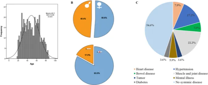
This study analysed the influence of several disease- and non-disease-related covariates on salivary levels of CSF-1. Demographics of the cohort of 441 participants are presented in Fig. 1. We also assessed a reference range for CSF-1 in saliva of a group of non-smokers, without manifest caries, that were periodontally and systemically healthy.
Influence of age on salivary levels of CSF-1.
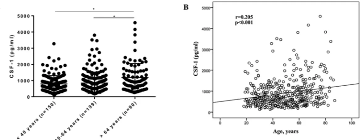
Age has been associated with CSF-1 levels in blood17.Therefore, we compared the salivary levels of CSF-1 in three age categories (<40 years old, 40–64 years old, and >64 years old). Mean (±SD) levels of CSF-1 were 793.0 (±511.8), 848.5 (±636.8) and 1185.0 (±868.9) pg/ml for participants <40 years old, 40–64 years old, and >64 years old respectively. Participants over 64 years (n = 90) old showed significantly higher levels of CSF-1 in comparison to participants of both the age group below 40 years (n = 150) and between 40–64 years (n = 199) old (Fig. 2A). Differences remained after CSF-1 normaliza-tion for total amount of protein in saliva (Suppl. Fig. 1A). Furthermore, age was significantly correlated to CSF-1 (r = 0.205; p < 0.001, Fig. 2B). Together, these results underscore that age is associated with CSF-1 levels in saliva.
Effects of sex and smoking on salivary levels of CSF-1.
We next sought out to address the possible impact of sex and smoking on salivary CSF-1 levels. Females showed mean (±SD) CSF-1 levels of 873.7 (±677.6) pg/ml and males of 927.3 (±661.5) pg/ml. There was no significant difference in CSF-1 levels between female (n = 221) and male (n = 218) participants, both uncorrected and corrected for total amount of protein (Fig. 3A, Suppl. Fig. 1B). Regarding smoking, mean (±SD) levels of CSF-1 were 928.8 (±682.2) and 759.6 (±596.5) pg/ml for, respectively, non-smokers (n = 365) and smokers (n = 74). Smokers showed significantly lower levels of CSF-1 compared to non-smokers (Fig. 3B). This difference was lost after CSF-1 normalization for the total amount of protein (Suppl. Fig. 1C).CSF-1 levels in saliva of participants with oral diseases.
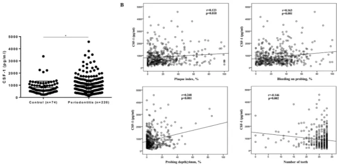
Based on our recent finding of higher CSF-1 levels in saliva of periodontitis compared to healthy subjects in a smaller cohort23, we next investigated the relationbetween CSF-1 levels and periodontitis in this large cohort. Participants without periodontitis (n = 74) exhibited mean (±SD) CSF-1 levels of 802.1 (±532.9) pg/ml, while participants with periodontitis (n = 220) showed mean (±SD) levels of 973.3 (±725.2) pg/ml. Salivary CSF-1 levels were significantly increased in periodontitis compared to without (Fig. 4A). This difference remained significant when restricted to patients without manifest caries (data not shown). However, differences between groups were lost when corrected for total protein (Suppl. Fig. 1D). Further, CSF-1 levels in saliva correlated significantly with PI (r = 0.123; p = 0.010), BOP (r = 0.163; p = 0.001), percentage of PD ≥4 mm (r = 0.248; p < 0.001), and number of teeth (r = −0.146; p = 0.002) (Fig. 4B).
Figure 1. Demographics of the study population. (A) Histogram of age (n = 441). (B) Sex (223 females and 218
males) and smoking distributions (75 smokers and 366 non-smokers). (C) Prevalence of systemic conditions: heart disease (n = 35), hypertension (n = 76), bowel disease (n = 31), muscle and joint disease (n = 102), mental illness (n = 26), tumor (n = 16), and diabetes (n = 16).
www.nature.com/scientificreports/
We next investigated the possible impact of caries on CSF-1 levels in saliva. Mean (±SD) levels of CSF-1 were, respectively, 852.2 (±626.2), 945.0 (±641.5) and 1158.2 (±964.5) pg/ml for participants with no MCL (n = 299), MCL 1–2 (n = 102) and MCL ≥3 (n = 38). Salivary CSF-1 levels were significantly higher in participants with MCL ≥3 as compared to MCL 0, whereas no difference was found between MCL 1–2 and MCL 0 or between MCL 1–2 and MCL ≥3 (Fig. 5A). The difference between groups was lost when corrected for total protein (Suppl. Fig. 1E). When participants having periodontitis were excluded, only a trend towards significant difference was observed (data not shown). Further, CSF-1 levels correlated significantly with MCL (r = 0.161; p = 0.001), DMFT (r = 0.164; p = 0.001) and caries risk (r = −0.365; p = 0.001). There was no significant correlation between CSF-1 and salivary flow rate (r = 0.055; p = 0.248) (Fig. 5B).
We also assessed correlations between the levels of CSF-1 and other inflammatory molecules previously ana-lyzed in saliva of this cohort4, 5 and found significant correlations to the levels of IL-1β (r = 0.467; p < 0.001), IL-8
(r = 0.413; p < 0.001), and MMP-8 (r = 0.459; p < 0.001).
Effects of systemic conditions on CSF-1 levels in saliva.
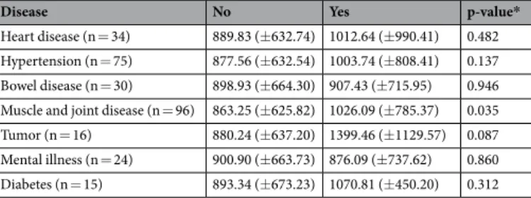
To address the possible relation between CSF-1 levels in saliva and systemic diseases we analysed CSF-1 levels with respect to several systemic conditions. Individuals having muscle and joint diseases (n = 96) showed increased levels of CSF-1 as compared to patients not having the disease (n = 335), but the difference was lost after CSF-1 correction for total amount of protein. Participants who reported tumors (n = 16) showed a trend towards increased levels of CSF-1, however this did not reach statistical significance. There was no significant difference in mean levels of CSF-1 between participants having heart disease, hypertension, bowel disease, mental illness and diabetes and participants not having the disease (Table 1). To address the possible confounding factor of comparing participants with a particular systemic disease to participants having other systemic diseases, a reference group, which was composed by subjects free of any of the systemic conditions (n = 241), was derived. The results were roughly the same when we compared CSF-1 levels of patients having systemic conditions to the reference group (data not shown).CSF-1 reference range in saliva.
In order to establish a reference range for CSF-1 in saliva, we derived a group of participants encompassing non-smokers, free of systemic diseases, without manifest caries lesions,Figure 2. Salivary levels of CSF-1 in participants of different age groups. (A) CSF-1 levels in saliva of
individuals <40 years (n = 150), 40–64 years (n = 199) and >64 years old (n = 90). *p-value < 0.05 (ANOVA with Bonferroni post-test). Data are presented as mean ± standard deviation. (B) Correlation scatterplot between CSF-1 levels and age (Pearson correlation).
Figure 3. Salivary levels of CSF-1 according to sex and smoking status. (A) CSF-1 levels in female (n = 221) and
male participants (n = 218). (B) CSF-1 levels in smokers (n = 365) and non-smokers (n = 74). *p-value < 0.05 (Student’s t test) Data are presented as mean ± standard deviation.
without bone loss and periodontal pockets ≥4 mm. This group was composed by 35 subjects. Mean (±SD) CSF-1 was 763.95 (±415.55) pg/ml, ranging from 224.55 to 2218.00 pg/ml. The 95% reference range was 234.42–1862.08 pg/ml. For the normalized values, mean (±SD) was 1083.72 (±459.70) pg/mg, ranging from 405.53 to 2174.98 pg/mg. The 95% reference range was 426.57–2187.76 pg/mg. Worth noting, CSF-1 levels were detected in 100% of the saliva samples of the whole cohort (n = 439).
Seeking to identify the determinants of CSF-1 levels in saliva, results from a linear regression analysis found that age, presence of tumors, percentage of PD ≥4 mm, and number of MCL were significantly associated with CSF-1 levels. Overall, these 4 variables accounted for 32.8% of the variance in CSF-1 levels (Table 2). For the normalized CSF-1, only age remained significantly associated with its levels, and it accounted for 13% of CSF-1 variance.
Figure 4. Impact of periodontal status on salivary levels of CSF-1. (A) CSF-1 levels in participants with
periodontitis (n = 220) and controls (n = 74). *p-value < 0.05 (Student’s t test). Data are presented as mean ± standard deviation. (B) Correlation scatterplots between CSF-1 levels and periodontal parameters (Pearson correlation).
Figure 5. Impact of cariological status on salivary levels of CSF-1. (A) CSF-1 levels in participants with no
manifest caries lesions (MCL) (n = 299), MCL 1-2 (n = 102) and MCL ≥3 (n = 38). *p-value < 0.05 (ANOVA with Bonferroni post-test). Data are presented as mean ± standard deviation. (B) Correlation scatterplots between CSF-1 levels, cariological parameters and salivary flow rate (Pearson correlation).
www.nature.com/scientificreports/
Discussion
This study, to the best of our knowledge, is the first one to comprehensively evaluate how disease- and non-disease-related variables affect salivary CSF-1 levels in a relatively large number of participants. We found that age, smoking, periodontitis, caries, and the presence of muscle and joint diseases are related to altered sali-vary levels of CSF-1. We also established a 95% reference range for salisali-vary CSF-1 from 234.42 to 1862.08 pg/ml in healthy individuals. The elucidation of variables that might impact CSF-1 levels in saliva increases its potential to be accurately used as a biomarker, as the variability unrelated to disease can be taken into account.
We found that age was associated with CSF-1 levels in saliva, particularly after 64 years old. We also identified, in a regression analysis, that each additional year of age is associated with an average increase of 4.69 pg/ml in CSF-1 levels. In line with the observed correlation between age and salivary CSF-1, other relations between age and inflammatory markers in saliva have been described5, 24. Age has an evident impact on cytokine responses,
although this effect is cytokine- and/or stimuli-dependent, such as defects in the production of T-helper-related cytokines25. Studies investigating the relationship between age and CSF-1 in blood have been conflicting. While
significant associations have been reported between CSF-1 and age in healthy individuals17, 26–28, another study has
not found age as a significant covariate for CSF-110. The significance of increased CSF-1 levels with age is unknown.
However, we speculate this increase might reflect the process of inflammaging that is associated with aging and that is evident in saliva as well, as highlighted by increased markers of protein oxidation with aging in saliva29.
In our study, there was no significant association between sex and CSF-1 levels in saliva. In line with our find-ing, no significant effect of sex has been reported on blood levels of CSF-110, 17. On the other hand, studies have
reported higher blood levels of CSF-1 in males18, 30, as well as in females31, 32. Circulating levels of CSF-1 is further
modulated by pregnancy, wherein up-regulated levels are measured in pregnant women33. As circulating CSF-1
levels seem to correlate with hormone concentrations (estradiol) in women, but not in men30, this might account
for the controversies found in the literature regarding its levels. Contrary to sex, smoking exerted a significant role in CSF-1 levels in saliva. Being a smoker was associated with an average decrease of 166.20 pg/ml in CSF-1 levels. Smoking has been related to decreased levels of other inflammatory markers in saliva such as IL-8 and MMP8/ TIMP1 ratio5. Liede et al. reported lower salivary MMP-8 levels in current smokers34. Decreased flow of gingival
crevicular fluid in smokers, which would lead to a lower influx of inflammatory mediators into saliva, might be one possible reason for this decrease35. Lower levels of CSF-1 in serum have been found in smoking patients with
long bone fractures32, whereas others found no significant effect of smoking on plasmatic CSF-110. Together, our
data indicates the need of taking age and smoking into account when relating salivary CSF-1 levels to diseases. We reported significantly higher levels of CSF-1 in participants diagnosed with periodontitis, even when restricted to patients not having manifest caries. We also found significant correlations to clinical periodontal parameters. These results confirm, in a larger group, recent findings of increased CSF-1 levels in saliva from per-iodontitis patients by our group23. Further, CSF-1 levels correlated significantly with the levels of IL-1β, IL-8, and
MMP-8, which were previously analysed in participants from this cohort5. This is also in line with our previous
finding of a significant correlation between the levels of CSF-1 and MMP-8 in saliva23. Blocking of the CSF-1
receptor has been shown to reduce alveolar bone loss and number of osteoclasts in experimental periodontitis in mice16. Further, in experimental arthritis in mice injections of CSF-1 have been shown to exacerbate disease
whereas CSF-1 blocking alleviates disease severity14. Along with the fact that CSF-1 stimulates RANKL-induced
Disease No Yes p-value*
Heart disease (n = 34) 889.83 (±632.74) 1012.64 (±990.41) 0.482 Hypertension (n = 75) 877.56 (±632.54) 1003.74 (±808.41) 0.137 Bowel disease (n = 30) 898.93 (±664.30) 907.43 (±715.95) 0.946 Muscle and joint disease (n = 96) 863.25 (±625.82) 1026.09 (±785.37) 0.035 Tumor (n = 16) 880.24 (±637.20) 1399.46 (±1129.57) 0.087 Mental illness (n = 24) 900.90 (±663.73) 876.09 (±737.62) 0.860 Diabetes (n = 15) 893.34 (±673.23) 1070.81 (±450.20) 0.312
Table 1. Mean (±SD) levels of CSF-1 (pg/ml) in saliva from patients having or not systemic conditions.
SD: standard deviation. *Student’s t test.
Variables Coefficient (β) 95% CI p-value
Age 4.69 0.84–8.54 0.017
Smoking −166.20 −329.24–−3.15 0.046
Tumor 417.12 90.27–743.97 0.012
Percentage of PD ≥4 mm 10.58 4.50–16.66 0.001 Manifest caries lesions 62.82 20.13–105.58 0.004
Table 2. Linear regression analysis of the association of demographic variables, oral and systemic conditions
with CSF-1 (pg/ml) in saliva (n = 439). Variables included, but not retained, in the model: Sex, bleeding on probing, heart disease, hypertension, bowel disease, muscle and joint disease, mental illness, and diabetes. R2 = 0.328.
osteoclast formation36, increased salivary levels of CSF-1 might be reflective of higher osteoclastogenic potential
in periodontitis.
Participants with high level of caries (MCL ≥3) exhibited higher levels of CSF-1 in saliva. Further, we found a significant correlation between salivary CSF-1 and caries risk. An association between MCL and MMP-8 has pre-viously been reported in this cohort37. However, when we restricted the analysis to participants not having
per-iodontitis, there was only a trend towards higher CSF-1 levels in subjects with manifest caries (p-value = 0.092), which suggest that the effects of caries on CSF-1 levels might partially be due to their periodontal condition. Dental pulp fibroblasts from dentinal caries sections immunostained for CSF-1 in about 38% of the teeth, while no immunostaining was observed in sections from non-carious teeth. Moreover, TNF-α-stimulate pulp fibro-blasts produced CSF-138. We can speculate that, at least partially, CSF-1 might leak out from the carious dentin
to saliva.
Inflammatory molecules in saliva have been investigated in relation to systemic diseases and their utility as biomarkers have been demonstrated4, 7. Our study found increased salivary levels of CSF-1 in participants having
muscle and joint diseases, which is in line with findings of higher CSF-1 in serum and urine from patients with systemic lupus erythematosus19. Increased expression of CSF-1 in plasma has also been reported in active as
compared to quiescent rheumatoid arthritis20. Our study also showed that the presence of tumors was
associ-ated with CSF-1 levels. Elevassoci-ated serum levels of CSF-1 have been found in patients with breast cancer, and even higher in patients with invasive cancer and concomitant lymph node metastasis21. Increased serum levels have
also been reported in patients with colorectal cancer39. Our result thus suggests that assessment of CSF-1 levels
in saliva might be valuable for monitoring patients with muscle and joint diseases and tumors however, further investigations are needed.
The main limitation of this study is its cross-sectional nature and, thus, no causal claim can be done. Prospective studies would be of great value to confirm hypotheses generated in this study, as well as the addition of gingival crevicular fluid. Besides, medical conditions were determined by self-report and, then the diseases were grouped together in broader categories. Therefore, we cannot rule out that particularities from each disease might influence differently on CSF-1 levels, as well as medications used to treat them and the disease status dur-ing saliva collection, such as active/quiescent and controlled/uncontrolled. We also cannot rule out that other covariates might play a role in determining salivary levels of CSF-1. Indeed, an impact of genetic factors has been reported on the variation of plasmatic CSF-1 levels26, thus we speculate that genetic factors might also play a role
in the variation of salivary CSF-1. As saliva is a complex biologic fluid, attention to its collection method is man-datory, which can have an impact on the concentration of proteins. Generally, levels of inflammatory biomarkers are higher in unstimulated than stimulated whole saliva40. However, we have shown no significant difference
in CSF-1 levels between unstimulated and stimulated saliva, either fasting or non-fasting23. We also reported
that the significant differences in CSF-1 levels were lost for smoking, periodontitis, caries, and muscle and joint diseases when correcting for total amount of protein. This might be explained by the higher amount of total pro-tein in these groups of patients, which could be a consequence of increased plasma propro-tein leakage during the inflammatory process. Higher protein concentration in periodontitis patients has been shown, which could not be attributed to increased salivary flow rate41, 42. On the other hand, decreased concentration of total protein has
been reported in smokers43.
In conclusion, this study found that age, smoking, periodontitis, manifest caries, and the presence of tumors are associated with the salivary levels of CSF-1. Furthermore, these results showed increased CSF-1 levels in patients reporting muscle and joint diseases.
Methods
Study sample.
This cross-sectional study enrolled 441 individuals (48.5 [±16.8] years; 50.6% women) living in Skåne, Sweden, who were orally examined and provided a saliva sample. Study protocol was approved by the Research Ethics Committee at the Lund University, Sweden, and all participants gave their written informed con-sent. All methods were performed in accordance with the relevant guidelines and regulations. Participants were asked to answer a questionnaire regarding oral health, and data concerning age, sex, smoking habits (including use of Swedish snus), presence of diseases, and use of medication were collected. Smoking was recording as yes or no. Diseases were self-reported and grouped as heart disease, hypertension, bowel disease, muscle and joint disease, mental illness, tumor, and diabetes. Details of the sample selection and the questionnaire were described previously44.Clinical examination.
90.5% of the clinical examinations were performed by four dentists employed at the Department of Oral Diagnostics, Faculty of Odontology, Malmö University. Periodontal parameters included visible plaque index (PI), bleeding on probing (BOP) and probing depth (PD), all recorded in four sites on each tooth using a periodontal probe (PCPUNC15, Hu-Friedy, IL, USA). In addition, digital bitewings and panoramic radiographs were taken for evaluation of alveolar bone loss. Periodontitis was defined as the presence of PD ≥4 mm and BOP >20%. Participants having no PD ≥4 mm and bone loss not exceeding 1/3 of the root length were selected as controls.Cariological parameters included DMFT (decayed, missing, filled teeth), number of manifest caries lesions (MCL), which encompassed lesions involving dentin, as seen on bitewing radiographs and cavitated lesions on other surfaces, and caries risk. Number of MCL was grouped as previously described: MCL 0: no MCL, MCL 1–2: 1 or 2 MCLs and MCL ≥3: 3 or more MCLs37. Caries risk was evaluated using the Cariogram, showing the
%-chance of avoiding caries45.
Saliva collection.
Stimulated saliva was collected during 5 minutes chewing 0.5 g of paraffin in a graded tube. Secretion rate (ml/min) was determined after excluding the foam. Samples were immediately frozen at −20 °Cwww.nature.com/scientificreports/
until processing. Saliva samples were then centrifuged and supernatants aliquoted and stored at −80 °C. Each aliquot was used only once for biomarker analysis. For the analysis of CSF-1, 439 samples were included.
CSF-1 immunoassay.
Levels of CSF-1 in saliva were determined using a commercial enzyme-linked immu-nosorbent assay (ELISA) according to the manufacturer’s instructions (R&D Systems, Minneapolis, MN, USA). Samples were thawed on ice and centrifuged at 10000 rpm for 5 min prior to analysis. The detection limit of the assay was 78.1 pg/ml and the sensitivity 11.2 pg/ml. Readings were made at 450 nm with wavelength correction set to 540 nm to subtract background using a microplate spectrophotometer (SpectraMAX 340, Sunnyvale, CA, USA). Total amount of protein was determined by the Bradford assay to enable normalization of CSF-1 levels to total salivary protein.Statistical analysis.
Data analyses were performed using Statistical Package for Social Sciences (SPSS) ver-sion 20 (IBM Corporation, Armonk, NY, USA). Continuous variables are presented as mean and standard devi-ation (SD) and categorical variables as frequencies. Group comparisons were performed with Student’s t-test or ANOVA with Bonferroni post-test. Correlations between CSF-1 levels and other variables were determined by Pearson correlation coefficient. A multiple linear regression was done with CSF-1 as dependent variable and age, sex, smoking, bleeding on probing, percentage of PD ≥4 mm, number of MCL, and the systemic conditions as independent variables. Statistical significance was set at P ≤ 0.05.In order to establish a reference range for CSF-1 in saliva, a 95% reference range was calculated as mean ± (1.96 × SD). For that purpose, CSF-1 values were log-transformed in order to achieve a normal distribu-tion, and then the anti-log was calculated to exhibit the reference range in pg/ml.
References
1. Proctor, G. B. The physiology of salivary secretion. Periodontol 2000 70, 11–25 (2016).
2. Zhang, Y. et al. The emerging landscape of salivary diagnostics. Periodontol 2000 70, 38–52 (2016).
3. Loo, J. A., Yan, W., Ramachandran, P. & Wong, D. T. Comparative human salivary and plasma proteomes. J Dent Res 89, 1016–1023 (2010).
4. Rathnayake, N. et al. Salivary biomarkers for detection of systemic diseases. PloS one 8, e61356 (2013).
5. Rathnayake, N. et al. Salivary biomarkers of oral health: a cross-sectional study. J Clin Periodontol 40, 140–147 (2013).
6. Gursoy, U. K. et al. Use of host- and bacteria-derived salivary markers in detection of periodontitis: a cumulative approach. Disease
markers 30, 299–305 (2011).
7. Zhang, L. et al. Discovery and preclinical validation of salivary transcriptomic and proteomic biomarkers for the non-invasive detection of breast cancer. PloS one 5, e15573 (2010).
8. Miller, C. S., King, C. P. Jr., Langub, M. C., Kryscio, R. J. & Thomas, M. V. Salivary biomarkers of existing periodontal disease: a cross-sectional study. Journal of the American Dental Association 137, 322–329 (2006).
9. Said, H. S. et al. Dysbiosis of salivary microbiota in inflammatory bowel disease and its association with oral immunological biomarkers. DNA Res 21, 15–25 (2014).
10. Enroth, S., Johansson, A., Enroth, S. B. & Gyllensten, U. Strong effects of genetic and lifestyle factors on biomarker variation and use of personalized cutoffs. Nat Commun 5, 4684 (2014).
11. Brodin, P. & Davis, M. M. Human immune system variation. Nat Rev Immunol 17, 21–29 (2017).
12. Pixley, F. J. & Stanley, E. R. CSF-1 regulation of the wandering macrophage: complexity in action. Trends Cell Biol 14, 628–638 (2004).
13. Marshall, D., Cameron, J., Lightwood, D. & Lawson, A. D. Blockade of colony stimulating factor-1 (CSF-I) leads to inhibition of DSS-induced colitis. Inflamm Bowel Dis 13, 219–224 (2007).
14. Campbell, I. K., Rich, M. J., Bischof, R. J. & Hamilton, J. A. The colony-stimulating factors and collagen-induced arthritis: exacerbation of disease by M-CSF and G-CSF and requirement for endogenous M-CSF. J Leukoc Biol 68, 144–150 (2000). 15. Lin, E. Y., Nguyen, A. V., Russell, R. G. & Pollard, J. W. Colony-stimulating factor 1 promotes progression of mammary tumors to
malignancy. J Exp Med 193, 727–740 (2001).
16. Kimura, K. et al. An anti-c-Fms antibody inhibits osteoclastogenesis in a mouse periodontitis model. Oral diseases 20, 319–324 (2014).
17. Larsson, A. et al. The effects of age and gender on plasma levels of 63 cytokines. J Immunol Methods 425, 58–61 (2015). 18. Ramsey, J. M. et al. Molecular sex differences in human serum. PloS one 7, e51504 (2012).
19. Menke, J. et al. Colony-stimulating factor-1: a potential biomarker for lupus nephritis. J Am Soc Nephrol 26, 379–389 (2015). 20. Rioja, I. et al. Potential novel biomarkers of disease activity in rheumatoid arthritis patients: CXCL13, CCL23, transforming growth
factor alpha, tumor necrosis factor receptor superfamily member 9, and macrophage colony-stimulating factor. Arthritis Rheum 58, 2257–2267 (2008).
21. Aharinejad, S. et al. Elevated CSF1 serum concentration predicts poor overall survival in women with early breast cancer. Endocr
Relat Cancer 20, 777–783 (2013).
22. Rallidis, L. S. et al. Raised concentrations of macrophage colony stimulating factor in severe unstable angina beyond the acute phase are strongly predictive of long term outcome. Heart 90, 25–29 (2004).
23. Martinez, G. L. et al. Salivary colony stimulating factor-1, interleukin-34, and matrix metalloproteinase-8 as markers of periodontal disease. J Periodontol 1–15 (2017).
24. Tobon-Arroyave, S. I., Isaza-Guzman, D. M., Restrepo-Cadavid, E. M., Zapata-Molina, S. M. & Martinez-Pabon, M. C. Association of salivary levels of the bone remodelling regulators sRANKL and OPG with periodontal clinical status. J Clin Periodontol 39, 1132–1140 (2012).
25. Ter Horst, R. et al. Host and Environmental Factors Influencing Individual Human Cytokine Responses. Cell 167, 1111–1124 e1113 (2016).
26. Livshits, G., Pantsulaia, I., Trofimov, S. & Kobyliansky, E. Genetic influences on the circulating cytokines involved in osteoclastogenesis. J Med Genet 41, e76 (2004).
27. Hasegawa, Y., Sawada, M., Ozaki, N., Inagaki, T. & Suzumura, A. Increased soluble tumor necrosis factor receptor levels in the serum of elderly people. Gerontology 46, 185–188 (2000).
28. Suehiro, A. et al. Age related elevation of serum macrophage colony stimulating factor (M-CSF) level. Arch Gerontol Geriatr 29, 13–20 (1999).
29. Wang, Z. et al. Age-related variations of protein carbonyls in human saliva and plasma: is saliva protein carbonyls an alternative biomarker of aging? Age (Dordr) 37, 9781 (2015).
30. Trofimov, S., Pantsulaia, I., Kobyliansky, E. & Livshits, G. Circulating levels of receptor activator of nuclear factor-kappaB ligand/ osteoprotegerin/macrophage-colony stimulating factor in a presumably healthy human population. Eur J Endocrinol 150, 305–311 (2004).
31. Ramsey, J. M., Cooper, J. D., Penninx, B. W. & Bahn, S. Variation in serum biomarkers with sex and female hormonal status: implications for clinical tests. Sci Rep 6, 26947 (2016).
32. Kottstorfer, J. et al. The influence of non-osteogenic factors on the expression of M-CSF and VEGF during fracture healing. Injury 44, 930–934 (2013).
33. Tsakonas, D. P., Nicolaides, K. H., Tsakona, C. P., Worman, C. P. & Goldstone, A. H. Changes in maternal plasma macrophage-colony stimulating factor levels during normal pregnancy. Clin Lab Haematol 17, 57–59 (1995).
34. Liede, K. E. et al. The association between smoking cessation and periodontal status and salivary proteinase levels. J Periodontol 70, 1361–1368 (1999).
35. Ustun, K. & Alptekin, N. O. The effect of tobacco smoking on gingival crevicular fluid volume. Eur J Dent 1, 236–239 (2007). 36. Bostrom, E. A. & Lundberg, P. The newly discovered cytokine IL-34 is expressed in gingival fibroblasts, shows enhanced expression
by pro-inflammatory cytokines, and stimulates osteoclast differentiation. PloS one 8, e81665 (2013).
37. Hedenbjork-Lager, A. et al. Caries correlates strongly to salivary levels of matrix metalloproteinase-8. Caries Res 49, 1–8 (2015). 38. Sawa, Y. et al. Production of colony-stimulating factor in human dental pulp fibroblasts. J Dent Res 82, 96–100 (2003).
39. Mroczko, B. et al. Serum macrophage-colony stimulating factor levels in colorectal cancer patients correlate with lymph node metastasis and poor prognosis. Clin Chim Acta 380, 208–212 (2007).
40. Miller, C. S. et al. Current developments in salivary diagnostics. Biomark Med 4, 171–89 (2010).
41. Sánchez, G. A., Miozza, V., Delgado, A. & Busch, L. Determination of salivary levels of mucin and amylase in chronic periodontitis patients. J Periodontal Res 46, 221–227 (2011).
42. Henskens, Y. M. et al. Protein composition of whole and parotid saliva in healthy and periodontitis subjects. Determination of cystatins, albumin, amylase and IgA. J Periodontal Res 31, 57–65 (1996).
43. Kolte, A. P., Kolte, R. A. & Laddha, R. K. Effect of smoking on salivary composition and periodontal status. J Indian Soc Periodontol 16, 350–353 (2012).
44. Lundegren, N., Axtelius, B. & Akerman, S. Oral health in the adult population of Skane, Sweden: a clinical study. Acta Odontol Scand 70, 511–519 (2012).
45. Hansel Petersson, G., Twetman, S. & Bratthall, D. Evaluation of a computer program for caries risk assessment in schoolchildren.
Caries Res 36, 327–340 (2002).
Acknowledgements
The authors thank the participants of the study and the operators who performed the clinical examinations. The authors thank Daniela Bureik and Gisele Martinez, both at the Department of Dental Medicine, Karolinska Institutet, for technical assistance with the CSF-1 ELISA in saliva. The study was initiated and financially supported by the Regional Board of Dental Public Health in the county of Skåne, Sweden. The study was also supported by the Swedish National Graduate School in Odontological Science, the Department of Dental Medicine, Division of Periodontology, Karolinska Institutet, Stockholm, Sweden and Stockholm County Council (SOF). RLJ is a recipient of a scholarship through the Rio de Janeiro State Agency for Research Support (FAPERJ), Brazil. EAB is a recipient of a grant for half-time position in clinical research environment from the Swedish Research Council (2012-07110). The funders had no role in the design, data collection and analysis, decision to publish, or preparation of the manuscript.
Author Contributions
S.Å., A.G., B.K., and E.A.B. conceived and designed the experiments. R.L.J. and E.A.B. performed experiments and analyzed the data. R.L.J., S.Å., A.G., B.K., and E.A.B. wrote and edited the manuscript.
Additional Information
Supplementary information accompanies this paper at doi:10.1038/s41598-017-07698-4 Competing Interests: The authors declare that they have no competing interests.
Publisher's note: Springer Nature remains neutral with regard to jurisdictional claims in published maps and
institutional affiliations.
Open Access This article is licensed under a Creative Commons Attribution 4.0 International
License, which permits use, sharing, adaptation, distribution and reproduction in any medium or format, as long as you give appropriate credit to the original author(s) and the source, provide a link to the Cre-ative Commons license, and indicate if changes were made. The images or other third party material in this article are included in the article’s Creative Commons license, unless indicated otherwise in a credit line to the material. If material is not included in the article’s Creative Commons license and your intended use is not per-mitted by statutory regulation or exceeds the perper-mitted use, you will need to obtain permission directly from the copyright holder. To view a copy of this license, visit http://creativecommons.org/licenses/by/4.0/.