DISSERTATION
ANALYSIS OF THE RELATIONSHIP BETWEEN GENOMIC INSTABILITY, HETEROZYGOSITY LEVELS AND PHENOTYPE
IN SACCHAROMYCES CEREVISIAE
Submitted by
Nadia Maria Vieira Sampaio
Graduate Degree Program in Cell and Molecular Biology
In partial fulfillment of the requirements For the Degree of Doctor of Philosophy
Colorado State University Fort Collins, Colorado
Fall 2018
Doctoral Committee:
Advisor: Juan Lucas Argueso Laurie A. Stargell
John K. McKay Kenneth F. Reardon
Copyrighted by Nadia Maria Vieira Sampaio 2018 All Rights Reserved
ABSTRACT
ANALYSIS OF THE RELATIONSHIP BETWEEN GENOMIC INSTABILITY, HETEROZYGOSITY LEVELS AND PHENOTYPE
IN SACCHAROMYCES CEREVISIAE
Understanding the forces that mediate genome evolution is a central problem in genetics, with implications for diverse processes that range from speciation, to
biotechnological applications, to human disease. The central theme of my dissertation was the characterization of two forces, genomic instability and natural selection, that significantly impact genome structure by influencing the levels of genomic
heterozygosity. While genomic instability processes can act to erode heterozygosity from the genome, natural selection may favor the maintenance of heterozygous alleles in cases where there is a positive correlation between heterozygosity and higher fitness.
In Chapter I, I reviewed different types of mitotic mutations that can result in the appearance of tracts of homozygosity in genomes and recent discoveries about the temporal accumulation of such events. I also introduce the concept of heterosis, a phenomenon characterized by a positive correlation between genomic heterozygosity and phenotype in many species, and its potential role in contributing to the long-term maintenance of genomic heterozygosity.
In Chapter II, I describe the characterization of a mechanism of systemic genomic instability in yeast that challenges the conventional model of gradual and independent accumulation of mutations. We showed that a subset of mitotic cells within
a population experience bursts of genomic instability, which results in multiple independent events of loss-of-heterozygosity (LOH) accumulating over one or a few generations of mitotic cell division. We named this outcome “systemic genomic instability”. The occurrence of this phenomenon was initially identified in the
heterozygous yeast strain JAY270, and then validated in a conventional laboratory strain background, whose genome is almost fully homozygous. Elevated rates of coincident LOH was also observed in mutant strains incapable of entering meiosis, indicating cryptic initiation of meiotic recombination followed by return-to-growth in a few cells in the population was not responsible for the higher than expected rates of
coincident LOH. This finding brings to light a novel and intriguing mechanism of
genomic instability in yeast that has relevant parallels to bursts of accumulation of copy number alterations in the human genome, providing a powerful experimental model system to dissect the fundamental mechanisms responsible for the generation of rapid changes in chromosome structure.
In Chapter III, we explored the role that genomic heterozygosity plays on the superior industrial traits of the JAY270 strain. In the previous Chapter we showed that mitotic recombination leading to LOH occurs at a high frequency during JAY270’s clonal propagation. These LOH events act against the long-term maintenance of genomic heterozygosity, yet about 60% of JAY270’s genome has remained heterozygous over time. We hypothesized that specific heterozygous alleles may have a positive impact on the traits of this strain and therefore were maintained through selection. We generated a collection of inbred strains derived from JAY270, and assessed them phenotypically under different growth conditions. Our results demonstrated that genomic
heterozygosity indeed has a substantial impact on two important industrial traits of this strain – heat stress tolerance and growth kinetics. We identified several genomic regions potentially associated with those traits and conducted experiments to investigate the bulk contributions of heterozygosity blocks in three specific
chromosomes. This study revealed candidate regions containing loci that potentially underlie important industrial traits of JAY270 and details on the extent to which heterozygosity may impact JAY270’s genome evolution and phenotype.
The combined results of these research projects provide important insights about the role of genomic instability mechanisms and their phenotypic outcomes in
determining genome evolution, contributing discoveries that may have important practical implications for diverse fields, including biotechnology, cancer development and evolution, as well as genome sciences.
ACKNOWLEDGMENTS
The work presented in this dissertation reflects the support and contributions of many people. First, I am thankful to my advisor, Dr. Lucas Argueso. His guidance and continuous excitement for our career and research have guided me through graduate school and will certainly continue to guide my future career. It was a privilege and a joy to be trained by him.
I thank the members of my graduate advisory committee, Dr. John McKay, Dr. Kenneth Reardon and Dr. Laurie Stargell, for pushing me to think critically about my projects and for all suggestions that helped to improve the quality of my research. I am also profoundly grateful to Dr. Carol Wilusz, a brilliant teacher, scientist and mentor. All lessons I have learned from you will remain an inspiration for the rest of my scientific career. Thank you for your support. I would also like to thank Dr. Cris Argueso for her support and continuous encouragement. Dr. Chris Allen, Dr. Claudia Wiese, Dr. Howard Liber, Dr. Mark Stenglein, Dr. David Maranon, thank you for sharing your knowledge with me and for making my research much easier.
I am grateful for the help and support of all current and past members of the Argueso Lab. I would like to especially thank Dr. Hailey Conover Sedam. I could not have completed my PhD without your daily dose of laughter. Thank you for your friendship and for teaching me so much, about science and life! I also thank our lab angel, Ruth Watson. Thank you for all your help and dedication during the completion of my projects.
I would like to thank the Cell and Molecular Biology Program, a great intellectual and cooperative environment. I especially thank Charlene Spencer and Lori Williams for being always so patient and willing to help.
I would like to give special thanks to Marcelo Bassalo. This work would not have been possible without his continuous encouragement and love. Thank you for being the biggest supporter of my career. It has been incredibly fun to walk this journey by your side.
Finally, I would like to express my profound gratitude to my family. I thank my parents, Sergio and Nilza Sampaio, and my siblings, Andre and Ana Sampaio. Thank you for your love, for believing in me and for all the personal sacrifice you have made to give me the chance to pursue my career goals. This dissertation is dedicated to you.
TABLE OF CONTENTS ABSTRACT ... II ACKNOWLEDGMENTS ... V LIST OF TABLES ... IX LIST OF FIGURES ... X CHAPTER I ...1
Introduction and Background ...1
General classes of mitotic mutations ...1
Mechanisms leading to loss-of-heterozygosity (LOH) ...2
Incremental steps or large jumps in the accrual of chromosomal alterations? ...5
A tug of war in the regulation of genomic heterozygosity in yeast ...10
REFERENCES ...14
CHAPTER II ...19
Mitotic systemic genomic instability in yeast ...19
Summary ...19
Introduction ...20
Results ...22
Appearance of altered colony morphology derivatives of JAY270 ...22
Genetic basis of the rough colony phenotype ...26
Analysis of Chr12 LOH in spontaneous rough colony isolates ...27
Analysis of selected Chr12 LOH ...31
Genome-wide analysis LOH and CNA in the JAY664 isolate ...35
Investigation of systemic genomic instability ...37
Validation and quantification of coincident LOH ...41
Discussion ...43
Rough colony phenotype and mutation in ACE2 ...43
mitSGI and precedents of coincident recombination ...44
mitSGI-like observations in human disease ...48
Possible mechanisms underlying mitSGI ...49
Materials and Methods ...52
Growth media ...52
Yeast genetic backgrounds and procedures...52
Isolation of spontaneous rough colonies derived from JAY270 ...53
Allele replacement and complementation tests ...54
Construction of strains used in single and double LOH assays ...54
Quantitative LOH rate assays ...55
Genome Sequencing Analyses ...56
Genotyping specific HetSNPs through PCR-RFLP and molecular karyotyping ...62
CHAPTER III ...69
Characterization of phenotypic consequences of heterozygosity in yeast ...69
Summary ...69
Introduction ...70
Results ...75
Controlled reduction of heterozygosity in the JAY270 genome through inbreeding ...75
Characterization of phenotypic variation in the inbred diploid collection ...79
Identification of genomic regions associated with phenotypic variation ...87
Phenotypic consequences of chromosome-scale LOH ...89
Discussion ...94
Heterozygosity and fitness in yeast hybrids ...94
Cryptic phenotypic variation in JAY270 ...95
UPD strategy for phenotypic testing of confined LOH ...97
Materials and Methods ...99
Growth media ...99
Yeast genetic backgrounds and microbiology procedures ... 100
Construction of a collection of partial inbred diploids derived from JAY270 .. 100
Construction of a GFP-tagged JAY270 derivative ... 101
Phenotypic assessment of the inbred collection ... 101
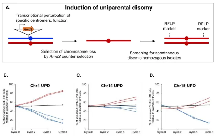
Identification of allele combinations enriched in high and low fitness strains . 103 Construction of UPD strains ... 105
REFERENCES ... 108
CHAPTER IV ... 114
Conclusions and future directions ... 114
Systemic genomic instability and resulting rapid karyotype evolution... 114
Effects of heterozygosity on phenotype and its influence in genome configuration 117 REFERENCES ... 119
APPENDIX A: SUPPLEMENTAL FIGURES ... 120
LIST OF TABLES
Table S2.1. Yeast strains used in this study. ... 140
Table S2.2. Oligonucleotides used in this study. ... 143
Table S2.3. List of phased JAY270 HetSNPs and hemizygous sequences and respective detection methods. ... 145
Table S2.4. Smooth and rough clones isolation specifics and analysis of chromosome size polymorphisms by PFGE. ... 146
Table S2.5. Summary of WGS analysis of rough colony isolates. ... 149
Table S3.1. Yeast strains used in this study. ... 152
Table S3.2. Oligonucleotides used in this study. ... 155
Table S3.3. Growth conditions tested through plate spotting assay. ... 157
Table S3.4. Summary of phenotypic data measure in competition assays (CA) and tolerance to high temperature assays (HT) for each inbred strain. ... 158
Table S3.5. List of phased JAY270 HetSNPs interrogated for confirmation of UPD strains. ... 162
Table S3.6. Details on genomic regions showing significant association to the growth kinetics and heat tolerance phenotypes. ... 163
LIST OF FIGURES
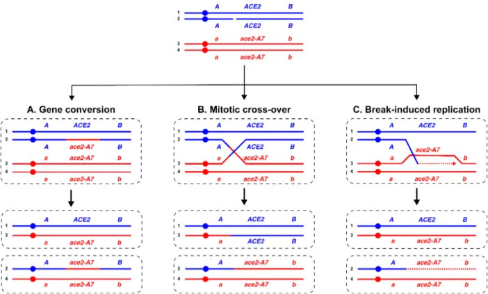
Figure 1.1. Genetic outcomes of interhomolog mitotic homologous recombination. ...5
Figure 1.2. Models of mutation accumulation. ...8
Figure 1.3. The levels of heterozygosity in the yeast genome are regulated by the balance between the counter-acting forces of heterosis and loss-of-heterozygosity. ....13
Figure 2.1. Smooth and rough colony morphologies, mother-daughter cell attachment, and phenotypes of diploids derived from mating specific haploids. ...23
Figure 2.2. LOH tract maps of Chr12 from five original rough colony isolates. ...30
Figure 2.3. Quantitative analyses of LOH. ...33
Figure 2.4. Analysis of unselected chromosomal changes in JAY664. ...36
Figure 2.5. Genome-wide map of LOH tracts in spontaneous rough colony isolates. ....40
Figure 3.1. Panel of inbred diploids derived from JAY270. ...79
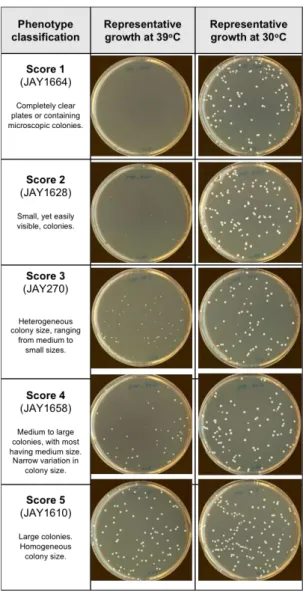
Figure 3.2. Inbred strains show heterogeneous levels of tolerance to high temperature stress. ...81
Figure 3.3. Phenotypic assessment through growth competition assays. ...83
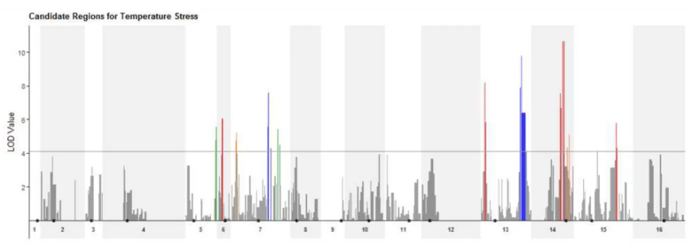
Figure 3.4. Genome-wide association analyses to identify potential loci underlying heat stress tolerance. ...88
Figure 3.5. Construction and relative growth kinetics profiles of Chr-UPD strain pairs. .93 Figure S2.1. Draft map of heterozygosity in the JAY270 genome. ... 120
Figure S2.2. HetSNP co-segregation genetic mapping approach and ace2-A7 complementation tests. ... 121
Figure S2.3. Sanger DNA sequencing analysis of ACE2 locus and genome-wide copy number in JAY664. ... 122
Figure S2.4. PCR genotyping of LOH and CNV in JAY664. ... 123
Figure S2.5. Analysis of Chr12 LOH in selected smooth and rough clones. ... 125
Figure S2.6. Detailed maps of Chr12 LOH in the 25 sequenced spontaneous rough colony isolates. ... 127
Figure S2.7. PFGE profiles of 29 independent smooth derivatives of JAY270. ... 129
Figure S2.8. PFGE profiles of smooth clones obtained through two bottlenecking lineages of JAY270. ... 130
Figure S2.9. PFGE profile of 27 independent rough colony derivatives of JAY270. .... 133
Figure S3.1. Genotype distribution in the inbred collection. ... 134
Figure S3.2. Inbred strains show uniform growth at 30oC and wide variation in tolerance to high temperature stress at 39oC. ... 135
Figure S3.3. Flow cytometry data analysis. ... 136
Figure S3.4. Venn Diagrams ... 137
CHAPTER I
Introduction and Background
General classes of mitotic mutations
A genome contains all of the information necessary for cellular function. Cells devote a repertoire of enzymatic pathways to accurately replicate and maintain their DNA molecules and the information encoded by its nucleotide sequence. Even though these pathways are extremely reliable, failure to faithfully replicate the DNA or to appropriately repair DNA damage does occur occasionally, and can lead to mutations that alter the DNA sequence and, consequently, the way cells behave and interact with an ever-changing environment.
When mutations occur in the germline they are passed on to the offspring and have long-term consequences on a species. On the other hand, mutations that occur in mitotically dividing somatic cells are not heritable, but they may have important
functional outcomes to a cell’s clonal lineage. For instance, mutations that occur in somatic cells are an underlying cause of cancer in humans (Hanahan and Weinberg 2000) and, in single celled microorganisms, they have a significant impact in adaptive evolution to new environmental conditions (Payen et al. 2016; Venkataram et al. 2016).
Spontaneous single nucleotide mutations and small insertions or deletions commonly result from DNA damage and replication errors that evade repair (Boiteux and Jinks-Robertson 2013; Zhu et al. 2014). They represent are an essential source of genetic variability, but in diploid cells, the potential detrimental effects of
non-synonymous mutations are usually compensated by the presence of a second intact wild type allele of the mutated gene. In contrast, mutational events that result in structural chromosomal alterations have a greater potential for leading to phenotypic changes because a single event can alter the genotype of multiple loci at once (Putnam and Kolodner 2017). These structural alterations can cause, for instance, gene dosage imbalances as a result of segmental deletions and amplifications (Putnam and Kolodner 2017). In addition, chromosomal alterations that result from recombination events
between homologous chromosomes often lead to extensive copy-neutral loss-of-heterozygosity (LOH) (Symington, Rothstein, and Lisby 2014; Putnam and Kolodner 2017), which may unmask recessive mutant alleles. This dissertation focuses on the characterization of mutational events that lead to structural chromosomal alterations, particularly LOH, and their phenotypic consequences.
Mechanisms leading to loss-of-heterozygosity (LOH)
Generally, structural chromosomal alterations arise as a result of inappropriate repair of DNA double-strand breaks (DSBs), triggered by local DNA lesions and/or replication fork collapse (Pâques and Haber 1999; Putnam and Kolodner 2017). In yeast, as well as in human cells, several repair mechanisms compete hierarchically to repair eventual DSBs and, although different organisms favor different repair pathways their genetic outcomes are essentially identical. Typically, these mechanisms are classified in two categories. Cells may repair DSBs by simply re-joining the two broken chromosome ends through a non-homologous end joining pathway (NHEJ) (Chang et al. 2017). Alternatively, cells may repair DSBs using pathways that rely on an unbroken
DNA template through homologous recombination (HR). In this scenario, if the perfectly identical and allelic sister chromatid is the chosen homologous template for repair, the initial DNA lesion will have no genetic consequence (Pâques and Haber 1999;
Symington, Rothstein, and Lisby 2014). However, in heterozygous diploid cells, the homologous chromosome (non-sister chromatid) may also be used as the donor sequence for repair, providing the potential for LOH tract accumulation and other changes in the chromosome structure to occur. For instance, interstitial and terminal copy-neutral LOH tracts, ranging from a few to hundreds of kilobases, may arise as a result of interhomolog allelic mitotic homologous recombination (Symington, Rothstein, and Lisby 2014).
The HR pathway is initiated by 5’-3’ resection at the DSB. One of the resulting 3’ single-stranded DNA (ssDNA) tails invades the double stranded DNA (dsDNA) of the homologous template to form a displacement loop (D-loop). The 3’ end of the resected tail is then used to prime the synthesis using the complementary donor strand as template. The displaced donor strand is then available for annealing to the other resected tail on the broken molecule. Through DNA synthesis, branch migration and ligation, these structures eventually progress to form a double Holliday junction (dHJ) intermediate. Resolution of the dHJ may occur by dissolution and re-annealing of recipient DNA, or by endonucleolytic cleavage. The orientation of the cleavage of the the dHJ may lead to the formation of either crossover (perpendicular orientation) or non-crossover (parallel orientation) outcomes. Formation of a non-non-crossover product, i.e. a gene conversion tract, will result in a daughter cell with an interstitial copy neutral LOH tract after the ensuing cell division (Chen et al. 2007) (Figure 1A). Alternatively, the dHJ
may be resolved to yield a crossover product, which can result in reciprocal terminal LOH tracts if the recombinant chromatids segregate to different daughter cells (Symington, Rothstein, and Lisby 2014) (Figure 1B).
An identical terminal LOH genetic outcome may also arise as a result of break-induced replication (BIR), a synthesis-dependent recombination pathway for repair of single-ended DSBs that results in the non-reciprocal transfer of DNA from the donor homologous chromosome to the broken, recipient molecule (Llorente, Smith, and Symington 2008; Malkova and Ira 2013; Donnianni and Symington 2013) (Figure 1C). This pathway is also initiated by single strand invasion and D-loop formation. However, in this case, the homologous chromosome is used as a template for conservative DNA synthesis that may replicate > 100 kb of the template until the end of the chromosome, resulting in extensive LOH. Although the genetic outcome of repair through BIR is indistinguishable from interhomolog allelic mitotic recombination resolved by crossover, it has been demonstrated that BIR is suppressed when both ends of a DSB are
available to initiate repair (Stark and Jasin 2003; Llorente, Smith, and Symington 2008) and the efficiency of this repair pathway is greatly reduced (<40%) when the length of DNA to be synthesized is greater than ~100 Kb (Donnianni and Symington 2013). In addition to the pathways described above, LOH can be generated by other types of mutational events. For example point mutations may lead to localized LOH and
segmental deletions and whole chromosome loss may lead to extensive LOH due to the loss of one haplotype (Symington, Rothstein, and Lisby 2014; Putnam and Kolodner 2017).
Figure 0.1. Genetic outcomes of interhomolog mitotic homologous recombination.
Cells may repair DSB lesions through different pathways that rely on homologous recombination. Circles indicate centromeres. Three different heteroalleles are
represented by A/a, ACE2/ace2-A7 and B/b. Duplicated homologous chromosomes in diploid cells are represented in colors blue and red. A) Gene conversion. Unidirectional genetic exchange between a donor homolog (red) and a recipient homolog (blue) leads to a non-reciprocal interstitial tract of LOH in only one of the resulting daughter cells. B) Crossover. Recombination resolved by a crossover yields extensive LOH if the
recombinant molecules segregate to opposite cells in the subsequent cell division. C) Break-induced replication (BIR). One-ended DSBs repaired through conservative synthesis lead to extensive LOH in one of the resulting daughter cells. Note: all LOH events shown above span the ACE2 locus, which will be relevant for the experiments described in Chapters II and III of this dissertation.
Incremental steps or large jumps in the accrual of chromosomal alterations?
Over the last four decades, the development and advancement of whole-genome sequencing technologies have enabled a revolution in our understanding of the
have revealed many details about the nature, frequency and distribution of mutations (Ciriello et al. 2013; Alexandrov and Stratton 2014; Peter et al. 2018). The landscape of somatic mutations found in most cancer genomes is astounding when compared to normal healthy cells. An average cancer genome contains about 1000- 10000 point mutations, 10 - 100 small insertions and deletions and 1 - 10 large scale chromosomal rearrangements (reviewed in Zhang and Pellman 2015). Large-scale genomic studies have revealed the existence of mutation hotspots, genomic regions including late replicating and heterochromatic regions that are more prone to accumulate mutations. In addition, most cancer types are associated with a characteristic mutational signature, which suggests cancers may be driven by different mutational processes (Ciriello et al. 2013; Alexandrov and Stratton 2014). For instance, colorectal carcinoma, uterine carcinoma and acute myeloid leukemia are frequently associated with point mutations, while ovarian and breast carcinoma are characterized by copy number changes (Ciriello et al. 2013). Although it is now evident that the type and distribution of mutations are not uniform throughout the genome, their frequency of appearance is estimated to be low and these events are generally thought to accumulate independently of each other over time, in a random and gradual fashion (Stratton, Campbell, and Futreal 2009). While literature overwhelmingly supports this general mechanism, mounting evidence is emerging for the co-existence of punctuated bursts of mutation accumulation, in which multiple mutational events take place over a single or a few cell division cycles (Zhang et al. 2015; Zhang and Pellman 2015; Sottoriva et al. 2015; Gao et al. 2016; Field et al. 2018). One relatively well understood example is chromothripsis, a mutational
single chromosome, a chromosome segment or occasionally a few chromosomes (Stephens et al. 2011; Leibowitz, Zhang, and Pellman 2015). These massive localized rearrangements have been shown to arise over a single cell division as a result of nuclear reincorporation of mis-segregated chromosomes (Zhang et al. 2015) and also as a result of telomere crisis caused by rupture and fragmentation of dicentric
chromosomes (Maciejowski et al. 2015). Other examples include chromoplexy (Baca et al. 2013), firestorms (Hicks et al. 2006) and kataegis (Roberts et al. 2012), all of which are associated to numerous point mutations or chromosomal rearrangements confined to one or a couple limited genomic regions. In addition to these examples of bursts of clustered mutations, a few recent studies have also reported evidence of punctuated bursts of copy number alterations (CNAs) distributed on a genome-wide scale in some types of cancer and neurodevelopmental disorders (Gao et al. 2016; Liu et al. 2017; Field et al. 2018).
Recent genomic analysis of thousands of individual cells extracted from tumors of patients with triple-negative breast cancer revealed that cells extracted from the same tumor could be broadly clustered into only 1 to ~3 discrete phylogenetic subpopulations based on their copy number profiles (Gao et al. 2016). Each phylogenetic branch was composed of tens of individual cells with highly rearranged genomes compared to normal cells, yet with very similar alterations to each other indicating stable clonal
expansion. Importantly, cells with intermediate rearranged karyotypes were not detected in any of the 12 tumor samples analyzed, which should have been observed if the CNAs accumulated gradually or sequentially. Instead, advanced mathematical modeling
of tumor evolution, in short punctuated bursts, followed by stable clonal expansions that formed the tumor mass.
Figure 0.2. Models of mutation accumulation.
Left) Traditional model of gradual accumulation of mutations, where new mutations emerge in incremental, sequential steps. Dashed lines indicate the time of appearance of new mutations. Right) Punctuated model of mutation accumulation, in which multiple mutations accumulate over a short period of crisis, followed by stasis and clonal
expansion of mutant cells. Insets represent the rate of mutation accumulation over time for each model.
Similar conclusions were drawn from single cell whole genome sequencing data and mathematical modeling approaches used to investigate genomic heterogeneity in uveal melanoma (UM), the most frequent type of primary eye cancer, which often leads to metastatic death (Field et al. 2018). UM is often associated with inactivation of one allele of the BAP1 tumor suppressor gene, followed by loss of the second functional allele through an LOH event spanning the BAP1 locus on chromosome 3. The patterns of genome-wide CNA and LOH of thousands of single cells from 151 different tumor samples were strikingly reminiscent of those found in breast cancer: single genomes from the same tumor sample could be clustered into few and narrowly defined
Combined, these results strongly imply that the rearrangements present in each tumor subpopulation most likely accumulated rapidly, through an early burst of genome-wide instability in one or a few founding cells. Those highly rearranged cells then expanded stably to give rise to genomically uniform subpopulations that were ultimately found in the advanced stage tumors.
The discovery of rare bursts of chromosomal alterations has the potential to transform our understanding of the initial steps of cancer formation and phenotype-genotype evolution in general. However, mechanisms and cellular pathways governing these bursts are yet well characterized and their investigation is technically very
challenging. In Chapter II of this dissertation, we provide experimental evidence that an analogous phenomenon of systemic genomic instability leading to punctuated bursts of chromosomal alterations also occurs in the yeast Saccharomyces cerevisiae. The significance of this finding is mainly threefold, (1) it indicates that the punctuated evolutionary model might be widespread among higher eukaryotes, (2) it provides a powerful model system that may facilitate the investigation of the underlying molecular mechanisms leading to punctuated bursts of genomic instability and (3) it reveals a novel mutational mechanism that can contribute to rapid changes in phenotype and adaptive evolution of natural populations of yeast. We discuss in Chapter II plausible underlying causes for transient genomic crises and, in Chapter IV, we propose future perspectives for the research in this field and some of the technical challenges that will need to be overcome.
A tug of war in the regulation of genomic heterozygosity in yeast
Loss of a functional tumor suppressor allele through LOH can lead to
uncontrolled cell proliferation and tumorigenesis (Ryland et al. 2015). For instance, this mechanism accounts for a large portion of the cases of retinoblastoma, which are frequently associated with loss of a functional copy of the Rb allele (Cavenee et al. 1983). LOH is not only an outcome of genomic instability that plays an important role in tumorigenesis, but these events are also a driving force in the evolution of unicellular microorganisms (Magwene et al. 2011). Mechanisms leading to LOH have an elevated potential for leading to phenotypic changes because one single mutational event has the capability of altering the genotype of long stretches of DNA harboring hundreds of genes. Population genomic analyses of many yeast isolates have shown that
heterozygosity is common among wild diploid strains and LOH might represent a path of easy access to new allelic combinations and phenotypes (Magwene et al. 2011). For example, LOH has been correlated to recurrent acquisition of drug resistance (Coste et al. 2006), adaptation to nutrient-limited conditions (Smukowski Heil et al. 2017), among others.
An important feature of this type of mutational event is that the resulting LOH tract can never be reverted back to the original heterozygous genotype. The high rates of LOH events during mitotic cell division and the irreversible nature of these events lead to many intriguing questions: How is heterozygosity maintained overtime in the genomes of wild yeast strains? How does heterozygosity influence phenotype? What are the phenotypic consequences of LOH?
One possibility is that human-associated environments facilitate outcrossing between unrelated strains, thus contributing to the maintenance of heterozygosity in strains isolated from those settings (Magwene et al. 2011). Another possibility, and the one explored in this dissertation, is that heterozygosity itself may be advantageous to natural yeast populations. A positive correlation between heterozygosity and fitness has been described in many other species, a phenomenon named hybrid vigor or heterosis (Birchler et al. 2010; Melchinger et al. 2007). This relationship has been characterized in a few studies showing evidence that S. cerevisiae strains also benefit from
heterozygous genomes. Using a yeast collection with strains originated from two major groups, the “domesticated” strains, which were selected from environments associated with human activity (laboratory, industry, clinic) and the “wild” strains, which were
isolated from natural habitats, Plech et al. 2014 showed that heterosis is prevalent in the group of domesticated yeast strains (Plech, de Visser, and Korona 2014). In addition, Shapira et al. 2014 successfully demonstrated that yeast heterosis is governed by the combined effects of different genetic interactions – dominance, overdominance and epistasis (Shapira et al. 2014).
Although these studies established the initial background necessary for understanding how this complex phenomenon manifests itself in yeast, the genomic regions and specific genes associated with the heterotic phenotype were not
investigated. In addition, the hybrid strains analyzed in those studies were artificially created by mating spores derived from homozygous unrelated strains, i.e. the heterotic phenotypes analyzed are not observed in nature. In Chapter III of this dissertation, we contribute insights into this problem by demonstrating that heterozygosity likely plays a
role in the fitness of a natural hybrid S. cerevisiae strain. We use different approaches to reduce the abundance and genomic distribution of heterozygous alleles and show that phenotype is altered each time heterozygosity is reduced.
In summary, my doctoral research focused on the investigation of two counter-acting forces that regulates the levels of heterozygosity on the genome and,
consequently, contribute to shaping genome structure. In Chapter II, I describe a novel phenomenon of genomic instability that can very rapidly erode heterozygosity from the genome. In Chapter III, I investigate the impact of heterozygosity on fitness, and how this relationship may contribute to the maintenance of heterozygous genomes in wild isolates of S. cerevisiae (Figure 3). The combined results presented here provide an experimental model system to further dissect the fundamental mechanisms responsible for bursts of systemic genomic instability that might underlie cancer and genomic
disorders. In addition, they also provide insights into the roles of heterozygosity and mitotic recombination in shaping the genome architecture of S. cerevisiae.
Figure 0.3. The levels of heterozygosity in the yeast genome are regulated by the balance between the counter-acting forces of heterosis and
loss-of-heterozygosity.
In Chapter II of this dissertation, we described the occurrence of genome instability episodes that lead to the accumulation of multiple tracts of loss-of-heterozygosity over one or a few mitotic cell divisions. In Chapter III, we investigated the role that
heterozygous alleles play on fitness, a relationship that could be contributing to the long-term maintenance of highly heterozygous genomes in many yeast strains.
REFERENCES
Alexandrov, Ludmil B., and Michael R. Stratton. 2014. “Mutational Signatures: The Patterns of Somatic Mutations Hidden in Cancer Genomes.” Current Opinion in
Genetics and Development 24 (1): 52–60. https://doi.org/10.1016/j.gde.2013.11.014.
Baca, Sylvan C., Davide Prandi, Michael S. Lawrence, Juan Miguel Mosquera,
Alessandro Romanel, Yotam Drier, Kyung Park, et al. 2013. “Punctuated Evolution of Prostate Cancer Genomes.” Cell 153 (3): 666–77.
https://doi.org/10.1016/j.cell.2013.03.021.
Birchler, James A, Hong Yao, Sivanandan Chudalayandi, Daniel Vaiman, and Reiner Veitia. 2010. “Heterosis.” The Plant Cell 22 (July): 2105–12.
https://doi.org/10.1105/tpc.110.076133.
Boiteux, Serge, and Sue Jinks-Robertson. 2013. “DNA Repair Mechanisms and the Bypass of DNA Damage in Saccharomyces Cerevisiae.” Genetics 193 (4): 1025–64. https://doi.org/10.1534/genetics.112.145219.
Cavenee, W. K., T. P. Dryja, R. A. Phillips, W. F. Benedict, R. Godbout, B. L. Gallie, A. L. Murphree, L. C. Strong, and R. L. White. 1983. “Expression of Recessive Alleles by Chromosomal Mechanisms in Retinoblastoma.” Nature 305 (5937): 779–84.
https://doi.org/10.1038/305779a0.
Chang, Howard H. Y., Nicholas R. Pannunzio, Noritaka Adachi, and Michael R. Lieber. 2017. “Non-Homologous DNA End Joining and Alternative Pathways to Double-Strand Break Repair.” Nature Reviews Molecular Cell Biology 18 (8): 495–506.
https://doi.org/10.1038/nrm.2017.48.
Chen, Jian-Min, David N. Cooper, Nadia Chuzhanova, Claude Férec, and George P. Patrinos. 2007. “Gene Conversion: Mechanisms, Evolution and Human Disease.”
Nature Reviews Genetics 8 (10): 762–75. https://doi.org/10.1038/nrg2193.
Ciriello, Giovanni, Martin L Miller, Bülent Arman Aksoy, Yasin Senbabaoglu, and Chris Sander. 2013. “Emerging Landscape of Oncogenic Signatures across Human Cancers.”
Nature Genetics 45 (10): 1127–33. https://doi.org/10.1038/ng.2762.Emerging.
Coste, Alix, Vincent Turner, Françoise Ischer, Joachim Morschhäuser, Anja Forche, Anna Selmecki, Judith Berman, Jacques Bille, and Dominique Sanglard. 2006. “A Mutation in Tac1p, a Transcription Factor Regulating CDR1 and CDR2, Is Coupled with Loss of Heterozygosity at Chromosome 5 to Mediate Antifungal Resistance in Candida Albicans.” Genetics 172 (4): 2139–56. https://doi.org/10.1534/genetics.105.054767.
Donnianni, R. A., and L. S. Symington. 2013. “Break-Induced Replication Occurs by Conservative DNA Synthesis.” Proceedings of the National Academy of Sciences 110 (33): 13475–80. https://doi.org/10.1073/pnas.1309800110.
Field, Matthew G., Michael A. Durante, Hima Anbunathan, Louis Z. Cai, Christina L. Decatur, Anne M. Bowcock, Stefan Kurtenbach, and J. William Harbour. 2018.
“Punctuated Evolution of Canonical Genomic Aberrations in Uveal Melanoma.” Nature
Communications 9 (1). https://doi.org/10.1038/s41467-017-02428-w.
Gao, Ruli, Alexander Davis, Thomas O. McDonald, Emi Sei, Xiuqing Shi, Yong Wang, Pei Ching Tsai, et al. 2016. “Punctuated Copy Number Evolution and Clonal Stasis in Triple-Negative Breast Cancer.” Nature Genetics 48 (10): 1119–30.
https://doi.org/10.1038/ng.3641.
Hanahan, D, and R A Weinberg. 2000. “The Hallmarks of Cancer.” Cell 100 (1): 57–70. https://doi.org/10.1007/s00262-010-0968-0.
Hicks, James, Alexander Krasnitz, B Lakshmi, Nicholas E Navin, Michael Riggs, Evan Leibu, Diane Esposito, et al. 2006. “Novel Patterns of Genome Rearrangement and Their Association with Survival in Breast Cancer Novel Patterns of Genome
Rearrangement and Their Association with Survival in Breast Cancer.” Genome
Research, 1465–79. https://doi.org/10.1101/gr.5460106.
Leibowitz, Mitchell L., Cheng-Zhong Zhang, and David Pellman. 2015. “Chromothripsis: A New Mechanism for Rapid Karyotype Evolution.” Annual Review of Genetics 49 (1): 183–211. https://doi.org/10.1146/annurev-genet-120213-092228.
Liu, Pengfei, Bo Yuan, Claudia M B Carvalho, Arthur Wuster, Klaudia Walter, Tomasz Gambin, Zechen Chong, et al. 2017. “An Organismal CNV Mutator Phenotype
Restricted to Early Human Development.” Cell 168 (5): 830–42. https://doi.org/10.1016/j.cell.2017.01.037.An.
Llorente, Bertrand, Catherine E. Smith, and Lorraine S. Symington. 2008. “Break-Induced Replication: What Is It and What Is It For?” Cell Cycle 7 (7): 859–64. https://doi.org/10.4161/cc.7.7.5613.
Maciejowski, John, Yilong Li, Nazario Bosco, Peter J. Campbell, and Titia De Lange. 2015. “Chromothripsis and Kataegis Induced by Telomere Crisis.” Cell 163 (7): 1641– 54. https://doi.org/10.1016/j.cell.2015.11.054.
Magwene, Paul M., Ömür Kayıkçı, Joshua A. Granek, Jennifer M. Reininga, Zackary Scholl, and Debra Murray. 2011. “Outcrossing, Mitotic Recombination, and Life-History Trade-Offs Shape Genome Evolution in Saccharomyces Cerevisiae.” Proceedings of
Malkova, Anna, and Grzegorz Ira. 2013. “Break-Induced Replication: Functions and Molecular Mechanism.” Current Opinion in Genetics and Development 23 (3): 271–79. https://doi.org/10.1016/j.gde.2013.05.007.
Melchinger, a. E., H. F. Utz, H. P. Piepho, Z. B. Zeng, and C. C. Schön. 2007. “The Role of Epistasis in the Manifestation of Heterosis: A Systems-Oriented Approach.”
Genetics 177 (November): 1815–25. https://doi.org/10.1534/genetics.107.077537.
Pâques, F, and J E Haber. 1999. “Multiple Pathways of Recombination Induced by Double-Strand Breaks in Saccharomyces Cerevisiae.” Microbiology and Molecular
Biology Reviews : MMBR 63 (2): 349–404. https://doi.org/<p></p>.
Payen, Celia, Anna B. Sunshine, Giang T. Ong, Jamie L. Pogachar, Wei Zhao, and Maitreya J. Dunham. 2016. “High-Throughput Identification of Adaptive Mutations in Experimentally Evolved Yeast Populations.” PLoS Genetics 12 (10): 1–24.
https://doi.org/10.1371/journal.pgen.1006339.
Peter, Jackson, Matteo De Chiara, Anne Friedrich, Jia-xing Yue, David Pflieger, Anders Bergström, Anastasie Sigwalt, et al. 2018. “Genome Evolution across 1,011
Saccharomyces Cerevisiae Isolates.” Nature 556. https://doi.org/10.1038/s41586-018-0030-5.
Plech, Marcin, J. Arjan G. M. de Visser, and Ryszard Korona. 2014. “Heterosis Is
Prevalent Among Domesticated but Not Wild Strains of Saccharomyces Cerevisiae.” G3
Genes Genomes Genetics 4 (2): 315–23. https://doi.org/10.1534/g3.113.009381.
Putnam, Christopher D, and Richard D Kolodner. 2017. Pathways and Mechanisms
That Prevent Genome Instability InSaccharomyces Cerevisiae. Genetics. Vol. 206.
https://doi.org/10.1534/genetics.112.145805.
Roberts, Steven A., Joan Sterling, Cole Thompson, Shawn Harris, Deepak Mav, Ruchir Shah, Leszek J. Klimczak, et al. 2012. “Clustered Mutations in Yeast and in Human Cancers Can Arise from Damaged Long Single-Strand DNA Regions.” Molecular Cell 46 (4): 424–35. https://doi.org/10.1016/j.molcel.2012.03.030.
Ryland, Georgina L., Maria A. Doyle, David Goode, Samantha E. Boyle, David Y.H. Choong, Simone M. Rowley, Jason Li, et al. 2015. “Loss of Heterozygosity: What Is It Good For?” BMC Medical Genomics 8 (1): 1–12. https://doi.org/10.1186/s12920-015-0123-z.
Shapira, R, T Levy, S Shaked, E Fridman, and L David. 2014. “Extensive Heterosis in Growth of Yeast Hybrids Is Explained by a Combination of Genetic Models.” Heredity, no. October 2013: 1–11. https://doi.org/10.1038/hdy.2014.33.
Shendure, Jay, Shankar Balasubramanian, George M. Church, Walter Gilbert, Jane Rogers, Jeffery A. Schloss, and Robert H. Waterston. 2017. “DNA Sequencing at 40: Past, Present and Future.” Nature 550 (7676). https://doi.org/10.1038/nature24286. Smukowski Heil, Caiti S., Christopher G. DeSevo, Dave A. Pai, Cheryl M. Tucker, Margaret L. Hoang, and Maitreya J. Dunham. 2017. “Loss of Heterozygosity Drives Adaptation in Hybrid Yeast.” Molecular Biology and Evolution 34 (7): 1596–1612. https://doi.org/10.1093/molbev/msx098.
Sottoriva, Andrea, Haeyoun Kang, Zhicheng Ma, Trevor A. Graham, Matthew P. Salomon, Junsong Zhao, Paul Marjoram, et al. 2015. “A Big Bang Model of Human Colorectal Tumor Growth.” Nature Genetics 47 (3): 209–16.
https://doi.org/10.1038/ng.3214.
Stark, Jeremy M, and Maria Jasin. 2003. “Extensive Loss of Heterozygosity Is Suppressed during Homologous Repair of Chromosomal Breaks.” Molecular and
Cellular Biology 23 (2): 733–43. https://doi.org/10.1128/MCB.23.2.733-743.2003.
Stephens, Philip J., Chris D. Greenman, Beiyuan Fu, Fengtang Yang, Graham R. Bignell, Laura J. Mudie, Erin D. Pleasance, et al. 2011. “Massive Genomic
Rearrangement Acquired in a Single Catastrophic Event during Cancer Development.”
Cell 144 (1): 27–40. https://doi.org/10.1016/j.cell.2010.11.055.
Stratton, Michael R., Peter J. Campbell, and P. Andrew Futreal. 2009. “The Cancer Genome.” Nature 458 (7239): 719–24. https://doi.org/10.1038/nature07943.
Symington, L. S., R. Rothstein, and M. Lisby. 2014. “Mechanisms and Regulation of Mitotic Recombination in Saccharomyces Cerevisiae.” Genetics 198 (3): 795–835. https://doi.org/10.1534/genetics.114.166140.
Venkataram, Sandeep, Barbara Dunn, Yuping Li, Atish Agarwala, Jessica Chang, Emily R. Ebel, Kerry Geiler-Samerotte, et al. 2016. “Development of a Comprehensive
Genotype-to-Fitness Map of Adaptation-Driving Mutations in Yeast.” Cell 166 (6): 1585– 1596.e22. https://doi.org/10.1016/j.cell.2016.08.002.
Zhang, Cheng-Zhong, and David Pellman. 2015. “From Mutational Mechanisms in Single Cells to Mutational Patterns in Cancer Genomes.” Cold Spring Harbor Symposia
on Quantitative Biology 80: 117–37. https://doi.org/10.1101/sqb.2015.80.027623.
Zhang, Cheng-Zhong, Alexander Spektor, Hauke Cornils, Joshua M Francis, Emily K Jackson, Shiwei Liu, Mattew Meyerson, and David Pellman. 2015. “Chromothripsis from DNA Damage in Micronuclei.” Nature 522: 179–84.
Zhu, Y. O., M. L. Siegal, D. W. Hall, and D. A. Petrov. 2014. “Precise Estimates of Mutation Rate and Spectrum in Yeast.” Proceedings of the National Academy of
CHAPTER II1
Mitotic systemic genomic instability in yeast
Summary
Conventional models of genome evolution generally include the assumption that mutations accumulate gradually and independently over time. We characterized the occurrence of sudden spikes in the accumulation of genome-wide
loss-of-heterozygosity (LOH) in Saccharomyces cerevisiae, suggesting the existence of a mitotic systemic genomic instability process (mitSGI). We characterized the emergence of a rough colony morphology phenotype resulting from an LOH event spanning a specific locus (ACE2/ace2-A7). Surprisingly, half of the clones analyzed also carried unselected secondary LOH tracts elsewhere in their genomes. The number of
secondary LOH tracts detected was 20-fold higher than expected assuming
independence between mutational events. Secondary LOH tracts were not detected in
1 This chapter is an adaptation of a preprint manuscript available on BioRxiv, the figures have been renumbered to indicate both chapter and figure number.
Reference for the full article:
Rodrigues Prause A, Sampaio NMV, Ajith VP, Gurol TM, Chapman MJ, Malc EP, Chakraborty P, Duarte FM, Aguirre GM, Tizei PA, Pereira GAG, Mieczkowski PA, Nishant KT, Argueso JL. Mitotic systemic genomic instability in yeast. BioRxiv 161869; doi: https://doi.org/10.1101/161869
Contributions to this research are as follows:
Identification and genetic characterization of rough colony phenotype: ARP, PAT, TMG, GAGP. Quantitative LOH assay: NMVS, MJC, GMA. Karyotype analysis: NMVS, FMD. Whole Genome Sequencing: NMVS, VPA, PC, PAM, KTN, JLA. Data Analysis: NMVS, ARP, VPA, KTN, JLA. Strain Construction: NMVS, ARP. Manuscript Preparation: NMVS, JLA
control clones without a primary selected LOH event. We then measured the rates of single and double LOH at different chromosome pairs and found that coincident LOH accumulated at rates 30-100 fold higher than expected if the two underlying single LOH events occurred independently. These results were consistent between two different strain backgrounds, and in mutant strains incapable of entering meiosis. Our results indicate that a subset of mitotic cells within a population experience systemic genomic instability episodes, resulting in multiple chromosomal rearrangements over one or few generations. They are reminiscent of early reports from the classic yeast genetics literature, as well as recent studies in humans, both in the cancer and genomic disorder contexts, all of which challenge the idea of gradual accumulation of structural genomic variation. Our experimental approach provides a model to further dissect the
fundamental mechanisms responsible for mitSGI.
Introduction
Heterozygosity is often associated with beneficial phenotypes in a variety of multicellular eukaryotes ranging from plants, to livestock, and even humans (Chen 2013). At the organismal level, heterozygosity can be promoted and maintained through breeding between unrelated individuals, and conversely, can be lost through inbreeding (Charlesworth et al. 2009). It can also be lost at the cellular level through allelic mitotic recombination between homologous chromosomes. Such loss-of-heterozygosity (LOH) events typically have negative consequences, such as somatic mosaicism or loss of tumor suppressor genes (Lapunzina and Monk 2011), but unless these mutations occur
in the germline, they are not heritable and do not have long term consequences for the species.
Single cell eukaryotes including various yeast species also benefit from
heterozygous genomes (Magwene 2014; D’Enfert et al. 2017). However, maintaining heterozygosity is more challenging in these cases as a mitotic LOH event leads to immediate fixation of the homozygous state in a clonal cell lineage. High levels of genomic heterozygosity have been described in several Saccharomyces cerevisiae strains (Argueso et al. 2009; Magwene et al. 2011; Borneman et al. 2011). One of the first examples to be characterized was the JAY270/PE-2 strain used in bioethanol production (Argueso et al. 2009). This heterothallic diploid was originally isolated as a robust and highly productive contaminant at a sugarcane distillery (Basso et al. 2008). Similarly isolated wild strains are also heterothallic and heterozygous (Babrzadeh et al. 2012), and genomic heterozygosity is suspected to contribute to their industrial traits. Interestingly, in most of the strains described above, including JAY270/PE-2,
heterozygosity is not evenly distributed across the genome. Heterozygous regions are interspersed with stretches of homozygosity indicating the occurrence of LOH events in clonal ancestors (Magwene et al. 2011). However, it is still unclear what, if any,
consequences these LOH events may have on their general fitness.
The relationship between genomic heterozygosity, LOH, and phenotypic
consequences in yeast is better understood in the human pathogen Candida albicans. In that system, LOH events have been shown to have a profound effect on clinically relevant traits, particularly drug resistance (D’Enfert et al. 2017; Bennett, Forche, and Berman 2014). For example, LOH leading to homozygosis of a hyperactive form of
Tac1, a transcription factor that regulates the multidrug transporter genes, results in increased efflux of azole antifungal drugs (Coste et al. 2006). In addition to providing a recurrent path to drug resistance, LOH has been shown to play a significant role in the evolution of the C. albicans genome (Hirakawa et al. 2015; Ford et al. 2015).
In this study, we identified and characterized a specific and easily discernible phenotypic transition in the S. cerevisiae JAY270/PE-2 strain, from smooth to rough yeast colony morphology, caused by an LOH event spanning the ACE2 locus on
chromosome XII (Chr12). Whole genome analyses of rough clones selected for carrying this specific Chr12 LOH event revealed that additional unselected recombination events were often present elsewhere in the genome. This initial observation was validated by direct measurements of coincident LOH rates at different chromosomes, suggesting the existence of a mitotic systemic genomic instability (mitSGI) process. The high rate of coincident LOH uncovered in our study resembles the bursts of accumulation of copy number alterations (CNAs) in human cancer (Gao et al. 2016) and genomic disorders (Liu et al. 2017). The results reported here have important ramifications for the
characterization of mitSGI mechanisms that contribute to structural genomic variation.
Results
Appearance of altered colony morphology derivatives of JAY270
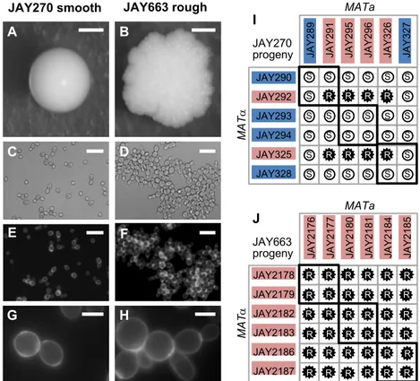
One of the most desirable features of the JAY270/PE-2 bioethanol production strain (henceforth referred to simply as JAY270) is that it does not normally aggregate during industrial sugarcane extract fermentation (i.e. cells stay in suspension in liquid culture). Accordingly, JAY270 produces normal hemispherical colonies with smooth
surfaces and edges when grown in solid agar medium (Fig. 2.1A). While this is the phenotype typically observed, over the course of our studies using this strain we noticed the sporadic occurrence of colonies clonally derived from JAY270 that displayed altered morphology: relatively flat-growing colonies with rough surfaces and edges (Fig. 2.1B). Under bright field microscopic examination, yeast cells derived from such rough
colonies appeared to grow in chains, showing a budding pattern consistent with a defect in the separation of the daughter cells from their mother (Fig. 2.1C-D). We stained these cells with calcofluor white to visualize the chitin-rich ring septa, confirming the
attachment of mother and daughter cells at the budding neck site (Fig. 2.1E-H).
Figure 0.1. Smooth and rough colony morphologies, mother-daughter cell attachment, and phenotypes of diploids derived from mating specific haploids. A-H show images of the JAY270 smooth parent diploid strain (left panels) and its
spontaneous rough derivative JAY663 (right panels). A and B, colony morphologies on YPD agar after 3 days growth at 30C. C and D, bright field, and E-H, fluorescence
A C E G B D F H JAY290 JAY292 JAY293 JAY294 JAY325 JAY328 R S JA Y 2 8 9 JA Y 2 9 1 JA Y 2 9 5 JA Y 2 9 6 JA Y 3 2 6 JA Y 3 2 7 S S S S S R R R R R R R S S S S S S S S S S S S S S S S S S S S S S MATa MA T a I JAY270 progeny JAY2178 JAY2179 JAY2182 JAY2183 JAY2186 JAY2187 R JA Y 2 1 7 6 JA Y 2 1 7 7 JA Y 2 1 8 0 JA Y 2 1 8 1 JA Y 2 1 8 4 JA Y 2 1 8 5 R R R R R R R MATa MA T a R R R R R R R R R R R R R R R R R R R R R R R R R R R R J JAY663 progeny JAY663 rough JAY270 smooth
daughter cell attachment. Scale bars are 1mm (A-B), 20µm (C-F) and 5µm (G-H). I and
J, Smooth (S, white circles) and rough (R, black stars) phenotypes of diploids formed by
crossing the indicated MATa and MATα haploids isolated from three tetrads of each JAY270 (I) and JAY663 (J). Thick black lines indicate the four diploids derived from matings of intra-tetrad sibling haploids. The colored backgrounds for each haploid correspond to their inferred genotype (Blue, dominant wild type allele; Red, recessive mutant allele). All 12 haploids from panel I had their whole genomes sequenced. Co-segregation analysis with JAY270 HetSNPs (Fig. S2.1) was used for identification of the causal mutation at the ACE2 locus (Figs. S2.2 and S2.3). Main results in this figure were generated by: ARP.
We initially isolated five independent examples of such rough colonies for genetic characterization (JAY663, JAY664, JAY665, JAY912 and JAY913), all of which were derived either directly from JAY270 or from JAY270-isogenic strains. The phenotype of these isolates was stably maintained and was not reversible over several clonal
generations, suggesting that it was likely hard-wired genetically and not caused by a transient transcriptional or post-transcriptional state. We estimated that these five rough colony isolates appeared spontaneously from a pool of ~50,000 smooth colonies. Assuming a genetic origin and based on this high frequency of occurrence in diploid cells, we reasoned that this phenotype was unlikely to be caused by a rare dominant de
novo nucleotide point mutation, but instead, mitotic recombination leading to
loss-of-heterozygosity (LOH) provided a more plausible mechanism.
In a parallel project, we observed that crossing two specific haploid descendants of JAY270 (JAY291 MATa and JAY292 MATa) resulted in diploid cells with the same rough colony morphology and mother-daughter cell attachment pattern observed in the five rough-colony isolates above. This was despite the fact that JAY291, JAY292, and all other haploid derivatives of JAY270 have the normal smooth colony phenotype. This indicated that the rough colony phenotype was diploid-specific, and the ability to
consistently reproduce the mutant phenotype in controlled crosses between specific haploids opened an avenue to investigate its genetic basis.
We previously reported the whole genome sequence of the JAY291 haploid(Argueso et al. 2009). Since then we have sequenced the genomes of 55 additional JAY270-derived haploids. This genome sequence dataset, comprising fourteen sets of four-spore tetrads, was generated in a project to characterize the abundance, distribution and phasing of heterozygous loci in the JAY270 genome, the full results of which will be described elsewhere. These haploid genomic sequences were used to create a draft map of phased heterozygous single nucleotide
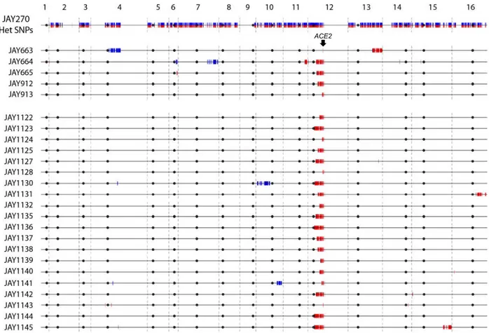
polymorphisms (HetSNPs) containing 12,023 loci unevenly distributed across the genome (Fig. S2.1).
We carried out crosses between twelve sequenced haploid descendants of JAY270 (3 tetrads; Fig. 2.1I). All possible MATa x MATa crosses were performed producing 36 different diploids. Among them, we found eight with rough and 28 with smooth colony surfaces, in a pattern that was consistent with recessive inheritance of a trait controlled by a single gene. Even though the rough colony phenotype was not observed in any of the haploid parents, the phenotypes of their respective diploid combinations allowed us to infer which allele was present in the parents: either the wild type dominant allele or the recessive mutant allele.
In addition, we induced sporulation of one of the spontaneous rough-colony isolates, JAY663, dissected tetrads, and examined the phenotypes of the haploid derivatives. None of the resulting haploids displayed the rough colony phenotype; they were all smooth (~100 examined). We then took twelve of these haploids (JAY2176
through JAY2187 comprising three full tetrads, determined their mating types, and conducted all possible mating combinations between them (Fig. 2.1J). In this case, all 36 crosses resulted in rough colony diploids. This result was consistent with JAY663 being homozygous for the causal recessive mutant allele, and supported the hypothesis that copy neutral LOH could be responsible for the sporadic appearance of the mutant phenotype in JAY270.
Genetic basis of the rough colony phenotype
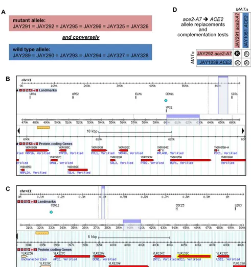
Based on the interpretation that the rough colony phenotype was associated with monogenic recessive inheritance of a diploid-specific trait, we divided the sequenced JAY270-derived haploids from Fig. 2.1I into two groups according to their inferred genotype. Group 1 included the six haploids inferred to carry the mutant recessive allele, whereas group 2 included the six haploids with the wild type dominant allele. We then compared the genome sequences of the twelve haploids to the draft JAY270 HetSNPs map. We interrogated each of the HetSNPs searching for alleles that co-segregated in all six individuals within group 1, and that conversely, had the other allele co-segregating in all six individuals within group 2. This analysis identified two candidate regions that fit the strict co-segregation criterion (Fig. S2.2A-C). One of the candidate regions corresponded to ~30 Kb on Chr11, including thirteen genes; and the other spanned ~15 Kb containing nine genes on the right arm of Chr12, located ~50 Kb centromere proximal to the ribosomal DNA genes tandem repeats (rDNA).
We reviewed the annotations of the 22 candidate genes, and identified a gene located in the Chr12 region, ACE2, which encodes a transcription factor that controls
the expression of genes involved in the mother-daughter cell separation process (Weiss 2012). In cells lacking Ace2p, the daughter cell remains attached to the mother cell wall at the bud neck, resulting in the accumulation of multicellular clusters. Importantly, a diploid-specific rough colony phenotype is observed in ace2/ace2 homozygous mutant strains in certain genetic backgrounds (Voth et al. 2005).
We inspected the genomic sequence of the ACE2 gene in JAY291, and
compared it to the sequence in the S288c reference genome. Only one difference was identified: The wild type ACE2 allele in S288c contains a homopolymer run of eight adenine nucleotides, while the mutant allele in JAY291 has seven adenines in this region, resulting in a -1 frameshift mutation and a stop codon shortly downstream. Hence, we named the mutant allele ace2-A7. We then conducted reciprocal
complementation tests to formally demonstrate that ace2-A7 was the causal mutation responsible for the rough colony phenotype. The mutant alleles in haploids JAY291 and JAY292 were replaced with the wild type allele, resulting respectively in the isogenic
ACE2 strains JAY1051 and JAY1039. When these allele replacement strains were
crossed to ace2-A7 strains (Fig. S2.2D), the resulting diploids displayed the smooth colony phenotype, thus confirming that the wild type ACE2 allele fully complemented the ace2-A7 mutation in heterozygous diploids.
Analysis of Chr12 LOH in spontaneous rough colony isolates
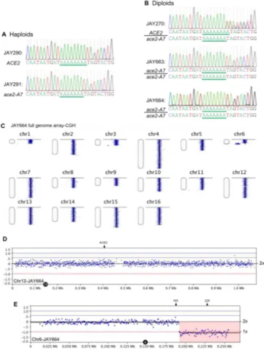
After identifying the association between the ACE2 locus and the rough colony phenotype, we determined its sequence in the five spontaneous rough colony
region containing the adenine homopolymer run in ACE2 from JAY270, from the haploid derivatives JAY290 and JAY291, and from the rough colony isolates (Fig. S2.3A-B). This analysis confirmed the presence of a run of 8 adenines in ACE2 (JAY290) and 7 adenines in ace2-A7 (JAY291). The chromatogram in the JAY270 heterozygous diploid was consistent with a mixture of ACE2 and ace2-A7 DNA templates being present in the sequencing reaction: single nucleotide peaks were observed at positions primer-proximal to the homopolymer run, and out-of-register double peaks were seen
downstream of the seventh adenine nucleotide. The chromatograms for all five rough colony isolates showed the presence of the ace2-A7 frameshift mutation and absence of the ACE2 allele. The loss of the ACE2 allele in the diploid rough-colony isolates may be explained by either a copy-neutral LOH mechanism such as inter-homolog mitotic recombination, by a segmental deletion spanning ACE2, or Chr12 monosomy. To distinguish between these scenarios, we conducted tetrad analysis with the five
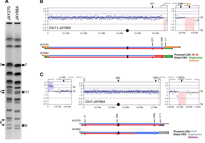
spontaneous rough colony isolates. Four of them produced tetrads that had four viable haploid spores, and each spore had a copy of the ACE2 locus as determined by PCR (data not shown). One of the isolates, JAY664, produced tetrads with two viable and two inviable spores, indicating the presence of a recessive-lethal mutation. We performed array-CGH analysis on JAY664 and determined that two copies of Chr12, including the ACE2 locus, were present (Fig. S2.3C-D). Together, these results showed that all five rough colony isolates were homozygous for ace2-A7, in agreement with the initial hypothesis that the high frequency of smooth to rough colony morphology
transitions among JAY270 derivatives was caused by interhomolog recombination leading to copy-neutral LOH. Unexpectedly, the JAY664 array-CGH also showed that
this rough colony isolate did carry copy number alterations in genomic regions other than Chr12. In particular, a terminal deletion on the right arm of Chr6 spanned multiple essential genes and explained the 2:2 spore viability phenotype (Fig. S2.3E).
Interestingly, the breakpoint for the Chr6 deletion occurred at a position
immediately distal to FAB1, where a tRNA gene and Ty1 retrotransposon sequences are found in the S288c reference genome and in the JAY270 maternal Chr6 homolog, which sustained the deletion. We analyzed the status of HetSNP markers flanking the breakpoint and found that a proximal marker (Chr6 - 185 Kb) remained heterozygous, while a distal marker (Chr6 - 229 Kb) lost heterozygosity through a deletion mechanism (Fig. S2.4A-B). Even though we did not characterize the precise sequences that were joined at the deletion breakpoint in JAY664, this pattern was consistent with non-allelic homologous recombination (HR) involving Ty retrotransposon repeats, a major class of gross chromosomal rearrangements observed in S. cerevisiae (Argueso et al. 2008; Putnam and Kolodner 2017).
LOH is typically a regional, rather than local, mutational mechanism. Interstitial tracts of homozygosity can span tens of kilobases, and terminal tracts are even longer, extending all the way to the telomeres (St Charles and Petes 2013). Therefore, in addition to being homozygous for ace2-A7, the rough colony isolates might also be homozygous for flanking HetSNPs. We tested this model initially at low resolution by determining the genotypes at eleven Chr12 HetSNPs using PCR (Table S2.3). The results of this analysis were compiled to produce the LOH tract maps shown in Fig. 2.2. As expected, JAY270 was heterozygous for all eleven markers tested. Notably, Chr12 in this strain is only heterozygous for positions to the left of the rDNA cluster (Fig. S2.1).
This pattern is similar to that described previously for other heterozygous diploid S.
cerevisiae genomes and is suggestive of ancestral LOH events mediated by rDNA
instability (Magwene et al. 2011).
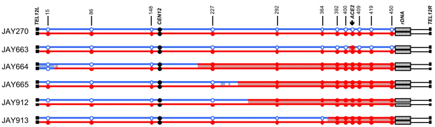
Figure 0.2. LOH tract maps of Chr12 from five original rough colony isolates.
The genotypes at twelve phased JAY270 Chr12 HetSNP marker loci were determined using PCR and RFLP or Sanger sequencing analyses (Table S2.3). The approximate coordinates of the markers are shown in Kb. The Chr12 homolog containing the
ace2-A7 allele was arbitrarily designated as maternal (Chr12-M, red) and the homolog
containing the wild type ACE2 allele as paternal (Chr12-P, blue). JAY270 was heterozygous at all markers, and all rough colony isolates were homozygous for the
ace2-A7 allele. White boxes distal to the 450 Kb HetSNP represent ~1.5 Mb of
ribosomal DNA repeats (rDNA). Chr12 regions distal to the rDNA do not contain any heterozygous markers in JAY270. The red or blue shading corresponds to the directions (M or P, respectively) and approximate breakpoint positions of the LOH tracts
determined at high resolution using whole genome sequencing (detailed in Fig. S2.6). Main results in this figure were generated by: ARP, MJC, NMVS.
Analysis of the JAY663 isolate showed that, while it was homozygous for the
ace2-A7 mutation, it remained heterozygous at all other flanking markers, including
those immediately proximal and immediately distal to the ACE2 locus. A mitotic gene conversion tract limited to the 8.7 Kb region between these HetSNPs could explain this result. Alternatively, a de novo -1 contraction mutation in the adenine homopolymer run of the ACE2 allele could also account the for the JAY663 genotype (Tran et al. 1997).
450 rDNA 419 400 409 392 292 364 CE N1 2 227 15 86 148 JAY270 JAY663 JAY664 JAY912 JAY913 TE L1 2 L TE L1 2 R ç ACE 2 JAY665
The four remaining isolates (JAY664, JAY665, JAY912, and JAY913) were
homozygous for regions well beyond the ACE2 locus. In this low-resolution map, all four LOH tracts were unidirectional, continuous, and homozygous for SNPs present in the Chr12 homolog that contained the ace2-A7 allele, which we arbitrarily designated the maternal homolog (Chr12-M; red in all figures). Subsequent high resolution LOH mapping using whole genome sequencing (WGS; below) confirmed the initial results and revealed additional complexities to the tracts. The centromere-proximal breakpoints of the LOH tracts were roughly mapped to positions ranging from 39 Kb (JAY913) to 184 Kb (JAY664) from the ACE2 locus. On the distal side, these isolates had an
additional 45 Kb of LOH that extended to the HetSNP at position 450 Kb, located 1.4 Kb proximal to the rDNA repeats. From this point, Chr12 contains ~1.5 Mb of rDNA repeats plus another ~0.6 Mb of distal homozygous single copy sequences. Since the 450 Kb HetSNP was the most distal marker in Chr12, we could not distinguish if these LOH tracts were generated as very long interstitial gene conversion events, or if they extended to the right telomere. This initial PCR-based analysis also revealed an
unexpected secondary LOH event on the left arm of Chr12 in JAY664, but in this case, it was associated with homozygosity for the SNPs from the paternal homolog (Chr12-P, blue in all figures; see below).
Analysis of selected Chr12 LOH
Taken together, the results described above showed that the majority of the isolates with altered colony morphology were homozygous not only at the ACE2 locus, but also for surrounding regions, indicating that interhomolog mitotic recombination was
frequent in JAY270 and that it likely had substantial effect on the genetic makeup of this strain. In addition, the distribution of HetSNPs in the genome is notably uneven (Fig. S2.1), with long tracts of homozygosity, suggesting that abundant LOH occurred in the JAY270 lineage.
To gain a deeper understanding of the impact of genome instability processes on the present genetic composition of JAY270, we conducted experiments to directly
measure the rate of LOH in this strain (Fig. 2.3A). Starting with a homozygous ura3/ura3 derivative of JAY270 (FGY050; gift from F. Galzerani), we introduced one copy of the
KlURA3-ScURA3-KanMX4 CORE2 counter selectable cassette (Zhang et al. 2013) at a
position immediately proximal to ACE2 (1.3 Kb from the adenine homopolymer run). We grew cultures of strains carrying this insertion and plated the cells in media containing 5-FOA to identify clones that had lost the cassette. We performed this assay in a
derivative of JAY270 carrying the hemizygous CORE2 insertion in Chr12-M. The frequency of homozygosity for the ACE2 allele was 1.2 x 10⁻⁴, comparable to the
unselected frequency of ace2-A7 homozygosity (5 in ~50,000) estimated earlier in the study. We also used hemizygous CORE2 insertions to measure LOH rates at two other positions in the genome (Chr4 near SSF2, and Chr13 near ADH6), and on Chr5 by deleting one allele of the CAN1 gene (can1D::NatMX4/CAN1), and selecting for loss of
the remaining WT allele in clones resistant to canavanine. In order to provide a reference for comparison of LOH rates from JAY270, we introduced these same four constructs in a standard laboratory yeast strain background routinely used to study genome instability mechanisms, including LOH (CG379; Fig. 2.3B) (Morrison et al. 1991; Conover et al. 2015). The rates of LOH were not significantly different between