SEIDA EROVIC-ADEMOVSKI
TREATMENT OF
INTRA-ORAL HALITOSIS
SEID A ER O VIC-ADEMO VSKI MALMÖ UNIVERSIT TREA TMENT OF INTR A -OR AL HALIT OSIS DOCT OR AL DISSERT A TION IN ODONT OL OG YDoctoral Dissertation in Odontology, 2017
© Copyright Seida Erovic Ademovski 2017 Fotografs/illustratörs namn
ISBN 978-91-7104-718-2 (print) ISBN 978-91-7104-719-9 (pdf) Holmbergs, Malmö 2017
Malmö University, 2017
Faculty of Odontology
Department of Periodontology
Kristianstad University
SEIDA EROVIC-ADEMOVSKI
TREATMENT OF
INTRA-ORAL HALITOSIS
Publikationen finns även elektroniskt, se www.mah.se/muep
CONTENTS
LIST OF PAPERS ... 9 ABSTRACT ... 11 Paper I ... 12 Paper II ... 12 Paper III... 13 Paper IV ... 13 POPULÄRVETENSKAPLIG SAMMANFATTNING ... 15 Studie I ... 15 Studie II ... 16 Studie III ... 17 Studie IV ... 18 ABBREVIATIONS ... 19 INTRODUCTION ... 20The olfactory system odour and scent ... 20
Halitosis ... 21
Social interaction and intra-oral halitosis ... 23
Prevalence of intra-oral halitosis ... 24
Factors associated with intra-oral halitosis ... 28
Tongue coating ... 30
Microbiota ... 30
Periodontal conditions and intra-oral halitosis ... 30
Other factors linked to intra-oral halitosis ... 31
Treatment of intra-oral halitosis ... 33
Periodontal treatment ... 33
Tongue cleaning ... 34
Mouth rinsing ... 34
OBJECTIVES ... 36
MATERIALS AND METHODS ... 37
Paper I and II ... 39 Paper II ... 40 Paper III ... 40 Paper IV ... 41 Statistics ... 42 Paper I ... 42 Paper II ... 42 Paper III... 43 Paper IV ... 43 Ethical approval ... 43 RESULTS ... 44 Paper I ... 44 Paper II ... 46 Paper III ... 48 Paper IV ... 50 DISCUSSION ... 53 CONCLUSIONS ... 58 ACKNOWLEDGEMENT ... 59 REFERENCES ... 61 PAPERS I - IV ... 73
LIST OF PAPERS
This thesis is based on the following four papers; they will be re-ferred by the Roman numerals in the text and are attached at the end of the thesis.
I. Erovic Ademovski S, Lingström P, Winkel E, Tangerman A, Persson G. R, Renvert S. Comparison of different treat-ment modalities for oral halitosis. Acta Odontologica Scandinavica 2012; 70: 224-233.
II. Ademovski S.E, Persson G.R, Winkel E, Tangerman A, Lingström P, Renvert S. The short-term treatment effects on the microbiota at the dorsum of the tongue in intra-oral halitosis patients- a randomized clinical trial. Clinical Oral Investigations 2013; 17: 463-473.
III. Erovic Ademovski S, Mårtensson C, Persson G.R, Renvert S. The effect of periodontal therapy on intra-oral halitosis: a case series. Journal of Clinical Periodontology 2016; 43: 445-452.
IV. Erovic Ademovski S, Mårtensson C, Persson G.R, Renvert S. The long-term effect of a zinc acetate and chlorhexidine diacetate containing mouth rinse on intra-oral halitosis- a randomized clinical trial. (In review) Journal of Clinical Periodontology.
Published papers are copied with the permission of the respective copyright holders. Paper I: © 2012 Informa Healthcare, Paper II: © 2012 Springer- Verlag. Paper III © 2016 John Wiley & Sons A/S.
ABSTRACT
Intra-oral halitosis (bad breath) is reported to affect 15-83 % of the adult population. Having intra-oral halitosis is a social and psycho-logical handicap, and may cause people in the person’s social circle to increase the physical distance or to turn their faces in another di-rection to avoid the unpleasant smell from the exhaled air. Such be-haviours may affect the individual’s self-confidence resulting in inse-curity in social and intimate relations. The oral health-related quality of life status has also been reported to be lower in individuals with halitosis. Approximately 90% of what is considered as bad breath is the result of the degradation of organic substrates (proteins) by an-aerobic bacteria of the oral cavity. Intra-oral halitosis can be as-sessed using both subjective and objective methods to evaluate the subject’s exhaled air. The most common one and the one often re-ferred to as the ”gold standard”, is the organoleptic scoring system (OLS). OLS is a subjective method evaluating the strength of halito-sis in exhaled air using a scale from 0-5. One objective method to assess the presence of volatile sulphur compounds in exhaled air is to use a sulphide monitor measuring the total sum of the volatile sul-phur compounds (T-VSC) in exhaled air. The three gases (hydrogen sulphide (H2S), methyl mercaptan (MM) and dimethyl sulphide
(DMS)) in exhaled air related to intra-oral halitosis can be assessed separately using a simplified gas chromatograph. Different treatment models such as periodontal treatment, tongue scraping and rinsing with Zn ion containing products have been used to reduce intra-oral halitosis. The present thesis has evaluated the efficacy of different treatment models in the treatment of intra-oral halitosis.
Paper I
In Paper I, the effects on intra-oral halitosis using a mouth rinse containing zinc acetate (0.3%) and chlorhexidine diacetate (0.025%) (ZN/CHX) with or without the adjunct use of a tongue scraper were assessed. Twenty-one subjects without a diagnosis of periodontitis were randomized in a cross-over clinical trial. Or-ganoleptic scores (OLS) total volatile sulphur compounds (T-VSC) and the presence of hydrogen sulphide (H2S), and methyl
mercap-tan (MM) were assessed. Evaluations were made before rinsing, immediately after rinsing, 30 minutes after rinsing, and 14 days af-ter rinsing. OLS scores were significantly lower following active rinse combined with tongue scraping (p< 0.001) at all time points. After 30 min, and at day 14, the T-VSC values, H2S and MM were
lower in the active rinse sequence than in the placebo rinse se-quence. The placebo rinse sequence with tongue scraping reduced T-VSC at 30 min but not at 14 days. Similar reductions of T-VSC, H2S and MM values following the active rinse sequence with or
without tongue scraping were found. In conclusion, the use of a tongue scraper did not provide additional benefits to the active mouth rinse, but reduced OLS scores, and tongue coating.
Paper II
In Paper II the effects on the microbiota at the dorsum of the tongue were studied following rinsing with a Zn/CHX containing mouth rinse with or without adjunct tongue scraping on volatile sulphur compounds (VSCs) in exhaled air. Bacterial samples from the dorsum of the tongue were assessed by checkerboard DNA– DNA hybridization. At day 14, 48% of the individuals rinsing with the active rinse and using the tongue scraper, and 14% of the indi-viduals in the placebo and tongue scraping sequence were consid-ered effectively treated for their intra-oral halitosis. At day 14 in the active rinse sequence, significantly lower bacterial counts were identified in samples from the dorsum of the tongue (p<0.001) for 15/78 species including Fusobacterium sp., Porphyromonas
gin-givalis, Pseudomonas aeruginosa, Staphylococcus aureus, and Tannerella forsythia. In successfully treated subjects a decrease in
bacterial counts from baseline to day 14 were found for 9/74 spe-cies. Data from study II showed that VSC values were not
associ-ated with bacterial counts in samples taken from the dorsum of the tongue. The active rinse alone had effects on intra-oral halitosis and reduced bacterial counts of species associated with intra-oral halitosis. Tongue scraping had no additional effects on the micro-biota studied.
Paper III
In Paper III the effects of non-surgical periodontal therapy on in-tra-oral halitosis was evaluated. Non-surgical periodontal therapy was performed in sixty-eight adults with confirmed intra-oral hali-tosis. Three months after therapy OLS scores (p<0.01), T-VSC (p<0.01), and MM (p<0.05) values were significantly lower. The non-surgical therapy resulted in a significant reduction of probing pockets (PPD), bleeding on probing (BOP) and plaque indices (PI). Successful periodontal therapy was defined as a BOP<20% and a ≥50% reduction of total PPD (total PPD was calculated by adding all pockets ≥ 4 mm from the entire dentition). The 34 individuals with successful periodontal treatment demonstrated reductions in OLS (p<0.01) scores, and T-VSC (p<0.01) values. Effective treat-ment for intra-oral halitosis (T-VSC value <160 ppb, H2S value
<112 ppb and MM value <26 ppb) was identified in 11/68 indi-viduals. Thus, non-surgical periodontal therapy resulted in few in-dividuals who were considered as effectively treated for intra-oral halitosis.
Paper IV
In Paper IV the long-term effects of a zinc acetate and chlorhexi-dine diacetate (Zn/CHX) mouth rinse on intra-oral halitosis were evaluated 3, and six months after treatment. Forty-six adults with intra-oral halitosis were randomized into a 6-month, double-blinded, placebo-controlled clinical study. Using assessments of or-ganoleptic scores (OLS), total volatile sulphur compounds (T-VSC), hydrogen sulphide (H2S), and methyl mercaptan (MM)
con-centrations in exhaled air the presence of intra-oral halitosis was evaluated. At three and six months, individuals rinsing with the Zn/CHX rinse presented with reductions of the OLS, T-VSC, H2S,
and MM in exhaled air. At six months 68.2% of individuals using the Zn/CHX rinse experienced a 1 or 2 category improvement in
OLS compared with 19.1% of placebo-treated subjects. In the group rinsing with the Zn/CHX mouth rinse 91% of subjects were categorized as being effectively treated for their intra-oral halitosis (i.e. H2S < 112 ppb). Compared to 43% in the placebo group. In
conclusion, a Zn/CHX mouth rinse provides effective long-term efficacy against intra-oral halitosis, assessed both objectively and subjectively.
POPULÄRVETENSKAPLIG
SAMMANFATTNING
Att leva med dålig andedräkt (Intra-oral halitosis) kan vara ett socialt och psykologiskt handikapp. Det kan resultera i att personer i omgiv-ningen vänder bort ansiktet för att undvika den obehagliga lukten. Ett sådant beteende kan påverka individens självförtroende och leda till osäkerhet i sociala och privata relationer. Personer med dålig ande-dräkt har sämre livskvalité. Att ha dålig andeande-dräkt är vanligt och mel-lan 15 och 83 % har rapporterats ha dålig andedräkt. Den huvudsak-liga anledningen till dålig andedräkt är att bakterier i munhålan bryter ner proteiner och bildar svavelinnehållande illaluktande gaser. Dålig munhygien, tungbeläggningar och ett inflammerat tandkött förklarar 80-90 % av all dålig andedräkt. En persons andedräkt kan undersö-kas med både subjektiva och objektiva metoder. Den vanligaste meto-den som ofta anges som "gold standard", är att lukta på patientens utandningsluft. Undersökaren bedömer sedan styrkan av dålig ande-dräkt, så kallad organoleptisk scoring (OLS) enligt en skala från 0 till 5 där 0 står för ingen dålig lukt och 5 står för extremt dålig ande-dräkt. En objektiv metod för att mäta den sammanlagda förekomsten av flyktiga svavelföreningar (T-VSC) i utandningsluften, är att använ-da en sulfidmätare. De tre gaser i utandningsluften som kopplas till dålig andedräkt är väte sulfid (H2S), metylmerkaptan (MM) och
dimetylsulfid (DMS). Dessa kan mätas separat i en portabel gaskro-matograf. Olika behandlingsmodeller har använts för att reducera då-lig andedräkt. Denna avhandling har utvärderat effekten av behand-ling av inflammerat tandkött, skrapning av tungan och sköljning med Zink-jon innehållande munsköljmedel på dålig andedräkt.
Studie I
I studie I, utvärderades effekten på dålig andedräkt av ett munskölj-medel innehållande zinkacetat (0,3%) och klorhexidindiacetat (0,025%) (Zn/CHX) eller ett placebo sköljmedel med eller utan tillägg av en tungskrapa. Tjugoen försökspersoner utan tandlossning randomiserades i en cross-over studie. Försökspersonernas ande-dräkt kontrollerades före sköljning, direkt efter sköljning, 30 minu-ter efminu-ter sköljning, och 14 dagar efminu-ter sköljning. OLS värden var sig-nifikant lägre efter aktiv sköljning i kombination med tungskrapa vid alla tidpunkter. Efter 30 minuter, och vid dag 14 var värdena för T-VSC, H2S och MM lägre efter sköljning med Zn/CHX jämfört
med sköljning med placebo. Efter sköljning med placebo kombinerat med användning av tungskrapa minskade värdet på T-VSC vid 30 minuters mätning, men inte vid dag 14. Efter användning av skölj-vätskan som innehöll Zn/CHX med eller utan användning av tung-skrapa minskade värdena för T-VSC, H2S och MM.
Tilläggsbehand-ling med tungskrapa gav ingen ytterligare reduktion av värdena jäm-fört med bara sköljning med den aktiva sköljsekvensen. Användning av tungskarapa minskade däremot värdena på OLS och mängden diagnosticerad tungbeläggning
Studie II
I studie II, utvärderades effekten av ett Zn/CHX innehållande munsköljmedel eller placebo med eller utan komplement av tung-skrapa på mikroorganismer på tungan och på flyktiga svavelföre-ningar (VSCs) i utandningsluften. Bakterieprover från tungan be-dömdes med en checker-board DNA-DNA-hybridiserings metod. VSC värdena som registrerades vid dag 0 var korrelerade till före-komsten av flera bakteriearter. Baserat på VSC värden vid 14 da-gar hade 57% av de som sköljt med den aktiva substansen ingen dålig andedräkt. Efter användning av den aktiva substansen kom-binerat med tungskrapa hade 48% ingen dålig andedräkt Sköljning med placebolösning resulterade i att 5% inte hade dålig andedräkt efter 14 dagars sköljning. Om placebosköljning kombinerades med användning av en tungskrapa var 14% klassade som effektivt be-handlade. I prover tagna från tungryggen vid dag 14 identifierades signifikant färre bakterier efter användning av den aktiva sköljse-kvensen. Femton av sjuttioåtta bakteriearter inklusive
Fusobacteri-um sp., Porphyromonas gingivalis, Pseudomonas aeruginosa, Staphylococcus aureus, och Tannerella forsythia var signifikant
lägre i prover från tungryggen (p<0,001), efter användning av det aktiva sköljmedlet. Hos individer som bedömdes effektivt behand-lade för sin dålig andedräkt vid dag 14 var 9/74 bakteriearter redu-cerade i prover från tungryggen. Flyktiga svavelföreningar var inte förknippade med bakterietal i prover tagna från tungan. Sköljning med ett Zn/CHX innehållande munsköljmedel hade effekt på dålig andedräkt och minskade förekomsten av bakteriearter som är för-knippade med dålig andedräkt. Tungskrapning hade inga ytterliga-re effekter på de mikroorganismer som studerades.
Studie III
I studie III utvärderades effekten av icke-kirurgisk parodontal be-handling (tandhygienistbebe-handling innefattande munhygieninstruk-tion, borttagning av tandsten samt polering av tänderna) på 68 vuxna individer med konstaterad dålig andedräkt. Tre månader ef-ter behandling konstaef-terades minskade värden för OLS (p<0,01), T-VSC (p<0,01), och MM (p<0,05). Den icke-kirurgiska behand-lingen resulterade i en signifikant reducering av parodontala fickor, blödning vid sondering och plackindex (bakteriebeläggningar på tänderna). Individernas totala fickdjup beräknades genom att ad-dera alla fickor ≥ 4 mm i hela bettet vid studiens början och slut. Framgångsrik parodontal behandling definierades om blödning vid sondering var <20% och om det sammanlagda värdet av de fickor som var över 4 mm hade reducerats med ≥50%. Hos de 34 indivi-der som på detta sätt bedömdes vara parodontalt effektivt behand-lade minskade OLS, och T-VSC värdena signifikant (p<0,01). Elva av 68 individer klassades som effektivt behandlade för sin dåliga andedräkt dvs hade T-VSC <160 ppb, H2S <112 ppb och ett värde
på MM <26 ppb). Icke-kirurgisk parodontal behandling resultera-de i få indiviresultera-der som ansågs effektivt behandlaresultera-de för sin dåliga an-dedräkt.
Studie IV
I studie IV utvärderades effekten av (Zn/CHX) munsköljmedel på dålig andedräkt efter sex månader. Fyrtiosex vuxna med dålig an-dedräkt randomiserades i en 6-månaders, dubbel-blind, kliniskt kontrollerad studie. Sköljning med ett Zn/CHX innehållande mun-sköljmedel resulterade i en signifikant reduktion av OLS, T-VSC, H2S och MM i utandningsluften vid både tre och sex månaders
uppföljning. Efter 6 månaders sköljning med den aktiva substansen uppvisade 68,2% en förbättring i OLS (ett eller två stegs förbätt-ring på den femgradiga skalan) jämfört med 19,1% av de individer som sköljt med en placebolösning. Hos gruppen som sköljde med ett Zn/CHX innehållande munsköljmedel var 91 % av individerna effektivt behandlade för sin dåliga andedräkt (H2S <112 ppb),
jäm-fört med 43% i placebogruppen. Användning av ett Zn/CHX in-nehållande munsköljmedel reducerar effektivt och har en långtids-effekt mot dålig andedräkt utvärderat med såväl subjektiva som objektiva metoder.
ABBREVIATIONS
BOP Bleeding on probing
CAL Clinical attachment level
CHX Chlorhexidine DMS Dimethyl sulphide DT Decayed teeth GI Gingival index H2S Hydrogen sulphide MM Methyl mercaptan NS No significant difference OLS Organoleptic scores PD Pocket depth PI Plaque index ppb Parts per billion PPD Probing pocket depth TC Tongue coating
T-VSC Total sum of volatile sulphur compounds VSC Volatile sulphur compounds
WTCI Winkel tongue coating index
INTRODUCTION
The olfactory system odour and scent
The olfactory system is the part of the sensory system used for smelling. Through the nasal cavity, scent molecules reach the re-ceptor cells located in the olfactory epithelium (Ward et al. 2003). There are between 6 and 10 million receptor cells located in the ol-factory epithelium (Ward et al. 2003). The human nose can recog-nize 10 000 different scents. The olfactory system cannot be turned off and prompts immediate emotional responses (Bradford et al. 2009). The olfactory receptors send information both to the neo-cortex for conscious processing and to the limbic brain system for emotional processing, allowing us to associate a particular smell with distinct memories (Grammer et al. 2005).
Body odour derives from different body fluids. The glandular sys-tem of the skin is the primary source for human body odours. Pheromones are chemical messengers (odour signals) produced by the human body. Such pheromones may activate specific psycho-logical and behavioural responses (Grammer et al. 2005). The scents are related to kin recognition, mate selection, menstrual cy-cle synchronicity and emotional contagion (Bader et al. 2002, Weisfeld et al. 2003, Pause 2012, Semin et al. 2013). Heterosexual males considered the odour of a female ovulatory period to be more pleasant and sexy than during the luteal period (Trouton et al. 2012). Human odours can be related to the person's emotional state such as stress, sadness, and fear (Bradford et al. 2009).
Human preferences and behaviours are influenced by scents. Indus-try is using different scents in marketing their products. It is well known that scents influence consumers into react in certain ways. Warm ambient scents may influence the consumers into increased purchasing of premium products (Madzharov et al. 2015). Scents have also been used in dental offices to reduce anxiety among the patients (Lehrner et al. 2000).
Halitosis
The term halitosis derives from the Latin word 'halitus' (breath), and the Greek word 'nosos' (disease). The term halitosis has been used since 1930s (Ramdurg & Mendigeri 2014). Halitosis is a gen-eral term describing an unpleasant odour from oral and systemic sources (Tonzetich 1977). Terms such as bad breath, breath malo-dour, oral malomalo-dour, intra-oral halitosis, fetor ex ore and fetor oris have also been used to describe an unpleasant odour originating from the oral cavity (Tangerman 2002, Van der Sleen et al. 2010). There are documents dating back to the Greek and Roman times describing halitosis. Also the Islamic theology describes the impor-tance to prevent bad breath (Mandel 1988, Fischman 1997). In the Talmud (a central text of Rabbinic Judaism) bad breath was con-sidered a major problem, so a woman was entitled to seek divorce if her husband had bad breath. According to the Talmud, priests with bad breath were not allowed to carry out holy duties in the temple. In 1898, Joseph Howe published the monograph “The breath, and the diseases which give it a fetid odour”.
In the Mediterranean countries, a resin from the Pistacia lentiscus tree (labdanum) has been used to refresh the breath for thousands of years. Other folk cures include the use of parsley (Italy), cloves (Iraq), guava peels (Thailand), and eggshells (China) (Mandel 1988, Rosenberg 1996, Fischman 1997, Huwez et al. 1998, Shif-man 2002).
Halitosis often classifies as Genuine halitosis, Pseudo halitosis or Halitophobia. In individuals with pseudo-halitosis others do not perceive an obvious malodor, although the patient complains of its existence. The condition can be improved by counseling (using
lit-erature support, education and explanation of objective examina-tion results) and simple oral hygiene measures (Yaegaki & Coil 2000, Amstrong et al. 2010, Seemann et al. 2014).
If after treatment for genuine or pseudo–halitosis, the patient is still convinced that he/she suffers from halitosis although no physi-cal or social evidence exists to suggest that halitosis is present, the condition is referred to as halitophobia. Most halitophobic patients interpret other people’s behavior, such as covering the nose, avert-ing the face or steppavert-ing back, as an indication of their own bad breath (Yaegaki & Coil 2000, Amstrong et al. 2010, Seemann et al. 2014)
In 2014 Seemann et al. recommended to use the word halitosis and to distinguish between intra- and extra-oral halitosis. Intra-oral halitosis originates from the mouth and extra-oral originates from other sources. Extra-oral halitosis can be subdivided in to blood-borne and non-blood-blood-borne halitosis. Tangerman and Winkel (2007) aimed to explain the origin and cause of intra-oral and ex-tra-oral halitosis. The degree of inex-tra-oral halitosis determined by organoleptic scoring of mouth breath and the concentration of H2S
and MM in mouth breath is strongly correlated (Tangermann 2002, Tangerman & Winkel 2007). MM is considered the main contributor to intra-oral halitosis and DMS the main contributor to extra-oral or blood-borne halitosis whereas MM and H2S
can-not be found in blood-borne halitosis (Tangerman 2002, Tangerman & Winkel 2007).
Intra-oral halitosis is accordingly used to describe bad breath from the oral cavity. Bacterial degradation of proteins in the oral cavity is the primary source of intra-oral halitosis (Rosenberg & McCulloch 1992, Tangerman 2002, Quirynen et al. 2009). Indi-viduals suffering from intra-oral halitosis present with higher con-centrations of volatile sulphur compounds (VSCs), including hy-drogen sulphide (H2S), and methyl mercaptan (MM) (Tonzetich
1977). Data have indicated that H2S and MM are the main VSCs
MM has a cooked cabbage smell, and DMS has cabbage, cauli-flower, and garlic aromas (Swiegers & Pretorius. 2007).
The oral cavity is considered as the primary source of intra-oral halitosis (Tangerman & Winkel 2007, Quirynen et al. 2009). Hali-tosis can also be related to diseases such as chronic sinusitis, pneumonia, liver diseases and cancer in the respiratory system (Tangerman & Winkel 2010, Seemann et al. 2014). Extra-oral blood borne halitosis originates from systemic diseases i.e.; liver cirrhosis, uremia, diabetic ketoacidosis, metabolic disorders, and medications i.e.; disulfiram, dimethyl sulfoxide and cysteamine (Tangerman & Winkel 2010 Seemann et al. 2014). Extra-oral non-blood borne halitosis also originates from pathological conditions such as throat infection; tonsillitis, nasal infection; sinusitis and postnasal drip, infection of respiratory system; lung infection, lung disease; lung cancer, and tuberculosis (Tangerman & Winkel 2010, Seemann et al. 2014). The most common source of extra- oral hali-tosis is; tonsillitis (71%) followed by sinusitis (9.5%), foreign body in the nose (9.5%) and diabetes mellitus (9.5%) (Seemann et al. 2006). Actions taken to treat intra-oral halitosis will not help those suffering from extra-oral halitosis. Such individuals should be re-ferred for medical examination and treatments.
Social interaction and intra-oral halitosis
Intra-oral halitosis is considered as an unattractive aspect of social interaction. In a Dutch survey, 14.5% out of 1000 individuals an-swered that they on a daily basis were exposed to individuals with halitosis. Sweat malodour was considered as the strongest ‘downer’ when meeting a person for the first time (de Jongh et al. 2014). To have halitosis is a social and psychological handicap and may cause people in the person’s social circle to increase the physical distance, or to turn their faces in another direction to avoid the unpleasant smell from their breath when communicating (Scully et al. 1997, de Jongh et al. 2016).
Decreased self-confidence and insecurity in social and intimate re-lations were major reasons for individuals seeking treatment at specialized breath odour clinic (McKeown 2003). If a person
per-ceives a constant bad breath problem, he/she may avoid social rela-tions affecting the person's well-being (McKeown 2003, Lu et al. 2016). Kursun et al. (2014) reported 62% to have anxiety caused by halitosis. Individuals with halitosis and with a strong social anxiety disorder were reported to have difficulties overcoming their halitosis anxiety (Zaitsu et al. 2011). The anxiety of having halito-sis is reinforced when others cover their noses or avert their faces in social contacts as such behaviours are interpreted as an indica-tion of halitosis (Yaegaki & Coil 1999).
In a Swedish population, the 49-item Oral Health Impact Profile (OHIP) was used to assess oral health-related quality of life in 1309 subjects. Bad breath was the most prevalent condition, and especially reported by young individuals (Oghli et al. 2017). In an-other study, the authors identified that individuals with intra-oral halitosis were reported to have poorer oral health related quality of life status as compared to those without halitosis (Lu et al. 2016).
Prevalence of intra-oral halitosis
The prevalence rates of intra-oral halitosis vary greatly between studies (from 1.5% to 100 %) and may be explained how halitosis has been assessed or defined. In some studies prevalence rates of intra-oral halitosis are based on self-perceived assessments. Self-percieved reports of halitosis should be interpreted with caution as the perceptions of halitosis vary between individuals. Self-assessment of intra-oral halitosis is difficult.
Many individuals with halitosis are, not aware of the fact that their breath may be obnoxious (Rosenberg 1996). In a study based on dental students, the perception of halitosis did not always reflect the actual situation (Rani et al. 2015). Cultural differences may, in part explain differences in reported prevalence rates of intra oral halitosis based on self-perceived assessments (Mumghamba et al. 2006, Seemann et al. 2006, Hammad et al. 2014, Lu et al. 2014, Villa et al. 2014, Kim et al. 2015, and Rani et al. 2015).
Age and gender may also be factors influencing the perception of intra-oral halitosis. Among male Swiss army recruits 1.5 %
per-ceived that they had halitosis frequently (Bornstein et al. 2009a) whereas 14 % of young mothers in Tanzania reported that they had bad breath, and that this was associated with gingival bleeding on tooth brushing, and periodontal pockets ≥ 6 mm (Mumghamba et al. 2006). Sixty-two percent of elderly individuals living in a nursing home in Sweden were convinced of having halitosis (Zell-mer et al. 2016). There are, however, conflicting results regarding the impact of age on intra-oral halitosis (Nadanovsky et al. 2007, Samnieng et al. 2012).
Socioeconomic factors have been reported to be related to self-reported halitosis (Lopes et al. 2016). Inadequate oral hygiene practices have also been strongly associated with self-reported hali-tosis (Al-Ansari et al. 2006). Other self-perceived factors reported to be related to halitosis are poor health status, overweight or obese, stress, lower economic levels, high intake of fast food, in-stant noodles and the low intake of fruits and vegetables (Kim et al. 2015). Among undergraduate dental students the prevalence of self-perceived halitosis was found to significant correlate with smoking and dryness of mouth (Setia et al. 2014).
Depending on methods used to assess intra-oral halitosis figures ranging from 15 % (Nadanovsky et al. 2007) to 83 % (Zürcheret al. 2012) have been reported (Table 1).
Intra-oral halitosis varies during the day, and higher levels have been reported in morning breath (Miyazaki et al. 1995). If 75 ppb of H2S in the exhaled air was considered as the socially acceptable
level, 23% in the late morning and 6% in the early afternoon had halitosis (Miyazaki et al. 1995). Another study, however, failed to show significant variation during the day (Samnieng et al. 2012).
26 T abl e 1. Pr ev al enc e of int ra or al ha lit os is . A ut hor L oc at ion Ag e Sa m ple s iz e (N) In tra -o ra l Ha lit os is ( % ) Aim et ti e t a l. 2 01 5 Ita ly 20 -75 744 55. 38 B or ns te in e t a l. 2009a Swit ze rla nd 18 -25 580 OL S 2 = 25 , T -V SC > 75 p pb = 42. 6 B or ns te in e t a l. 2009 b Swit ze rla nd 18 -94 419 OL S ≥ 2 = 31. 5 > 75 p pb = 72. 1 C he n e t a l. 2016 C hi na 22 -70 720 > 110 pp b = 33. 2 L iu e t a l. 2006 C hi na 15 -64 2000 27. 5 L u e t a l. 2014 C hi na 18 -82 911 OL S ≥ 2 = 77. 3 Mi yaz ak i e t al . 1 99 5 Jap an 18 -64 2672 6-23 H am m ad e t a l. 2014 Jo rd an 18 -68 205 78 N alç ac i e t a l. 2 00 8a T ur ke y 7-11 628 14. 5 N alç ac i e t a l. 2 00 8b T ur ke y 7-15 30 V SC > 11 0 ppb = 26. 7 N ada nov sky e t a l. 2007 B ra zil 1-87 344 15 V illa e t a l. 2 01 4 Ita ly 6-16 101 OL S 2 = 15 , > 100 pp b=3 7. 6
27 A ut hor L oc at ion Ag e Sa m ple s iz e (N) Sa m ni eng e t a l. 2012 T ha ila nd >60 428 H2 S 60. 5 MM 62. 9 DM S 80. 7 Se em an n e t a l. 2006 G erma ny 6-76 407 66. 8 Q ui ry ne n e t a l. 2009 B elg iu m 2-90 2000 75. 8 T an ge rm an & Wi nk el 2 00 7 Ne th erl an d 58 81 T ake uc hi e t a l. 2010 Jap an Me an 4 6 823 61. 3 O ho et a l. 2001 Jap an Me an 4 7 155 45 Z ür che r e t a l. 2012 Swit ze rla nd 6– 83 465 82. 7 Z ellm er e t a l. 2 01 6 Sw ed en 66 -99 124 54 Y okoy am a e t a l. 2010 Jap an 16 474 39. 6
28
Fa
cto
rs
a
ss
oc
ia
te
d w
ith
int
ra
-o
ra
l ha
lit
os
is
Se ve ra l fa ct ors h av e b ee n a sso ci ate d w it h i ntra -or al ha lit os is . I n a m aj or it y of s tudi es e xa m ini ng f ac tor s l in ke d t o i nt ra -or al ha lit os is t ong ue c oa ti ng a nd pe ri odont it is ha ve be en i de nt if ie d a s p re do mi na nt fa ct ors (se e T ab le 2 ). T ab le 2 . F ac to rs a sso ci ate d w ith in tra -o ra l h alit os is . C orre la ti on to V SC o r OL S A ut hor Sa m ple s iz e (N) TC B OP PI DT C AL PD GI Smo king Pe ri o-dont it is C ari es Aim et ti e t a l. 2 01 5 744 x x B or ns te in e t a l. 2009 a 580 x x B or ns te in e t a l. 2009 b 419 x x C alil e t a l. 2 00 9 72 x Fi gue ir edo e t a l. 2002 PD > 3 m m n=21 x PD ≤ 3 mm n= 20 x H am m ad e t a l. 2014 205 x x L ee et a l 2 00 3 40 x x x x Mi yaz ak i e t al . 1 99 5 2672 x x x x x29 C or re la ti on t o V SC o r O L S A ut hor Sa m ple s iz e (N) TC B OP PI DT C AL PD GI Smo king Pe ri o-dont it is C ari es M or it a e t a l. 2001 82 x x x x N alç ac i e t a l. 2 00 8 a 628 x L iu e t a l. 2006 2000 x x x L u e t a l. 2014 911 x x T ake uc hi e t a l. 2010 823 x x x x x T sa i e t a l. 2008 72 x x Pha m e t a l. 2012 Gin giv it is n =8 0 x x x Pe ri od on ti ti s n=13 7 x x x x x x Q ui ry ne n e t a l. 20 09 2000 x x Y okoy am a e t a l. 2010 474 x x x T C : T ong ue c oa ti ng , B O P: B le ed in g o n p ro bin g, P I: P la qu e in de x, DT : De ca ye d t ee th , C AL : C lin ic al a tt ac hm en t le ve l, PD: P oc ke t d ep th , GI : Gin giv al i nde x
Tongue coating
Normally, the tongue has a pink colour. If dead cells and bacteria remain on the surface of the tongue a white or pale yellow layer is formed on the surface, and referred to as tongue coating. Individu-als with intra-oral halitosis appear to have more tongue coating than individuals without intra-oral halitosis (Oho et al. 2001). The thickness of tongue coating has been associated with levels of H2S
(Samnieng et al. 2012). Tongue coating has been reported as a ma-jor reason for intra-oral halitosis (Yaegaki & Sanada 1992a, Quirynen et al. 2009). Miyazaki et al. (1995) concluded that in the younger generation intra-oral halitosis was caused primarily by tongue coating and in the older individuals by periodontal diseases in combination with tongue coating. Tongue coating is associated with a more pronounced bacterial diversity and to the prevalence of Leptotrichia wadei and Peptostreptococcus stomatitis was re-ported to be higher in tongue coating samples from children with intra-oral halitosis than in children without intra-oral halitosis (Ren et al. 2016). Tongue coating has been closely associated with clinically confirmed intra-oral halitosis whereas high PI scores are associated with self-perceived intra-oral halitosis (Rani et al. 2015).
Microbiota
Intra-oral halitosis is the result of bacterial degradation of proteins in the oral cavity (Rosenberg & McCulloch, 1992, Tangerman & Winkel 2007, Quirynen et al. 2009). The degradation of sulphur-containing amino acids (i.e. cysteine and methionine), by the mi-crobiota produce volatile sulphur compounds (VCS´s) such as hy-drogen sulphide (H2S), methyl mercaptan (MM) (CH3SH) and
di-methyl sulphide (DMS) [(CH3)2S] (Kleinberg & Westbay 1990, Yageaki & Sanada 1992a, De Boever et al. 1994 & 1995, Hughes & McNab 2008). Examples of microorganisms involved in this process are Porphyromonas gingivalis, Prevotella intermedia,
Tanerella forsythia, Fusobacterium nucleatum and Treponema denticola.
Periodontal conditions and intra-oral halitosis
Studies have demonstrated that intra-oral halitosis is associated with both gingivitis, and periodontitis (Quirynen et al. 2009, Pham
et al. 2012, Zellmer et al. 2016). Quirynen et al (2009) reported gingivitis to be the cause in 4 % of cases and periodontitis in 7 % of cases. Gingival bleeding and periodontal pockets have been as-sociated with high levels of MM (Samnieng et al. 2012). High lev-els of MM and H2S were also reported in periodontitis patients by
Yaegaki & Sanada (1992 a,b). In addition, elevated VSC levels have been linked to progression of periodontitis (Makino et al. 2012).
Other factors linked to intra-oral halitosis
Data suggest, that hormonal cause of intra-oral halitosis may occur but this is not common (Quirynen et al. 2009). It appears that in-dividuals who drink coffee have lower OLS and VSC´s scores (Lu et al. 2014). In contrast, cigarette smokers have significantly higher VSC scores. Intra-oral halitosis is also associated with mouth breathing (Motta et al. 2011), hypo salivation, fixed prosthodon-tics and dementia (Zellmer et al. 2016).
Methods used for the assessment of intra-oral halitosis
Intra-oral halitosis can be assessed using both subjective and objec-tive assessments of exhaled air. The organoleptic scouring method (OLS) is often referred to as the “gold standard” for the assessment of intra-oral halitosis (Greenman & Rosenberg 2005, Vandekerck-hove et al. 2009). This method is a subjective classification of an individual’s intra-oral halitosis. The sense of smell can be trained and verified using an identification test (Sensonics Inc., Haddon Heights, NJ, USA) (Laleman et al. 2014, Seemann et al. 2014). One or several individuals trained to evaluate the exhaled air grade the exhaled air using an organoleptic scoring index. Different types of OLS indexes have been proposed (Rosenberg et al. 1991a, b, Yaegaki & Coil 2000, Murata et al. 2002, Bornstein et al. 2009b). Rosenberg et al. in 1991 proposed an OLS score using a 0-5 scale. To perform the test the patient is asked to hold his/her breath for approximately one minute and then slowly exhale the air from the mouth. The investigator judging the smell of the exhaled air should be positioned close to the patient (approximately at a distance of 10 centimetres). The investigator then grades the smell of the ex-haled from 0 to 5: 0 = no odour, 1 = barely noticeable odour,
2 = slight but clearly noticeable odour, 3 = moderate odour, 4 = strong odour, 5 = extremely strong odour close to saturation (Rosenberg et al. 1991a,b). The patient could also exhale the air into a tube that is inserted through a privacy screen. In this way the examiner does not see the patient. The examiner then judges the odour on the other side of the screen at the other end of the tube/straw (Murata et al. 2002).
Different objective measurements for the detection of VSC can also be used. A portable gas chromatograph (OralChroma™), and two
portable sulphur monitors (Halimeter® and Breathtron®) are
cur-rently available (Laleman et al. 2014). The Halimeter® gives an
in-formation of the combined amount of VSC, H2S, MM and DMS
(Rosenberg et al. 1991a,b, Tangerman & Winkel 2008, Vande-kerckhove et al. 2009). The instrument draws air from a plastic straw placed inside the patient’s mouth. The total amount of VSC´s is determined in parts per billion (ppb). The Halimeter® is more
sensitive for H2S, whereas the detection sensitivity for MM and
DMS is lower (Furne et al. 2002, Vandekerckhove et al. 2009). This instrument is primarily used for the analysis of intra-oral hali-tosis (Tangerman & Winkel 2008). Different threshold limits have been used to indicate halitosis (Nalçaci et al. 2008a, Villa et al. 2014, Bornstein et al. 2009 a,b, Lu et al. 2014,). A VSC level ≤ 75 ppb has been proposed as a socially acceptable cut-off level (Yaegaki & Sanada 1992c). VSC values obtained by the Breathtron® device arecorrelated to organoleptic registrations and
VSCs measured by gas chromatography (Ueno et al. 2008). The OralChroma™ measures VSC`s (H
2S, MM and DMS)
sepa-rately and at low concentrations. The results can be graphically shown on a computer screen (Laleman et al. 2014). The sample of air is collected via a 1 ml syringe, which is injected in the Oral-Chroma™, after 8 minutes the concentration of the three gases is
displayed in ppb or ng/10 ml (Murata et al. 2006, Tangerman & Winkel 2008). Thus, Yaegaki et al. (2012) reported that the detec-tion limit for each gas was 0.54 ng/10 ml. According to the manu-facturer of the OralChroma™ device the following cut-off values for
ppb and for DMS ≥ 8 ppb (Vandekerckhove et al. 2009, Laleman et al. 2014).
Several studies have reported a correlation between organoleptic scores and Halimeter® readings (Rosenberg et al. 1991b,
Tanger-man & Winkel 2007, Bornstein et al. 2009b Quirynen et al. 2009, Vandekerckhov et al. 2009, Dadamio et al. 2013a). Correlations between organoleptic scores and OralChroma™ readings have been
reported by Tsai et al. (2008) and Vandekerckhove et al. (2009). With regard to the organoleptic score to detect patients with or without intra-oral halitosis, the sensitivity and specificity for the Halimeter® was 63% and 98%, respectively, and for the
Oral-Chroma™ 69% and 100% by using the threshold suggested by the
manufacturer. By lowering the values, sensitivity could be im-proved without a significant decrease in specificity for both devices (Vandekerckhove et al. 2009).
Treatment of intra-oral halitosis
Individuals suffering from intra-oral halitosis may try to mask their bad breath. Mint lozenges and, chewing gums are commonly used in attempts to reduce intra-oral halitosis. It has, however, been shown that the effect is of short duration (Reingewirtz et al. 1999). It has also been demonstrated that the levels of MM increase after the use of a sugarless chewing gum and that mint does not reduce the amount of MM (Yaegaki et al. 2002). In a recent review, data supporting the use of hydrogen peroxide, baking soda, essential oils and flavours in the management of oral malodour are incon-clusive (Dadamio et al. 2013b). Other treatment strategies that may have long lasting effects on intra-oral halitosis should there-fore be evaluated.
Periodontal treatment
Periodontitis has been documented as one of the causes of intra-oral halitosis (Quirynen et al. 2009). It therefore seems logical that a reduction of the periodontal inflammation would reduce the lev-els of VSCs in exhaled air. Tooth brushing alone is, however, not efficient in reducing intra-oral halitosis (Aung et al. 2015). In con-trast, treatment of gingival inflammation in children (Kara et al.
2006), and non-surgical periodontal treatment in adults are effec-tive methods reducing the extent of intra-oral halitosis (Quirynen et al. 1998, Tsai et al. 2008). Periodontal treatment combined with tongue scraping/brushing has also been shown to reduce VSC levels in the oral cavity (Quirynen et al. 2005, Takeuchi et al. 2010).
Tongue cleaning
There are several studies suggesting that tongue cleaning/scraping can reduce the levels of VSCs in exhaled air (Lee et al. 2003, Casemiro et al. 2008, Tsai et al. 2008, Pham et al. 2011, Lu et al. 2014). One published review of the literature reported that the ef-ficacy of tongue cleaning/scraping is conflicting (Van der Sleen et al. 2010) whereas another systematic review demonstrated that there was a minor superiority of tongue scrapers as compared to brushing in reducing halitosis (Outhouse et al. 2006).
Mouth rinsing
Different mouth rinse solutions have also been used in the treat-ment of intra-oral halitosis (Fedorowicz et al. 2008, Blom et al. 2012). Mouth rinses containing metal salts, essential oils, chlor-hexidine, chlorine dioxide and cetylpyridinium chloride in different combinations have been shown to reduce VSCs in exhaled air (Pitts et al. 1983, Rosenberg et al. 1992, Kozlovsky et al. 1996, Frascella et al. 2000, Silwood et al. 2001, Young et al. 2001, Borden et al. 2002, Winkel et al. 2003, Peruzzo et al. 2007, Shinada et al. 2010). Metal salts, essential oils, chlorhexidine, chlorine dioxide and cetylpyridinium chloride are also known to have an antibacterial effects (Young et al. 2001 & 2003, Roldan et al. 2003 & 2004, Sreenivasan et al. 2005 & 2013, Shinada et al. 2010, Gu et al. 2012). Data have shown that zinc (Dadamio et al. 2013b), clorin-dioxid (Silwod et al. 2001) and CHX (Quirynen et al. 2005) re-duces VSC levels in exhaled air. CHX splits the disulphide bonds contained within H2S and MM to release sulphur ions, which are
successively bound by Zn ions to form insoluble and non-odorous zinc-sulphides (Young et al. 2003). In a recent systematic review, the authors concluded that due to very limited evidence, the poten-tial effect of a specifically formulated dentifrice and mouth-washes,
or a tongue scraper for the treatment of intra-oral halitosis is un-clear (Slot et al. 2015).
Lactobacillus salivarius WB21 containing tablets have been shown
to reduce halitosis (Iwamoto et al. 2010, Suzuki et al. 2014). Two recent reviews have identified that probiotic therapy also can be used to reduce intra-oral halitosis (Anusha et al. 2015, Janczarek et al. 2016). Probiotic therapy following oral disinfection with CHX may reduce the severity of halitosis over longer periods in children (Jamali et al. 2016). Adjunctive use of probiotics in addition to lo-cal debridement also results in clinilo-cal benefit in terms of pocket depth reduction and reduced intra-oral halitosis (Penala et al. 2016). Lack of effectiveness has also been reported (Marchetti et al. 2015).
OBJECTIVES
The objectives were:
• In Paper I, to compare the efficacy of four intervention modali-ties to control for intra-oral halitosis in study individuals with a diagnosis of intra-oral halitosis but without a diagnosis of periodontitis.
• In Paper II, to investigate in study individuals with confirmed intra-oral halitosis but without evidence of periodontitis if (1) the microbiota at the dorsum of the tongue is related to VSC and (2) if any of four different treatment modalities employed over 14 days reduced the counts of individual bacteria at the dorsum of the tongue.
• In Paper III, to evaluate the effects of non-surgical periodontal therapy on intra-oral halitosis 3 months after therapy.
• In Paper IV, to evaluate the long-term (6 month) effect of a Zn/CHX containing mouth rinse in periodontally healthy indi-viduals with genuine intra-oral halitosis, compared to a pla-cebo mouth rinse.
MATERIALS AND METHODS
The four papers included in the present thesis are based on re-search performed at the Dental Hygiene Clinic, Kristianstad Uni-versity under the umbrella of the Oral Health, General Health and Quality of Life Research Center, Kristianstad University. The microbiological analyses were performed at the University of Bern, Switzerland. The research activities were performed between 2008-2016.
In Papers I and II, a cross-over, double-blinded short term study design was used including individuals with confirmed intra-oral halitosis but with no evidence of periodontitis. In paper III, a case series including individuals with confirmed intra-oral halitosis and chronic periodontitis without surgical treatment needs were studied over three months. In paper IV, a randomized clinical trial ap-proach was employed with a follow-up period of six months. In all four papers intra-oral halitosis was evaluated using subjective assessments of the exhaled air using the OLS score as proposed by Rosenberg et al. (1991a,b). Additionally, objective registrations of exhaled air was performed using the Halimeter® (Interscan
Corpo-ration, Chatsworth, CA, USA) to assess T-VSC, the OralChroma™
(ABIMEDICAL Corporation, Kawasaki City, Japan), to assess H2S,
MM and DMS. The Winkel tongue coating index (WTCI) was used to assess the extent of tongue coating. All evaluations of intra-oral halitosis were performed by one and the same investigator (SEA).
All individuals included in the studies were carefully instructed to adhere to the following rules before returning to the clinic for as-sessments of their breath odour; (I) not to consume food contain-ing onions, garlic, or hot spices within 48 hours before assess-ments, (II) not to drink alcoholic beverages within 12 hours before assessments, (III) not to eat or drink within 4 hours before assess-ments (subjects were allowed to drink water until 3 hours before assessments), (IV) not to perform oral hygiene measures, tongue cleaning or use any mouth rinse in the morning of the examination day, and (V) not to use scented cosmetics, perfume or after-shave lotions in the same morning as the study assessments were per-formed.
In Papers I, II, and IV the mouth rinse used contained water, glyc-erin, sorbitol, alcohol (1.8%), zinc acetate (0.3%), chlorhexidine diacetate (0.025%), sodium fluoride (0.05%), hydrogenated castro oil, citric acid, acesulfame potassium, menthol and mentha piperita. The mouth rinse was administered by rinsing 10 ml in the mouth for 1 minute, twice daily. Morning rinsing was performed after tooth brushing, post-breakfast, and evening rinsing was done before bed-time. The mouth rinse solutions were distributed in identical coded bottles. The study subjects, and the examiner (SEA) were unaware of sequence of assignment.
Advertisements in the local newspaper, on message boards, and on the web page at Kristianstad University, Sweden, were used to re-cruit subjects. Participants were also rere-cruited from the Dental Hy-giene Clinic at Kristianstad University, Sweden. The study indi-viduals were screened for the presence of intra-oral halitosis. In the present research series, all study individuals were diagnosed with intra-oral halitosis. All study participants enrolled in the re-search resulting in papers I and II did not have a diagnosis of pe-riodontitis defined as not having any PPDs > 5 mm.
All individuals enrolled in the research resulting in paper III had a diagnosis of moderate periodontitis deemed not to require surgical intervention.
All individuals in paper IV had been effectively treated for perio-dontitis.
Paper I and II
Twenty-one adults fulfilling the criteria below were included in a cross-sectional study.
Inclusion criteria: (1) halitosis of intra-oral origin, (2) OLS ≥ 2 and (3) T-VSC ≥160 ppb, as determined with a Halimeter®.
Exclusion criteria: (1) untreated periodontitis defined as the pres-ence of more than one periodontal pocket with a probing pocket depth ≥ 6 mm, (2) open caries lesions, (3) pregnancy, (4) systemic medications known to cause hypo-salivation, (5) systemic antibi-otic therapy within the preceding 3 months before the study, (6) current smoker, or (7) a medical history with a disease known to be associated with extra-oral halitosis.
Subjects were randomly assigned to a protocol sequence order (Latin square). The following sequences were included: (I) the ac-tive test mouth rinse alone, (II) the acac-tive test mouth rinse with the use of a tongue scraper, (III) the inactive mouth rinse alone and (IV) the inactive mouth rinse with the use of a tongue scraper. The different test sequences were separated by a wash-out period of one week.
Study assessments were performed as follows: (I) Day 1: baseline values before intervention, (II) Day 1: immediately after interven-tion, (III) Day 1: 30 min after intervention and (IV) Day 14: 8–12 h after the last intervention the evening before.
Paper II
This paper was based on additional analyses of microbial samples collected from individuals included in paper I. Bacterial samples were taken with Catch all swabs (CatchAll™, Sample collection
swab, Epicentre, Madison, WI, USA). The swab was moved across the dorsum of the tongue in several strokes back to forward as well as across the tongue. The samples were processed by checkerboard DNA–DNA hybridization. A software program (ImageQuant, Am-ersham Pharmacia, Piscataway, NJ, USA) was used to analyse the digitized information. Signals were compared against standard lanes of known bacterial amounts (105 cells). A total of 74 bacte-rial species were studied. The microbiological processing of the study material was performed at the Oral Microbiology Labora-tory, University of Bern, Switzerland.
Paper III
A total of 300 individuals were screened for enrolment. The fol-lowing inclusion criteria were used: (1) an OLS score ≥2 (Rosenberg et al. 1991a,b), (2) a level of T-VSC >110 ppb deter-mined with a portable sulphur compound detector (Halimeter®),
(3) ≥20 teeth, (4) the presence of bleeding on probing (BOP) ≥30%, (5) a minimum of four periodontal sites at different teeth with periodontal pockets ≥4 mm and (6) completion of the study. Seventy healthy adults (34 females) fulfilled the inclusion criteria and were included in the study.
Oral examinations were performed at baseline, and 3 months after treatment. Once the baseline registrations were performed, all study individuals received oral hygiene instructions, and non-surgical periodontal treatment were provided within a period of 2 weeks. At 4 and 8 weeks, PI scores were recorded and individual-ized re-enforcement of oral hygiene procedures were given. Two licensed dental hygienists performed non-surgical periodontal treatments adjusted to the participant’s individual needs. Partici-pants received supra- and sub-gingival debridement using ultra-sonic (NSK, Varios2, Nakanishi Inc, Kanuma Tochigi, Japan), and hand instruments (LM-Instruments Oy, Parainen, Finland). Each treatment session was completed with professional tooth cleaning
using a prophylactic paste (Abrasion RDA 170, Clean Chemical Sweden CCS®, Borlänge, Sweden), and Air-Flow treatment
(Air-Flow Master Piezon®, Electro Medical Systems S.A, Nyon,
Switzer-land). Following the registrations at 3 months, study participants were defined as being effectively treated for intra-oral halitosis if they met the following criteria: T-VSC values <160 ppb, H2S value
<112 ppb, and a MM value <26 ppb (Ademovski et al. 2013). In addition, and based on BOP values <20%, and a ≥50% reduction of the total sum of PPD measurements (calculated from the total sum of probing depths ≥4 mm for the entire dentition) at 3 months, the participants were categorized as having received pe-riodontally successful treatment.
Paper IV
Forty-six periodontally healthy adults recruited among the 70 indi-viduals from study III were included in the present study.
The subjects were required to have (1) ≥ 20 teeth, (2) bleeding on probing (BOP) ≤ 20%, (3) halitosis of intra-oral origin, (4) an OLS ≥ 2, and (5) a T-VSC concentration >160 parts per billion (ppb) to be included in the study.
The study comprised a 6-month treatment period, with study visits at baseline, month 3, and month 6. Before entering the study, one dental hygienist (SEA) performed non-surgical periodontal de-bridement according to individual needs. All participants received supra- and/or sub-gingival debridement by an ultrasonic device (NSK.Varios2, Nakanishi Inc, Kanuma Tochigi, Japan), and/or by hand instruments (LM-Instruments Oy, Parainen, Finland). The treatment session at baseline ended with Air-Flow treatment of the teeth (Air-Flow Master Piezon®, Electro Medical Systems S.A,
Nyon, Switzerland) and a professional tooth cleaning with a pro-phylactic past (Abrasion RDA 170, Clean Chemical Sweden CCS®,
Borlänge, Sweden). At baseline, the study individuals were ran-domized using a computer-based randomization software program SPSS 22.0 (IBM Corp., Armonk, NY, USA) into two groups, one rinsing with the active mouth rinse and the other group rinsing with a non-active placebo rinse. The clinical examiner, and the
in-dividuals participating in the study were blinded to group assign-ment.
Statistics
Paper I
The Kolmogorov-Smirnov test was used to identify that data for all variables failed to demonstrate a normally distribution pattern. The Kruskal-Wallis ANOVA and Univariate ANOVA with the Bonferroni post-hoc tests were used to compare baseline sequence conditions. Further data analysis between and within study se-quences for the study group sese-quences were studied by Wilcoxon signed rank test, by Kruskal-Wallis ANOVA and by repeated Mann Whitney U-tests. Data were also assessed by Spearman rank correlation. Significance was declared at p <0.05. IBM® SPSS® Statistics Standard 18.0 software package for PC, IBM Corp., So-mers, NY, USA was used for the statistical analyses.
Paper II
Statistical analysis by Kolmogorov–Smirnov tests identified that the study data did not present with a normally distribution pattern. Statistical analysis was performed using nonparametric test includ-ing Mann–Whitney U tests and related samples Wilcoxon signed-rank test to assess differences in bacterial counts between and within study groups. Correlations between bacterial counts and VSC scores were assessed with Pearsons’ and Spearman rank bivariate correlation. Due to multiple observations, significance was declared at p<0.001. The statistical analysis was performed with a statistical software package (IBM® SPSS® Statistics Stan-dard 18.0 software package for PC, IBM Corp., Somers, NY, USA).
Paper III
The statistical package SPSS 22.0 (IBM®, Corporation Somers, NY,
USA) for Windows was used for statistical analyses. The Kolmo-gorov Smirnov test identified that the data set did not have a nor-mal distribution pattern and should be analysed with non-parametric statistical methods to determine whether significant dif-ferences existed within individuals for the variables tested before and after treatment (Wilcoxon sign rank test). To determine whether there was a significant correlation between tongue coating and intra-oral halitosis the Spearman Rank Correlation test was used. Reliability between assessment methods was performed by kappa α- analysis for intra-class correlation. Significance was de-clared at p <0.05.
Paper IV
The intention to treat (ITT) population was defined as all subjects randomized and exposed to study treatment who hat at least one follow-up assessment for efficacy. The Kolmogorov-Smirnov test was used to test for normal distribution of the data. Wilcoxon Signed Rank Test was employed to determine whether significant differences existed within individuals for the variables tested at the different time points. Mann-Whitney U tests were used to deter-mine difference between the active mouth rinse, and the control placebo rinse at baseline, three and six months after rinsing. Sig-nificance was declared at p<0.05. All statistical analyses were done using SPSS 22.0 (IBM Crop., Armonk, NY, USA).
Ethical approval
The Regional Ethics Committee at Lund, Sweden approved the studies (protocol IDs: 150/2007 & 2011/424).
RESULTS
Paper I
After two weeks significantly lower OLS scores were obtained fol-lowing the sequence using active rinse alone (p <0.01), or in com-bination with a tongue scraper (p<0.001). Lower OLS scores were also obtained following the sequence using the placebo rinse and tongue scraping (p<0.01). No differences in OLS values were found between baseline and 14 days for the placebo rinse sequence. The propotion of the different OLS scores asessed after 14 days are presented (Figure 1).
Figure 1. Organoleptic scores 0-5 at day 14. A = Active rinse alone
A+T =Active rinse + tongue scraper P= Placebo rinse alone
P+T = Placebo rinse + tongue scraper
T-VSC, H2S and MM values were significantly lower following the
active rinse sequense compared to the placebo rinse sequense at day 14. Statistical analysis failed to demonstrate differences for T-VSC, H2S and MM values between the placebo rinse sequences.
Statistical analysis also failed to demonstrate differences for T-VSC, H2S and MM values between the the active rinse sequences.
In figure 2 the mean percentage reduction at day 14 compared to baseline values for T-VSC, H2S and MM by treatment sequenses
Figure 2. Mean reduction (%) of Total sum of volatile sulphur compounds (T-VSC), Hydrogen sulphide (H2S) and Methyl mercaptan (MM).
A= active rinse alone
A+T = active rinse + tongue scraper P= placebo rinse alone
P+T = placebo rinse + tongue scraper
Paper II
Statistically significant differences between baseline and day 14 were observed for the active rinse and active rinse and tongue scraping sequences (p<0.01). At baseline VSC levels were signifi-cantly correlated to the following bacterial species: Actinomyces.
israelii, Actinomyces neuii, Actinomyces odontolyticus, Aggregati-bacter actinomycetemcomitans (serotype a), Atopobium parvulum, Prevotella bivia, Prevotella disiens, Prevotella nigrescens, Pseudo-monas. aeruginosa, Staphylococcus epidermis, Staphylococcus con-stellatus, Streptococcus mitis, Tannerella forsythia, and Veillonella parvula.
Two weeks following therapy the subjects were considered as suc-cessfully treated for intra-oral halitosis if the T-VSC values were < 160 ppb and H2S values < 112 ppb, and MM values < 26 ppb. The
number of individuals successfully treated are presented in table 3. The number of individuals having an OLS value ≤ 1 are also pre-sented in table 3.
Table 3. Subjects considered as successfully treated for intra-oral halitosis at day 14. T-VSC < 160, H2S < 112 & MM < 26 N (%) OLS value ≤ 1 N (%) A 12 (57.1) 8 (38.1) A+T 10 (47.6) 14 (66.6) P 1 (4.8) 5 (23.8) P+ T 3 (14.3) 10 (47.6)
Statistical analysis failed to demonstrate differences in bacterial counts at day 14 for the placebo rinse with or without the use of a tongue scraper, and for the active rinse combined with the tongue scraper between those who were effectively treated for intra-oral halitosis in comparison to those who had intra-oral halitosis also at day 14. In the active rinse sequence, the bacterial counts at day 14 were lower for 15/78 bacterial species (p <0.001) in individuals who were effectively treated for intra-oral halitosis as compared to those who were still identified as having intra-oral halitosis at day 14 (table 4). The species presented in table 4 decreased in counts at day 14 compared to baseline for subjects with a successful treatment outcome (active rinse sequence).
Table 4
Active rinse sequence
Lower bacterial counts in suc-cessfully treated cases
compered to non-successfully treated cases at day 14
Changes between baseline and day 14 in successfully treated cases
Aerococcus christensenii
Aggregatibacter actinomy-cetemcomitans (Y4) Actinomyces israelii Capnocytophaga gingivalis Actinomyces naeslundii Campylobacter rectus
Campylobacter. Gingivalis Fusobacterium. nucleatum sp. Naviforme
Eubacterium saburreum Parvimonas micra Fusobacterium .nucleatum sp.naviforme Porphyromonas gingivalis Fusobacterium . nucleatum sp.polymorphum Prevotella melaninogenica Mobiluncus mulieris Staphylococcus. aureus ATCC Peptostreptococcus anaerobius Ttreponema. Denticola Porphyromonas gingivalis
Pseudomonas aeruginosa Staphylococcus aureus ATCC Staphylococcus. aureus yellow strain
Staphylococcus. aureus white strain
Tannerella forsythia
Paper III
Three months after non-surgical parodontal therapy a significant reduction of BOP, PI and the number of PPD ≥4 mm, was ob-served. If the provided therapy resulted in a BOP value below 20% and a 50% reduction of the total probing pocket depth (calculated from the total sum of probing depths ≥4 mm for the entire denti-tion) the individuals were considered effectively treated. Using cri-teria to define successful treatment, 50% of the individuals were considered effectively treated. For the 34 periodontally successfully Table 4
Active rinse sequence
Lower bacterial counts in suc-cessfully treated cases
compered to non-successfully treated cases at day 14
Changes between baseline and day 14 in successfully treated cases
Aerococcus christensenii
Aggregatibacter actinomy-cetemcomitans (Y4) Actinomyces israelii Capnocytophaga gingivalis Actinomyces naeslundii Campylobacter rectus
Campylobacter. Gingivalis Fusobacterium. nucleatum sp. Naviforme
Eubacterium saburreum Parvimonas micra Fusobacterium .nucleatum sp.naviforme Porphyromonas gingivalis Fusobacterium . nucleatum sp.polymorphum Prevotella melaninogenica Mobiluncus mulieris Staphylococcus. aureus ATCC Peptostreptococcus anaerobius Ttreponema. Denticola Porphyromonas gingivalis
Pseudomonas aeruginosa Staphylococcus aureus ATCC Staphylococcus. aureus yellow strain
Staphylococcus. aureus white strain
Tannerella forsythia
Paper III
Three months after non-surgical parodontal therapy a significant reduction of BOP, PI and the number of PPD ≥4 mm, was ob-served. If the provided therapy resulted in a BOP value below 20% and a 50% reduction of the total probing pocket depth (calculated from the total sum of probing depths ≥4 mm for the entire denti-tion) the individuals were considered effectively treated. Using cri-teria to define successful treatment, 50% of the individuals were considered effectively treated. For the 34 periodontally successfully Table 4. Bacterial changes at day 14.
considered effectively treated. For the 34 periodontally successfully treated individuals, reductions in OLS (p<0.01) and T-VSC (p <0.01) scores were observed three months after therapy. In con-trast, the statistical analysis failed to show improvements for OLS, T-VSC, H2S or MM scores for the 34 participants who were
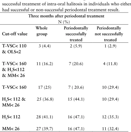
cate-gorized not successfully treated from a periodontal perspective. Successfully treated for intra oral- halitosis using different cut-off values for halitosis definitions are presented in table 5.
Table 5. Distributions of individuals using different definitions of successful treatment of intra-oral halitosis in individuals who either had successful or non-successful periodontal treatment result.
Three months after periodontal treatment N (%) Cut-off value Whole group Periodontally successfully treated Periodontally not successfully treated T-VSC< 110 & OLS<2 3 (4.4) 2 (5.9) 1 (2.9) T-VSC< 160 & H2S<112 & MM< 26 11 (16.2) 7 (20.6) 4 (11.8) T-VSC< 160 17 (25) 7 ( 20.6) 10 (29.4) H2S< 112 & MM< 26 25 (36.8) 15 (44.1) 10 (29.4) H2S< 112 28 (41.1) 16 (47.1) 12 (35.3) MM< 26 27 (39.7) 16 (47.1) 11 (32.4)
Paper IV
In this double blinded randomised clinical trial, the effects of an active mouth rinse (A) in comparison to a placebo mouth rinse (P) were evaluated over six months with an interim assessment at three months. Over time increases in PI and BOP scores, as well as the numbers of 4 mm and 5 mm probing depths were observed for both study groups (NS). Compared to baseline, data analyses at three and six months identified significant reductions of OLS (p<0.01), T-VSC (p<0.01), H2S (p<0.001) and MM (p<0.01) values
in the active treatment group (A). Six months after treatment, dif-ferences between treatment A and P were found for OLS (p<0.05), T-VSC (p<0.05), H2S (p<0.01), MM (p<0.01) values, and with
lower values in the test group. Changes from baseline to six months in OLS scores are presented in figure 3.
Figure 3. Changes from baseline to 6 months after treatment. Re-duction of the organoleptic scores by 1 category, 2 categories in-crease and no change presented for the active (A) and placebo (P) treatments.
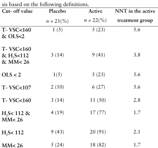
In table 6 the number of individuals successfully treated for intra-oral halitosis using different definitions are presented. In addition the number of individuals needed to treat to achieve one successful-ly treated individual for intra-oral halitosis after six months of treatment is also presented.
Table 6. Six months after successful treatment of intra-oral halito-sis based on the following definitions.
Cut- off value Placebo
n = 21(%) Active n = 22(%) NNT in the active treatment group T- VSC<160 & OLS<2 1 (5) 5 (23) 5.6 T- VSC<160 & H2S<112 & MM< 26 3 (14) 9 (41) 3.8 OLS < 2 1(5) 5 (23) 5.6 T- VSC<107 2 (10) 6 (27) 5.6 T- VSC<160 3 (14) 11 (50) 2.8 H2S< 112 & MM< 26 4 (19) 17 (77) 1.7 H2S< 112 9 (43) 20 (91) 2.1 MM< 26 5 (24) 18 (82) 1.7
In figure 4 the mean values of T-VSC, H2S and MM at baseline, 3
months after treatment and 6 months after treatment are presented for the active and placebo treatments.
Figure 4. Presenting the mean T-VSC; Total sum of volatile sul-phur compounds, H2S; Hydrogen sulphide an MM; Methyl mer-captan.