UPTEC K 20027
Examensarbete 30 hp
Augusti 2020
Nanocellulose surface functionalization
for in-situ growth of zeolitic
imidazolate framework 67 and 8
Teknisk- naturvetenskaplig fakultet UTH-enheten Besöksadress: Ångströmlaboratoriet Lägerhyddsvägen 1 Hus 4, Plan 0 Postadress: Box 536 751 21 Uppsala Telefon: 018 – 471 30 03 Telefax: 018 – 471 30 00 Hemsida: http://www.teknat.uu.se/student
Abstract
Nanocellulose surface functionalization for in-situ
growth of zeolitic imidazolate framework-67 and 8
Beyar Abdulla
This master’s thesis was conducted at the Department of
Nanotechnology and Functional Materials at Ångström Laboratory as part of an on-going project to develop hybrid nanocomposites from Cladophora cellulose and a sub-type of metal-organic frameworks; zeolitic imidazolate frameworks (ZIFs).
By utilizing a state-of-the-art interfacial synthesis approach, in-situ growth of ZIF particles on the cellulose could be
achieved. TEMPO-mediated oxidation was diligently used to achieve cellulose nanofibers with carboxylate groups on their surfaces. These were ion-exchanged to promote growth of ZIF particles in a nanocellulose solution and lastly, metal ions and organic linkers which the ZIFs are composed of were added to the surface
functionalized and ion-exchanged nanocellulose solution to promote ZIF growth. By vacuum filtration, mechanical pressing and furnace drying; freestanding nanopapers were obtained. A core-shell morphology between the nanocellulose and ZIF crystals was desired and by adjusting the metal ion concentration, a change in
morphologies was expected. The nanocomposites were investigated with several relevant analytical tools to confirm presence,
attachment and in-situ growth of ZIF crystal particles upon the surface of the fine nanocellulose fibers.
Both the CNF@ZIF-67 and CNF@ZIF-8 nanocomposites were successfully prepared as nanopapers with superior surface areas and thermal
properties compared to pure TEMPO-oxidized cellulose nanopapers. The CNF@ZIFs showcased hierarchical porosities, stemming from the micro- and mesoporous ZIFs and nanocellulose, respectively. Also, it was demonstrated that CNF@ZIF-8 selectively adsorbed CO2 over N2. Partial formation of core-shell structure could be obtained, although a relationship between increased metal ions and ZIF particle morphology could not wholly be observed.
Tryckt av: UPPSALA
ISSN: 1650-8297, UPTEC K 20027 Examinator: Peter Broqvist Ämnesgranskare: Maria Strømme Handledare: Chao Xu
Populärvetenskaplig sammanfattning
Under många år har mänskligheten och vår natur utsatts för industrirelaterade utsläpp. Allt eftersom industrin växer, kommer dessa utsläpp att öka i mängd och enligt WHO befann vi oss, under 2018, i ett stadium där ca 90% av all världens population andades förorenad luft till viss mån.
För att motverka dessa utsläpp och minimera mängden giftiga gaser, såsom CO2, har man flitigt
varit på jakt efter material och produkter vilka skulle kunna implementeras inom industriella områden. Detta bidrar alltså till att b.la. etablerade produktionsmetoder blir mer hållbara för framtiden eller att nya produkter skapas för att ersätta befintlig teknologi inom separering, lagring eller omvandling av miljöfarliga utsläpp. Exempelvis används s.k. zeoliter flitigt inom industriella sammanhang. Dessa material är uppbyggda av aluminat (AlO4) och silikat (SiO4), vilka agerar
byggstenar som bundna tillsammans bidrar till skapandet av ett enda stort, öppet nätverk av mikroporer. Denna struktur möjliggör att materialet kan agera som b.la. molekylsikt för selektiv separering av gaser. Sedan 2019 har det skapats ca. 235 typer av zeoliter för olika sammanhang, varvid många av dessa förekommer naturligt. Däremot finns det fortfarande begränsningar i hur man kan manipulera zeoliter för att erhålla nya strukturer vid syntes, för olika applikationer. En relativt ny klass av porösa material som uppmärksammats under senaste åren är metall-organiska material (metal-organic frameworks eller MOFs på engelska). Dessa material besitter mycket stor ytarea och har inom forskningen diskuterats som användbara inom industrin för diverse applikationer. Exempelvis har de konstaterats kunna användas som lagringsmaterial för gaser eller som katalysatorer. Generellt består dessa utav metalljoner som koordinerar med organiska ligander och bildar ett kristallint, mikroporöst nätverk. Möjligheten till att använda olika kombinationer av metalljoner och organiska ligander har bidragit till att över 6000 MOFs syntetiseras årligen sedan dess upptäckt. På laborativ skala erbjuds en kontrollerad process som öppnar upp möjligheten för en skräddarsydd syntetisering av ett material med olika egenskaper som exempelvis optimal katalytisk förmåga, selektiv separering av gaser eller infoga elektrokemiska egenskaper i MOF:en. En begränsning som hindrar materialet från industriell tillämpning är dess mekaniska egenskaper, mer specifikt dess höga grad av sprödhet.
För att gynna MOF:ens implementering inom industrin har man försökt kombinera dessa med ett annat material via nanoteknologiska engagemang. Exempelvis kan man utnyttja ett polymert substrat som man inkorporerar MOF partiklar i för att bilda flexibla nanokompositer som erhåller det bästa av två världar. Problemet har dock dels varit att MOF-partiklarna endast fysiskt är inblandade i kompositen och att polymererna har petrokemiska ursprung, vilket dessvärre bidrar till miljöbelastning. Däremot har ny forskning visat att biopolymerer skulle kunna vara goda kandidater som substratmaterial.
I detta projekt användes en gränsskiktssyntes i lösning för att skapa en nanokomposit genom att växa en sub-typ av MOF (zeolitic imidazolate frameworks eller s.k. ZIFs) på cellulosa från en grön alg, Cladophora. Metoden gick ut på att introducera funktionella grupper på cellulosan och samtidigt defibrillera den till fina nanofibrer (CNFs) via en kemisk metod, s.k. TEMPO-oxidering. Dessa funktionella grupper kunde sedan agera som aktiva säten på vilka ZIF- partiklar
kunde fästa sig vid och växa till nanopartiklar i en lösning. Lösningen filtrerades och kvar erhöll man ett flexibelt papper bestående av en nanokomposit som benämndes CNF@ZIFs. Syftet var att undersöka huruvida ZIF kristallernas morfologi på cellulosafibrerna kunde manipuleras genom att ändra koncentrationen metalljoner i lösningen för att gynna skapandet av ZIF-lager istället för individuella nanopartiklar.
Contents
1. Introduction ... 1 1.1 Background ... 1 1.2 Project aim ... 2 2. Theory ... 2 2.1 Cladophora Cellulose ... 22.2 TEMPO/NaBr/NaClO Oxidation (TEMPO-CNFs) ... 4
2.3 Ion-exchange of the TEMPO-CNFs ... 5
2.4 Polyvinylpyrrolidone (PVP) ... 6
2.5 Metal-organic frameworks (MOFs) ... 7
2.6 Zeolitic Imidazole Frameworks (ZIFs) ... 7
2.7 CNF@MOFs ... 10
2.8 Gas sorption for porosity and surface area analysis (N2 gas sorption) ... 11
3. Experimental ... 12
3.1 Materials ... 12
3.2 TEMPO-mediated oxidation the cellulose ... 12
3.3 Synthesis of CNF@ZIF-67 and CNF@ZIF-8 ... 13
3.3.1 Solvent-exchange of the TEMPO-oxidized CNFs ... 13
3.3.2 Ion-exchange ... 13
3.3.3 CNF@ZIFs: Synthesis and controlling morphology of ZIF-67 and ZIF-8 crystals upon CNFs 14 3.4 Methods of characterization of CNF@ZIFs ... 16
3.4.1 X-ray diffraction (XRD)... 16
3.4.2 Fourier transform infrared spectroscopy (FTIR) ... 17
3.4.3 Scanning electron microscopy (SEM) ... 17
3.4.4 Thermogravimetric analysis (TGA) ... 17
3.4.5 Nitrogen gas sorption ... 17
4. Results and Discussion ... 17
4.1 TEMPO-mediated oxidation of Cladophora cellulose and subsequent solvent-exchange ... 17
4.2 Cross-linking due to ion-exchange of the TEMPO-CNFs solutions as preparation for synthesis of CNF@ZIF-67 and CNF@ZIF-8 ... 18
4.3 Analysis of samples ... 19
4.3.1 Structural- and chemical analysis of CNF@ZIFs nanocomposites ... 19
4.3.3 Porosity analysis of CNF@ZIFs nanocomposites ... 27
5. Conclusions ... 29
6. Future perspectives ... 30
7. Bibliography ... 32
Acknowledgement
I would like to thank my subject reader; Professor Maria Strømme for allowing me to perform my masters thesis at the Department of Nanotechnology and Functional materials in Uppsala University, Ångströms laboratory.
My project supervisor, Dr. Chao Xu, had his door always open for me whenever I had questions or doubts regarding specific matters related to the project. Your passion and ambition were infectious and discussions with you were always insightful. Secondly, Shengyang Zhou and Xueying Kong; Thank you both for being there for me during laborations and if I had questions regarding the report. Shengyang your energy and positive attitude was very uplifting. Xueying, I am very grateful for your help preparing my samples prior the SEM imaging. Both of you are very bright minds and you always made me feel as if I was a part of the team.
To my beloved parents (Jowan Hafeed and Parosh Aziz Abdulla), sister (Yara Abdulla), cousins (Miran- and Mandy Aziz and little Josef Mohammed), aunts (Naza- and Chra Hafeed),
grandparents (Farok- and Akhter Hafeed); You have always been an integral part of my life and this journey was certainly not an exception. My love for all of you is unconditional.
To my friends, Andreas Abdelki and Mikael Svensson which were doing their respective thesis works at the department too; I am glad and thankful to have had you guys by my side throughout my thesis work. The discussions we had were always fruitful and the mental support you two gave me was very encouraging. Andreas, your kind and supportive nature towards me is beyond measure. Thank you for being you. Mikael, your thoughtful words and ability to carefully listen to my thoughts are admirable traits.
Lastly, to all the fellow workers at the department; I wish you well and good luck with your respective researches!
1
1. Introduction
1.1 Background
Our modern world is in strong symbiosis with the technological industry, which provides us with a high quality of life. Although, as our demands grow and technology always has to keep up, our environment suffers due to inevitable emissions and pollution from the expansion of our industries. Striving for a more sustainable future by addressing and trying to resolve issues such as environmental degradation from emissions has been a collective aim of humankind for many years. For instance, according to a report by the World Health Organization (WHO), as of 2018 nearly 90% of the human population unfortunately breathes polluted air to some degree [1]. Much effort has been put into trying to resolve the impact that the environment and humans have had to endure from emissions, such as carbon dioxide from usage of fossil fuels. Such advances within the field, as an example, is using porous materials as suitable receptors to encapsulate and /or diminish the toxicity of pollutants.
Porous materials, especially as we tread down on the nanoscale, have the intrinsic property of retaining high surface areas due to the presence of small voids which occur throughout the structure, on micro- and nanoscale. Examples of porous materials which have been used commercially are zeolites. These materials are composed of aluminosilicates and retain large surface areas. Unfortunately, their lack of a simple tunable structure has inhibited their evolution and applicability in new avenues. Thus, another porous material which has caught much attention is the emerging family of crystalline, 3-dimensional metal-organic frameworks (MOFs). These consist of metal ion clusters that are bound to organic linkers via coordination bonding to form microporous networks, with unique properties that depend on the structure and coordination of the organic linkers and metal ions used. Some of the more attractive features of these materials are their potential application within energy storage, catalysis and gas filtration due to their structural tunability and affinity towards certain molecules. A lot of research has been put into the synthesis of these porous materials and over 6000 MOFs have been published each year since 2016. Although, according to [2] there are still significant challenges in manipulating their morphologies, dimensions and processability as they often are insoluble and brittle, rendering them very difficult to handle during post-process and manufacturing. It has been shown that a substrate as template for the growth of a MOF could be used in order to obtain chemically integrated hybrids with MOF crystals grown upon their modified surface [3]. For example, there have been reports of configuring MOF crystals within matrices of metal oxides or polymeric substrates in order to process them as thin film membranes and porous beads. Albeit, these hybridized MOFs still lack the flexibility and shapeability necessary to effectively limit their manufacturing shortcomings [4]. Thus, there has been a rising interest in functionalizing biopolymers which are environmentally friendly, ductile and shapeable to act as templates upon which MOF crystals would grow upon and potentially use them within the industry, e.g., as filter membranes for gas separation.
The project at hand will focus on nanoengineering of metal-organic framework materials by surface modifying Cladophora cellulose to develop freestanding hybrid nanocomposite papers of
2
MOFs attached to the surface of cellulose. The results given by the study to be conducted may open new avenues on how nanomaterials may be implemented in the industry.
1.2 Project aim
The main goal of this thesis project is to utilize a state-of-the-art interfacial synthesis approach to try and synthesize two sub-types of MOFs; zeolitic imidazolate framework-67 (ZIF-67) and zeolitic imidazolate framework-8 (ZIF-8) on the surface of cellulose nanofibers (CNF) to develop nanocomposites of CNF@ZIF-67 and CNF@ZIF-8. The ZIFs have been proven to showcase increased surface area versus comparable zeolites and showcase chemical- and thermal robustness. The substrate used for the integrated nanocomposites will be surface modified nanofibrous cellulose stemming from a specific type of green algae, Cladophora. The surface modification will be a combined result from TEMPO-mediated oxidation and subsequent exchange. These ion-exchanged surfaces of the cellulose nanofibrils will act as anchoring sites, on which ZIF-67/ZIF-8 crystals may nucleate and grow upon in-situ. A series of experiments will be conducted to observe if there is a correlation between the morphology of the ZIFs crystals upon the CNFs and metal salt concentration. Desirably, a core-shell structure between the nanocellulose fibers and ZIF crystals will be devised. Furthermore, a proof-of-concept to use one of these nanocomposites as selective CO2 filter membranes is demonstrated for potential applicative use.
2. Theory
2.1 Cladophora Cellulose
Cladophora algae are a type of green algae which bloom and grow prominently in coastal regions, their growth being mainly triggered by industrial pollutants in water [5]. The aquatic life is affected the most when these algae age and die, forming dense mats on the surface of the water and drift away as a consequence of wind action. As time passes, the filamentous algae eventually drop down to the seabed where they decompose. The decomposition process is initiated by nearby bacteria and the dead algae are perceived to be very nutritious to them, allowing for the bacteria to thrive. The combination of over-nutrition and loss of oxygen supply, as the decomposition process consumes oxygen, results in a weakening of functioning marine ecosystems in the nearby region. According to [5], the physical removal of such algal mats on the seabed was proven to be beneficial to the marine life and restored balance to any present ecosystem. Subsequently, there was a rising interest to explore whether or not the removed Cladophora algae could potentially be used as a resource, especially their cellulose.
3
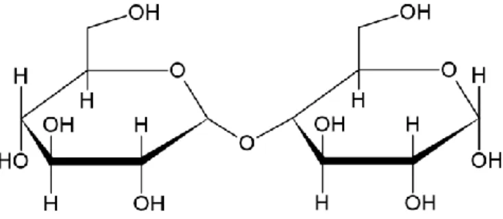
Cellulose is a renewable and abundantly available biopolymer that is found as a major constituent in plant fibers, bacteria’s and in different families of algae, such as the green algae Cladophora. It is one of the most occurring biopolymers on Earth and was first extracted from tissues of plants by Anselme Payen in 1838 [6]. The biopolymer is composed of polysaccharides with a repeating molecular unit defined as D-anhydrous glucopyranose ring unit (AGU). One of the monosaccharides, which composes the polysaccharide as a whole, is bound to the other monosaccharide via a covalent β(1→4) glycosidic oxygen linkage which the polymer chain is able to rotate, bend and twist around. There is a total of three alcohol groups per monosaccharide. The primary alcohol group is located on C6, while the other two secondary alcohol groups are on C2 and C3, respectively, as can be seen in Figure 1. The oxygen atoms which are present on the cellulose chain will, together with the hydroxyl groups, generate strong hydrogen bonds and give rise to hydrophilic properties in aqueous solutions. The hydrogen bonds may occur between adjacent polymer chains too, which yield cellulose microfibrils and is the main cause as to why this biomaterial is rather insoluble in most solvents [7].
Cellulose from Cladophora has a native hierarchical structure due to the reoccurring hydrogen bonds between adjacent chains. The polymer chains are continuously stacked around each other in crystalline and amorphous domains [8]. The accumulation of these polymer chain stacks contributes to a hierarchical, fibrous structure that is defined by interfiber voids, giving rise to a mesoporous structure with a high surface area of ~100 m2/g. This is a larger value than cellulose from different origin. These structures are then assembled into larger microfibrils which constitutes the cellulose fibers in plants and algae. Measurements revealed that the degree of crystallinity in Cladophora cellulose could be as much as 95%, which is higher compared to cellulose from a variety of plants and bacteria.
Disintegrating cellulose fibers to obtain the building block fibrils is called fibrillation and has found industrial interest as it opened up for an effective way of surface modifying individual cellulose fibers and functionalizing them [9]. Fibrillation of Cladophora cellulose may yield very long, rigid nanofibrils with high aspect ratios that may be dispersed in aqueous solutions, which provide an excellent solution-based approach to organize and prepare the nanocellulose for surface modification. Obtaining nanocellulose from Cladophora often yields fibers with diameters ranging from ~25-30nm. The high degree of crystallinity makes the cellulose an excellent candidate for chemical modification and ultrasonication defibrillation without completely destroying its structure.
4
2.2 TEMPO/NaBr/NaClO Oxidation (TEMPO-CNFs)
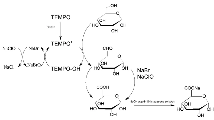
As mentioned in 2.1, Cladophora nanocellulose exhibits excellent properties to be used as a template for growth of nanomaterials on it. One interesting property that all type of investigated cellulose have in common is the abundancy of hydroxyl groups that may act as chemical sites for surface modification of the cellulose in question [10]. In the past decade, high-energy ultrasonication has been used to obtain fibrillar structures of the cellulose denoted as micro- and nanocellulose, although this alone has never yielded a very high degree of individualized fibrils. Instead, the fibers were found to be bundled together non-homogenously to some degree, suggesting remaining presence of hydrogen bonds [11]. Another route consists of chemical modification of the cellulose surface to facilitate the separation of bundled fibrils at their amorphous regions by treating cellulose in sulfuric acid, although the fibril length and yield is significantly reduced by 30-50%. This treatment yields crystalline nanocellulose (CNC). In order to increase the yield of nanofibrils and attempt to retain their lengths, another chemical modification has been proposed. This method conceptualizes the selective oxidation of the primary alcohol on the C6 position to a carboxylate group. This method allows for cellulose microfibrils to be effectively liberated into elementary nanofibrils, by using TEMPO (2,2,6,6-tetramethylpiperidin-1-oxyl radical) and was first utilized by De Nooy et al. [12]. The aforementioned method was diligently used in this master’s thesis for surface modification of the Cladophora cellulose to obtain cellulose nanofibers (CNFs) in combination with ultrasonication. TEMPO, a nitroxyl radical, is in itself a rather stable molecule due to the presence of bulky side chains. It may be converted into a nitrosonium cation (TEMPO+) in the presence of stoichiometric amounts of a primary oxidant, sodium hypochlorite (NaClO), and sodium bromide (NaBr) as co-catalyst in an aqueous solution. The TEMPO+ is the true oxidant of the -OH group and oxidation yields an aldehyde intermediate that is further converted into a carboxylate group. Following oxidation of the alcohol, the TEMPO+ is reduced to a hydroxylamine which may be re-oxidized in the presence of NaBrO, being NaBr that has been oxidized by NaClO, to regenerate the oxidation cycle. A reaction mechanism suggesting how C6 hydroxyl groups are converted into carboxylates is presented in Figure 2.
5
Figure 2. TEMPO-mediated oxidation of -OH at C6.
The end-result of the oxidation process is heavily pH dependent as the carboxylate moiety may either be in protonated form at lower pH or in deprotonated form in alkaline conditions [13]. In general, acidic carboxyls are formed during the reaction and it was shown that the most effective oxidation occurred within a pH window of 10-11. Thus, it is of importance to maintain a stable pH by adding a certain amount of sodium hydroxide (NaOH) that may be consumed during the reaction until the pH remains at a static value of ~10. For aqueous solutions of cellulose being oxidized under alkaline conditions (~pH 10), the C6 -OH groups on the surface of the cellulose is converted into sodium carboxylate groups due to presence of Na+ which the negatively charged carboxylate groups may bind with. Some chemical bonds may still be present between neighboring fibrils as the degree of oxidation may vary depending on the duration of the reaction. These may be broken via ultrasonication, yielding fine individual TEMPO-oxidized nanocellulose fibrils (TEMPO-CNFs) dispersed in the aqueous solution.
2.3 Ion-exchange of the TEMPO-CNFs
Native cellulose, from any of the aforementioned sources, are initially free from viable functional groups which may promote growth of material upon the surface. Thus, anchoring capabilities with potential metal ions for in-situ growth of ZIF nanoparticles are severely inhibited. Following the TEMPO-mediated oxidation, the formed carboxylate groups will be attached to the sodium ion via ionic bonding as can be seen in Figure 3. This sodium ion will be feasible for an ion-exchange with other metal ions. By allowing the TEMPO-CNFs to soak in metal salt solutions over a period of time, ion-exchange may be initiated [14].
6
This opens up venues for the TEMPO-CNFs to act as a substrate for potential growth of material, such as in this case; growth ZIFs. The sodium ion can be ion-exchanged with a metal ion which the particular ZIF is composed of and act as an anchor site for the attachment of organic ligands. This subsequently leads to the growth of the ZIFs following addition of more metal salt and organic linkers into the ion-exchanged TEMPO-CNFs solution [15].
Figure 3. Introduction of a sodium carboxylate group on C6 thanks to TEMPO-mediated oxidation. The sodium ion may be viable for ion-exchange.
2.4 Polyvinylpyrrolidone (PVP)
Polyvinylpyrrolidone (PVP) is an amphiphilic and non-toxic polymer that may be used in a variety of ways during nanoparticle synthesis, such as an encapsulating agent of various metal oxides or capping agent to promote growth of nanoparticles to specific shapes by capping specific surfaces of the particle nuclei in question [16]. The repeating unit is composed of two constituents; a hydrophilic pyrrolidone moiety that is polar due to the amide group and a hydrophobic alkyl group which is non-polar. Thus, its amphiphilic nature stems from these two respective components. It has been shown that it can act as an excellent stabilizer, making sure that nanoparticles in a solvent do not aggregate. In this work it was used as a crystallization- and interfacial stabilizing agent between the ZIFs and the TEMPO-CNFs as per methods utilized by Zhou et al [17].
7
2.5 Metal-organic frameworks (MOFs)
Metal-organic frameworks (MOFs) are crystalline materials with extensive surface areas, which stems from their microporous nature. They consist of metal cations, linked to an organic linker via coordination bonding [18]. This, subsequently, leads to the formation of a network that extends with repeating coordination clusters in a periodic manner through space, forming a long-range ordered framework. Common metals used are the ones from the transition series such as Fe3+, Co2+ and Cr2+. Alkali- and alkaline earth metal ions have also been reported for the formation of various MOFs with different properties [19]. During most synthesis routes, the metal ions are commonly present as precursors such as nitrates, acetate, chlorides or oxides. The organic linkers, by which the metal ions or clusters are attached to coordinatively, may contain functional groups such as carboxylates, imidazole’s, sulfonates, phosphates, amines or nitriles. Therefore, the choice of metal ion and organic linker dictates not only the structure, but the MOFs intrinsic properties as well.
The reason as to why MOFs have garnered so much attention over the past few years is due to the fact that their surface areas, according to [18], most often exceeds the value for other comparative porous compounds such as activated carbons, zeolites and mesoporous silica by large values. One of the most prominent merits of MOFs is their topological diversity. By exploiting the philosophy which conceptualizes reticular chemistry, the science of combining molecular building blocks, scientists have been able to produce various tailored MOFs using a plethora of different metal ion-organic linker combinations [20].
There are different routes to synthesize MOFs. Common methods are solvothermal, hydrothermal and non-solvothermal methods, such as ball-milling and mechanical grinding. A few other unconventional methods have been reported such as microwave-assisted synthesis, layer-by-layer synthesis and sonochemical synthesis. Room temperature synthesis have also been explored due to its potential for less energy consumption and ease of performance. [21, 22].
Many factors affect the synthesis of MOFs, thus, trying to manipulate parameters to enhance or stabilize certain properties has been a challenge for many [24]. The major synthesis parameters that has been subjects of interest are the type of solvent used, pH, metal-ligand ratio and temperature. The solvent in question has been deduced to play a part on the morphology of the crystal/formation of the MOF. Most often, choice of a specific solvent is based on the solubility of the starting materials and proteolysis property of the organic linker.
2.6 Zeolitic Imidazole Frameworks (ZIFs)
As stated in previous sections, microporous aluminosilicate-based materials known as zeolites have had a big impact in the industry mostly due to their surface areas resulting in high catalytic abilities, sensing and gas storage [25]. Even though their use has been established commercially, modifying their structures to enhance their properties for, e.g. CO2 absorption, has been largely
unrealized due to difficulties fine-tuning their structures to promote CO2 affinity. Instead, during
recent years, Yaghi et al [26] discovered a plethora of framework compounds which had superior properties compared to other porous materials with industrial potential as, e.g., CO2 capture.
8
Zeolitic imidazolate frameworks, also known as ZIFs, have recently emerged as a sub-class of the MOFs. They are porous, crystalline and share a similarity in structure with zeolites. ZIFs consist of metal ions which coordinate tetrahedrally, such as Zn2+, Co2+, Fe2+, with imidazole-type organic linkers [25]. The angle at which the organic linker bridges with the metal ion is ~145o, which is
similar to the silicon-oxygen-silicon angle found in zeolites, resulting in their isomorphic porous structure. This has led to the synthesis of different ZIFs with remarkable properties, exhibiting permanent porosity, exceptional thermal- and chemical stabilities, making them excellent potential candidates for industrial applications. As they share traits stemming from both zeolites and MOFs, ZIFs combine the structural tunability of a MOF with the chemical- and thermal stability of a zeolite. Alas, they do retain weaknesses similar to other MOFs, such as low post-processability and high brittleness. The pristine product obtained is most often a powder, which precipitates out of solution during solvent-based synthesis.
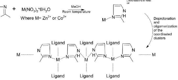
There have been a handful of ZIFs which have been synthesized since their discovery. During this thesis project ZIF-67 and ZIF-8 will be synthesized in room temperature and used as the components, respectively, to be grown in-situ upon the TEMPO-CNFs. These ZIFs will be synthesized in methanol (MeOH) and composed of tetrahedrally coordinated 2-methylimidazole (2-MIM) linkers bridged with a Co2+-ion for ZIF-67 and Zn2+-ion for ZIF-8. A proposed reaction scheme has been deduced, which can be seen in Figure 5, and the reaction steps involved may be categorized as:
- Linker coordination of Zn2+/Co2+ centers. - Deprotonation of the 2-MIM linker.
- Oligomerization of Co2+-(deprotonated 2-MIM) complexes leading to complete structure.
Figure 5. Proposed general reaction mechanism for the formation of ZIF-67 and ZIF-8 in MeOH, the former obtained by using cobalt ions while the latter requires zinc ions.
9
The synthesis of ZIF nanoparticles have usually been performed in an organic solvent, thus the choice of using MeOH in this thesis work. Synthesizing in a mixture of an organic solvent with deionized water has also been done [27, 28]. Although, the latter and even pure deionized water was proven to yield very different morphologies for ZIF-67 with e.g. lower BET surface areas and dense structures compared to its counterpart synthesized in MeOH. One of the causes was the apparent sensitivity to moisture these compounds retain during synthesis, especially ZIF-67. Pure-phase ZIF-8 has been obtained with certain crystallinity but lower yield during room temperature aqueous synthesis.
Recent studies involving the deduction of the degradation mechanism, in order to understand their sensitivity, when MOFs and ZIFs are exposed to water revealed that hydrolysis or ligand displacement are the culprits for structure failure [29]. The ligand displacement reaction follows as:
𝑀𝑛+− 𝐿𝑛−+ 𝐻
2𝑂→𝑀𝑛+− (𝑂𝐻2) … 𝐿𝑛− (1)
As can be seen from the reaction formula, a water molecule is inserted between the metal-ligand bond, leading to a hydrated cation and release of a ligand, collapsing the framework. During hydrolysis, the metal-ligand bond is fully broken while a water molecule dissociates, forming a hydroxylated cation and a protonated ligand that is released. The hydrolysis reaction scheme follows as:
𝑀𝑛+− 𝐿𝑛−+ 𝐻2𝑂→𝑀𝑛+− (𝑂𝐻)−+ 𝐻𝐿(𝑛−1)− (2)
In order for proper formation of ZIF nanocrystals and control over particle size, one of the most important parameters related to synthesis of ZIFs has been found to be the molar ratio between metal ions and organic linkers [30]. An increased amount of organic linker results in a higher number of deprotonated ligands which the metal ion may coordinate with accordingly. Additionally, by adding a heftier number of organic linkers, the excess ligands may cover nucleated crystal seeds and allow inhibition of prolonged crystal growth. Consequently, this leads to rapid nucleation, favoring smaller ZIF nanocrystals over larger ones. However, the amount of organic linker needed to form nanocrystals of ZIF-8 and ZIF-67 have been reported to be at least ~20-100 times more than the metal ions, especially in water. Hence, the more deprotonated ligands obtained, the more feasible the ZIF- nanoparticle formation will be.
By addition of a deprotonating agent with basic character, the 2-MIM linker used to synthesize ZIF-8 and ZIF-67 have been shown to rapidly deprotonate and allow for a higher nucleation rate to take place, simultaneously inhibiting prolonged crystal growth. Hence, this method favors the nucleation and formation of small crystals, at lower required amounts of 2-MIM for formation of ZIF-8/ZIF-67 from Zn(NO3)2•6H2O/ Co(NO3)2•6H2O and 2-MIM in the presence of triethylamine
(TEA). This suggests that a minimum amount of 2-MIM may be used for successful formation of ZIFs when TEA is included during synthesis.
10
2.7 CNF@MOFs
Structuring and assembling of MOFs on templates in order to facilitate their application uses in an industrial scale has been a topic of high interest. Such modifications may result in composites fit for air filtration, gas storage and catalytical endeavors. Research has been put into using different templates and substrates in order to facilitate both the assembly during synthesis and their manufacture processing. The results were hybridization of MOFs with either polymers or metal oxide matrices in order to assemble them as a variety of structures ranging from thin films, porous beads, dense pellets and hierarchical monoliths [31]. Although, most of these polymers used are synthetic and a product of derivatives from the petrochemical industry and possess risks of health- and environmental degradation if scaled-up. Beside their toxic nature, one of the major difficulties regarding polymer-MOF composites have been the absence of desirable rigidity and flexibility. Another difficulty has been the fact that most of these hybrid nanocomposites have been developed with complicated experimental procedures, which effectively hampers their rate of production on an industrial scale. For example, there have been attempts to synthesize MOF particles upon polyacetonitrile nanofibers using microwave radiation, albeit the nanofibers melted due to the irradiation and the composites structure was heavily affected [32]. These difficulties have caused significant setbacks of advancing such hybrid materials from lab scale to industrial scale.
Therefore, there has been a wide interest in implementing biopolymers as a means to be used as templates for incorporation of MOFs, allowing for enhanced post-processability of the MOFs and simultaneously decreasing the composites inherent environmental toxicity. These originate from natural resources with intrinsic ductile properties. They come in abundant amounts and add flexibility alongside shapeability to the nanocomposite and allow MOFs to be applied in various applicable ways. Examples of biopolymers that have been used to produce composites of MOFs are surface modified chitin and cyclodextrin as effective encapsulating-, gas sequestering- and drug-delivery agents [31].
Recently, cellulose has been proven to be able to be used as a substrate for facile incorporation of MOF particles within the cellulose fiber matrix. By combining properties stemming from both the cellulose and the MOF particles, the composites could be used for effective filtration of organic dyes, polluted solutions containing heavy metals and gas separation [15, 33, 34]. Albeit, the MOF particles are only randomly dispersed within the cellulose matrix and are primarily held in place merely due to the physical entanglement arising from the fibrous network and weak van der Waals forces [15]. Thus, the shapeability and processability of these composites would be further enhanced if controlled growth of the actual MOF particles could be induced upon the individual nanocellulose fibers via chemical bonds which would allow for the fabrication of freestanding membranes.
Zhou et al [17] succeeded in producing sophisticated nanocomposites of a plethora of MOFs with Cladophora cellulose as the substrate of choice, defined as CNF@MOFs. The nanocomposites were synthesized by assembling MOFs onto TEMPO-oxidized and ion-exchanged nanocellulose fibers (TEMPO-CNFs). By using a process of filtration, drying and mold pressing, freestanding
11
CNF@MOF nanopapers were also achieved. The MOF crystals had a layered morphology, wrapping the individual TEMPO-CNFs.
2.8 Gas sorption for porosity and surface area analysis (N
2gas sorption)
Gas sorption may be used as a means to characterize the porosity, pore size distribution and surface area of porous materials [35]. There are two different types of surface adsorption; physisorption and chemisorption. The former occurs when molecules attach to the surface via London-dispersion forces. Chemisorption occurs when adsorbents bind themselves by chemical bonding with the atoms present on the open surface and is therefore much stronger than
physisorption. Lowering the temperature facilitates the adsorbents to attach to the surface through London forces, instead of forming ionic and/or covalent bonds which are difficult to break. Commonly, nitrogen gas is used at a temperature of 77 K as this allows the nitrogen molecules to attach to the surface of the material via physisorption in a reversible fashion for the sake of characterization.
According to IUPAC there is a set of proposed definitions for different types of adsorption isotherms, relating the number of adsorbed molecules and the relative pressure to a given porosity type which can be seen in Figure 6. While performing adsorption-desorption
measurements, the obtained isotherm may be characterized accordingly. The Brunauer-Emmet-Teller, BET, surface area and pore distribution of a sample may also be calculated using this type of measurement.
Figure 6. IUPAC classification on adsorption isotherms [35]. Type I refers to a microporous adsorbent with pore sizes smaller than 2 nm, type II, III and VI non-porous/microporous materials with pore sizes of 2-50 nm while type IV and V is characteristic for mesoporous materials which have a pore size greater than 50 nm.
12
3. Experimental
3.1 Materials
Cladophora cellulose powder provided by Dr. Chao Xu. Sodium bromide (NaBr), 2, 2, 6, 6- tetramethylpiperidine-1-oxyl (TEMPO), sodium hypochlorite solution (NaClO, 10%), 2- methylimidazole 97% was purchased from Alfa Aesar, polyvinylpyrrolidone (PVP, Mw = 3.6 × 10⁵ g/mol), triethylamine (≥ 99%), sodium hydroxide, hydrochloric acid, cobalt(II) nitrate hexahydrate, Zinc(II) nitrate hexahydrate 98+% were purchased from Sigma-Aldrich without further purification. Methanol of isocratic grade was purchased from VWR Chemicals.
3.2 TEMPO-mediated oxidation the cellulose
Initial experiments conducted for the synthesis of the composites were mostly considered as failures, many of which stemmed from several of the TEMPO-oxidized nanocellulose batches that were made in the beginning due to difficulties calibrating the concentration of the TEMPO-CNFs solution. Thus, the first few batches of TEMPO-CNFs were considered a part of a trial and error approach until viable batches were produced. Below is an in-depth description of methods utilized for the final TEMPO-CNFs batch which was used for successful synthesis of CNF@ZIFs nanocomposites.
In order to remove any present impurities before the oxidation, 1 g of the Cladophora cellulose powder was washed in a 200 mL NaOH aqueous solution (0,5 mol/L) for 24 hours prior to the TEMPO-mediated oxidation. The solution was stirred at 450 rpm using a magnetic stirrer. As the NaOH was readily available in pellet-form, to prepare a 200 mL 0,5 mol/L solution, 4 g of NaOH pellets were dissolved in deionized water.
The 200 mL solution was divided into 4, 50 mL tubes and centrifuged repeatedly at 3,800 rpm for 15 minutes with deionized water in order to neutralize the cellulose and prepare it for the TEMPO-oxidation. After each cycle, a litmus paper test was conducted in each tube to assure that the pH was as close to 7 as possible. The sedimented cellulose flocculates were collected from each tube and put into a sonication beaker along with 100 mL of deionized water. Ultrasonication with a probe was conducted for 10 minutes with a pulse length of 10 seconds and a pause of 10 seconds with a power amplitude of 60 % under running, cool water. After sonication, the thick cellulose hydrogel was transferred into another beaker for TEMPO oxidation.
By adding a 10 mL aqueous solution of 16 mg TEMPO and 100 mg NaBr to the cellulose, the TEMPO-mediated oxidation was initiated with the addition of 3,1 mL NaClO. The solution was continuously stirred at 650 rpm. Immediately, the pH rose to 12 after adding the NaClO due to its alkaline nature. After a while, the pH started slowly decreasing, hence confirming the initiation of the oxidation reaction. The oxidation occurred for 6 hours. Solutions of NaOH and HCl with the concentrations 0,5 mol/L were prepared and added to the solution every 30 minutes to ensure that
13
the pH of the solution was at ~10 during the whole oxidation process, as it tended to continuously drop until no further NaOH was able to be consumed. In order to observe the pH, a pH meter was used.
The TEMPO-CNFs was then washed with deionized water by vacuum filtration with a PVDF membrane with a pore size of 0,1 µm several times in order to try and remove any unreacted reagents, inorganic salt etc. The obtained hydrogel of oxidized cellulose was then collected and put into the sonication beaker to be sonified once again for 10 minutes with 10 second pulses and 10 second pauses with a power amplitude of 60% in 100 mL deionized water. The concentration of the TEMPO-CNFs suspension was then calibrated by allowing 1 mL of the solution dry in a furnace at 70o C for several hours until a dry film was obtained. The dry cellulose film was weighted and had a mass of 7,33 mg. Thus, the suspension had an estimated concentration of 7,33 mg/mL and was put into a fridge for further experiments.
3.3 Synthesis of CNF@ZIF-67 and CNF@ZIF-8
As outlined in section 2.3, there are many ways to synthesize MOFs and ZIFs. The synthesis of pristine ZIF-67 and ZIF-8 has been performed and reported in ambient conditions with MeOH as solvent and TEA as a deprotonating agent [30, 36].
3.3.1 Solvent-exchange of the TEMPO-oxidized CNFs
5 mL (7,33 mg/mL) of the TEMPO-CNFs solution was solvent-exchanged by centrifugation 3 times with MeOH at 11,000 rpm for 30 minutes. Between each pass, the cellulose was dispersed in MeOH and shaken 20 minutes in order to achieve as homogenous of a dispersion as possible. The solvent exchanged TEMPO-CNFs was then redispersed in 10 mL of MeOH for ion-exchange. If any flocculates were seen in-between the centrifugation passes even after extensively shaking the TEMPO-CNFs/MeOH solution, sonication was commenced for 15 minutes in order to further homogenize the suspension.
3.3.2 Ion-exchange
The ion-exchange was commenced by mixing a select, excess, amount of Co(NO3)2•6H2O in 2
mL of MeOH until dissolved. The slight pink solution was put into the 10 mL solvent-exchanged TEMPO-CNFs solution and stirred at 300 rpm for 1 hour. Later, the solution with the ion-exchanged CNFs was washed several times with MeOH by centrifugation at 11,000 rpm for 30 minutes. The washed and ion-exchanged TEMPO-CNFs was redispersed in 10 mL MeOH. The same procedure was commenced when ion-exchanging for growth of ZIF-8. The only difference being the metal salt used; Zn(NO3)2 •6H2O for ZIF-8. The reason why an excess amount of metal
14
many sodium carboxylate groups were ion-exchanged before washing. For the one-step process, the washing step described above was skipped.
3.3.3 CNF@ZIFs: Synthesis and controlling morphology of ZIF-67 and ZIF-8 crystals upon CNFs
A set of experiments were conducted in order to determine whether the morphology of the grown ZIF-67 and ZIF-8 crystals upon the TEMPO-CNFs could be controlled or not, in relation to the amount of metal salt used. As stated in previous sections, the synthesis of MOFs in general depend on many factors and are complex. Thus, in order to enhance the probability of being able to determine whether or not the amount of metal salt affected the morphology of the ZIF-67/ZIF-8 on the TEMPO-CNFs, other parameters remained as static as possible during synthesis. These
included the amount of PVP, molar ratio between TEA, 2-MIM and
Co(NO3)2•6H2O/Zn(NO3)2•6H2O, concentration of TEMPO-CNFs and temperature. The final
molar ratio between metal salt and organic linker was set to 1:5 while for organic linker and TEA was set to 1:1.
After washing the ion-exchanged TEMPO-CNFs, the calculated amount of metal salt and PVP were dissolved in 2 mL of MeOH in a 15 mL test tube (Component A). In another 15 mL test tube, 2 mL of 2-MIM and TEA (Component B) were mixed vigorously. Component A was put into the ion-exchanged TEMPO-CNFs solution and stirred for approximately 20 minutes, allowing the PVP to activate the surface of the nanocellulose fibrils. Subsequently, Component B was poured into the solution and stirred overnight. The amount of each reagents used for the synthesis of successful CNF@ZIF-67 and CNF@ZIF-8 samples are listed in Tables 1 and 2. Finally, the products were washed with MeOH 3 times in order to remove as much of any unreacted reagent as possible.
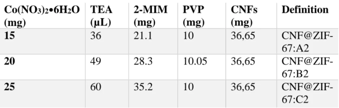
Table 1. Reagents used for synthesis of CNF@ZIF-67 using two-step process.
Co(NO3)2•6H2O (mg) TEA (µL) 2-MIM (mg) PVP (mg) CNFs (mg) Definition 15 36 21.1 10 36,65 CNF@ZIF-67:A2 20 49 28.3 10.05 36,65 CNF@ZIF-67:B2 25 60 35.2 10 36,65 CNF@ZIF-67:C2
15
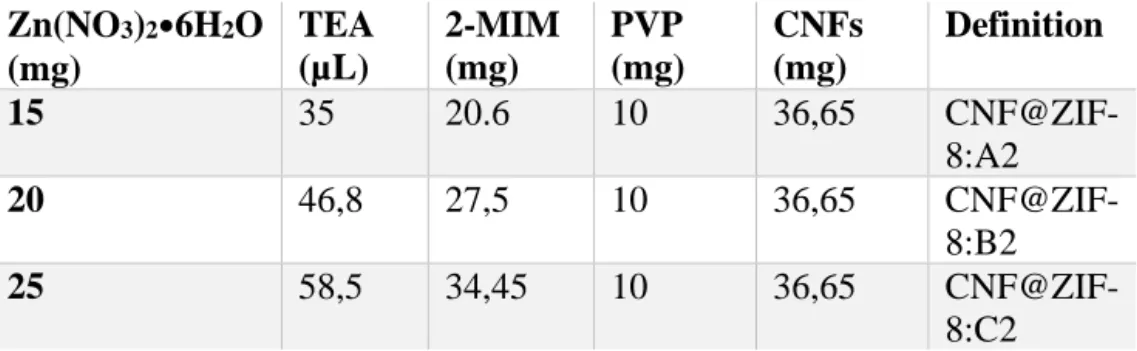
Table 2. Reagents used for synthesis of CNF@ZIF-8 using two-step process.
Zn(NO3)2•6H2O (mg) TEA (µL) 2-MIM (mg) PVP (mg) CNFs (mg) Definition 15 35 20.6 10 36,65 CNF@ZIF-8:A2 20 46,8 27,5 10 36,65 CNF@ZIF-8:B2 25 58,5 34,45 10 36,65 CNF@ZIF-8:C2
For each of the above-mentioned experiments, a dark purple solution was obtained for CNF@ZIF-67, while a milky white solution was obtained for CNF@ZIF-8. These colors are characteristic of ZIF-67/ZIF-8 formation in solution and could be seen in their respective paper formats too, which may be observed in Figure 7 [33]. The freestanding nanopaper of the CNF@ZIFs nanocomposites were obtained by putting respective solution of CNF@ZIF through a vacuum filtration device with a PVDF membrane which had a pore size of 0.1 µm. The filter cake was set to dry for an hour with the vacuum on. Later, the PVDF membrane with the filter cake was removed and another PVDF membrane was put upon it. The sandwiched filter cake was then pressed between two metal plates and put into a furnace at 70o C for 5 hours. The formed nanocomposite paper was then carefully removed by manually peeling it off the filter paper with a pair of tweezers.
Figure 7. (a) Pure TEMPO-CNFs nanopaper. (b) CNF@ZIF-8 scaled up nanopaper. (c) CNF@ZIFs “Lab”-scale nanopapers. (d) The CNF@ZIF papers are very shapeable and flexible. Notice the characteristic colors of ZIFs on the individual nanopapers.
16
In order to demonstrate the scalability, each type of nanopaper was scaled up once, thus the papers obtained were bigger in comparison to the “lab”-scaled ones produced for structural- and thermal characterizations only. The reason also being that the smaller versions were too small and, e.g. the N2-sorption requires a certain mass in order to yield measurable results. Unfortunately, the
CNF@ZIF-67 nanopaper was too thin to peel off in one whole piece and be showcased.
Also, during this thesis project, experiments for CNF@ZIF-67 were initially conducted in a two-step process involving an extra washing two-step between the ion-exchange and the final synthesis of the nanocomposites, while at a later stage they were conducted in a one-step process and then, finally, once again in a two-step process. The two-step process was the official approach.
The one-step process was conducted only to figure out whether or not it was possible to form ZIF-67 crystals in the solvent-exchanged TEMPO-CNFs solution. Formation of ZIF-ZIF-67 was particularly difficult in the beginning when the two-step process was used. Thus, it was a necessity to follow the one-step process in order to control the formation of ZIF-67 crystals upon the CNFs, before the two-step process could be commenced which washes away most of the excess metal-ions in the ion-exchanged solution. Hence, the reason as to why the formation of ZIF-67 failed was speculated to be that there were far too few metal ions left for favorable formation of ZIF-crystals the first few times the two-step process was used.
By properly recalculating molar ratios between the metal salt and organic linker and the inclusion of TEA, the two-step process could be properly utilized to limit the amount of potentially un-anchored ZIF nanoparticles within the CNF@ZIF-67 solution after the ion-exchange, yielding fewer freestanding nanoparticles that would not be anchored the TEMPO-CNFs. Specifically, this meant that there would be less brittleness to the paper, which would help enhance its flexibility and shapeability due to less amount of excess ZIF particles physically mixed within the TEMPO-CNFs.
When the two-step process was deemed a success for formation of CNF@ZIF-67, CNF@ZIF-8 was immediately synthesized via the two-step process. In regard to experiments conducted with the one-step process, amounts of reagents used for this particular method of synthesis will be listed in Table A1 in the Appendix alongside SEM micrographs that were taken of them in Figure F1. Also, solutions of successful CNF@ZIF-67 and CNF@ZIF-8 vs. failed CNF@ZIF-67 are shown in Figure F2.
3.4 Methods of characterization of CNF@ZIFs
3.4.1 X-ray diffraction (XRD)
XRD was used in order to confirm the proper synthesis of crystalline CNF@ZIFs nanocomposites with each component constituting the hybrid nanomaterial and ensure whether or not any
17
contaminants may be present and have caused the formation of unknown phases. Specifically, CNF@ZIF nanopapers were taped onto a metallic holder to create as flat of a surface as possible and put into the XRD instrument. The analysis was conducted in a Bruker D8 diffractometer with Cu-Kα X-ray radiation (λ = 0.154 nm).
3.4.2 Fourier transform infrared spectroscopy (FTIR)
FTIR was utilized in order to inspect the chemical properties of the nanocomposites and their constituents. The concept behind the method is obtaining a spectrum with wavelengths within the infrared interval for different materials. Raw material was analyzed and, in relation to it, the spectra for the nanocomposites was studied in order to confirm possible chemical changes. The instrument used to record the spectra was a Bruker Tensor 27 spectrometer in attenuated total reflectance mode (ATR) between 400-4000 cm-1 with a resolution of 4 cm-1.
3.4.3 Scanning electron microscopy (SEM)
SEM micrographs were obtained in order to observe the surface of the nanopapers to confirm the presence of ZIFs crystals and inspect their morphology on nano- and microscale. Micrographs were obtained with a Leo Gemini 1530 FEG SEM instrument with an acceleration voltage of 3kV.
3.4.4 Thermogravimetric analysis (TGA)
This is a method used in order to verify the thermal stability of samples by applying a certain thermal profile. The profile may, in summary, be dynamic or static. The samples are then subjugated to the heat in a controlled chemical environment. It is a versatile method that may be used to, e.g. detect mass changes at certain temperatures. Here, TGA was performed in order to observe the thermal stability of the nanocomposites and determine how the in-situ growth of ZIFs crystals may have affected the TEMPO-CNFs substrate. The experiments were conducted in an inert N2-atmosphere with a gas flow of 60 mL/min over a dynamic heating range from
25-900o C with a heating rate of 10o C/min. The instrument used was a Mettler Toledo TGA/DSC 3+.
3.4.5 Nitrogen gas sorption
Nitrogen gas sorption was used in order to characterize properties related to the porosity of the nanocomposites. The obtained isotherms were recorded in an ASAP 2020 V4.03 instrument with N2 gas at 77 K. Also, isotherms were obtained for CNF@ZIF-8 at 293K with N2 and CO2. Prior
to analysis the samples were degassed for 10 hours in 100o C. The surface areas would be calculated using Brunauer-Emmet-Teller (BET) method in the relative pressure ranges of 0.04-0.15 while the pore size distribution was calculated using density functional theory (DFT).
4. Results and Discussion
4.1 TEMPO-mediated oxidation of Cladophora cellulose and subsequent
solvent-exchange
One of the more frustrating challenges during this thesis project was to prepare a homogenous dispersion of TEMPO-CNFs in MeOH following solvent-exchange from the aqueous environment in which the oxidation had taken place. Both Cladophora cellulose in its natural state and
TEMPO-18
CNFs retain very good hydrophilicity [14]. The former, due to the abundant amount of -OH groups present on the fibrils while for the latter it is mostly because of the introduction of sodium carboxyl groups that also are hydrophilic in nature. Thus, transferring the TEMPO-CNFs from an aqueous solution, in which they already are well dispersed in, caused difficulty to the solvent-exchange process and their dispersion was quite poor. The consequence was TEMPO-CNFs loss between each MeOH washing cycle after centrifugation, resulting in less amount of TEMPO-CNFs for each sample than the theoretically desired value.
Another plausible explanation is that due to the fact that MeOH and water are polar, separating a solute in a mixture of these is relatively difficult. Originally, the centrifugation was supposed to occur at a lower rpm value of 10,000 for 15 minutes. But the amount of CNFs mass that was lost between each cycle was deemed too high, thus a higher rpm and longer centrifugation time was preordained to at least inhibit the amount of TEMPO-CNFs being lost between each centrifugation pass. This made the fabrication of the nanopapers very difficult and tedious in the beginning, due to higher amount of ZIFs particles compared to CNFs in synthesis solution. This caused brittleness and thinning of the manufactured paper and removing it from the PVDF membrane was difficult, which might have been the unfortunate case for the scale-up of the CNF@ZIF-67 nanopaper. In this case, prolonged centrifugation and sonication was used to minimize CNFs loss between each washing cycle.
4.2 Cross-linking due to ion-exchange of the TEMPO-CNFs solutions as
preparation for synthesis of CNF@ZIF-67 and CNF@ZIF-8
In order to promote the ZIF particles growth upon the individual CNFs, instead of being physically mixed within the TEMPO-CNFs matrix, metal ions which the ZIFs are, respectively, composed of had to be introduced onto the surface modified CNFs which was explained in Section 2.3 previously.
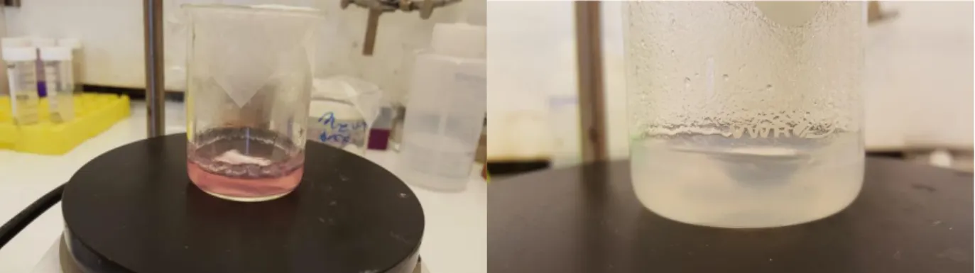
To ensure that as many sodium carboxylate groups on the surface of the CNFs were ion-exchanged with the respective metals, an excess amount of the specific metal salt was added to the solution to saturate it. This resulted in the solution turning thick and gel-like almost immediately which could be explained by the reaction phenomenon behind the ion-exchange procedure. Metal ions of divalent nature will react ionically with the carboxylic TEMPO-CNFs and promote the formation of cross-linking between carboxylate groups of neighboring cellulose nanofibrils. This is a phenomenon which has also been observed before during a study conducted by Zhu et al when exposing sodium carboxylated CNFs to other multivalent ions such as Cu2+ etc. [15]. Thus, the transition from a light suspension to a thicker MeOH gel indicated ion-exchange behavior of the carboxylated CNFs prior to further analysis, as can be seen in Figure 8.
19
.
Figure 8. (Left) TEMPO-oxidized CNFs being exchanged with cobalt ions. (Right) TEMPO-oxidized CNFs being exchanged with zinc ions. These images represent the gelation of the CNFs suspension due to cross-linking initiated by the
ion-exchange procedure. A thickening has occurred and bits of CNFs-COOCo2+/Zn2+ MeOH gel can be seen stuck to the walls of
respective beaker as the content is stirred.
4.3 Analysis of samples
In this section, the synthesis and growth of the ZIFs crystals upon the ion-exchanged and surface modified CNFs were to be analyzed using the series of analytical tools denoted in Section 3.4. Specifically, the presence, attachment and growth of the crystals on the nanocellulose fibers were assessed in order to verify the proper synthesis of the nanocomposites.
4.3.1 Structural- and chemical analysis of CNF@ZIFs nanocomposites
The chemical and structural changes which had occurred during the synthesis of CNF@ZIF nanocomposites were analyzed, in order to verify the synthesis of the nanocomposites and, especially, the incorporation of ZIF crystals on the surface of the TEMPO-CNFs. XRD, FTIR and SEM were diligently used for this portion of the thesis.
20
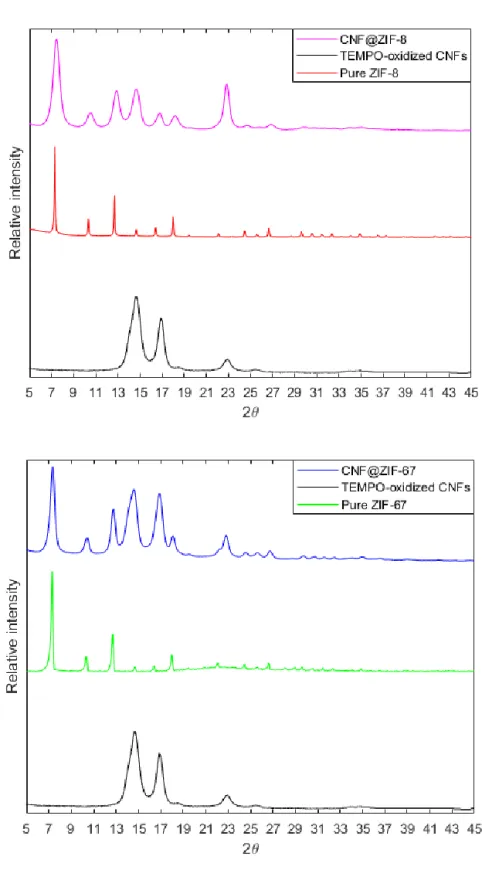
Figure 9. (Top): XRD patterns of TEMPO-oxidized cellulose, pure-phase ZIF-67 and CNF@ZIF-67. (Bottom): XRD patterns of TEMPO-oxidized cellulose, pure-phase ZIF-8 and CNF@ZIF-8
21
The XRD pattern shown in Figure 9 depicts the TEMPO-CNFs versus pure ZIF-67, ZIF-8 and the hybrid nanocomposites. Per literature, the characteristic peaks for ZIF-67 were present and could be observed [37]. The prominent peaks lie at 2θ-values of 7o, 10o, 12o, 14o, 16o and 18o respectively. The peaks were sharp and defined, indicating presence of pure-phase formation of ZIF-67 crystals. The characteristic peaks for the TEMPO-CNFs could also be found at 14,9o, 16,9o and 22,7o which is in accordance with 2θ-values obtained in previous literature [38]. The diffraction patterns for pure ZIF-67 and pure TEMPO-CNFs aligned very well in the XRD pattern for the nanocomposite, suggesting successful incorporation of ZIF-67 crystals on the TEMPO-CNFs without any unknown phases having been formed during synthesis.
The same conclusions could be drawn for the CNF@ZIF-8 nanocomposite. The XRD peaks which may be correlated to pure ZIF-8 had intense reflections at approximately 7o, 10o, 12o, 14o, 16o and 18o which were also in alignment with previous studies conducted on pristine ZIF-8, pointing towards that pure-phase ZIF-8 had been obtained [30]. The peaks from both the TEMPO-oxidized CNFs and pure-phase ZIF-8 is aligned and overlapped well in the CNF@ZIF-8 diffraction pattern. Thus, it could be concluded that successful incorporation of ZIF-8 crystals onto the TEMPO-CNFs had been achieved.
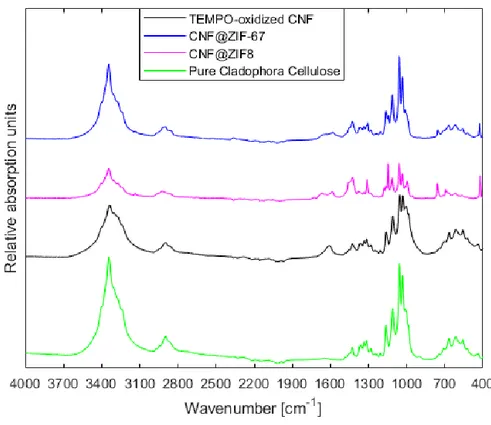
Figure 10. ATR-FTIR of the CNF@ZIFs vs. pure Cladophora cellulose powder and TEMPO-oxidized CNFs. Notice the introduction of the peak at ~1612 cm-1representingthe stretching of the carboxylate group the surface of the TEMPO-oxidized
Cladophora cellulose. Then, after a change in chemical environment of the carboxylate group on the CNFs due to ion-exchange, it shifts and decreases for both composites.
22
Characteristic absorption bands in the ATR-FTIR spectra for the Cladophora cellulose could be readily observed at 3344 cm-1 , 2897 cm-1, 1315 cm-1, 1161 cm-1, 1059 cm-1 and 898 cm-1, depicting the -OH stretching, C-H stretching, H-C-H wagging vibration and C-O-C asymmetric bridge stretching of the β-glycosidic linkage between anhydroglucose units, C-O-C pyranose ring skeletal vibrations and deformation of the β-glycosidic linkage in Figure 10 [39]. Comparing these absorption bands with those observed for the TEMPO-oxidized CNFs, a difference in the absorption spectra was found at ~1612 cm-1. The existence of this absorption band corresponded to the stretching vibration of the carboxylate group which had been introduced after oxidation of the C6 hydroxyl groups on the Cladophora cellulose. This finding confirmed that the TEMPO-mediated oxidation had been a success. Furthermore, looking over at the spectra depicting the CNF@ZIFs nanocomposites, new absorption bands had appeared signifying several chemical changes. One of the major changes was correlated to the carboxylate stretching vibration absorption peak at ~1612 cm-1 that was discussed earlier, which had shifted and dampened for both
CNF@ZIF-8 and CNF@ZIF-67. As mentioned in Section 2.3, the concept behind the interfacial synthesis of ZIFs and MOFs on CNFs overall is that there needs to be specific sites at the surface of the cellulose on which the MOF/ZIF can attach to and grow upon. Thus, either Co2+ or Zn2+ may ion-exchange with the sodium ion and bond ionically to the carboxylates. The explanation as to how metal ions bonded with the carboxylates after ion-exchange was through the free, delocalized electron of the carboxylate group. This change in chemical environment, from being bound with Na+- to either Co2+ or Zn2+ ions, affected the stretching vibrational mode of the carboxylate group and is seen as a shift of the peak, consequently indicating ion-exchange [40]. Additionally, for TEMPO-CNFs, CNF@ZIF-67 and CNF@ZIF-8, the -OH peak at 3344 cm-1 had
undergone a slight broadening and shifted, compared to pure TEMPO-CNFs which is consistent with the study conducted by Zhu et alas they prepared MOFs aerogels templated by ion-exchanged TEMPO-CNFs.
Also, characteristic peaks for respective ZIFs could be located in each absorption spectra of the nanocomposites. The absorption bands at 3135 cm-1,1448 cm-1, 1580 cm-1, 1142 cm-1, 756 cm-1 and 426 cm-1 were correlated to the stretching of the aliphatic methyl group in 2-MIM, C=N
stretching in 2-MIM, stretching and bending of the imidazole ring and the Co-N stretching for ZIF-67 and Zn-N for ZIF-8 [41]. These findings not only confirmed the introduction of carboxylate groups on the surface of the nanocellulose fibers, but also a change in the chemical environment of the carboxylate groups after the sodium ion was exchanged with either Co2+ or Zn2+ after addition of metal salts. Due to the fact that ZIF-8 is isostructural to ZIF-67 and is composed of the same constituents, excluding zinc ions instead of cobalt, the FTIR spectra were rather similar, also confirming the presence of ZIF-8 on the TEMPO-CNFs.
23
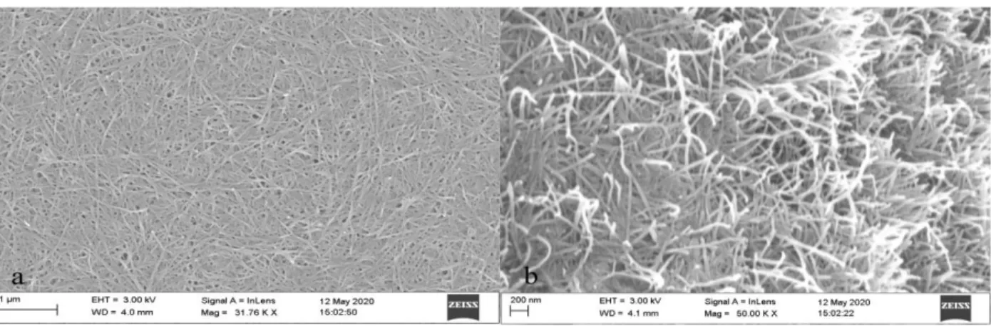
The images presented in Figure 11 are SEM micrographs of nanopaper composed of pure Cladophora cellulose which had endured 6 hours of TEMPO-mediated oxidation. As mentioned in previous sections, during the oxidation, -OH groups at C6 were selectively oxidized into sodium carboxylate groups, essentially depolymerizing and inhibiting formation of hydrogen bonds between adjacent chains, rendering the polymer with long, individualized and thinner fibers instead [42]. As can be seen in the figure, the individualized nanofibrils are clearly visible as a direct result of the TEMPO-oxidation.
Figure 11. (a) Pure TEMPO-CNFs seen from above. (b) TEMPO-CNFs at a higher magnification. Notice the individualized fibers as a direct result from oxidation.
Unfortunately, the SEM micrographs of the CNF@ZIFs nanocomposites were difficult to obtain, many of them were perceived to be blurry to be able to distinguish direct, in-depth, results from. The reason is speculated to be due to poor conductivity between the substrate used for SEM imaging and the freestanding nanopapers. Thus, those that were deemed acceptable to draw any conclusion from were selected and are presented in Figure 12.
Figure 12(a-c) depicts the CNF@ZIF-8 nanofibers with different metal salt concentrations used during synthesis. At first glance, the surfaces for all the nanocomposites were somewhat reminiscent of pure TEMPO-CNFs. Although, the inset images which depict magnified versions of the respective micrographs showcased that there were clear and distinct crystals which were distributed in an assembled manner along the individual nanocellulose fibers. This signified not only successful formation of ZIF-8 crystals, but also successful attachment of these crystals along the outer surface of the nanofibrils. Although, for the CNF@ZIF-8:A2, there seemed to be a coalesced layer instead of individual nanoparticles. This coalesced layer made it very hard to determine whether there were individually wrapped ZIF-8 layers on the nanocellulose fibers or not. A viable reason for the formed layer may have been due to heavy amounts of remaining organic solvent.
Inspecting the surface, it seemed rather rough, and some ZIF-8 particle agglomeration could be seen distributed in random locations for all CNF@ZIF-8 samples which may have pointed toward an insufficient washing after the ion-exchange, leading to a higher accumulation of metal ions than the calculated amount, thus, allowing formation for excess ZIF-8 crystals. Compared to CNF@ZIF-8 nanocomposites, the CNF@ZIF-67 nanocomposites were deemed to have obtained a much smoother surface with less agglomerated crystal particles. Although, the inhomogeneity
24
throughout the papers, in regard to ZIFs morphology, seemed to be a reoccurring trait; CNF@ZIF-67 exhibited an overall varied fibril thickness, especially CNF@ZIF-CNF@ZIF-67:B2, as can be seen in Figure 12e. The smooth surface may be explained by the fact that the majority of free particles have been removed by the multiple washings, diminishing the occlusion of CNF@ZIF-67 hybrid nanofibrils in the micrographs. For CNF@ZIF-67:A2, the overall morphology was that of a layered structure and partially agglomerated spherical particles at random locations. The difference
Figure 12. (a-c) CNF@ZIF-8:A2, B2 and C2. (d-f) CNF@ZIF-67:A2, B2 and C2.
in the morphologies of both nanocomposites, despite the same washing steps, indicates that ZIF-67 crystals seem to be easier to wash away than ZIF-8. Another reason for the low amount of spherical particles may be that ZIF-67 crystals under conditions given, perhaps favored the layered morphology over the spherical, agglomerated morphology.
25
Pure TEMPO-CNFs has a typical fiber diameter of ~25-30 nm as mentioned in Section 2.1. Although, the fibers of CNF@ZIFs nanocomposite which could be roughly measured, without spherical particles in the way or too much a blurriness in the micrograph, had diameters ranging from ~32-47 nm suggesting successful growth of ZIF-crystal with layered morphology had been achieved at random locations in all the samples. The only exception being CNF@ZIF-8:A2 due to the coalesced layer which made measuring impossible. In essence, all the samples did have areas of inhomogeneity where spherical morphology of ZIF crystals could be observed to a certain degree. The fact that there was inhomogeneity throughout the samples with mixed morphologies made it difficult to determine whether there was an increase in probability of gaining layered morphologies for an increased metal salt concentration. The relation was deemed to be random.
4.3.2 Thermal analysis of CNF@ZIFs nanocomposites
As can be seen from the TGA analysis in Figure 13, the initial thermal degradation, Td, of
TEMPO-CNFs occurred at ~245o C. The initial Td for the CNF@ZIFs nanocomposites,
corresponding to the degradation of the TEMPO-CNFs component due to pyrolysis, occurred directly at ~324o C for CNF@ZIF-67 nanocomposites and ~330o C for CNF@ZIF-8
nanocomposites. Furthermore, the major mass loss of TEMPO-CNFs was significantly lowered in the nanocomposites, especially for CNF@ZIF-8. It is known that pristine ZIF-67 has a lower thermal stability than pristine ZIF-8, which explained the difference in their ability to protect the TEMPO-CNFs from degradation in the CNF@ZIF-67- and CNF@ZIF-8 nanocomposites [43]. The mass loss occurring from 0-100o C for pure TEMPO-CNFs is attributed to be due to water evaporation.
These observations support the SEM findings that there should have been formation of core-shell structure between the TEMPO-CNFs and ZIFs with the layered morphology, at least to some extent, as Zhou et al observed the same trend in their CNF@MOFs with continuous,
homogenous layers of MOF crystals [17]. As was commented on the SEM micrographs, this layered morphology might not have been the case fully throughout the samples. From what could be observed, the morphology of the ZIF crystals were a mix between reoccurring spherical particles and core-shell structure. Interestingly, a trend in the TGA curves could be observed for each of the nanocomposites. Also, the major weight loss seen for the TEMPO-CNFs was further inhibited as the metal salt increased during synthesis of CNF@ZIF.
Also, it could be speculated that obtaining mixed morphologies of ZIF crystals growing upon the TEMPO-CNFs may be sufficient to induce a thermal barrier strong enough to decrease the degradation of the cellulose fibers as a whole. Simultaneously, the trend emphasized that a higher amount of metal salt yielded a higher loading of ZIFs within the nanocomposite. Thus, a higher loading of ZIFs incorporated an increased thermal protection to the TEMPO-CNFs component. Nonetheless, these results indicated that the nanocomposites are viable for various endeavors in room temperature and perhaps flue gas treatments, where temperature ranges are
26
within ~47-127o C, without risking severe thermal decomposition [44]. Corresponding DTG
curve may be seen in Figure F3 in the Appendix.
Figure 13. TGA curve for pure TEMPO-oxidized CNFs and CNF@ZIF-8/CNF@ZIF-67 with varying amounts of Zn/Co-salt used during synthesis.
![Figure 6. IUPAC classification on adsorption isotherms [35]. Type I refers to a microporous adsorbent with pore sizes smaller than 2 nm, type II, III and VI non-porous/microporous materials with pore sizes of 2-50 nm while type IV and V is characteristic](https://thumb-eu.123doks.com/thumbv2/5dokorg/4267838.94589/18.918.248.673.661.931/classification-adsorption-isotherms-microporous-adsorbent-microporous-materials-characteristic.webp)