THESIS
ACURRACY ASSESSMENT OF FOUR DIAGNOSTIC TESTS FOR THE DETECTION OF GIARDIA AND CRYPTOSPORIDIUM IN THE ABSENCE OF GOLD STANDARD: A
BAYESIAN APPROACH
Submitted by
Jairo Enrique Palomares Velosa Department of Clinical Sciences
In partial fulfillment of the requirements For the Degree of Master of Science
Colorado State University Fort Collins, Colorado
Fall 2014
Master's Committee:
Advisor: Mo D Salman Lora Ballweber
Copyright by Jairo Enrique Palomares Velosa 2014 All Rights Reserved
ABSTRACT
ACURRACY ASSESMENT OF FOUR DIAGNOSTIC TESTS FOR THE DETECTION OF GIARDIA AND CRYPTOSPORIDIUM IN THE ABSENCE OF GOLD STANDARD: A
BAYESIAN APPROACH
Giardia and Cryptosporidium are important parasites that cause gastrointestinal disease in numerous animal species including dogs and cats. The accurate diagnostic of this diseases is cucial for the aplication of preventive measures and precise treatment. Estimation of test accuraccy is not difficult when a reference test (gold standard) is available. However, when a gold standard test is not available the Bayesian Latent Class (BLC) Analysis is an effective analytical tool for the estimation of diagnostic accuracy. The aim of this study was to estimate the sensitivity (Se) and specificity (Sp) of four commercial diagnostic kits using BLC. The four diagnostic tests were (1) Merifluor®Direct Fluorecence Antigen (DFA; Giardia
/Cryptosporidium; Meridian Diagnostics, Inc., Cincinnati, Ohio), (2) IVD®DFA (Giardia /Cryptosporidium; IDV Research Inc., Carlsbad, CA), (3) IVD Microwell ELISA® (Giardia ; IDV Research Inc., Carlsbad, CA), (4) and IDEXX SNAP® (Giardia ; IDEXX Laboratories Inc., Westbrook, ME). The results from 201 laboratory analysed samples, the prior distributions elicited from three experts, and the consistency of samples as splitting covariate were used as inputs for the BCL models. The estimated Se and Sp of the tests were 87.7% and 97.3% (Merifluor-Cryptosporidium), 68.0% and 99.1% (IVD-Cryptosporidium), 93.6% and 97.9% (Merifluor-Giardia ), 96.1% and 97.9% (IVD-Giardia ), 86.0% and 98.2% (ELISA-Giardia ), and 84.8% and 98.0% (SNAP-Giardia ) respectively. The prevalence for non-diarrheic versus
diarrheic samples were 2.3% and 4.8% (Cryptosporidium), and 6.9% and 13.5% (Giardia ) respectively. We were able to use BLC to assess the sensitivity and specificity of the four commercial diagnostic tests. We ran 36 models and used objective indicators of the per formances of the models to choose the best model for estimation of parameters. The results of the study indicated that Merifluor, IVD, and ELISA are equally suitable as diagnostic tests for detection of Giardia. For detection of Cryptosporidium, Merifluor was more accurate than the IVD test.
TABLE OF CONTENTS
ABSTRACT ... ii
LIST OF FIGURES ... viii
LIST OF TABLES ... viii
1 LITERATURE REVIEW ... 1
1.1 GIARDIASIS IN CATS AND DOGS ... 1
1.1.1 Etiology ... 1 1.1.2 Morphology... 3 1.1.3 Life cycle ... 5 1.1.4 Pathogenesis ... 5 1.1.5 Epidemiology ... 7 1.1.6 Clinical Findings ... 8 1.1.7 Diagnosis ... 9 1.1.8 Treatment ... 14 1.1.9 Prevention ... 15
1.1.10 Public health significance ... 16
1.2 CRYPTOSPORIDIOSIS IN CATS AND DOGS ... 17
1.2.1 Etiology ... 17 1.2.2 Morphology... 18 1.2.3 Life cycle ... 20 1.2.4 Pathogenesis ... 21 1.2.5 Epidemiology ... 23 1.2.6 Clinical findings ... 24 1.2.7 Diagnosis ... 25
1.2.8 Treatment ... 27
1.2.9 Prevention ... 29
1.2.10 Public health significance ... 29
1.3 DIAGNOSTIC TEST ASSESSMENT... 30
1.3.1 Notation and definitions... 30
1.3.2 Applied probability for diagnostic tests ... 32
1.3.3 Diagnostic performance ... 34
1.3.4 Practical applications of diagnostic tests ... 38
1.4 ASSESSMENT OF DIAGNOSTIC TESTS WHEN THE TRUE DISEASE STATUS IS UNKNOWN ... 40
1.4.1 Bayesian approach for assessment of diagnostic test and disease prevalence ... 42
1.4.2 Assumptions ... 49
1.4.3 Identifiability and analysis of the model ... 51
2 MATERIALS AND METHODS ... 53
2.1 SAMPLES ... 53 2.1.1 Source of samples ... 53 2.1.2 Sample size ... 53 2.2 LABORATORY TESTS ... 53 2.2.1 Diagnostic tests ... 53 2.2.2 Sample processing ... 54
2.3 BAYESIAN STATISTICAL ANALYSIS ... 59
2.3.1 Prior distribution elicitations ... 60
3 RESULTS ... 65
3.1 LABORATORY TESTS ... 65
3.1.1 Ease of use of the kits. ... 65
3.2 PRIOR DISTRIBUTIONS ... 66
3.3 POSTERIOR DISTRIBUTIONS INFERENCES ... 67
3.3.1 Test 1 and Test 2 estimates for Cryptosporidium detection ... 68
3.3.2 Estimates of Se and Sp for tests detecting Giardia ... 68
3.3.3 Test-2 for detection of Giardia ... 68
3.3.4 Test-3 test for detection of Giardia ... 69
3.3.5 Test-4 test for detection of Giardia ... 69
3.3.6 Prevalence of Giardia ... 69
3.4 PERFORMANCE OF THE MODELS ... 69
3.4.1 Convergence ... 69
3.4.2 Autocorrelation ... 70
4 DISCUSSION ... 82
4.1 LABORATORY TESTS RESULTS ... 82
4.2 PRIOR DISTRIBUTIONS ... 82
4.3 POSTERIOR DISTRIBUTIONS INFERENCES ... 84
4.3.1 Test-1 and Test-2 for Cryptosporidium detection model ... 84
4.3.2 Sensitivity and Specificity of Test-1 for detection of Giardia ... 85
4.3.3 Sensitivity and Specificity of Test-2 for detection of Giardia ... 86
4.3.4 Sensitivity and Specificity of Test-3 for detection of Giardia ... 86
4.3.5 Sensitivity and Specificity of Test-4 for detection of Giardia ... 87
4.4 FINAL COMMENTS ... 89
5 BIBLIOGRAPHY ... 91
LIST OF TABLES
Table 1. Drug therapy used for the treatment of giardiasis in dogs and cats; modified from
Tangtrongsup & Scorza, 2010 ... 14
Table 2. Prevalence of Cryptosporidium in dogs ... 23
Table 3. Table 2Drug therapy used for the treatment of cryptosporidiosis in Dogs and Cats; modified from Scorza & Tangtrongsup (2010). ... 28
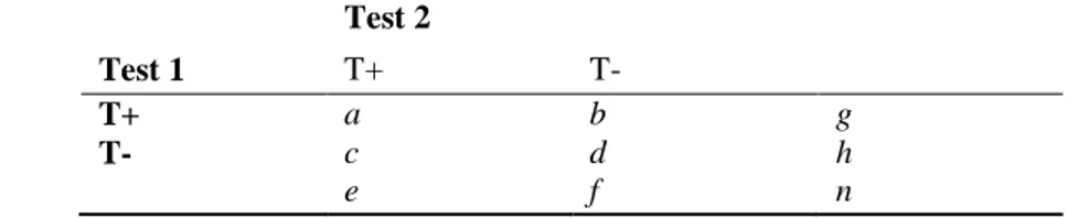
Table 4. Test results states as positive (T+) or negative (T-). From Enoe, Geordais, & Johnson, 2000. ... 40
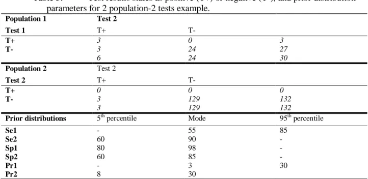
Table 5. Test results states as positive (T+) or negative (T-), and prior distribution parameters for 2 population-2 tests example. ... 47
Table 6. Summary statistics for illustration of Bayesian estimation of Se and Sp with no reference test. ... 48
Table 7. Tests results of four diagnostic tests for the detection of Giardia and Cryptosporidium. ... 70
Table 8. Elicited values of sensitivity from three experts (lower confidence 5th percentile and mode). ... 71
Table 9. Elicited values of specificity from three experts (minimum confidence 5th percentile and mode). ... 71
Table 10. Elicited values of prevalence of Giardia and Cryptosporidium from three experts. Comparison according to consistence of the sample (mode and maximum confidence 95th percentile). ... 71
Table 11. Estimates of Se and Sp for Test-1 and Test-2 when detecting Cryptosporidium (Median and 95%PI). ... 72
Table 12. Estimates of prevalence of Cryptosporidium (Median and 95%PI). ... 72
Table 13. Estimates of Sensitivity and Specificity for tests detecting Giardia ... 72
Table 14. Estimated values for prevalence of Giardia. ... 75
Table 15. Area under the autocorrelation plot, models 1-2-(C,E1-3)with Cryptosporidium detection results. ... 76
Table 16. Area under the autocorrelation plot for sensitivity (Giardia detection results and consensus prior-distribution) ... 76
Table 17. Area under the autocorrelation plot for specificity (Giardia detection results and consensus prior) ... 77 Table 18. Area under the autocorrelation plot for Giardia prevalence (consensus prior)... 77
LIST OF FIGURES
Figure 1. Scheme of a Giardia trophozoite anatomy (Google Image search;
http://www.vetlive.com/2011/07/12/Giardia -in-dogs/). ...3
Figure 2. This scanning electron micrograph (SEM) clearly shows the ventral surface of a Giardia muris trophozoite. The adhesive disk facilitates adherence of the protozoan to the intestinal surface. Created: 2000 (Public Health Image Library Photographer: Dr. Stan Erlandsen). ...4
Figure 3. The pathophysiological manifestations of giardiasis (Elsevier Licensed 3317710976907) ...6
Figure 4. Scheme of the morphologic characteristics of a Cryptosporidium zoite. (Elsevier license 3416010271077)... 19
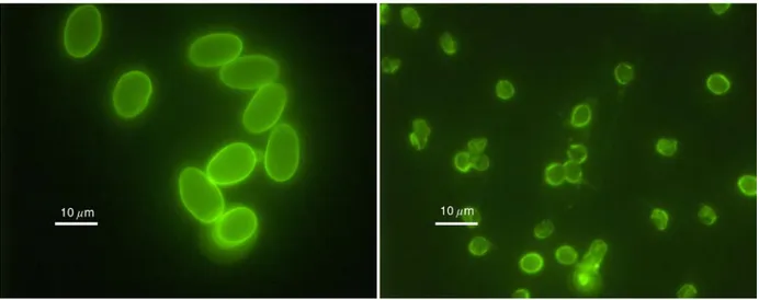
Figure 5. Giardia cysts (left) and Cryptosporidium oocysts (right) under the fluorescent microscope. ... 57
Figure 6. Nestlé PURINA fecal scoring system. ... 59
Figure 7. Schematic representation of conditional dependence of Se and Sp. ... 62
Figure 8. Posterior inferences of Test-1 sensitivity; median (±95% PI) ... 77
Figure 9. Posterior inferences of Test-1 specificity; median (±95% PI) ... 78
Figure 10. Posterior inferences of Test-2 sensitivity; median (±95% PI)... 78
Figure 11. Posterior inferences of Test-2 specificity; median (±95% PI) ... 79
Figure 12. Posterior inferences of Test-3 sensitivity; median (±95% PI)... 79
Figure 13. Posterior inferences of Test-3 specificity; median (±95% PI) ... 80
Figure 14. Posterior inferences of Test-4 sensitivity ; median (±95% PI). ... 80
Figure 15. Posterior inferences of Test-4 specificity; median (±95% PI). ... 81
Figure 16. Estimates of Giardia Prevalence in the non-diarrheic vs. diarrheic populations; median (±95% PI; black = PI for non-diarrheic, red = PI for diarrheic). ... 81
1 LITERATURE REVIEW
1.1 GIARDIASIS IN CATS AND DOGS 1.1.1 Etiology
Giardia duodenalis (syn. intestinalis, lamblia) is a primitive eukaryotic species of the Phylum Metamonada and order Giardia (Pluzer, Ongerth, & Karanis, 2010; Cavalier-Smith, 2003). The following is the taxonomic classification of the genus Giardia according to the systematic taxonomy based on genetic, structural, and biochemical data:
Kingdom Eukaryote
Phylum Metamonada
Subphylum Trichozoa – flagellated protozoans
Superclass Eopharyngia
Class Trepomonadea
Subclass Diplozoa
Order Giardiida
Family Giardiae
Genus Giardia Cavalier-Smith, 2003 (Pluzer, Ongerth, & Karanis, 2010)
The organisms of the genus Giardia are a very unusual kind of ancient eukaryotes as they share many characteristics with anaerobic prokaryotes. Giardia does not have the common intracellu-lar organelles such as mitochondria, peroxisomes, or even a traditional Golgi apparatus that characterizes most of eukaryotes (Pluzer, Ongerth, & Karanis, 2010; Ankarklev, Jerlstrom-Hultqvist, Ringqvist, Troell, & Svard, 2010). However, during encystation, large secretory
com-Golgi cisternae, this pseudo-organelles contain the essential compound for the cyst wall devel-opment (Pluzer, Ongerth, & Karanis, 2010; Ankarklev, Jerlstrom-Hultqvist, Ringqvist, Troell, & Svard, 2010).
In the past, the light microscopy was the most common tool for differentiating species of micro-organisms. Then, the use of electro-microscopy increases the amount of morphologic infor-mation available for species identification. Six species of Giardia have been identified based on morphologic characteristics as feature of ventrolateral flange, marginal groove, ventral disc, and flagellum (Pluzer, Ongerth, & Karanis, 2010). Five from the six species were isolated from am-phibians (G. agilis), birds (G. ardeae, G. psittaci), mice (G. muris), and voles (G. microti). The sixth species included Giardia strains isolated from large range of others mammalian hosts. The-se strains share The-several morphological features and were named as G. duodelanlis (Pluzer, Ongerth, & Karanis, 2010). Later on, with the use of modern molecular techniques such as RNA gene sequencing, all species have been defined (Pluzer, Ongerth, & Karanis, 2010).
The stains of Giardia derived from human isolates were earlier assigned to a separate species (G. lamblia) and the major lineages defined on these human-derived isolates were designated as as-semblages A and B (Pluzer, Ongerth, & Karanis, 2010). Giardia duodenalis, derived from ani-mal isolates, shows a similar genetic spectrum. Some isolates appear to be identical to genotypes found in humans, while others represent genotypes that are apparently host specific (Pluzer, Ongerth, & Karanis, 2010). These findings are relevant when the possibility of giardiasis as a zoonosis is taking in to account (see 1.1.10 section below).
The different assemblages of G. duodenalis have been assigned after finding substantial se-quence differences in the genes, such as the glutamate dehydrogenase/gdh, triosephosphate isomerase/tpi, and β-giardin/bg genes (Pluzer, Ongerth, & Karanis, 2010). Assemblages A to G
have been defined by molecular techniques within the G. duodenalis morphological group. It has been determined that dogs are primarily infected by assemblages C and D, whereas cats are pri-marily infected by assemblage F. Assemblages A and B have also been identified in feces from dogs and cats by DNA amplification (Pluzer, Ongerth, & Karanis, 2010; Scorza & Lappin, 2012).
1.1.2 Morphology
Giardia has two main life forms: trophozoite and cyst
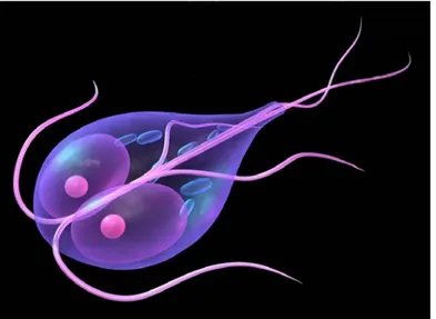
The trophozoite (Figure 1), which is the active and motile form that habits the lumen of the intes-tinal tract, is approximately 15 µm long, 8 µm wide, and 3 µm thick (Kirkpatrik, 1987). One of the most relevant trophozoite morphologic characteristic is its drop shape and the organization of its organelles: two nuclei, the axomeres, and the median bodies, which resemble a smiley, face (Scorza & Lappin, 2012).
Figure 1. Scheme of a Giardia trophozoite anatomy (Google Image search; http://www.vetlive.com/2011/07/12/Giardia -in-dogs/).
organelles may be visible in light microscopy preparations, such as four pair of flagella, two nu-clei, the axomeres, and the median bodies (Kirkpatrik, 1987). The cell tapers posteriorly where the two caudal flagella rise; all flagella are directed posteriorly. The trophozoite adheres on the brush border of the intestinal epithelial cells and the sucking force is generated by the beating of the ventral enlarged flagella (Scorza & Lappin, 2012).
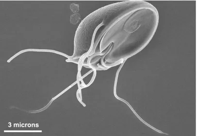
Figure 2. This scanning electron micrograph (SEM) clearly shows the ventral surface of a Giardia muris trophozoite. The adhesive disk facilitates adherence of the protozoan to the intes-tinal surface. Created: 2000 (Public Health Image Library Photographer: Dr. Stan Erlandsen).
The cyst, which is the environmental resistant stage of the parasite, has an oval or ellipsoidal form with approximately 12 µm long and 7 µm wide. This cyst contains two incompletely sepa-rated trophozoites. This stage is resistant to some environmental conditions and can last several months in wet and cold conditions (Ballweber, Xiao, Bowman, Kahn, & Cama, 2010;
Ankarklev, Jerlstrom-Hultqvist, Ringqvist, Troell, & Svard, 2010). This stage is the most com-mon form of the parasite used for diagnostic, and most of the diagnostic tests are designed to de-tect or identify some of the cyst wall components.
1.1.3 Life cycle
After ingestion of the cyst, it becomes metabolically active. The excystation process takes ap-proximately 15 minutes. The gastric acid and pancreatic enzymes trigger the excystation process on the duodenum. The liberated excyzoite undergoes cytokinesis separating the trophozoites (Ankarklev, Jerlstrom-Hultqvist, Ringqvist, Troell, & Svard, 2010).
After a short, not fully understood, biochemical mediated maturation process, the two released trophozoites attach to the brush border of the villous epithelium by its ventral discs (specific characteristic of the genus Giardia) (Ankarklev, Jerlstrom-Hultqvist, Ringqvist, Troell, & Svard, 2010).
The trophozoites multiply by binary fission and encyst in the intestinal tract. The mechanisms of encystation have yet been described completely. The encystation process is an induced response triggered by several host factors such as high levels of bile, low levels of cholesterol, and in-crease in the pH (Ankarklev, Jerlstrom-Hultqvist, Ringqvist, Troell, & Svard, 2010). The first step that takes place for encystation is the internalization of the flagella. Additionally to this, the fragmentation of the ventral disk favors the loss of ability to attach to the intestinal wall. The parasite gradually rounds up and decreases its metabolism to enter in a stage of dormancy. Final-ly, the encystation specific vesicles selectively transport the cyst wall proteins to the surface and form the cyst wall. Before encystation, the trophozoite starts a division cycle that ends after excystation. This division produces two nuclei pairs that are observable in the formed cyst (Ankarklev, Jerlstrom-Hultqvist, Ringqvist, Troell, & Svard, 2010).
1.1.4 Pathogenesis
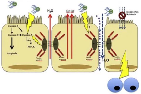
un-combination of both host and parasitic factors. One of the main clinical signs of giardiasis is the diarrhea, which appears to be caused by a combination of malabsorption and hypersecretion (Scorza & Lappin, 2012; Cotton, Beatty, & Buret, 2011). The common clinical sings found in patients with giardiasis are often related to four main pathological events: i) the increase of apop-tosis of epithelial cells, ii) the increase of intestinal permeability, iii) the disruption of cellular apical junctions, and iv) the shortening of the brush border microvilli (Figure 3) (Cotton, Beatty, & Buret, 2011).
Figure 3. The pathophysiological manifestations of giardiasis (Elsevier Licensed 3317710976907)
The increase of epithelial cell apoptosis rates could occur via activation of Caspases-9 and 3. However, the precise mechanisms are still unknown. Some other parasitic factors may activate hypersecretion of chloride, which may contribute to diarrhea (Cotton, Beatty, & Buret, 2011). Giardia also increases the intestinal permeability during giardiasis by disrupting apical
junctional complex components (including F-actin, ZO-1, claudin-1, and α-actinin) in a Caspase-3 dependent manner. The increase of the epithelial permeability is due, at least in part, to the
activation of Myosin Light Chain Kinase (MLCK). The lack of impermeability allows the trans-location of antigens into the subepithelial tissue (Cotton, Beatty, & Buret, 2011).
Another important event is the shortening of the brush border microvilli; an effect mediated by host CD8+ T lymphocytes. Consequently, the absorptive surface area is reduced during giardia-sis, resulting in digestive deficiencies and contributing to maldigestion; signs commonly associ-ated with giardiasis. Additionally, the microvillus lesion is unable to absorb glucose and electro-lytes effectively, resulting in the decrease of water uptake and subsequent malabsorptive diarrhea (Cotton, Beatty, & Buret, 2011).
1.1.5 Epidemiology
Giardia infects several mammalian species worldwide including humans. Many studies have es-tablished the prevalence of Giardia in dogs and cats (Ballweber, Xiao, Bowman, Kahn, & Cama, 2010; Mohamed, Glickman, Jr., Lund, & Moore, 2013). However, results tend to vary consider-ably because of the difference in the tests used, and the differences in population and region where the study was done (Thomson, Palmer, & O'Handley, 2008; Ballweber, Xiao, Bowman, Kahn, & Cama, 2010).
The affected patients acquire the environmental resistant Giardia cysts by oral ingestion --commonly from contaminated food or water-- or by grooming when the coat is contaminated with feces (Mohamed, Glickman, Jr., Lund, & Moore, 2013). Carnivorism is another possible way of acquiring Giardia, if the organism is present in the prey intestine (Kirkpatrik, 1987). The prepatent period of giardiasis ranges from 6 to 16 days in cats and from 4 to 12 days in dogs (Payne & Artzer, 2009). The number of cysts shed by an infected patient varies considerably, ranging from undetectable amounts to thousands of cysts per gram of feces. The peaks of cyst
1984). In a recent large-scale study (Mohamed, Glickman, Jr., Lund, & Moore, 2013), living in crowded and unsanitary conditions was identified as an important risk; factor for Giardia infec-tion in dogs. In addiinfec-tion, young puppies and intact individuals have more are on higher risk of having the disease than when compared to other populations (Mohamed, Glickman, Jr., Lund, & Moore, 2013) . An identified risk factor of giardiasis is to live in places that favors the environ-mental conditions that allow the cysts to survive longer, thereof, favoring higher contact and con-tagion rates (Mohamed, Glickman, Jr., Lund, & Moore, 2013). In other studies, only age and liv-ing in community were significant risk factors (Yang, et al., 2014; Bajer, Bednarska, & Rodo, 2011; Mark-Carew, et al., 2013).
1.1.6 Clinical Findings
Most of infected cats and dogs with Giardia do not show any clinical manifestation of the dis-ease. However, some patients may present with serious illness (Thomson, Palmer, & O'Handley, 2008; Payne & Artzer, 2009). The clinical signs may occur continuously or intermittently, or they may disappear after initiate treatment with nonspecific antidiarrheics (Rossignol, 2010; Thomson, Palmer, & O'Handley, 2008). The clinical signs can range from slight abdominal dis-comfort to severe abdominal pain (Payne & Artzer, 2009). Predominant signs of giardiasis in-clude those expected from maldigestion and malabsorption of nutrients: pale and malodorous feces, steatorrhea, chronic diarrhea, and weight loss or poor weight gain despite normal appetite (Thomson, Palmer, & O'Handley, 2008; Kirkpatrik, 1987; Payne & Artzer, 2009).
Since Giardia is not usually entero-invasive, very watery or hemorrhagic diarrhea is rare; it may occur if co-infection with other pathogens can occur organisms present (Ankarklev, Jerlstrom-Hultqvist, Ringqvist, Troell, & Svard, 2010). Most affected cats and dogs are not-febrile, do not
vomit, and have serum total protein concentration and complete blood counts values within ref-erence limits (Thomson, Palmer, & O'Handley, 2008; Payne & Artzer, 2009).
1.1.7 Diagnosis
There are numerous tests for the diagnosis of Giardia in dogs and cats. These tests range from the most conventional fecal microscopic examination (ME) to the modern quantitative Polymer-ase Chain Reaction (q-PCR) used for the identification of genetic markers (Tangtrongsup & Scorza, 2010; Koehler, Jex, Haydon, Stevens, & Gasser, 2013).
The most serious problem of diagnostic tests identifying Giardia is that none of them is sensitive enough to detect all the true positive cases when just one sample is examined. The combination of tests or the examination of interval samples is an option to increase sensitivity. However, this kind of process increases the medical costs by at least twice than using a more sensitive tool (Tangtrongsup & Scorza, 2010). Below, we describe the common diagnostic techniques for the detection of Giardia.
1.1.7.1 Conventional Microscopy
In some cases, with patients that have very watery diarrhea and hypermotility, some trophozoites may be found in the fresh fecal samples immediately examined after collection (100x for motility and 400x for morphologic details; light microscopy). This procedure uses a small quantity of the diarrheic sample or mucus mixed with warm (37°C) NaCl normal saline solution and covering with a cover-slip (Koehler, Jex, Haydon, Stevens, & Gasser, 2013; Tangtrongsup & Scorza, 2010). This is a highly specific detection tool but not very sensitive, since the detection of trophozoites of Giardia is confirmatory of its presence, but not finding trophozoites does not in-dicate its absence. Further tests need to be done to confirm a negative sample (Goka, Rolston,
room temperatures. The motility pattern allows the examiner to differentiate Giardia
trophozoites from trichomonads that are similar in size. The trichomonads can be differentiated by the presence of the undulating membrane, the rolling form of motility, the lack of concave surface, and the presence of a single nucleus. The use of stains such as iodine and
iron-haematoxylin, giemsa, or trichrome may enhance the ability to identify cellular structures of the trophozoites (Koehler, Jex, Haydon, Stevens, & Gasser, 2013). If the microscopic examination is inconclusive, detection of fecal antigen can be used for confirmation. In addition, nucleic acids amplification may be used whether for identification of species or identification of specific ge-netic markers is a matter of interest (Tangtrongsup & Scorza, 2010).
The most of the available diagnostic tools for the detection of Giardia are based on the detection and/or identification of the cysts in the fecal specimens. Concentration techniques have been rou-tinely used in order to increase the detection rates over wet mounts. In the same manner, staining procedures have been incorporated to the laboratory protocols in order to decrease misdiagnosis (Tangtrongsup & Scorza, 2010). One of the most common procedures for identification of Giar-dia cysts is staining procedure with Lugol's iodine after centrifugation/flotation in Zinc-Sulfate media (Koehler, Jex, Haydon, Stevens, & Gasser, 2013; Tangtrongsup & Scorza, 2010). Sensi-tivity of Zinc-Sulfate has been repoted as low as 45% in one sample (Rishniw, Liotta, Bellosa, Bowman, & Simpson, 2010). Then, it is recommended to examine at least three fecal samples from every other day, to increase the probability of detecting a true positive sample (Berghoff & Steiner, 2011), However, this imply that the time of delivering laboratory results would increase and the practicality of the diagnostic may be a matter of concern if we take the willingness of the clients to pursue this procedure.
The duodenal aspirate examination has been considered the most sensitive test for detection of Giardia. This technique requires general anesthesia, special endoscopic or surgical equipment, more complex expertise, and immediate examination of the sediment of duodenal content in warm slides (37°C), looking for motile trophozoites. However, this test is not widely used be-cause its complexity and bebe-cause it is invasive compared to other techniques invasiveness (Goka, Rolston, Mathan, & Farthing, 1990; Koehler, Jex, Haydon, Stevens, & Gasser, 2013).
1.1.7.2 Immunochemical antigen detection
Direct immuno-fluorescent antigen (DFA) detection tests are one of the most common tech-niques for the detection of Giardia cysts in fecal samples; considered as the reference tests by some researchers (Aziz, Beck, Lux, & Hudson, 2001; Garcia & Shimizu, 1997; Johnston, Ballard, Beach, Causer, & Wilkins, 2003). This technique uses fluorescein-labeled monoclonal antibodies to target cyst wall proteins (Koehler, Jex, Haydon, Stevens, & Gasser, 2013;
Tangtrongsup & Scorza, 2010). This test has low rate of false positives (high specificity), which is one of its more significant features as diagnostic tool. Its high specificity is due to both, the specific target of the monoclonal antibodies and the morphology recognition of the cysts by the technician (Koehler, Jex, Haydon, Stevens, & Gasser, 2013; Rishniw, Liotta, Bellosa, Bowman, & Simpson, 2010; Johnston, Ballard, Beach, Causer, & Wilkins, 2003; Aziz, Beck, Lux, & Hudson, 2001; Garcia & Shimizu, 1997). Another important feature of this diagnostic test is that the available commercial kits detect Cryptosporidium spp. as well. This is a relevant feature since Cryptosporidium and Giardia are frequently found as confections and both can be associ-ated with small intestine pathology (Thomson, Palmer, & O'Handley, 2008). One of the disad-vantages of this technique is that requires the use of a fluorescent microscope, which is not
usually available in common practices. This feature needs to be taken into account when decid-ing what test is more suitable for a particular situation.
Another popular technique is the enzyme linked immunosorbent assay (ELISA). Some of these assays are commercialized as point-of-care testing (POCT)kits due to their practicality and ra-pidity, and because they do not require specific training and, in most of the cases, they do not require complex equipments (Scorza & Lappin, 2012; Tangtrongsup & Scorza, 2010). One of the main concerns of these tests is the inconsistency of the results in different populations, showing different sensitivity values. Some of these discrepancies could be explained by the use of differ-ent reference tests to calculate the values (Zimmerman & Needham, 1995; Johnston, Ballard, Beach, Causer, & Wilkins, 2003; Aziz, Beck, Lux, & Hudson, 2001; Garcia & Shimizu, 1997; Mekaru, Marks, Felley, Chouicha, & Kass, 2007).
Since none of those tests has enough sensitivity to confirm the infection with Giardia, the Com-panion Animal Parasite Control (CAPCwww.capcvet.org) recommends testing the suspicious fecal samples from dogs and cats with a combination of direct smear, fecal centrifugation flota-tion, and any antigen detection test. In addiflota-tion, it is recommended to perform tests throughout several days to increase the probability of finding the cysts (Tangtrongsup & Scorza, 2010; Strand, Robertson, Hanevik, Alvsva, & Langeland, 2008).
1.1.7.3 Molecular Techniques
The use of molecular techniques has not been extensively used for regular diagnosis of Giardia. However, the molecular techniques have played a crucial role in the research and understanding of the biology, epidemiology, ecology, and population genetics of the genus Giardia (Ankarklev, Jerlstrom-Hultqvist, Ringqvist, Troell, & Svard, 2010; Feng & Xiao, 2011; Thomson R. , 2004). Most of the available techniques rely on the specific amplification of one or more loci in small
amounts of samples (Koehler, Jex, Haydon, Stevens, & Gasser, 2013). PCR-based methods are common molecular tools used for the identification and research of Giardia assemblages
(Koehler, Jex, Haydon, Stevens, & Gasser, 2013). The isolation of nucleic acids is crucial for the effective utilization of PCR-based methods. Some of the methods that have been assessed in-clude sonication, freeze/thaw cycling, phenol clorophormchloroform, among others (Adamska, Leońska-Duniec, Maciejewska, Sawczuk, & Skotarczak, 2010; Babaei, Oormazdi, Rezaie, Rezaeian, & Razmjou, 2011; Koehler, Jex, Haydon, Stevens, & Gasser, 2013). Gene markers beta-giardine (bg), triose-phosphate isomerase, and glutamate dehydrogenase, have been studied with the small subunit (SSU) of the nuclear ribosomal RNA (rRNA) gene to provide the basis for the molecular research of Giardia (Feng & Xiao, 2012).
Random amplification of polymorphic DNA analysis (RAPD) had been used because of its abil-ity to amplify small amounts of DNA and its capabilabil-ity to rapidly screen for variation without requiring previous sequencing (Deng & Cliver, 1999; Pelayo, Fraga, Núñez, Mendoza, Torres, & Finlay, 2003). However, this technique presents significant problems of specificity and reproduc-ibility, due to the stringency variability of the genomic material (MacPherson, Eckstein, Scoles, & Gajadhar, 1993). Restriction fragment length polymorphism (RFLP), specific PCR and se-quencing are the most common tools for identification and classification of Giardia (Caccio, Beck, Almeida, Bajer, & Pozio, 2010; Feng & Xiao, 2011; Thompson & P.T. Monis, 2004). RFLP has demonstrated to be useful for classification and research of Giardia. However, some of its limitations are that not all restriction enzymes detect all variations in a marker (Koehler, Jex, Haydon, Stevens, & Gasser, 2013). The gold standard for recognition of gene variations is the sequence-based analysis. This tool allow for comparisons within and among populations with
Bajer, & Pozio, 2010). Real time PCR is a molecular tool that not only allows for specific identi-fication of assemblages and subassemblies but also allows for quantiidenti-fication of the concentration of organisms in the samples (José L. Alonso, 2011; Guy, Payment, Krull, & Horgen, 2003). Novel molecular tools are often being designed or refined according to overcome technical and logistical limitations. In addition, the increase of computational analysis tools broadens the scope of the molecular tools usage to better understand the biology of Giardia.
1.1.8 Treatment
In practice, the treatments for Giardia are based on those used for humans (see Table 1)
(Tangtrongsup & Scorza, 2010; Gardner & Hill, 2001). The first goal for the treatment of giardi-asis is to stop the diarrhea; a secondary goal should be the elimination of the parasite, which is important when the assemblage found has zoonotic implications. When dietary manipulation has been used as an adjuvant to drug therapy, it may have beneficial results controlling weight loss, resolving diarrhea, and preventing cyst shedding. The addition of fiber, probiotics, and protect-ants (intestinal wall protectprotect-ants or liver protectprotect-ants) may be also used as co-adjuvprotect-ants in the treatment of giardiasis (Scorza & Lappin, 2012).
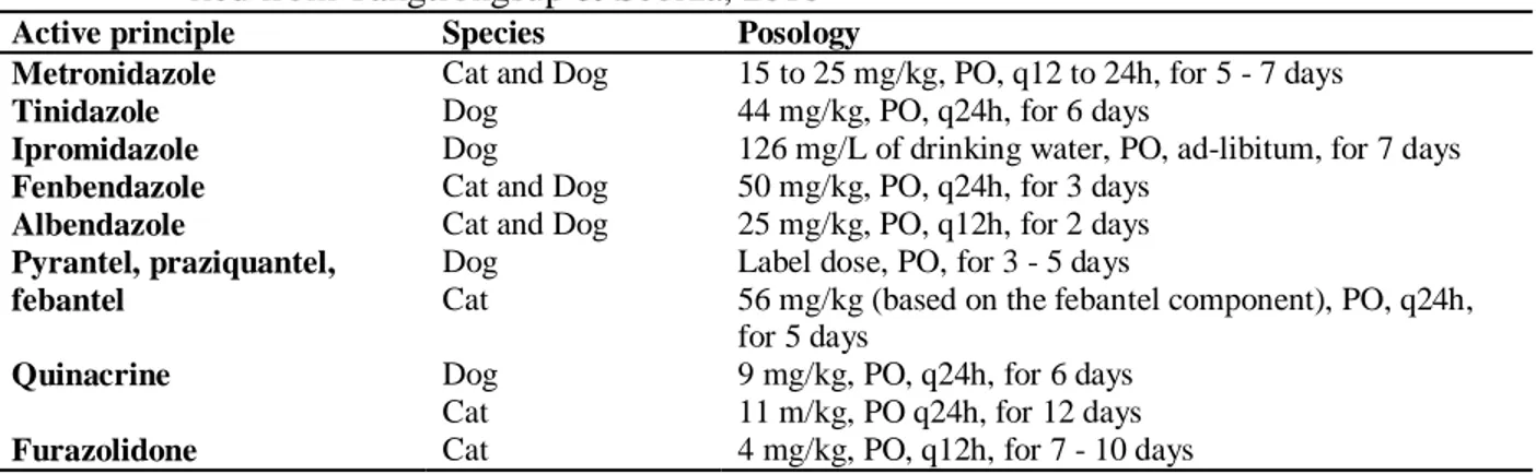
Table 1. Drug therapy used for the treatment of giardiasis in dogs and cats; modi-fied from Tangtrongsup & Scorza, 2010
Active principle Species Posology
Metronidazole Cat and Dog 15 to 25 mg/kg, PO, q12 to 24h, for 5 - 7 days
Tinidazole Dog 44 mg/kg, PO, q24h, for 6 days
Ipromidazole Dog 126 mg/L of drinking water, PO, ad-libitum, for 7 days
Fenbendazole Cat and Dog 50 mg/kg, PO, q24h, for 3 days
Albendazole Cat and Dog 25 mg/kg, PO, q12h, for 2 days
Pyrantel, praziquantel, febantel
Dog Cat
Label dose, PO, for 3 - 5 days
56 mg/kg (based on the febantel component), PO, q24h, for 5 days
Quinacrine Dog
Cat
9 mg/kg, PO, q24h, for 6 days 11 m/kg, PO q24h, for 12 days
The nitroimidazoles family, which includes metronidazole, has anti-protozoan properties in hu-mans and animals. Its mechanism of action is damaging the structure of the DNA of the parasite (Miller, Howes, Kasubick, & English, 1970). Metronidazole is well absorbed after oral admin-istration and inhibitory concentrations can be found in many tissues and secretions.
Nitroimidazoles are primarily metabolized by the liver and excreted in the urine (Lau, Lam, Piscitelli, & L. Wilkes, 1992). Metronidazole should be administered if concurrent infection with Clostridium perfringens is suspected because of the known antibiotic activity against this bacte-rium (Tangtrongsup & Scorza, 2010; Scorza & Lappin, 2004).
Several studies demonstrate the efficacy of benzimdazoles against Giardia (Barr, Bowman, Heller, & Erb, 1993; S. Barr, 1994). The mechanism of action of benzimidazoles is based on the disruption of the architectures of the cytoskeleton microtubules (Navarrete-Vázquez, et al., 2001; Morgan, J.A., & R.C.A., 1993). This drugs has generally broad spectrum of activity and low tox-icity (Gokbulut, Bilgili, Hanedan, & McKellar, 2007) Fembendazole, as a known anthelmintic, is recommended for treatment when co-infection with nematodes is suspected (Rossignol, 2010). The combination of pyrantel/praziquantel/febantel can be used as well when co-infection with nematodes is present. Febantel has been demonstrated to be effective for the treatment of dogs and cats with Giardia. However, there exist some discrepancies among efficacy studies, proba-bly due to the different formulation used on those studies (Rossignol, 2010; Olson & Heine, 2009).
1.1.9 Prevention
Taking into account the primary mode of transmission of Giardia and its associated risk factors, the prophylactic measures to prevent or, at least, decrease the ingestion of infective cysts can be
1. Cyst free environments: maintaining the areas clean from feces will decrease the chance of cyst ingestion. The use of steam cleaners or chemical disinfectants is highly recom-mended. A 1:30 dilution of 5% sodium hypochlorite or quaternary ammonium used at the manufacturer concentration effectively inactivates Giardia cysts. In addition, because the cysts are susceptible to drying, allowing the area to dry after the cleaning is recommend-ed. (Scorza & Lappin, 2012).
2. Cleaning cysts from coats: Grooming is a factor that increases the probability of infection or re-infection. Thus, animals at risk or in treatment may be bathed with regular pet shampoo. In addition, the use of non-irritant disinfectant may be used to clean the perinea area (Scorza & Lappin, 2012).
3. Keep the Giardia outside: In the case of large animal populations such as kernels or shel-ters, it is recommended that new dogs or cats to be bathed, as presented above, regardless if they are Giardia negative (Scorza & Lappin, 2012). The fomite transmission is a known way of spreading these infections, thus the use of basic biosecurity measures is recommended (Scorza & Lappin, 2012).
In conclusion, the prevention of Giardia, as it is for most of the infectious diseases, is a battle-ground with different fronts. Thus, the strategic integrated approach is probably the best way to prevent infection or re-infection with Giardia.
1.1.10 Public health significance
Current advances in molecular techniques have improved the understanding of the taxonomy and further the assemblage arrangement of Giardia isolates among species. This opens the discussion regarding the zoonotic potential of some of those assemblages. Particularly, the assemblage AI have been identified in humans, dogs, and cats (Ballweber, Xiao, Bowman, Kahn, & Cama,
2010; Thomson, Palmer, & O'Handley, 2008; Scorza & Lappin, Giardiasis, 2012).However, to conclude that the zoonotic potential of Giardia is a tangible risk, both biological and epidemio-logical information should be congruent (Ballweber, Xiao, Bowman, Kahn, & Cama, 2010). The molecular techniques of identification have to be analyzed with caution, because the identifica-tion of a particular assemblage depends on the chosen genetic marker, thus the multi-locus anal-ysis is more suitable for establishing any actual connection (Ballweber, Xiao, Bowman, Kahn, & Cama, 2010). Also in the review by Ballweber et al. (2010) it is stated that
"A robust molecular tool for consistent taxonomic classification and sufficient data on the population genetic structure of G. duodenalis are currently lacking, which are needed to understand more completely the transmission dynamics and zoonotic potential of this parasite."
This may imply that, with the actual available tools, there is not enough evidence to conclude that human outbreaks of giardiasis comes from animal source or vise versa.
Even though, there are some reports indicating that the same type of Giardia was found in sam-ples from dogs, cats, and humans interacting closely, there are still uncertainties in the epidemio-logic triangle connecting giardiasis from pets to giardiasis in humans, and the pathway of causa-tion is unclear (Ballweber, Xiao, Bowman, Kahn, & Cama, 2010).
1.2 CRYPTOSPORIDIOSIS IN CATS AND DOGS 1.2.1 Etiology
Ernest Edward Tyzzer was the first to name and describe Cryptosporidium in 1907 using charac-teristics such as the host species, location, and morphologic particularities (Fayer, 2010). Since Dr. Tyzzer discover Cryptosporidium, the host specificity, location in the host, and morphology characteristic have been the basis for taxonomy classification for species of the phylum
Cryptosporidium parvum parasitized the intestine of mammals (Fayer, 2010). With the develop-ment of novel molecular techniques, it was finally understood that there were two different cy-cles of transmission related to the genotype: the human (human-to-human) and bovine (animals-to-humans) genotypes (Fayer, 2010). The naming of a new species occurs now if the biological and genetic information is sufficient to identify an isolate as unique (Fayer, 2010).
Below is the taxonomic classification of the genus Cryptosporidium:
Kingdom Protozoa Phylum Apicomplexa Class Conoidasida Order Eucoccidiorida Suborder Eimeriorina Family Cryptosporidiidae
Genus Cryptosporidium Tyzzer, 1907 (Integrated Taxonomic Information System, 2013).
In 1979, Iseki described Cryptosporidium felis, the species that affects mainly cats. In addition, it was reported to be infective in both bovines and humans (Fayer, 2010). Cryptosporidium muris was identified in naturally infected cats (Pavlasek & Ryan, 2007). In the same manner, C. canis was identified to be the dog genotype and was established as an independent species based on transmission and molecular experiments; this genotype can infect young bovine as well (Fayer, 2010).
1.2.2 Morphology
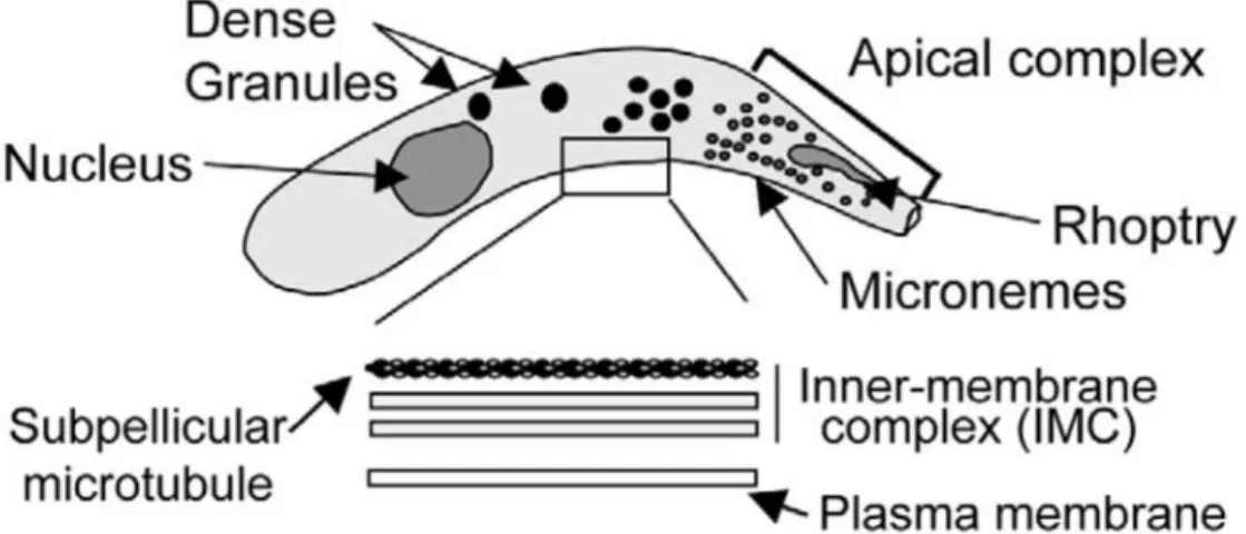
The typical zoites (merozoites or sporozoites) of Cryptosporidium are similar to other apicomplexans; they present crescent shaped cell body, apical rhoptry and micromeres, and
dense granules distributed throughout the cytoplasm ¡Error! No se encuentra el origen de la referencia. (O’Hara & Chen, 2011). The parasite surface (pellicle) is a multilayer membrane; the outer and inner membranes are each composed from two membranes and sub-pellicular microtu-bules (O’Hara & Chen, 2011).
Figure 4. Scheme of the morphologic characteristics of a Cryptosporidium zoite. (Elsevier license 3416010271077)
The endogenous stages of the parasites are closely associated with the luminal surface of the epi-thelial cells; they protrude from the cell surface. These bodies have spherical or elliptical shapes with sizes ranging from 2 to 6µm. Their location has been determined to be intracellular but ex-tra-cytoplasmic within the parasitophorus vacuoles membranes (O'Donoghue, 1995). The pellicle folds repeatedly forming a structure that adheres to the microvilli (O’Hara & Chen, 2011).
The oocyst is the exogenous, infective, and environmental-resistant form of the parasite. Mature oocysts contain 4 sporozoites enclosed within a oocyst. This configuration provides some of the characteristics for its visual classification. The oocysts vary in size and shape depending on the species, ranging from 4.5 to 8 µm in length by 4 to 6.5 in width (O'Donoghue, 1995).
1.2.3 Life cycle
After ingestion of the infective oocyst, excystation of the four sporozoites is triggered mainly by the change in temperature and pH. The sporozoites migrate along the surface of the epithelium until they find a place to attach. This process is driven by a complex biochemical mechanisms that include interaction of Cryptosporidium sporozoites with the host cell’s cytoskeleton. This process has been called gliding motility (Wetzel, Schmidt, Kuhlenschmidt, Dubey, & Sibley, 2005; O’Hara & Chen, 2011). The formation of the parasitophorus vacuole occurs after being encapsulated by a parasite modified host membrane. This process is known as internalization (O’Hara & Chen, 2011). During internalization, the feeder organelle is formed between the para-site and host cytoplasm. This organelle confers selective transport properties between host and parasite for nutrients uptake (O’Hara & Chen, 2011).
Type I, followed by TypeII meronts develop next. These are derived from the asexual reproduc-tion of the trophozoite in the process known as endopolygeny. The formareproduc-tion of the daughter cells occurs while still in the mother cell (O’Hara & Chen, 2011). The type I meront produce merozoites that are morphologically and biologically similar to the sporozoites. These
merozoites invade the surrounding enterocytes and can produce meronts type I and II (O’Hara & Chen, 2011; Scorza & Lappin, 2012).
Merozoites, derived from Type II meronts, differentiate into gametocytes to complete the sexual stage of development. These gametocytes can be either male or female reproductive stages, known as microgametocyte and macrogametocyte respectively (O’Hara & Chen, 2011). The fertilizationof the macrogametocyte by the microgametocyte results in the only diploid stage of development (the zygote), which undergoes sporogony process (meiosis-like process) resulting in the production of a sporulated oocyst containing four sporozoites. This oocyst can be thin or
thick-walled, the thick-walled oocysts are shed in the feces, and the thin-walled oocyst excysts within the intestinal lumen starting a process of autoinfection and escalating the infection level (O’Hara & Chen, 2011).
1.2.4 Pathogenesis
After excystation process, the free sporozoites adhere to the mucous membrane of the small in-testine by a carbohydrate-lectin mediated mechanism (O’Hara & Chen, 2011). Multiple proteins, localized in the apical surface of the zoite, have been identified to be importantly involved in the attachment process; gp40, gp15, gp900, and Circumsporozite-like glycoprotein (CSL) are some (O’Hara & Chen, 2011). Furthermore, a Gal/GalNAc-specific lectin (p30) was identified having lectin activity. Another sporozoite protein (cp47) localized in the apical region of the parasite, was found to be highly correlated with the efficiency of in vitro infectivity. It has been demon-strated that this protein interacts with a 57kDa (p57) protein of the host cell which is abundant in the ileum. This explains, in part, its affinity for this tissue (O’Hara & Chen, 2011).
The motility possess of aplicomplexans undergoes a unique method that is defined by the ab-sence of any obvious modification of the shape of the moving cell (O’Hara & Chen, 2011; Smith, Nichols, & Grimason, 2005). The structural stability and polarity is maintained by the mi-crotubules, while the locomotion and invasion mechanism is provided by the actomyosin system. The investigation of the gliding mechanisms in Toxoplasma gondii and Plasmodium have shown that trophozoites left a trail of proteins that are released (shed) trough the posterior pole of the cell (O’Hara & Chen, 2011; Smith, Nichols, & Grimason, 2005). The process of gliding motility, then, comprises three main steps: i) the secretion of adhesive molecules from the apical pole of the parasites that adhere to the host cell receptors; ii) the posterior translocation of the adhesive
molecules; and iii) the proteolytic cleavage and release of the parasite molecules in motility trails (O’Hara & Chen, 2011).
After the zoite has found its niche in the luminal surface of the host, the process of invasion is initiated by the fusion of both parasite and host membranes. The rhoptry is in close relation with the site of attachment and other organelles associated with the process (micronemes and dense granules) migrate to the parasite-host interface. The cytoplasm of the zoite vacuolize and a tun-nel-like structure is formed in this location (O’Hara & Chen, 2011; Smith, Nichols, & Grimason, 2005). The process of internalization-invasion starts with the clustering of vacuoles that ultimate-ly encloses the parasite. A unique condition is derived from this process; the zoites remains ex-tra-cytoplasmic yet intra-membranous (intracellular) (O’Hara & Chen, 2011). In addition, a structural support is formed at the base of the parasite-host interface by a network of recruited host actin (O’Hara & Chen, 2011). After internalization, the parasite also recruits the host cell channels and transporters to the parasite-host interface, which further serve to nourish and sup-port the sporozoite (Smith, Nichols, & Grimason, 2005). .
It was demonstrated the altered expression of over 200 genes in infected cultured human cells; the main altered genes include those associated with apoptosis, cyto-skeletal dynamics, and pro-inflammatory signaling cascades (O’Hara & Chen, 2011). One of the most important mecha-nisms for the proliferation of the infection is the inhibition of apoptosis, because the parasite re-quires viable host cells for the completion of its life cycle (O’Hara & Chen, 2011). Perhaps, the epithelial cell apoptosis mechanism is protective, limiting the parasites number (O’Hara & Chen, 2011).
The loss of epithelial brush in cryptosporidiasisis most likely caused by the immune host re-sponse rather than by any direct effect of the parasite (Scorza & Lappin, 2012).
1.2.5 Epidemiology
Cryptosporidium is distributed throughout the world. Its transmission is related to crowded and unsanitary conditions; immunocompromised individuals are specially affected by this kind of parasites (Fayer, 2010; O'Donoghue, 1995).
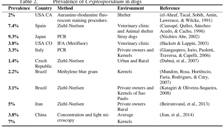
The prevalence of Cryptosporidium in dogs and cats is variable throughout the different reports (¡Error! No se encuentra el origen de la referencia.). The variation of these findings may be due, at least in part, to the uses of different tests that have different detection thresholds or in other words different sensitivity and specificity values, In such reports the number of true posi-tive or true negaposi-tive is unknown, which makes, the necessity for a reference test, even more evi-dent.
Table 2. Prevalence of Cryptosporidium in dogs
Prevalence Country Method Environment Reference
2% USA CA Auramine-rhodamine fluo-rescent staining procedure.
Shelter (el-Ahraf, Tacal, Sobih, Amin, Lawrence, & Wilcke, 1991)
7.4% Spain Ziehl-Neelsen Veterinary clinic and Animal shelter
(Causapé, Quílez, Sánchez-Acedo, & Cacho, 1996)
9.3% Japan PCR Stray dogs (Niichiro Abe, 2002)
3.8% USA CO IFA (Merifluor) Veterinary clinic (Hackett & Lappin, 2003)
3.3% Italy PCR Private owners and
Kernels
(Giangaspero, Iorio, Paoletti, Traversa, & Capelli, 2006)
1.4% Czech
Republic
Ziehl-Neelsen Urban and Rural (Dubná, et al., 2007)
2.2% Brazil Methylene blue gram Kernels (Mundim, Rosa, Hortêncio, Faria, Rodrigues, & Cury, 2007)
3.1% Brazil Ziehl-Neelsen Private owners and Kernels of Sao Paulo
(Katagiri & Oliveira-Sequeira, 2008)
5% Iran Ziehl-Neelsen Private owners
Rural
(Beiromvand, et al., 2013)
3.8% China Concentration and light mi-croscopy
Average (Jian, et al., 2014)
7% Kernels
con-disinfectants (Scorza & Lappin, 2012). In human populations, the contamination of public water supplies can lead to large outbreaks of cryptosporidiosis (Scorza & Lappin, 2012)
The risk factors associated with cryptosporidiosis in humans includes contact with contaminated water (recreational or drinking), exposure to infected animals (mainly bovines), travel to disease endemic areas, and ingestion of contaminated food (Yoder & Beach, 2010). Groups implicated with higher risks of infection include children and staff in day care centers, farmers and animal handlers, health care workers, and travelers to endemic zones (Ramirez, Ward, & Sreevatsan, 2004). Other commonly mentioned factor that increases the risk for cryptosporidiosis is the pre-tense of any type of immunodeficiency including but not limited to HIV infection and AIDS, drugs, organ transplantation, cancer chemotherapy, etc (Ramirez, Ward, & Sreevatsan, 2004). In pets, cats specifically, some of the reported associated factors are age (<1 year), presence of oth-er entoth-eric parasites (Giardia), feeding with not commoth-ercial diet, and diarrhea (Luisa Rambozzi, 2007; Ballweber, Panuska, Huston, Vasilopulos, Pharr, & Mackin, 2009). In a study in dogs in the province of Rio de Janeiro, Brazil, sporadic diarrhea and vomiting, living with cats, and the owner socioeconomic status were found to be significantly associated with canine cryptosporidi-osis (Ederli, Ederli, Oliveira, Quirino, & Carvalho, 2008).
1.2.6 Clinical findings
Many infections caused by Cryptosporidium in cats and dogs are subclinical or cause only mild clinical sign (Scorza & Tangtrongsup, 2010). The most common clinical signs associated with cryptosporidiosis are small bowel diarrhea, anorexia, and weight loss (Scorza & Lappin, 2012; Scorza & Tangtrongsup, 2010). In some animals, particularly animals affected by any type of immunodeficiency (viral, iatrogenic, stress, malnutrition, etc.) or co-infection with other enteric parasites, the infection may cause chronic diarrhea and malabsorption syndrome (Scorza &
Tangtrongsup, 2010). For some cases, it is difficult to establish if Cryptosporidium is the primary cause of the clinical signs, because of the presence of other etiologies: other parasites, viral in-fections, bacterial infections or inflammatory processes (Scorza & Lappin, 2012).
1.2.7 Diagnosis
As well as for diagnostic of Giardia, there are a number of available laboratory techniques for the detection of Cryptosporidium, which are summarized in the following section.
1.2.7.1 Conventional Microscopy
The direct microscopic examination of wet mounts is not used regularly. Even with the addition of concentration, the recognition of the oocysts in direct mounts is difficult due to the small numbers of oocysts in the feces of dogs, cats, and humans and can lead to false negative results. For this reason, Use of staining procedures can be used to increase the sensitivity of the micro-scopic tests; the most frequently used staining techniques are modified Ziehl-Neelsen (MZN) acid fast, safranin-methylene blue stain, Kinyoun acid fast, and DMSO-carbol fushin. With the MZN staining, the oocysts are stained with carbol-fuchsin and the dye is retained in the decolor-izing step with acid alcohol. One major disadvantage of this technique is its low sensitivity (70%) (Marks, Hanson, & Melli, 2004). However, this is a test that can be performed in small practices with a light microscope, and can serve as initial screening test (Scorza & Tangtrongsup, 2010).
1.2.7.2 Immunochemical antigen detection
Some commercial DFA tests are available for the simultaneous detection of Cryptosporidium oocysts and Giardia cysts. This technique can be more sensitive and specific than other micro-scopic techniques; its detection threshold is as low as 104 oocysts/gram of concentrated feline
Bishop, Wahlquist, Sullivan, & Juranek, 1991). As with Giardia, one of its advantages is that the results are based not only on the specific antibody link but also in the recognition of the mor-phology by the examiner. One of the major disadvantages is that this technique requires the use of a microscope with a fluorescent lamp for the examination of the slides, which is not often a regular equipment in private practices.
A number of ELISA tests for the detection of Cryptosporidium fecal antigens are available for use with human feces. One; one of its major advantages these techniques is that does not require the use of complex equipments or specific training. In kits, the readings of results can be per-formed by the comparison of the colorimetric change against a scale. On the other hand, most of the available commercial ELISA tests used in veterinary medicine have been developed for the diagnosis of cryptosporidiosis in humans, with C. parvum as its principal target. Antigenic dif-ferences amongst C. parvum, C canis, and C. felis exist which can explains why the results of these assays when used with dogs or cat feces are inconsistent (Scorza & Tangtrongsup, 2010; Marks, Hanson, & Melli, 2004).
1.2.7.3 Molecular techniques
The use of molecular techniques has help to elucidating the complex research questions about the biology, taxonomy, pathogenesis, and epidemiology of Cryptosporidium (O’Hara & Chen, 2011; Thomson R. , 2004; Fayer, 2010). Moreover, the use of molecular techniques for detection of oocysts has been increasing (Scorza, Brewer, & Lappin, 2003). The amplification of Cryptos-poridium DNA in feces can be a useful tool. This has shown to be more accurate than ELISA tests and Ziehl-Neelsen (Uppal, Singh, Chadha, & Jha, 2014; Omoruyi, Nwodo, Udem, & Okonkwo, 2014; Scorza, Brewer, & Lappin, 2003). In addition, when the sequencing is added to the analysis of samples, the association of particular species with the infection can be determined
. (Thomson R. , 2004). However, since these kinds of techniques are more expensive and lack from extensive or well-controlled studies that allow us to estimate its performance. The use of this technique has been limited to identification of cases with chronic-unexplained diarrhea that are negative to other tests, or when genotyping is the goal (Scorza & Tangtrongsup, 2010).
1.2.7.4 Other diagnostic tools
Other possible available tools for the diagnosis of Cryptosporidium are detection of serum anti-bodies (ELISA or FA), inoculation of mice, and intestinal biopsy; nonetheless, those techniques are not being used routinely in the diagnostic laboratory (Scorza & Tangtrongsup, 2010).
Immuno-PCR is a technique has been used for detection of low concentration of oocyts in water sources. This technique is based on the primary attachment by antigen-antibody complexes to a gold matrix that afterwards is used to perform PCR (Deng, et al., 2014).
1.2.8 Treatment
Over 100 compounds have been evaluated for the treatment of cryptosporidiosis. However, none of them has shown clear remission of signs or elimination of infection (Scorza & Lappin, 2012; Rossignol, 2010). Thus, the primary goal of the treatment should be to stop diarrhea. Palliative support should be given, according to practitioner discretion. The use of high digestible diet, hy-dration solutions, mucosal protectors, and antibiotic for secondary bacterial infection may be necessary as part of the treatment of cryptosporidiosis (Scorza & Tangtrongsup, 2010).
Chemotherapy in cats and dogs lacks of extensive studies showing the efficacy therapy to control the clinical signs of Cryptosporidium infection. . (Scorza & Tangtrongsup, 2010; Thomson, Palmer, & O'Handley, 2008; Armson, Reynoldson, & Thompson, 2003). In companion animals, positive results have been reported for treating infections with Cryptosporidium using
Tangtrongsup (2010), shows the treatment porotocols used on cats and dogs with cryptosporidiosis.
Table 3. Table 1Drug therapy used for the treatment of cryptosporidiosis in Dogs and Cats; modified from Scorza & Tangtrongsup (2010).
Active principle Posology what is this?
Azithromycin 10 mg/kg, PO, q24 hours, until remission of clinical signs.
Nitazoxanide 25 mg/kg, PO, q12 hours, for at least 7 days.
Paromomycin 125 - 165 mg/kg, PO, q12 - 24 hours, for at least 5 days.
Tylosin 10 - 15 mg/kg, PO, q8 - 12 hours, for 21 days.
Paromomycin is an antibiotic, part of the amino-glycoside group; its mechanism of action is based on the disruption of the protein synthesis pathway targeting the ribosome (Gargala, 2008). Its absorption is limited at the intestinal level, but can be absorbed in small amounts at the apical membrane of the epithelial cell (Gargala, 2008; Scorza & Lappin, 2012). Paromomycin has been evaluated in cats, showing decreased oocyst shedding to below detection limits (Scorza & Tangtrongsup, 2010; Lappin, 2004). When there is uncertainty of the integrity of the mucosal membrane, however, its use should be avoided, because of increased absorption rates, which re-sult in renal and ototoxicity (Scorza & Tangtrongsup, 2010).
Azithromycin is an azalide antibiotic, which interferes with the microbial protein synthesis, and is considered the most active among the macrolides (Gargala, 2008). Azitrhtomicin has been evaluated in animals. It has been reported that the administration to infected calves, improves the clinical signs and reduces the oocyst shedding (Elitok, Elitok, & Pulat, 2008).
Nitazoxanide (NTZ) is a 5-nitrothiazolyl salicylamide derivative with well-known activity against protozoa and helminthes (Gargala, 2008). NTZ has been administered to cats and dogs resulting in remission of clinical signs. However, NTZ also causes intestinal irritation, and it is not effective when the patient is not immuno-competent (Scorza & Tangtrongsup, 2010)
Tylosin has been administrated to cats and dogs empirically resulting in improvement of clinical signs. However, these observations were uncontrolled and it is possible that the results of tylosin administration were related to the control of bacterial co-infection or anti-inflammatory effects. In addition, tylosin can be a gastrointestinal irritant and it is not well tolerated by cats because of its taste (Scorza & Tangtrongsup, 2010; Westermarck, et al., 2005).
1.2.9 Prevention
Cryptosporidium oocysts are resistant to extreme temperatures and most frequently used disin-fectants. Concentrated ammonia solution (50%) has been effective for inactivation of oocysts. Steam (>55°C), freezing thawing, and drying are effective preventive measures for the inactiva-tion of oocyts (Scorza & Tangtrongsup, 2010).
Cryptosporidium oocysts and Giardia cysts have similar characteristics of resistance to the envi-ronmental conditions. Both agents share many epidemiologic features and thus the measures of control may be work for preventing their infection. Furthermore, maintaining the areas clean from feces plus the use of chemical disinfectants, and low humidity floors will decrease the chance of oocyst ingestion. Quarantine or isolation may be recommended for infected individu-als. Suspected animals may be bathed with regular pet shampoo to decrease the risk of infection by grooming. Screening test and regular baths are recommended for new members of a popula-tion.
1.2.10 Public health significance
In the past, it was believed that each Cryptosporidium species or genotype infects a particular host species. Cryptosporidium parvum was considered to infect humans, but later, with the inclu-sion of genotyping techniques, C. parvum was separated into two genotypes: C. parvum—the
2008). Additionally, the species affecting cats and dogs (C. felis and C. canis) have been identi-fied in human samples. However, the zoonotic roll C. felis and C. canis seems to be limited, be-cause the infection rates of those species in humans are low (0.26% and 0.02% respectively), and many studies have failed to show strong association between human cryptosporidiosis and pet contact (Scorza & Tangtrongsup, 2010; Lucio-Forster, Griffiths, Cama, Xiao, & Bowman, 2010). In the case of HIV-infected people, it should be recommended to avoid any contact with infected pets, and the sanitization practices should be emphasized in order to decrease the risk of trans-mission (Scorza & Lappin, 2012; Scorza & Tangtrongsup, 2010; Lucio-Forster, Griffiths, Cama, Xiao, & Bowman, 2010).
1.3 DIAGNOSTIC TEST ASSESSMENT 1.3.1 Notation and definitions
In this review, the terms probability and proportion are used synonymously and will be defined by relative frequency. Let A denotes the event that a randomly selected subject from a population has a defined characteristic. N denotes the total number of people in one population, thus NA the number of subject that has characteristic A. Then P(A) denotes the proportion of all subjects that have the A characteristic or, likewise, P(A) is the probability that a randomly selected subject has the characteristic A; 𝑃(𝐴) = 𝑁𝐴⁄ . Thus P(A) should be a real number contained between 0 and 𝑁 1 (0 ≤ P(A) ≤ 1). Let P(Ā) denotes the proportion of subjects that do not have the characteristic A, then 𝑃(𝐴̅) = 𝑁𝐴̅⁄ and 𝑃(𝐴̅) = 1 − 𝑃(𝐴), so P(Ā) is denominated the complementary pro-𝑁 portion to one of P(A). In the same manner, if NB denotes the number of subjects that have char-acteristic B, 𝑃(𝐵) = 𝑁𝐵⁄ , and its complementary 𝑃(𝐵�) = 𝑁𝑁 𝐵�⁄ = 1 − 𝑃(𝐵). 𝑁
Additionally, if NAB is the number of subjects that have both characteristics, we can describe the proportions of subjects having these two characteristics at the same time as 𝑃(𝐴 𝑎𝑛𝑑 𝐵) =
𝑁𝐴𝐵⁄ . The so-called conditional probability is defined by P(A|B), which is the probability that 𝑁
a randomly selected subject has a characteristic A given that it has characteristic B, or is condi-tional on having characteristic B. As stated above, P(A and B) represent the proportion of all subjects that possess both characteristic A and characteristic B, then
𝑃(𝐴|𝐵) = 𝑁𝑁𝐴𝐵⁄𝑁 𝐵⁄ =𝑁 𝑁𝐴𝐵 𝑁𝐵 = 𝑃(𝐴 and 𝐵) 𝑃(𝐵) , similarly 𝑃(𝐵|𝐴) =𝑁𝑁𝐴𝐵⁄𝑁 𝐴⁄ =𝑁 𝑁𝐴𝐵 𝑁𝐴 = 𝑃(𝐴 and 𝐵) 𝑃(𝐴) .
The association of two characteristics means that the probability of having one characteristic is affected by the probability of having other characteristics. In contrast, the independence or lack of association of two characteristics means that given that the subject have one characteristic does the probability of having the other characteristic is not affected. Then,
𝑃(𝐴 and 𝐵)
𝑃(𝐵) = 𝑃(𝐴) ⇒ 𝑃(𝐴 and 𝐵) = 𝑃(𝐴) × 𝑃(𝐵). This equation is often taken as the definition of independence.
When two proportions are matter of studies, the aim is often to establish or discard any type of association. The inclusion of the conditional proportion to the equation allow for that as
𝑃(𝐴 and 𝐵) = 𝑃(𝐴|𝐵) × 𝑃(𝐵).
With the rule of total probability, it is possible to know the probability of having one characteris-tics including conditional and complimentary probabilities as
𝑃(𝐵) = 𝑃(𝐴 and 𝐵) + 𝑃(𝐵 and 𝐴̅), then
The Bayes' Theorem, from the frequentist point of view, connects the conditional probabilities of A given B and vice versa by the probabilities of each event, this is
𝑃(𝐵|𝐴) =𝑃(𝐴|𝐵)𝑃(𝐵)𝑃(𝐴) .
Bayes' Theorem is a theorem of probability theory, it was originally stated by the Reverend Thomas Bayes. We can explain it as the way of how the probability of a true event can be affect-ed by the inclusion of the probability of other event as a piece of evidence (Feiss, Levin, & Paik, 2003).
1.3.2 Applied probability for diagnostic tests
For this part of the review, let T denotes the positive result of a diagnostic test, then 𝑇� denotes the complimentary negative result. In the same way, let D denotes the presence of disease in an individual, and 𝐷� its complimentary absence of disease indicator.
The sensitivity of a test, denoted Se, is the probability that a true positive or diseased sample tests positive, thus, following the probability notation above,
𝑆𝑒 = 𝑃(𝑇|𝐷).
The specificity of a test, denoted Sp, is the probability that a true negative or non-diseased sample tests negative, thus
𝑆𝑝 = 𝑃(𝑇�|𝐷�).
The disease prevalence (true prevalence) in the source population, denoted Pr, is the proportion of subjects from the source population that have the disease, thus
𝑃𝑟 = 𝑃(𝐷).
The term apparent prevalence (APr), is given to the proportion of subjects that have positive test result,
The predictive value is calculated using the Bayes' Theorem, for those subjects with positive test results this probability is called the Positive Predictive Value (PPV), defined by:
𝑃𝑃𝑉 = 𝑃(𝐷|𝑇) =𝑃(𝑇|𝐷)𝑃(𝐷)𝑃(𝑇) =𝑆𝑒 × 𝑃𝑟 + (1 − 𝑆𝑝)(1 − 𝑃𝑟).𝑆𝑒 × 𝑃𝑟
Knowing the Se and Sp and Pr, the rule of total probability is used to obtain the proportions of positive results or apparent prevalence, this is
𝑃(𝑇) = 𝑃(𝑇|𝐷)𝑃(𝐷) + 𝑃(𝑇|𝐷�)𝑃(𝐷�) = 𝑆𝑒 × 𝑃𝑟 + (1 − 𝑆𝑝)(1 − 𝑃𝑟). In the same way, the Negative Predictive Value (NPV) is defined by
𝑁𝑃𝑉 = 𝑃(𝐷�|𝑇�) =𝑃(𝑇�|𝐷�)𝑃(𝐷�)
𝑃(𝑇�) =
𝑆𝑝 × (1 − 𝑃𝑟)
𝑆𝑝 × (1 − 𝑃𝑟) + (1 − 𝑆𝑒)𝑃𝑟.
Confidence intervals (CI) can be calculated using the formula for estimating the Standard Error (SE) and the CI for a single proportion
𝑆𝐸(𝑝) = �𝑝(1 − 𝑝)𝑁 ,
then
𝜃 ∓ 𝑍1−∝ 2⁄ × 𝑆𝐸(𝑝),
where p is the proportion or probability of interest, N is the number of subjects of interest, θ is the upper or lower CI, and 𝑍1−∝ 2⁄ is the 1 − ∝ 2⁄ percentile of the normal distribution.
The sample size for the estimation of Pr, Se and Sp would depend on the desired confidence lev-el for the estimates and the allowed error in the estimates, then for estimation of the number of samples (n) the formula is