O R T H O D O N T I C A N C H O R A G E – E V I D E N C E - B A S E D E V A L U A T I O N O F A N C H O R A G E C A P A C I T Y A N D P A T I E N T S ’ P E R C E P T I O N S
Swedish Dental Journal, Supplement 191, 2007
© Ingalill Feldmann, 2007 ISBN 91-7104-294-6
INGALILL FELDMANN
ORTHODONTIC ANCHORAGE
Evidence-based evaluation of anchorage capacity and
patients’ perceptions
Department of Orthodontics
Faculty of Odontology, Malmö University, Sweden
Publikationen finns även elektroniskt, se www.mah.se/muep
CONTENTS
PREFACE ... 9 ABSTRACT ... 10 SUMMARY IN SWEDISH ... 13 INTRODUCTION ... 15 Orthodontic anchorage ...15 Skeletal anchorage ...16Patients’ perceptions of orthodontic treatment ...19
Evidence-based evaluation ...21
Final remarks ...22
AIMS ... 23
HYPOTHESES ... 24
MATERIALS AND METHODS ... 25
RESULTS ... 39 DISCUSSION ... 50 CONCLUSIONS ... 59 ACKNOWLEDGEMENTS ... 61 REFERENCES ... 64 APPENDIX ... 78 PAPER I ... 87 PAPER II ... 99 PAPER III ...109
To Hartmut, Andreas and Antonia
Give me a place to stand and I will move the earth” Archimedes 287 – 212 BC
PREFACE
This thesis is based on the following papers, which are referred to in the text by their Roman numerals I-IV.
I. Feldmann I, Bondemark L. Orthodontic anchorage: A systematic review. Angle Orthod 2006;76:493-501. II. Feldmann I, List T, John MT, Bondemark L. Reliability
of a questionnaire assessing experiences of adolescents in orthodontic treatment. Angle Orthod 2007;77:311-317. III. Feldmann I, List T, Feldmann H, Bondemark L. Pain and discomfort following surgical placement of ortho-dontic anchoring units and premolar extraction. A randomized controlled trial. Angle Orthod 2007;77:578-585.
IV. Feldmann I, Bondemark L. Anchorage capacity of os-seointegrated and conventional anchorage systems – A randomized controlled trial. Am J Orthod and Dentofa-cial Orthop. Accepted for publication (July 2007). These papers are reprinted with kind permission from the copyright holders.
ABSTRACT
Orthodontic anchorage is the ability to resist unwanted reciprocal forces and reinforcement of anchorage by supplementary appliances, in or outside the mouth, is often needed to obtain successful results. In the last 10 years, interest in appliances that use implants has been growing.
Successful orthodontic treatment demands effective methods and systematic evaluation of different treatment approaches is therefore essential. Several studies on the efficiency of various anchorage sys-tems have been published, but a critical appraisal or interpretation of evidence that systematically considers validity, results, and rele-vance has not been made. Analysis of treatment modalities must also include patients’ perceptions and potential side-effects.
The overall aim of this thesis was to evaluate a new anchorage technique that incorporates osseointegration and compare it with conventional methods concerning effects on tooth movements in adolescents and their acceptance and experience of the additional surgical procedures that osseointegration involves. The following anchorage systems were analyzed: Onplant system, Orthosystem implant, headgear and transpalatal bar.
This thesis was based on four studies:
Paper I systematically reviewed the efficiency of orthodontic an-chorage systems and interpreted the methodological quality of the selected studies from an evidence-based perspective. The literature search spanned January 1966 – December 2004 and was later ex-tended to July 2007.
Paper II, a methodological study involving 60 adolescent patients, examined the validity and reliability of a new questionnaire for as-sessing adolescent patients’ perceptions of orthodontic treatment. The questionnaire was based on focus group interviews.
Papers III and IV were randomized controlled trials involving 120 adolescent patients in orthodontic treatment. Paper III evaluated and compared adolescent patients’ perceptions of premolar extractions and surgical placement of Onplants and Orthosystem implants. Pa-per IV compared anchorage capacities of the four systems.
These conclusions were drawn:
• The scientific evidence, found in the review, was too weak to evaluate the efficiency of various anchorage systems (con-ventional and osseointegrated) during space closure after premolar extraction, and most studies have quality prob-lems. Future randomized controlled trials are recommended. • The new questionnaire, developed from focus group inter-views, had overall acceptable to good reliability and high face validity. It can therefore be recommended for use in the assessment of adolescents’ experiences of orthodontic treat-ment.
• Pain intensity after surgical placement of an Orthosystem implant was less than after Onplant installation and premo-lar extraction. Pain intensity after Onplant installation and premolar extractions were comparable.
• With respect to pain intensity, discomfort, and analgesic consumption, the Orthosystem implant can be recom-mended over the Onplant system.
• The Onplant system and the Orthosystem implant provided stable anchorage throughout the observation period. Head-gear anchorage was stable during the leveling/aligning phase but anchorage loss occurred at the end of the observation period. Transpalatal bar anchorage was never sufficient at any point in the observation period.
• Headgear and the transpalatal bar can only be recom-mended when need for anchorage reinforcement is limited.
• For maximum anchorage and if patients’ perceptions are considered, the osseointegrated Orthosystem implant is the system of choice.
POPULÄRVETENSKAPLIG
SAMMANFATTNING
När en tand utsätts för en kraft genereras alltid en motriktad (reci-prok) kraft av samma dimension. Ortodontisk förankring definieras som förmåga att avleda oönskade reciproka krafter och förankrings-förstärkning ingår ofta som en viktig del i behandlingsplaneringen för att säkerställa att behandlingsmålet uppnås. Under den senaste 10-års-perioden har stort intresse visats för tekniker som använder benförankrad förankringsförstärkning.
Ett stort antal studier om olika förankringssystem (konventio-nella och benförankrade) finns publicerade men en kritisk gransk-ning av resultat och klinisk relevans ur ett evidensbaserat perspektiv saknas i allmänhet. När ny teknik introduceras är det viktigt att ef-fektivitet utvärderas och jämförs med konventionella metoder men också att patientens upplevelser analyseras, speciellt om kirurgiska moment ingår.
Det övergripande syftet med denna avhandling var att utvärdera ny förankringsteknik, baserad på osseointegrering, och jämföra med konventionella metoder med avseende på förankringskapacitet hos en grupp ungdomar som genomgår ortodontisk behandling. Dessut-om analyserades patienternas upplevelser av de tillkDessut-ommande kirur-giska momenten och jämfördes med tandextraktioner.
Följande fyra förankringssystem system har analyserats: Onplant, Orthoimplantat samt extraoralt drag (EOD) och transpalatinal bar. Avhandlingen är baserad på följande studier
Studie I var en systematisk litteraturöversikt över effektiviteten av olika förankringssystem, ur ett evidensbaserat perspektiv. I syftet för
studien ingick också att göra en kvalitetsbedömning av de utvalda studierna. Översikten omfattade tidsperioden från januari 1966 till december 2004 och har därefter utökats att gälla tom juli 2007.
Studie II var en metodstudie som inkluderade 60 ungdomar och där reliabiliteten och validiteten av ett nytt frågeformulär, baserat på fokusgruppintervjuer, analyserades.
Studie III och IV var randomiserade kontrollerade studier med 120 ungdomar som genomgick ortodontisk behandling. Studie III utvärderade och jämförde ungdomars upplevelser av kirurgisk in-sättning av två olika osseointegrerade förankringselement och pre-molar extraktioner. Studie IV analyserade och jämförde förank-ringskapaciteten för de fyra olika förankringssystemen.
Konklusioner:
• Översikten visade att det inte var möjligt att dra några evi-densbaserade slutsatser om förankringsförstärkning på mo-larer efter premolarextraktioner. Studiernas kvalitet var mes-tadels låg och mera forskning behövs.
• Det nya frågeformuläret, baserat på fokusgruppintervjuer, uppvisade acceptabel till god reliabilitet och hög validitet. Det kan därför i framtiden rekommenderas när ungdomars erfarenheter av ortodontisk behandling skall utvärderas. • Smärta efter kirurgisk insättning av Orthoimplantet var
läg-re än efter insättning av ett Onplant eller pläg-remolaläg-rextrak- premolarextrak-tioner. Smärta efter kirurgisk insättning av ett Onplant var jämförbar med premolarextraktioner.
• Onplant och Orthoimplantat-systemet utgjorde stabil för-ankring under observationsperioden medan både EOD och transpalatinal bar uppvisade förankringsförluster.
• EOD och transpalatinal bar kan endast rekommenderas när begränsad förankring eftersträvas.
• Vid behov av maximal förankring och om hänsyn tas till pa-tientens upplevelser så är det osseointegrerade Orthoimplan-tatet det rekommenderade förankringssystemet.
INTRODUCTION
Orthodontic anchorage
Actioni contrariam semper et æqualem esse reactionem: sive corporum duorum actiones in se mutuo semper esse æquales et in partes contrarias dirigi
The law of Nature that underlies orthodontic tooth movement is Newton’s third law of motion (1687): For every action there is an opposite and equal reaction. This natural law guides orthodontic treatment thought in the planning of (1) what forces and moments are needed to reach treatment objectives and (2) what reactive forces need to be diverted.
In most cases anchorage is produced within the orthodontic appli-ance with the strategy to dissipate the reaction forces over as many teeth as possible and thereby control anchorage. Pressure in the periodontal ligaments of the anchor teeth are thereby kept to a minimum.1, 2 The anchor value of each tooth can be mathematically estimated as a function of root-surface area, and magnitudes of the forces of orthodontic tooth movements can be predicted. But this principle is not always reliable since anchorage capacity is also in-fluenced by attachment level, density and structure of the alveolar bone, periodontal reactivity, muscular activity, occlusal forces, cra-niofacial morphology, and friction resulting from tooth movement.3-5 The stability of biological anchorage may thus be enhanced by selec-tively modifying various moments and forces to alter root inclina-tion or torque of the anchorage teeth.6-8 These techniques are effec-tive, but they depend on the dexterity and biomechanical skills of the operator and the capability to deal with possible adverse side ef-fects like rotation and vertical forces.
Satisfactory reinforcement of anchorage may therefore require supplementary use of extraoral units, like headgear. Headgear can be effective and is easily adjustable for dissipating adverse vertical and rotational side effects9
but also limited: headgear is seldom used for more than part of the day, and its metallic bow has been associ-ated with injury in the facial area.10
Another strategy is the use of intermaxillary elastics to distribute reaction forces to the opposite jaw, thereby spreading forces over a larger area of the periodontal ligament and reducing pressure on the anchor units. Risk of vertical side effects and tipping, can however limit the use of elastics as routine anchorage. Use of headgear or elastics is highly dependent on patient cooperation, which implies a risk of prolonged treatment time and jeopardizes the treatment goal if patients do not cooperate. Patient cooperation is also considered difficult to predict.11-13
To minimize the need for compliance, various transpalatal bars and lingual arches—passive and active—have been widely used as alternatives to headgear and elastics.8,14-16 These bars and arches pro-duce anchorage by blocking molars in combination with pressure from the tongue, but the literature contains little or no evidence that such anchorage reinforcement is substantial.5,17 Another appliance that is widely used as anchorage during molar distalization—the Nance holding arch—produces anchorage via an acrylic button placed on the anterior palate. But studies have demonstrated that when molars are moved distally, anchorage is lost and overjet in-creases.18-21 Furthermore, insufficient hygiene under the button has caused soft tissue in contact with the button to become inflamed. 22,23 All of these anchorage arrangements discussed produce acceptable to good anchorage, but none provide completely secure anchorage in all three dimensions and 100% resistance to reactive forces, in other words, absolute anchorage. So interest in appliances that use implant-based anchorage has been great.
Skeletal anchorage
Most dental implant research focuses on the prosthodontic use of endosseous implants,24,25 but several experimental studies and case reports indicate that osseointegrated endosseous implants are also resistant to applied orthodontic forces.26,27 Conventional dental
im-plants require edentulous space in the dental arch and are most use-ful when combined orthodontic and prosthodontic treatment mo-dalities are needed.28-30 Since most orthodontic patients are adoles-cents with complete dentitions, alternative placements and designs for using implantable devices to reinforce anchorage are needed. Such devices, when used in orthodontic treatment, are temporary and should be removed after treatment.
In recent years, numerous, novel ways of reinforcing anchorage have appeared in the literature.31-41 These new methods use a variety of devices temporarily anchored in bone. The devices can be located subperiosteally or endosteally and be fixed to bone, osseointegrated or non-osseointegrated. Skeletal anchorage can be loaded in two ways:
• directly: forces needed for desired tooth movements are ap-plied to the skeletal device.
• indirectly: the teeth that act as reactive units are indirectly stabilized by the skeletal device via a transpalatal arch or wire.
With indirect anchorage, the stability of the anchoring teeth also de-pends on the rigidity of the connecting unit.
Osseointegrated anchorage systems
Several manufacturers have modified restorative implant designs to produce customized orthodontic titanium fixtures such as the Fri-alit-2 Implant system34
and the Orthosystem® implant, which is most documented33,42-46 The Orthosystem implant is an endosseous titanium implant of screw-type with a sandblasted, large-grit, acid-etched surface (SLA) that can be placed in the palate or the retromo-lar area.
The Onplant™ system, designed by Block and Hoffman,31,47-49 is an osseointegrated anchorage system that is placed subperiosteally in the palate where vertical bone height is limited. The device in this system, the Onplant, is a titanium disc coated with a thin layer of hydroxyapatite to facilitate osseointegration. Surgical placement and removal of an Onplant involves a larger area of the palate than does an implant, and a second-stage surgery to uncover the Onplant is required. However, no drilling is involved in placement. All tempo-rary osseointegrated anchorage devices need a healing period which
was initially set to 10-12 weeks. Recent research suggest that shorter healing period for palatal implants is possible.50
Non-osseointegrated anchorage systems
Ideally, implant anchorage should be easy to insert and remove and inexpensive. This goal led to the development of orthodontic mini-implants, which can be inserted by the orthodontist and were first described in the late 1990s.35,36 Mini-implants derive from maxillofa-cial fixation techniques and rely on mechanical retention for an-chorage, but their ends are specifically modified to engage orthodon-tic axillaries. The Aarhus Anchorage System®,51 the Spider screw®,38 the Absoanchor® Micro Implant,37
and the IMTEC® Ortho Im-plant52
are examples of mini-implants available in different lengths and diameters for placement at various sites in both jaws. Smaller diameters make insertion between roots possible, but a sufficient di-ameter is more important than length for mechanical interlocking in bone. Complications lie predominately in the potential for iatrogenic roots lesions and poor soft tissue response. Mini-screws have been found to extrude and tip in the direction of orthodontic loading.53
In 1998 Umemori37 introduced an orthodontic titanium mini-plate system—the Skeletal Anchorage System (SAS)—for stable anchorage with immediate loading. Since then, other variations in mini-plate design such as the OrthoAnchor™ System (tube plate and C-Palate Plate)40
and the Zygoma Anchorage System41
have been intro-duced. Surgical placement of mini-plates is more invasive than of mini-implants, and infections occur, but the advantage of these plates is that they are located away from the dentition and do not interfere with tooth movements.
Most publications on osseointegrated and non-osseointegrated skeletal anchorage systems are method descriptions of new designs, case reports, or small case series, so not all aspects of these new con-cepts have been explored from an evidence-based viewpoint. New prospective studies with sufficient sample size that compare various anchorages systems—skeletal and conventional—are therefore needed.
Patients’ perceptions of orthodontic treatment
Successful orthodontic treatment demands effective methods, so sys-tematic evaluations of new treatment approaches are essential. But besides analyses of the effectiveness of new treatment methods, it is necessary to explore patients’ acceptance and experiences and possi-ble side effects, especially when the new approach involves invasive techniques.
Pain has been reported to be patients’ major concern during or-thodontic treatment,54,55 and studies on adults and adolescents reveal that 95% of patients reported pain experiences during orthodontic treatment.56,57 The International Association for the Study of Pain de-fines pain as “an unpleasant sensory and emotional experience asso-ciated with actual or potential tissue damage or described in terms of such damage”.58 Experiences of pain are always complex and multidimensional and have a sensory and an affective aspect ex-pressed as intensity and discomfort.
Pain perception is subjective, and the objective strength of a pain stimulus has a limited association with the response and personal experience of pain. Perception is also influenced by several factors such as emotional, cognitive, environmental, and cultural aspects. It has been stated that elevated anxiety levels increase pain reports59 while high motivation for orthodontic treatment reduces pain re-ports.60,61
Adolescence appears to be a sensitive time in life, which coincides with the most common age for orthodontic treatment.62,63
Further-more, gender has been shown to be a risk factor for developing chronic pain as well as acute pain. Previous studies have shown that girls are more sensitive to pain and more likely to use analgesics than boys.56,57,64 Studies on adults also show that women are more likely to experience pain than men are.65 Studies have also pointed out that chronic and acute pain may have an impact on daily life by causing, for example, disturbed sleep and difficulties in chewing and biting foods of firm consistencies, which cause patients to change their eating habits.56, 57, 66
Since pain is a subjective experience, self-report has been suggested to be the criterion standard for pain assessment.67 A common method of assessing patients’ experiences of pain, discomfort, and functional impairment during treatment is use of self-administrated
questionnaires. Two commonly used scales are the visual analogue scale (VAS) and the verbal rating scale (VRS). The VAS allows pa-tients to indicate pain intensity precisely and maximizes personal expression68,69
while the VRS contains a list of adjectives that de-scribe various intensities for the patient to choose between. Both scales are valid for children and adolescents. 70, 71
Drawing generally applicable conclusions from self-report ques-tionnaires requires that the reliability and validity of these subjective measurements has been determined. So it is important to analyze whether questionnaires are adequate, well understood, and easy for the patients to complete. Qualitative methods can be complementary and useful tools when orthodontic treatment—from a patient’s per-spective—is explored through a questionnaire. Focus group inter-views are one example of such a qualitative method, until recently, when they began to be used in medicine and dentistry, they had been predominantly used in sociological research.72,73
Few studies have however evaluated the reliability and validity of questionnaires in a young population receiving regular orthodontic treatment.73-75
Except for third molar removal, patients’ perceptions of surgical procedures associated with orthodontic treatment have been sparsely evaluated. In 2004 Chaushu published two articles on pa-tients’ perceptions of recovery after exposure of impacted upper ca-nines.76,77 Nearly one-third of the adolescent patients participating in these studies reported severe pain (8-10 on a VAS from 1-10) one day after surgery, and analgesic consumption was high. Surgical placement of palatal implants was demonstrated to be well tolerated by adult patients, and they got used to the implants in about 2 weeks.78 Moreover, postoperative pain and discomfort after place-ment of two different mini-implants and a mini-plate have been compared and patients’ pain and discomfort was significantly re-duced during mini-implant placement without mucoperiostel inci-sion or flap surgery.79
Extractions on orthodontic indications, most frequently premo-lars, are common but often a source of anxiety for the patients, so extractions can be an important factor for declining orthodontic treatment. A newly published study revealed that 17% of adolescent
patients reported severe pain (8-10 on a VAS from 1-10) 1 day after extraction while only 3% reported such pain after 2 days.80
Overall, few studies have been published on patients’ perceptions and experiences of surgical procedures and tooth extractions associ-ated with orthodontic treatment.
Evidence-based evaluation
Evidence-based decision making is a combination of the best avail-able scientific evidence, clinical experience, and the patients’ desire. The process begins with a clinically relevant question, followed by an efficient literature search and finally an evaluation of the evi-dence, applying strict rules for reliability and validity. These findings are then integrated with clinical experiences and the patient’s desire. Risks of ineffective treatment methods and of variation in treatment care and outcome are thus minimized.81,82
The randomized controlled trial (RCT) has become the criterion standard for evaluation in an evidence-based approach and is sidered to generate the highest level of evidence, followed by con-trolled trials, trials without controls, case series, case reports, and finally expert opinions.83,84 RCTs are considered to provide the least biased assessment of differences in effects between two or more treatment alternatives since allocation to different study groups is made randomly. This assures that known and unknown determi-nants of outcome are evenly distributed between groups. In a com-parison between randomized trials and nonrandomized prospective studies, differences in estimated magnitude of treatment effects are common.85-88
Anyhow, the RCT study design is not appropriate for answering all questions, and ethical issues can arise, especially if untreated con-trols are used. The RCT is also expensive and time consuming due to the recruitment procedures which must be considered. It is there-fore important to realize that well-designed prospective and retro-spective studies can also provide valuable evidence, although their results must be carefully analyzed, considering their limitations.89
Much new information has become available in all fields in recent decades and orthodontic literature is no exception. As a practitioner, it can be an almost impossible assignment to deal with this new
in-formation and to classify different levels of evidence to determine the best possible treatment. Meta-analysis and systematic reviews are therefore helpful tools and have grown in importance.90-92 The meta-analysis statistically combines results from several studies to strengthen evidence for a specific question while a systematic review locates and evaluates evidence from scientific studies with outcome measures relevant for the patient, including making a comprehensive summary.93
Thus, in the process of systematically assessing evidence from sci-entific studies to value results and clinical relevance, a critical ap-proach can be recommended, and this also applies to evaluation of different orthodontic anchorage systems.
Final remarks
Anchorage is a vital part of orthodontic treatment, and several stud-ies on various anchorage systems have been published concerning application, function, and effectiveness. But it can be difficult for practitioners to interpret the information and evidence presented in these studies because they use a variety of study designs, sample sizes, and research approaches. In view of this, and because evi-dence-based medicine has grown in importance, a systematic review of present knowledge is helpful.
To reach a high level of evidence, RCTs that compare anchorage capacities of osseointegrated systems with traditional anchorage sys-tems need to be done, and since placement of units designed for os-seointegration is invasive, patients’ perceptions should be explored. A common method for assessing patients’ experiences of pain, dis-comfort, and functional impairment during treatment is use of self-administrated questionnaires. Generally applicable conclusions can-not be drawn from self-reported questionnaires unless the reliability and validity of these subjective measurements has been determined. So it is important to analyze whether questionnaires are adequate and well understood by the patients. Few studies have evaluated the reliability and validity of questionnaires used in young populations that receive regular orthodontic treatment.
AIMS
Paper I
In a systematic review of the literature, to:
• Determine what orthodontic anchorage systems have been evaluated in an evidence-based manner.
• Evaluate the anchorage efficiency of the systems in the se-lected studies.
• Analyze the methodological quality of the selected studies.
Paper II
In two groups of consecutive adolescent patients, one group to enter orthodontic treatment, and one group in active treatment:
• To evaluate the reliability and validity of a questionnaire that assessed the expectations and experiences of orthodon-tic treatment.
Paper III
In a group of adolescent patients undergoing orthodontic treatment: • To evaluate and compare perceived pain intensity and dis-comfort following installation of two orthodontic anchoring units designed for osseointegration and premolar extraction.
Paper IV
In a group of adolescent patients undergoing orthodontic treatment: • To evaluate and compare anchorage capacity following in-sertion of two osseointegrated and two conventional an-chorage systems.
HYPOTHESES
Paper I
Despite numerous studies on various orthodontic anchorage sys-tems, the level of evidence for anchorage capacity is low or non-existent.
Paper II
A questionnaire with a design based largely on focus group in-terviews will be reliable and valid.
Paper III
Differences in perceived pain intensity and discomfort between surgical installation of orthodontic anchoring units and premo-lar extractions will be nonsignificant.
Paper IV
Osseointegrated anchorage systems provide higher maxillary molar anchorage than conventional systems during level-ing/aligning and space closure after maxillary premolar extrac-tions.
MATERIALS AND METHODS
SUBJECTS
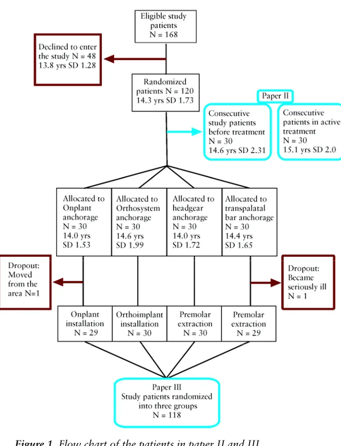
The study participants in papers II–IV were consecutively recruited from the Orthodontic Clinic in the Public Dental Service, Gävleborg County Council, Gävle, Sweden from January 2003 to March 2005. Figures 1 and 2 present flow charts of the patients in these studies.
Two groups of patients were selected for paper II: 30 consecutive patients (i.e allocated for paper III and IV) yet to begin treatment (19 girls and 11 boys, mean age 14.6 years, SD 2.31) and 30 con-secutive age-matched adolescent patients in active orthodontic treatment with fixed appliances (17 girls and 13 boys, mean age 15.1 years, SD 2.00).
Of the 168 patients invited to enter studies III and IV, 48 declined to participate. Thus, 120 patients (mean age 14.3 years, SD 1.73) were randomized to receive one of four anchorage systems; each group contained 15 girls and 15 boys. Paper III compared the two osseointegrated anchorage groups (Onplant and Orthosystem im-plant) with the Premolar extraction group, which comprised study patients allocated to the Headgear group and Transpalatal bar group.
All patients met these inclusion criteria: healthy, non-smoking adolescents in need of orthodontic treatment; no previous experi-ence of orthodontic treatment; permanent dentition; no transversal discrepancies; treatment plan involving extraction of two maxillary premolars (most patients also needed two premolars in the lower jaw extracted) followed by fixed appliances in both jaws; and addi-tional anchorage on the upper first molars considered necessary. The
study sample involved patients with large overjets and patients with crowding; need for additional anchorage varied from moderate to maximum (Figure 3). Treatment objectives for each patient were considered to be reachable with all four techniques.
The 48 patients who declined to participate comprised 26 boys and 22 girls (mean age 13.8 years, SD 1.28) and were not signifi-cantly different concerning gender and age compared to those who entered the study. The main reasons for declining were either fear of the surgical procedures or reluctance to wear headgear. After ran-domization but before treatment start, one patient from the Onplant anchorage group moved from the area and one patient in the Transpalatal bar group became seriously ill and dropped out of the study. These two patients were excluded because they did not start the allocated intervention. The study sample in papers III and IV thus comprised 118 patients.
Ethical considerations
The ethics committee of Uppsala University, Uppsala, Sweden, which follows the guidelines of the Declaration of Helsinki, ap-proved the informed consent form and study protocol.
Consent and randomization
Before studies II, III, and IV, two orthodontists provided the patients and parents with oral and written information of details about the study.After written consent was obtained from the patient and par-ent, the patients were randomized in blocks of four into one of four anchorage groups. The randomization process per se distributes sub-ject characteristics equally between groups, but since more girls than boys requested and received orthodontic treatment in Gävle, it was decided to stratify for gender so that each group would contain an equal number of girls and boys. The allocation sequence was com-puter generated by a statistician at Gävleborg County Hospital, Gävle, Sweden and concealed in envelopes until randomization.
29
Figure 3a. Study patient number 16 with severe crowding in the
up-per jaw before treatment.
Figure 3b. Study patient number 61 with 10mm overjet before
treatment
29 Figure 3a. Study patient number 16 with severe crowding in the up-per jaw before treatment.
Figure 3b. Study patient number 61 with 10mm overjet before treatment
Figure 3a. Study patient number 16 with severe crowding in the
up-per jaw before treatment.
Figure 3b. Study patient number 61 with 10mm overjet before
treatment
Figure 3a. Study patient number 16 with severe crowding in the
up-per jaw before treatment.
Figure 3b. Study patient number 61 with 10mm overjet before
treatment
29 Figure 3a. Study patient number 16 with severe crowding in the
up-per jaw before treatment.
Figure 3b. Study patient number 61 with 10mm overjet before
METHODS
Paper I
To identify studies that examined orthodontic anchorage systems or applications and their effectiveness, a survey of the Medline data-base (Entrez Pub Med, www.ncbi.nim.nih.gov) and the Cochrane Collaboration Oral Health Group Database of Clinical Trials (www.cochrane.org) was conducted. The search covered the period from January 1966 to December 2004 and used the Medical Subject Heading (MeSH) term “orthodontics” combined with the term “an-chorage”.
The search included human studies written in English but ex-cluded in vitro studies and articles on surgical treatment or cleft lip and palate treatment. Study design requirements were RCTs, pro-spective and retropro-spective controlled studies, and clinical trials that compared at least two anchorage applications and reported quanti-tative data on the effects of various anchorage devices (anchorage loss). Two reviewers independently assessed all of the articles with respect to the inclusion and exclusion criteria, and any inter-examiner conflicts were resolved by discussion to reach consensus.
Data were extracted on these items: author, year of publication, study design, material, gender and age, treatment time, anchorage unit used, ratio between anchorage loss and active movement. To document the methodological soundness of each article, quality was evaluated using a modification of the method described by Antczak et al.86 and Jadad et al.87 These eight variables were evaluated: study design (RCT = 3 points; prospective study = 1 point; retrospective study = 0 points); adequate sample size (1 point); adequate selection description (1 point); valid measurement methods (1 point); use of method error analysis (1 point); blinding in measurement (1 point); adequate statistics (1 point); no confounders included in analysis (1 point). Of the eight variables, a study could score a maximum of 10 points; study quality was categorized as low (0–5 points), medium (6–8 points), or high (9–10 points).
Several articles have been published since December 2004. So pa-per I was supplemented with a new survey from December 2004 to
July 2007 using the same search strategy, selection criteria, and quality evaluation.
Paper II
Development of the questionnaire
A new questionnaire was developed to assess adolescent patients’ opinions and experiences of orthodontic treatment. To ensure that the questionnaire items were relevant, four focus groups were formed: three with four age-matched adolescent patients each who had recently completed active orthodontic treatment and a fourth group with four parents of patients who had completed treatment. Two investigators conducted interviews using an open-ended inter-view style and collected 4 hours of audio-taped information. The participants were asked to describe why they had sought orthodon-tic treatment and how they had experienced the various phases of orthodontic treatment such as insertion of the fixed appliance, the first week of wearing braces, changes of arch wires, and so on. Spe-cific questions about various anchorage systems and experiences from premolar extractions and surgical procedures, mainly exposure of impacted upper canines, were also included.
Transcripts of the audiotapes were analyzed and used when the questionnaire was being constructed. Information from the four fo-cus groups was similar. Pain and soreness, difficulties when eating specific foods together with concern about facial appearances when wearing braces were important issues, but most patients stated that they “got used to it” and “relied strongly on the clinician’s profes-sional judgment”. Most patients also claimed that they had made an independent decision to undergo orthodontic treatment, although mostly after advice from clinical dental staff: “they should, of course, know what is best”.
The questionnaire comprised 46 questions divided into five sepa-rate domains; the questions were based on results of the focus group interviews and on other questionnaires (Appendix, Table A).11, 94, 95
Questionnaire
All patients in the group that was to enter orthodontic treatment were instructed to assess all 46 questions while the 30 patients
al-ready in active treatment assessed the questions that pertained to treatment (questions 12–42, 45, and 46).
The questionnaire comprised five domains:
Treatment motivation: This domain contained 7 questions (1–7) as-sessed on a VAS with the end phrases "not at all" and "very much" or "not at all" and "completely".
Treatment expectations: This domain contained 4 questions (8–11) assessed on a VAS with the end phrases “not at all” and “very much”.
Pain and discomfort from teeth, jaws, and face: This domain con-tained 13 questions (12–24): 10 questions on a VAS with the end phrases “none at all” and “worst imaginable” and 1 question on a 2-point scale (yes or no) with 2 follow-up questions.
Functional jaw impairment: This domain, a freestanding question-naire, has previously been used in other populations,95 but not for regular orthodontic patients, and contained 18 questions (25–42). Eight were related to mandibular function, 3 to psychosocial activi-ties, and 7 to eating specific foods. Each question was assessed on a 4-point scale with the alternatives not at all, slightly, much, or ex-tremely difficult.
Questionnaire validity: This domain contained 4 questions (43–46), one for each domain, assessed on a VAS with the end phrases "not at all" and "very well".
Reliability and Validity
In paper II, the reliability of the self-reported base-line questionnaire was determined in a test-retest96 study by administering the ques-tionnaire on two separate occasions at an interval of 1–2 weeks. Re-liability was tested on all individual questions and for the separate domains within the questionnaire. Internal consistency,97 which characterizes the homogeneity of the separate questions and ex-presses how well the separate questions within each domain relate, was also evaluated. Face validity was established by asking the pa-tients if they considered the items were relevant and reflected their motivation for orthodontic treatment and their expectations and ex-perience of orthodontic treatment.
Paper III
Surgical placement and premolar extractions
All Onplants (Nobel Biocare, Göteborg, Sweden) and Orthosystem implants (Institut Straumann AG, Basel, Switzerland) were placed according to standard surgical procedures under local anesthesia (see paper III for details). Onplants and Orthosystem implants were placed para median but close to the midline in the anterior palate at the level of the premolars. A guide stent was used to help position the Orthosystem implant. After surgical placement of the Onplants, a trimmed, vacuum-formed stent was placed over the surgical site to prevent hematoma formation and facilitate adaptation of the On-plant to the bone surface. Patients wore the stent 24 hours a day for 1 week. Orthosystem implant patients received a similar stent, which they wore 24 hours a day for 2 weeks to protect the implant from parafunctional activity from the tongue.
Fifty-one patients in the premolar extraction group had one maxil-lary and one mandibular premolar extracted on one occasion and maxillary and mandibular premolars on the other side extracted 1-2 weeks later. Eight patients had two premolars in the upper jaw ex-tracted during one session. Two maxillofacial surgeons at the Maxil-lofacial Unit, Public Dental Service, Gävleborg County Council, Gävle, Sweden, performed the surgery and premolar extractions.
Questionnaires
The questionnaires included self-report items developed in paper II and a few questions modified for this study (Appendix, Table B,C).
Patients’ perception of pain intensity and discomfort and analgesic consumption were evaluated on the first evening and 1 week after surgical placement of an Onplant or an Orthosystem implant. The premolar extraction group was evaluated on the first evening after the first two extractions and 1 week after the last premolars were extracted (Appendix, Table B). The questionnaire that was adminis-trated 1 week after the surgical interventions and premolar extrac-tion had addiextrac-tional quesextrac-tions about daily activities and funcextrac-tional jaw impairment (Appendix, Table C).
Paper IV
All patients received orthodontic appliances based on McLaughlin, Bennett, Trevisi´s straight-wire concept98 with a .022-inch slot size and designed for light forces. Leveling and aligning were carried out with laceback ligatures, coil springs, or both depending on the de-gree of crowding with this arch wire sequence: .016-inch Heat-Activated Nickel-Titanium (HANT), .018-inch stainless steel (SS), and .019 x .025-inch HANT. Spaces were closed with active tie-backs and .019 x .025-inch SS arch wires. The leveling/aligning phase was defined as the time in months from treatment start until insertion of a .019 x .025-inch SS arch wire while the space closure phase was the time in months from the end of the leveling/aligning phase until extraction spaces were closed or (in cases with planned anchorage loss) until anterior space closure with a correct Class I occlusion canine relationship was established. Two orthodontists at the Orthodontic Clinic, Gävleborg County Council, Gävle, Sweden performed the orthodontic treatments.
Anchorage systems
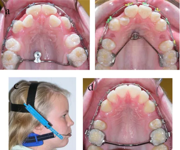
Nobel Biocare’s Onplant system is a subperiosteal implant in the shape of a 7.7-mm titanium disc. The disc is coated with a layer (75 µm) of bioactive hydroxyapatite to help osseointegration. The disc is slid through a subperiosteal tunnel into a position near the palatal midline via a surgical procedure under local anesthesia. After a heal-ing period of 16 weeks, an abutment was connected to the Onplant under local anesthesia and a 1.3-mm SS transpalatal bar was fabri-cated and bonded to the maxillary first molars (Figure 4a).
The Orthosystem implant is a short titanium implant (3.3 x 4 mm) that was inserted under local anesthesia near the midline of the pal-ate. After a healing period of 16 weeks, a 1.2-mm SS transpalatal bar was fabricated, connected to the implant, and bonded to the maxillary first molars (Figure 4b).
Headgear consisted of bands on the maxillary first molars and a short outer bow with the direction of force (400 g) corresponding to medium pull (Figure 4c). Force was checked on each visit to the clinic, and adjustments were made when necessary. Patients were in-structed to wear the headgear 10–12 hours a day.
The transpalatal bar consisted of bands on the maxillary first mo-lars with a 2.0 x 1.0-mm soldered SS bar. There was a 2-mm space between the bar and the palate (Figure 4d).
Figure 4. Occlusal view of the Onplant bar (a) and Orthosystem
implant bar (b). Lateral view of the headgear anchorage (c) and occlusal view of the transpalatal bar (d).
Outcome measures
The main outcome measures that were assessed after the level-ing/aligning (T1) and space closure (T2) phases were:
• Position of the maxillary first molars and distance moved • Position of maxillary central incisors and distance moved • Skeletal sagittal position of the maxilla
• Treatment time of each phase
Data on all patients who were randomly assigned to the four groups were analyzed on an intention-to-treat (ITT) basis. Thus, as soon as the anchorage system had been placed, all results—
a
regardless of outcome—were analyzed. Successful anchorage was defined as an anchorage loss of ≤ 1 mm, no osseointegration failures or failures during anchorage system placement, and no dropouts af-ter treatment start (insertion of anchorage).
Analysis of lateral head radiographs
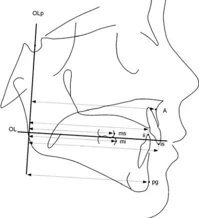
Lateral head radiographs in habitual occlusion were made at base-line (T0), after completion of the leveling/aligning phase (T1), and after the space closure phase (T2). Measuring points, reference lines, and measurements used were based on those defined and described by Björk99 and Pancherz.100, 101 Dental and skeletal changes as well as dental changes within the maxilla and mandible were determined by Pancherz SO analysis (analysis of changes in sagittal occlusion, Fig-ure 5).
Figure 5. SO-analysis according to Pancherz.
Measurements were made by hand to the nearest 0.5 mm or 0.5 de-grees. Images of bilateral structures were bisected. No correction was made for linear enlargement (10%). Changes in the various measuring points during treatment were calculated as differences in the after-minus-before positions. One orthodontist (Dr Feldmann)
made all measurements. Blinding during measurement was not pos-sible since the four anchorage systems were easily recognizable on the lateral cephalograms.
Statistical analysis
Inter-rater agreement between reviewers in assessing articles with respect to inclusion and exclusion criteria and quality evaluations in paper I was assessed with the kappa statistic (Cohen’s kappa, κ). Kappa values > 0.80 were considered excellent, 0.61–0.80 good, 0.41–0.60 moderate, 0.21–0.40 fair, and ≤ 0.20 poor.102
Test-retest reliability: Test-retest reliability in paper II was assessed by the kappa statistic when the questionnaire variable was measured on an ordinal or dichotomous scale. Kappa scores were supple-mented with percentage of total agreement.
When the questions were evaluated on a continuous scale or when summary scores for questionnaire domains were measured in paper II, reliability was assessed by calculating the intraclass correlation coefficient (ICC) based on a two-way mixed analysis of variance (ANOVA). This is an estimate of the precision in the data obtained by multiple measurements, relating the amount of measurement er-ror to subject variability. An ICC above 0.75 indicates excellent reli-ability, between 0.4 and 0.75 fair to good relireli-ability, and below 0.4 poor reliability.103
Internal consistency: In paper II, Cronbach’s alpha (α) 104 was cal-culated to estimate how consistently the subjects responded to the separate questions within each domain. Alpha values 0.70 or higher were considered sufficient.
Descriptive statistics including median value, interquartile range, and range were calculated for each variable in paper III, and the arithmetic mean and standard deviation (SD) were calculated for each variable in paper IV.
Differences between groups for pain and discomfort in paper III were tested with the nonparametric Kruskal-Wallis and Mann-Whitney tests. Chi-square tests were used to determine differences between groups in functional jaw impairment, affected daily activi-ties, and use of analgesics and, in paper IV, for success rate of an-chorage capacity.
Differences in means within groups in paper IV were tested by one-way ANOVA. A repeated measure ANOVA with a Bonferroni correction was used to analyze differences between groups at T0, T1, and T2.
Differences in papers III and IV with a P value less than 5 % (P<0.05) were considered statistically significant.
In paper IV, a sample size calculation was performed and based on an alpha significance level of 0.05 and a beta of 0.1 to achieve 90 % power to detect a clinically meaningful difference of 1.5 mm (SD 1.5) in anchorage loss between the four groups. The calculation revealed that 21 patients in each group were sufficient, but to com-pensate for conceivable dropouts during the trial, 30 patients were enrolled in each group.
Method error: In paper IV, 20 randomly selected cephalograms were traced on two separate occasions. No significant mean differ-ences between the two series of records were found using paired t-tests. The method error105 did not exceed 0.5 mm or 1.0 degree ex-cept for molar inclination where the error did not exceed 1.5 de-grees.
RESULTS
Paper I
The search strategy yielded 494 articles, but only 14 articles19,106-118 fulfilled the inclusion/exclusion criteria. The most common reasons for exclusions were case-report or small case-series, technical presen-tations and articles with objectives that did not follow this review (for details see paper I). Two main anchorage situations were found, anchorage of molars during space closure after premolar extractions and anchorage in the incisor and/or premolar region during distal movement of molars.
Inter-rater agreement was excellent for inclusion/exclusion assess-ment and good to excellent for data extraction and judgassess-ment of the selected articles’ quality.
Anchorage effectiveness of molars during space closure
Seven studies remained for the final analysis. Two were RCTs,113,118 two prospective comparative “split-mouth” studies,108,109
and three retrospective comparative studies.106,107,114 Various techniques and auxiliary holding appliances were used for either active movement or anchorage. The two RCTs reported conflicting results. Usmani et al.113 reported no difference in anchorage loss of molars during level-ing of the upper jaw with or without laceback ligatures (0.49 mm and 0.50 mm, respectively) while Irvine et al.118
demonstrated a sig-nificantly larger anchorage loss when laceback ligatures were used for leveling the lower jaw (0.75 mm and -0.08 mm, respectively). The other five studies106-109,114
revealed vast heterogeneity, and it was therefore difficult to value the efficiency of various anchorage sys-tems.
Anchorage effectiveness during distal movement of molars
The primary concern in the seven studies was to demonstrate distal molar movement, and the secondary concern was to show anchor-age loss. One study was an RCT,112
two studies were prospective comparative studies,111,115 one study was a retrospective controlled study,110 and three were retrospective comparative studies.19,116,117 A Nance or a modified Nance appliance was the main method of an-chorage during the intraoral distalization procedure while the active unit varied. Anchorage loss, measured at the incisors or premolars, varied from 0.2 to 2.2 mm, and the anchorage loss/distal molar movement ratio ranged from 0.2 to 0.8.
Study quality
Research quality and methodological soundness were high in two studies,113,118 medium in three studies,19,110,112 and low in nine stud-ies.106-109,111,114-117
The most obvious shortcomings were retrospective study design with inadequate selection description and small sample sizes, which mostly implied low power. In all studies, the methods used to detect and analyze anchorage loss and active tooth move-ments were valid and generally well known. But nine studies did not include a method error analysis, and only three studies used blinding in measurements. Moreover, three studies did not consider the risk of confounding factors influencing the outcome.
New literature search
In a supplementary literature search between December 2004 and July 2007, another 257 articles on orthodontic anchorage were found. Eleven119-129 studies merited final analysis, and the two main anchorage situations found in the primary search - (1) molar chorage during space closure after premolar extraction and (2) an-chorage in incisal and premolar regions during distal movement of molars - were the same in these studies.
Data of the four new articles on molar anchorage during space closure are summarized in Appendix, Table D. Three were prospec-tive comparaprospec-tive “split mouth” studies120-122 and one study was retro-spective.119
Seven articles on anchorage during distal movement of molars are listed in Appendix, Table E. Three studies were
prospec-tive, comparative125,127,129
and four were retrospective.123,124,126,128
The Nance button was still the most frequently used anchorage unit dur-ing molar distalization.
Quality assessments of the eleven articles are presented in Appen-dix, Table F. Five studies were of medium and six of low quality. The main weaknesses were retrospective study design, small sample sizes and inadequate selection description in combination with lack of blinding during measurement.
Skeletal anchorage
The first literature search up to December 2004 contained 149 arti-cles (out of 494) on skeletal anchorage with various implant sys-tems. Since most of the implant studies were technical presentations of new approaches and/or small case series, they did not qualify for the final analysis and were thus excluded. The expanded search up to July 2007 contained an additional 176 (of 257) articles, and two of them qualified for the final analysis. The prospective comparative study by Thiruvenkatachari121 reported absolute anchorage (i.e. an-chorage loss = 0 mm) with micro-implants during canine retraction after premolar extractions in comparison with a control group without any auxiliary anchorage unit. The study comprised just 10 patients and was overall judged to be of low quality. Gelgor129
com-pared the anchorage of a transpalatal bar system and a modified Nance, both enhanced by intraosseous screws, during molar distali-zation. Anchorage loss for the transpalatal bar was 0.5 mm and neg-ligible for the modified Nance (0.1 mm).
Paper II
Reliability and validity
All 60 patients filled in the questionnaire twice at an average inter-val of 12 days.
Reliability of the questionnaire, based on summary scores, was ex-cellent (ICC = 0.85–0.92) for all five domains. Differences in do-main reliability between the group of patients yet to enter treatment and those already in treatment were minor. But a discrepancy for the treatment motivation domain was observed between girls and boys (good and excellent, respectively).
42
Overall, reliability of the separate questions within each domain was good to excellent. But three questions (6, 18, and 19) had poor reliability and two questions (8 and 15) fair reliability. Figure 6a,b presents questions 18 and 19. In the functional jaw impairment do-main, question 31 and 34 exhibited fair reliability and question 27, 29, and 35 moderate reliability (� = 0.58, 0.52, and 0.48). But these questions were considered acceptable since percent total agreement was comparable with the other questions in this domain. Internal consistency for the five separate domains varied between 0.67 and 0.87 at the first assessment and between 0.63 and 0.94 at the second assessment, which implies that consistency was sufficient for all do-mains.
Figure 6a. Plot of first and second assessment for the question 18; “
Do you have pain from your molars when they are in contact?”
Figure 6b. Plot of first and second assessment for the question 19; “
Do you have pain from your molars when they are not in contact?”
42
Overall, reliability of the separate questions within each domain was good to excellent. But three questions (6, 18, and 19) had poor reliability and two questions (8 and 15) fair reliability. Figure 6a,b presents questions 18 and 19. In the functional jaw impairment do-main, question 31 and 34 exhibited fair reliability and question 27, 29, and 35 moderate reliability. But these questions were considered acceptable since percent total agreement was comparable with the other questions in this domain. Internal consistency for the five separate domains varied between 0.67 and 0.87 at the first assess-ment and between 0.63 and 0.94 at the second assessassess-ment, which implies that consistency was sufficient for all domains.
Figure 6a. Plot of first and second assessment for the question 18;
“Do you have pain from your molars when they are in contact?”
Figure 6b. Plot of first and second assessment for the question 19; “
Do you have pain from your molars when they are not in contact?”
Overall, reliability of the separate questions within each domain was good to excellent. But three questions (6, 18, and 19) had poor reliability and two questions (8 and 15) fair reliability. Figure 6a,b presents questions 18 and 19. In the functional jaw impairment do-main, question 31 and 34 exhibited fair reliability and question 27, 29, and 35 moderate reliability (� = 0.58, 0.52, and 0.48). But these questions were considered acceptable since percent total agreement was comparable with the other questions in this domain. Internal consistency for the five separate domains varied between 0.67 and 0.87 at the first assessment and between 0.63 and 0.94 at the second assessment, which implies that consistency was sufficient for all do-mains.
Figure 6a. Plot of first and second assessment for the question 18; “
Do you have pain from your molars when they are in contact?”
Figure 6b. Plot of first and second assessment for the question 19; “
Do you have pain from your molars when they are not in contact?”
42
Overall, reliability of the separate questions within each domain was good to excellent. But three questions (6, 18, and 19) had poor reliability and two questions (8 and 15) fair reliability. Figure 6a,b presents questions 18 and 19. In the functional jaw impairment do-main, question 31 and 34 exhibited fair reliability and question 27, 29, and 35 moderate reliability (� = 0.58, 0.52, and 0.48). But these questions were considered acceptable since percent total agreement was comparable with the other questions in this domain. Internal consistency for the five separate domains varied between 0.67 and 0.87 at the first assessment and between 0.63 and 0.94 at the second assessment, which implies that consistency was sufficient for all do-mains.
Figure 6a. Plot of first and second assessment for the question 18; “
Do you have pain from your molars when they are in contact?”
Figure 6b. Plot of first and second assessment for the question 19; “
Very high scores (Md 80–94) were obtained at the VAS assess-ment for face validity, which indicates that the patients considered the questions to be valid, and since the questions originated from the focus group interviews, the questionnaire also had content valid-ity.130
Paper III
The response rates for the questionnaires the first evening after the interventions were 97%, 93%, and 98% and after 1 week 90%, 90%, and 100% for the Onplant, the Orthosystem implant, and the Premolar extraction groups, respectively. Overall, gender differences were few.
Pain intensity, discomfort, and
analgesic consumption
Pain intensity was significantly higher the first evening after surgical placement of an Onplant (Md=38.5, P=0.002) and after premolar extraction (Md=29, P=0.007) compared to placement of an Ortho-system implant (Md=4). One week after the interventions, pain in-tensity was still significantly higher in the premolar extraction group compared to the Orthosystem implant group.
Discomfort was significantly higher the first evening after surgery in the Onplant group (Md=34.5, P=0.04) than after extraction in the Premolar extraction group (Md=22). The Onplant and Ortho-system groups were comparable concerning discomfort, but the Or-thosystem patients reported in an open-ended question that they had experienced drilling during surgery to be especially unpleasant. One week after the intervention, Orthosystem patients reported signifi-cantly less discomfort compared to the other two groups.
The protective stent caused more discomfort (Md=48, P=0.002) than did the actual surgery site (Md=13.5) in the Orthosystem group the first evening and in both groups 1 week after surgery.
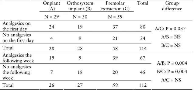
On the first day, significantly more patients in the Onplant group than in the Premolar extraction group took analgesics (P=0.037). In the week following intervention, analgesic consumption was signifi-cantly lower in the Orthosystem group compared to the other two groups (P=0.004). After all interventions, analgesic consumption on
the first day differed depending on gender (girls consumed more than boys, P=0.042).
Table I. Analgesic consumption on the first day and the following
week after surgical placement of an Onplant, an Orthosystem im-plant and premolar extractions.
Onplant (A) Orthosystem implant (B) Premolar extraction (C) Total Group difference N = 29 N = 30 N = 59 Analgesics on
the first day 24 19 37 80
No analgesics
on the first day 4 9 21 34
Total 28 28 58 114 A/C: P = 0.037 A/B = NS B/C = NS Analgesics the following week 19 9 39 67 No analgesics the following week 7 18 20 45 Total 26 27 59 112 A/B: P = 0.004 B/C: P = 0.004 A/C = NS NS = Not significant
Daily activities and functional jaw impairment
During the first week after the intervention, the Onplant group re-ported disturbed sleep more often than the Premolar extraction group (P=0.033). The Onplant and Orthosystem groups also re-ported more affected speech than the Premolar extraction group (P<0.001). Differences between groups in eating specific foods were significant, with the Onplant and Orthosystem groups being more inconvenienced than the Premolar extraction group, but with no ap-parent pattern.
Paper IV
This study comprised 118 patients. Five patients dropped out from the trial, but these patients were included and analyzed on an ITT basis as failures; thus, while 118 patients were included in the analy-sis, only 113 completed all phases of the study (Figure 2, Table II).
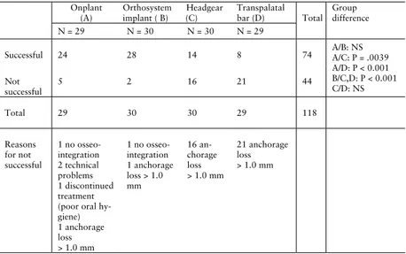
Table II. Distribution of success rate concerning anchorage capacity
for the Onplant group (A), Orthosystem implant group (B), head-gear group (C), and transpalatal bar group (D).
Onplant (A) Orthosystem implant ( B) Headgear (C) Transpalatal bar (D) N = 29 N = 30 N = 30 N = 29 Total Group difference Successful 24 28 14 8 74 Not successful 5 2 16 21 44 A/B: NS A/C: P = .0039 A/D: P < 0.001 B/C,D: P < 0.001 C/D: NS Total 29 30 30 29 118 Reasons for not successful 1 no osseo-integration 2 technical problems 1 discontinued treatment (poor oral hy-giene) 1 anchorage loss > 1.0 mm 1 no osseo-integration 1 anchorage loss > 1.0 mm 16 an-chorage loss > 1.0 mm 21 anchorage loss > 1.0 mm NS = Not significant
Because no significant differences in pretreatment cephalometric characteristics between the four groups or between girls and boys were found, data for girls and boys were pooled and analyzed to-gether.
The leveling/aligning phase
Maxillary molars within the maxilla in the Onplant, Orthosystem implant, and Headgear groups were stable during leveling and alignment. In the Transpalatal bar group, the molars on average moved forward 1.0 mm. Mesial movement of the molars (anchorage loss), which occurred in the Transpalatal bar group, was signifi-cantly different compared to the other three groups (P<0.001). The amount of mesial molar tipping was small in all four groups but sig-nificantly larger in the Transpalatal bar group (mean 4.1 degrees,
P<0.001).
45 Table II. Distribution of success rate concerning anchorage capacity for the Onplant group (A), Orthosystem implant group (B), head-gear group (C), and transpalatal bar group (D).
Onplant (A) Orthosystem implant ( B) Headgear (C) Transpalatal bar (D) N = 29 N = 30 N = 30 N = 29 Total Group difference Successful 24 28 14 8 74 Not successful 5 2 16 21 44 A/B: NS A/C: P = .0039 A/D: P < 0.001 B/C,D: P < 0.001 C/D: NS Total 29 30 30 29 118 Reasons for not successful 1 no osseo-integration 2 technical problems 1 discontinued treatment (poor oral hy-giene) 1 anchorage loss > 1.0 mm 1 no osseo-integration 1 anchorage loss > 1.0 mm 16 an-chorage loss > 1.0 mm 21 anchorage loss > 1.0 mm NS = Not significant
Because no significant differences in pretreatment cephalometric characteristics between the four groups or between girls and boys were found, data for girls and boys were pooled and analyzed to-gether.
The leveling/aligning phase
Maxillary molars within the maxilla in the Onplant, Orthosystem implant, and Headgear groups were stable during leveling and alignment. In the Transpalatal bar group, the molars on average moved forward 1.0 mm. Mesial movement of the molars (anchorage loss), which occurred in the Transpalatal bar group, was signifi-cantly different compared to the other three groups (P<0.001). The amount of mesial molar tipping was small in all four groups but sig-nificantly larger in the Transpalatal bar group (mean 4.1 degrees, P<0.001).
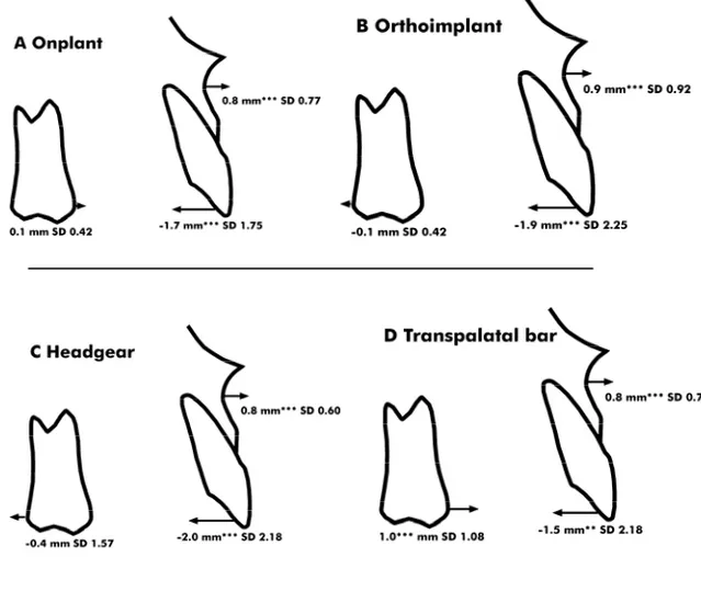
Figure 7a-d. Maxillary dental and skeletal changes (in mm) and
standard deviations (SD) contributing to alterations in the four groups during the levelling/aligning phase.
Average sagittal forward growth was 0.8–0.9 mm for the maxilla and 0.9–1.4 mm for the mandible. These changes were significant for all groups but with no differences between the four groups (Fig-ure 7a–d).
The space closure phase
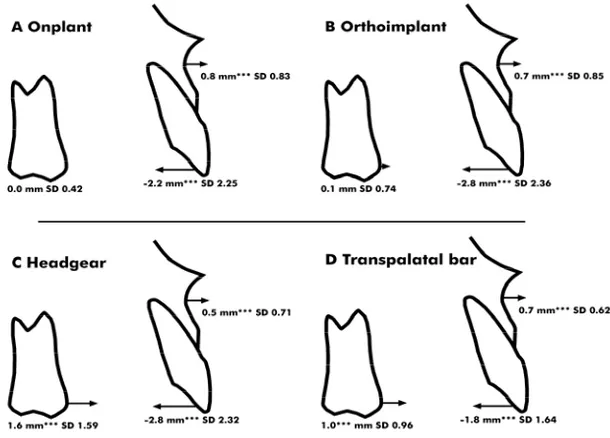
Maxillary molars in the two osseointegrated anchorage groups were stable during space closure. In the Headgear group the molars on average moved forward 1.6 mm (P<0.001) and in the Transpalatal bar group, anchorage loss continued and averaged another 1.0 mm (P<0.001). Mesial tipping of the molars during space closure was small and nonsignificant within and between the four groups.
Sagittal forward growth was 0.5–0.8 mm for the maxilla and 0.7– 0.9 mm for the mandible. These changes were significant for all groups but with no differences between the four groups (Figure 8a– d).
Figure 8a-d. Maxillary dental and skeletal changes (in mm) and
standard deviations (SD) contributing to alterations in the four groups during the space closure phase.
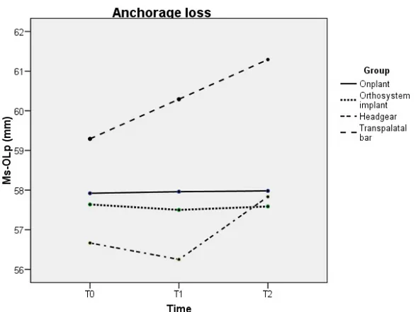
Total observation period
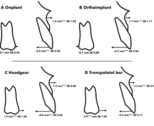
The maxillary molars were stable in the Onplant and Orthosystem groups, whereas they moved mesially in the Headgear and Transpala-tal bar groups, mean 1.2 and 2.0 mm, respectively (Figure 9). The an-chorage loss/incisor retraction ratio was 0.05 for the Onplant group, 0.02 for the Orthosystem group, 0.15 for the Headgear group, and 0.54 for the Transpalatal bar group (Figures 10a–d).
Figure 9. Maxillary upper first molar movements during
level-ling/aligning (T0-T1) and space closure (T1-T2) for all four groups.
Success of anchorage capacity
The Onplant and the Orthosystem implant had significantly higher success rates of anchorage than the headgear and transpalatal bar, and differences between the two osseointegrated groups and be-tween the two conventional groups were nonsignificant (Table II). Differences in success rate between the two orthodontists who per-formed the treatments were also nonsignificant.
In the Headgear group 14/30 patients had successful anchorage and ten of them were girls, which was a significant difference in comparison to boys (P = 0.028). There were no difference in mean
age between patients in the Headgear group with respect to success rate.
In the Transpalatal bar group there were no differences in gender, age, pre-treatment morphology or treatment time between the pa-tients who had successful anchorage (8/29) and those who had not.
Treatment time
Average treatment time for the leveling/aligning phase varied be-tween 8.0 and 8.7 months and for the space closure phase bebe-tween 8.6 and 9.7 months. Differences between the four groups were non-significant.
Figure 10a-d. Maxillary dental and skeletal changes (in mm) and
standard deviations (SD) contributing to alterations in the four groups during the total observation period.