Örebro University
School of Health and Medical Sciences Division of Clinical Medicine
Program: Biomedicine and Methods in Medical Diagnostics Experimental Medicine Course: Degree Project in Medicine
Date: 25th May 2016
Characterization of tick-born encephalitis and West
Nile virus non-structural 5 protein interactions with
host factors involved in immune evasion and cellular
apoptosis.
Author: Ngenya, Abdallah Athumani Supervisor: Samir Abdurahman, PhD Assistant Professor Inst. för Naturvetenskap Örebro Universitet
Abstract
Flaviviruses are human pathogens that cause large epidemics and tens of thousands of deaths annually in many parts of the world. They have a significant public health impact in different parts of the world. Organization of these viruses and their associated structural proteins has provided insight into the molecular transitions that occur during the viral life cycle and immune evasion. Many studies identify flaviviral RNA polymerase NS5 as potential therapeutic target. In this study the interaction between the viral NS5 protein of TBEV and WNV with the high temperature requirement protein A2 (HtraA2/Omi) have been assessed. Hela cells were cultured and transfected with plasmids (pKH3-WNS5-HA, pKH3-TNS5-HA, C1-DENVNS5, pEYFP-C1-WNVNS5, pEYFP-C1-TBEVNS5, pcDNA-HtrA2 – FLAG, pcDNA-HtrA2∆133 -GFP and pcDNA-OMP25-HA). Followed by co- immunoprecipitation and Immunoblotting. HtrA2/Omi was found to be toxic to cells. The study suggested that twelve hours post transfection was not good enough for co- immunoprecipitation. There was weak interaction between viral NS5 and HtrA2 in some membranes while other membranes there were no bands observed. Inconclusive and open results we had could be due to many factors such as pull down effects (buffers), antibodies and limited time we had.
Key words; Apoptosis, Culex, Flaviviruses, HtrA2/Omi, NS5, OMP25, Tick-borne Encephalitis, TBEV, Viruses, West Nile Virus,
1. Introduction
Protein-protein interactions are essential events that play an important role in a series of biological processes. Recent studies identify novel interactions between the NS5 of West Nile virus (WNV) and tick borne encephalitis and two mitochondrial proteins; the high temperature requirement protein A2 (HtraA2/Omi) and the outer membrane protein 25 (OMP25) [1]. Both Tick-borne encephalitis virus (TBEV) and West-Nile virus are arthropod-borne flaviviruses that have a major impact on global health for example climate change, lack of medical surveillance etc. have greatly contributed to the increase in flaviviral infections worldwide.
1.2 Flaviviruses
Flaviviruses belong to the family flaviviridae. Depending on the vector used for virus transmission flaviviruses are divided into three groups:
- Tick-borne (e.g. tick-borne encephalitis virus “TBEV”)
- Mosquito-borne (e.g. Yellow fever virus, Japanese encephalitis virus, Dengue viruses and West Nile virus)
- Not known Vector Flaviviruses (NKV) (e.g. Chimeric yellow fever virus 17D (YFV-17D) and dengue virus type 2 (DENV2) [2].
This study focused mainly on tick-borne encephalitis virus (TBEV) and West Nile virus (WNV).
1.3 Tick-borne encephalitis virus (TBEV)
Tick-borne encephalitis (TBE) is a central nervous system viral infection caused by TBEV. TBE is endemic in South-Eastern Sweden as well as in the Baltic regions, Central Europe and Russia [3,4]. The virus is transmitted by Ixodes ticks with high incidences of cases between the months of April and November due to high tick activity. [4,5]. An abundance of ticks containing a sufficient dose of infectious TBEV is a major factor contributing to the incidence of the disease. Studies on infected individuals showed that more than 70 % patients are asymptomatic or experience subclinical symptoms [6]. The remaining percentage of patients experience febrile illness that might progress to encephalitis with mortality rate reaching as high as 20-30% [7]. Usually those who progress to encephalitis are accompanied by symptoms such as sudden fever, nausea and vomiting, neck stiffness, headache, changes in mental state; such as confusion, drowsiness, or disorientation, seizures (fits) sensitivity to bright light (photophobia) and inability to speak. However, it has been reported that TBE can be successfully prevented by vaccine [4].
1.4 West Nile virus (WNV)
WNV which is vectored by Culex mosquito cycles between mosquitos and birds [8]. Some infected birds, can develop high-levels of the virus in their blood stream and mosquitos become infected by biting these infected birds. New birds then get infected through subsequent infected mosquito bites, and thus the spread. Mosquitos with WNV also bite and infect people, horses and other
mammals. However, humans, horses and other mammals are dead end hosts, which mean they do not develop high levels of viruses in their blood stream [9]. With incubation period of 2 to 14 days’ majority of humans infected with WNV usually have no or very mild symptoms [10]. Approximately only 20% of infected patients develop a febrile illness with malaise, myalgia, headache and lymphadenopathy [10]. Currently there is neither specific antiviral therapy nor licensed vaccine for treatment of humans.
1.5 Structure and Replication of Flaviviruses
Flaviviruses have a positive sense single stranded RNA genome (ssRNA (+) genome) of 10-11 kb which is capped in the 5’ end [1]. They have three structural and seven none structural (NS) proteins lined in the following order, C-prM-E-NS1-NS2A-NS2B-NS3-NS4A-NS4B-NS5 (fig. 1) that are required for replication in the host cell [1,11]. The NS5 is about 900 Amino acids long. It comprises the RNA cap methyltransferase (Mtase) domain at its N-terminus and an RNA‑dependent RNA polymerase domain (RdRp) at its C-terminal end [12]. Both of these enzymatic activities (Mtase and RdRp) form attractive targets for antiviral development [1,13]. Flaviviruses replication occurs at replication complexes (RCs) present within induced invaginations at the endoplasmic reticulum (ER) membrane, found in the cytoplasm [11]. However, not all flaviviruses RdRps localize to the cytoplasm. For example, in dengue and yellow fever viruses the NS5 protein is primarily localized within the nucleus [1,11]. The rationalization for a dengue and yellow fever RdRp localizing to the nucleus when its actual enzymatic functions in the virus life cycle are required in the cytoplasm is currently unknown [1].
Figure 1. The flaviviral genome with 1 open reading frame (ORF) that translates into a polyprotein, which is cleaved into 3 structural and 7 nonstructural proteins. (Taken from: Bidet K, 2014 [ 13])
1.6 Apoptosis
Apoptosis is a programmed cell death that occurs in multicellular organisms. Inhibitors of apoptosis (IAP) are a generation of proteins that are involved in cell cycle, immunity, inflammation, death and cell migration [14]. IAPs were first discovered in 1993 in baculoviruses as proteins were able to protect insect cells from apoptosis [14,15].
I- High temperature requirement protein A2 (HtrA2/Omi)
The high temperature requirement protein A2 also known as Omi (HtrA2/Omi) is a mitochondrial serine protease that is released into the cytosol during apoptosis to antagonize IAPs [16]. When released to the cytosol HtrA2/Omi contributes to caspase and finally cause cell death [17]. The unprocessed form of (HtrA2/Omi) is attached to the inner mitochondrial membrane through N-terminal of transmembrane domain while the more processed form HtrA2 is found in the cell nucleus [16]. A study was done where by overexpression of HtrA2/Omi was found to sensitize cell apoptosis while the removal of endogenous HtrA2/Omi proteins from cultured cells by RNA interference showed reduced cell death [14,16]. However, apart from inducing apoptosis or apoptotic like features the protease activity of the HtrA2/Omi is required for mitochondrial homeostasis [18].
2. Aim
Many studies have pinpointed NS5 as a key protein involved during viral RNA translation, replication and pathogenesis. However, the physical interaction and the molecular biology of NS5 are yet to be clearly understood. In this study the interaction between the viral NS5 protein of TBEV and WNV with the high temperature requirement protein A2 (HtraA2/Omi) have been assessed.
3. Material and methods
3.1 PlasmidsDuring the study period, the following plasmids were used pKH3-WNS5, pKH3-TNS5, pEYFP-C1-DENVNS5, pEYFP-C1-WNVNS5, pEYFP-C1-TBEV NS5, pcDNA-HtrA2, pcDNA-HtrA2 ∆133 and pcDNA-OMP25. The plasmids were tagged either a triplicate sequence of influenza hemagglutinin (HA), the FLAG peptide fusion consisting of eight amino acids, an enhanced yellow-green variant of the Aequorea Victoria green fluorescent protein (GFP) or just GFP. A schematic figure of each plasmid used in the study are shown in Figure 2.
Fig. 2; Schematic representation of the plasmids involved in the study
3.2 cell culture
Henrietta Lacks, (HeLa cells) were cultured in 25cm2 flasks (Sarstedt, Nümbrecht). Cells were maintained in DMEM (Gibco, Life Technologies) supplemented with 10% fetal bovine serum (Gibco, Life Technologies) and 3% antibiotics [Penicillin (0.1U/ml) + Streptomycin (100ng/ml)-PEST, Gibco, Life Technologies]. The cultures were kept at 37°C and 5% CO2 environment and
3.3 Transfection
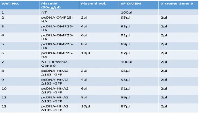
A six and twelve well culture plates (Sarstedt, Nümbrecht) were used for transfection. Since the mitochondrial proteins are toxic [14,16] the first transfection was done to identify the optimal concentration of OMP25 and HtrA2 encoding plasmids (Fig. 3). Hela (1 x 10⁵ cells / well) were seeded in a 12 well culture plate containing 2ml of growth medium DMEM (Gibco, Life
Technologies) supplemented with 10% fetal bovine serum (Gibco, Life Technologies) and 3%
antibiotics [Penicillin (0.1U/ml) + Streptomycin (100ng/ml)-PEST, (Gibco, Life Technologies)] followed by overnight incubation at 37°C and 5% CO2 environment. The following day cells were
transfected followed by 72h’s, incubation at 37°C and 5% CO2. X-tremeGENE9 DNA transfection
reagent (Roche diagnostics) was used for all transfections.
Table 1. The first Transfection plan. Abbreviations: NT (not transfected), SF-DMEM (serum free medium)
Trypan blue exclusion tests were performed to test cells viability [19]. Transfection reagent used was X-tremeGENE9 DNA Transfection reagent (Roche diagnostics). Second transfection was done as describe previously but this time a six well culture plate was used. It was done using the
optimal concentration of pcDNA-HtrA2 was identified (fig. 3). Cells were harvested 24h, 48h, and 72h post transfection.
Table 2. The second Transfection plan. Abbreviations: Not transfected, SF-DMEM (serum free medium)
Cell harvesting was done in cold environment. Culture plates and cell lysates were kept in an ice box during whole period of cell lysis and harvesting [20]. Cold PBS (137 mM NaCl, 2.7 mM KCl, 4.3 mM Na2HPO4, 1.47 mM KH2 PO4, pH 7.4) was used for washing. RIPA buffer (5M NaCl,
0.5M EDTA, pH 8.0, 1M Tris, pH 8.0, NP-40 [IGEPAL CA-630],10% sodium deoxycholate,10% SDS, dH2O) was prepared and supplemented with protease inhibitor (Roche diagnostics). 400μl
of RIPA supplemented with cocktail of protease inhibitor was added to each well and incubated on a shaker table for 15minutes. Lysates were transferred to micro centrifuge tubes followed by centrifugation at 4°C for 20 minutes at maximum speed. Precipitates (pellet) were discarded; 75μl of clear lysates were mixed with 25μl of Lameli buffer (% SDS, 20% glycerol, 10% 2-mercaptoethanol, 0.004% bromophenol blue and 0.125M Tris HCl, pH approx. 6.8). The mixtures were heated at 92°C for 5min and then stored at -20°C until used for western bloating. The remaining lysates were also at stored for immunoprecipitation at -20°C.
3.4. Western Bloating
During the study period western blot technique was performed several times for different purposes after different procedures. As per following reasons the first blot was done in order to confirm protein expression in the lysates while the second blot was done after streptavidin pull down and
the third blot was done after co-immunoprecipitation (Co-IP). Equal amounts of protein (20μg) were loaded into the wells of a mini (8cm x 8cm) format SDS-PAGE (10% Bis-Tris Plus Gels [Life Technologies]), along with molecular weight markers. The electrophoresis voltage used was 100V DC for 84minutes. Dry blotting system iBlot 2 (Life Technologies) was used for protein transfer to membrane for 12 minutes. 5% none-fat dry milk- TBST solution was used for blocking membrane for at least 30 minutes. The primary antibodies used were Mouse HA, Rabbit Anti-GFP (Abcam) and mouse anti-Flag (Sigma–Adrich). Secondary antibodies used were anti mouse IgG-HRP whole Ab and anti-rabbit IgG-HRP whole Ab (GE Health care). 1.5ml super signal western blot enhancer (Life Technologies) was used per membrane. Odyssey ® CLx Imaging System (LI-COR Biosciences) with Image Studio™ software was used for taking pictures and membrane analysis.
3.5. Immunoprecipitation (A) Streptavidin Pull down
Dynabeads M-280 streptavidin (life technologies) was used to co-purify proteins using biolinylated antibody. The Dynabeads were washed using wash buffer (10 mM Tris-HCl (pH 7.5) 1 mM EDTA 2M NaCl). Tubes containing dynabeads and equal amount of wash buffer were mixed, placed briefly on a magnet and the supernatant were discarded. This purification method was repeated five times. Dynabeads were then added into the cell lysates supplemented with Anti-HA-Biotin, High Affinity (3F10) (Sigma Aldrich) followed by overnight incubation on rotator at 4°C. Following overnight incubation western blot was run as described previously, however this time was loaded with equal amounts of protein 8μg into 15 wells of mini (8cm x 8cm) SDS-PAGE. Mouse anti-GFP (Santa Cruz) was used for detection. Secondary anti body used was anti mouse IgG-HRP whole Ab (GE Health care).
(B) Co-immunoprecipitation (Co-IP)
12h and 24h post transfection lysates were used. Primary antibody used was mouse anti-HA, rabbit anti-GFP (Abcam) and mouse ant-FLAG (Sigma Aldrich). Plasmids were immunoprecipatated using anti-HA, anti-GFP and anti-Flag (Sigma Aldrich).
Table 3 – Tagged protein immunoprecipitation plan
40μl Protein A/G-agarose beads (Santa Cruz Biotech) per sample were prepared by centrifuging for 1 min at 12,000 x g and washed twice with 1ml RIPA buffer at 4°C. Washed protein A/G-agarose beads were then added to the overnight incubated samples (Table 3) followed by 2h incubation at 4°C. After incubation the beads were span down and washed five times with 1ml RIPA buffer.45μl of 1 x lameli buffer (% SDS, 20% glycerol, 10% 2-mercaptoethanol, 0.004% bromophenol blue and 0.125M Tris HCl, pH approx. 6.8) were added into the samples then heated at 96°C for 5minutes and spanned down ready for immunoblotting. Western blot was done as described previously by loading equal amount of samples 20μl from immunoprecipitation into the 15 wells of mini (8cm x 8 cm) format SDS-PAGE. Samples immunoprecipatated with anti-HA and anti-GFP were detected with mouse anti-HA and rabbit anti-GFP (Abcam) respectively. While samples immunoprecipatated with anti-FLAG were detected with mouse anti-FLAG and vice versa. Secondary antibodies used were anti mouse IgG-HRP whole Ab and Anti Rabbit IgG- HRP whole Ab (GE Health care).
4. Results
Figure 3. is a trypan blue staining of; (1) OMP25 negative control (2) Transfected with 2μl of OMP25 (3) Transfected with 4μl of OMP25 (4) Transfected with 6μl of OMP25 (5) Transfected with 8μl of OMP25 (6) Transfected with 10μl of OMP25 (7) HtraA2 negative control (8) Transfected with 2μl of HtraA2 (9) Transfected with 4μl of HtraA2 (10) Transfected with 6μl of HtraA2 (11) Transfected with 8μl of HtraA2 (12) Transfected with 10μl of HtraA2. 20 x magnification.
OMP25 Negative control Control
OMP25(100ng/μl) OMP25 (200ng/μl) OMP25 (300 ng/μl)
OMP25 (500 ng/μl)
OMP25 (400 ng/μl) Neg Control with
transfection reagent
HtRA2(100ng/μl)
HtRA2 (200ng/μl) HtRA2 (300 ng/μl) HtRA2 (400 ng/μl) HtRA2 (500 ng/μl)
1 2 3 4
5 6 7 8
Fig 5. Co-expression of HA and GFP-tagged various NS5 clones with Flag or GFP tagged HtrAΔ133 in HeLa cells. Cells were harvested 24, 48, and 72 hrs. post-transfection and detected with rabbit anti-GFP antibody.
Fig 4. Expression of various NS5 clones in HeLa cells. Cells were harvested 24, 48, and 72 hrs. post-transfection and detected using mouse monoclonal anti-HA(upper) and rabbit polyclonal anti-GFP antibodies (lower).
Fig 6. Co-expression of various NS5 clones with FLAG-tagged HtrA2 construct in HeLa cells. Cells were harvested 24, 48, and 72 hrs. post-transfection and detected with mouse anti-FLAG antibody.
It is well known that as a cell “Henrietta Lacks” they do have mitochondria which poses HtrA2, yet in this study they were transfected with optimal concertation of HtrA2. When cells die they spill their contents all over their neighbors [26], a process called “Apoptosis Initiates”. This study suggests to have had more HtrA2 spillage to neighboring cells resulting to more cell death with time. Same effects have been observed following western blot analysis of various NS5 clones on fig. 5 and fig. 6. Analysis on fig.5 and fig.6 suggests strong NS5 bands on 24h post transfection but they were fading with time and none at 72h. Since bands intensity decreases as time increases it can there be hypothesized that 12hr post transfection might have more protein expression than 24h<48h<72h. Based on this hypothesis it was decided to work with cells from 12h and 24h post transfection for co-immunoprecipitation.
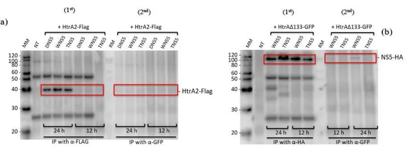
Fig.7. Western bloat analysis of co-immunoprecipitation complexes of various NS5 with HtrA2. (A-1st) YFP tagged plasmids were co-transfected with
HtrA2-Flag. They were co-immunoprecipatated with anti-HtrA2-Flag. (A-2nd) YFP tagged plasmids were co-transfected with HtrA2-Flag. They were co-immunoprecipatated
with anti-GFP. (B-1st) HA tagged plasmids were co-transfected with HtrA2 ∆133-GFP. They were co-immunoprecipatated with anti-HA. (B-2nd) HA tagged
Fig 7. Above both membranes were detected with rabbit anti-GFP. On the left side of membrane fig.(7A1st) there was no NS5-YFP bands detected while the right side of membrane fig. 7 (A2nd) various NS5-YFP bands expressed on 24h and fainting bands on the 12h. Same results observed on fig. 7 (B1st) and 7 (B2nd) were by the left side (B1st) there was no HtrA2 bands detected while the right side of membrane (B2nd) HtrA2 ∆133-GFP bands were expressed. These observations suggested that there was no interaction between viral NS5 and HtrA2.
Fig 8. Western bloat analysis of co-immunoprecipitation complexes of various NS5 with HtrA2. (A-1st) YFP tagged plasmids were co-transfected with
HtrA2-Flag. They were co-immunoprecipatated with anti-HtrA2-Flag. (A-2nd) YFP tagged plasmids were co- transfected with HtrA2-Flag. They were co-immunoprecipatated
with anti- GFP. (B-1st) HA tagged plasmids were co-transfected with HtrA2 ∆133-GFP. They were co-immunoprecipatated with anti-HA (B-2nd) HA tagged
Similar observations on fig.8. (A1st and A2nd) which the membrane was detected with mouse anti-Flag. The left side of membrane fig.8. (A1st) HtrA2-Flag bands were detected while the right side of membrane fig.8. (A2nd) no bands were detected. These observations also suggested that no interaction exists between viral NS5 and HtrA2. However, on fig.8. (B1st) and (B2nd) which the membrane was detected with mouse anti-HA; strong NS5-HA bands were detected on the left side of the membrane fig.8. (B1st) and weak NS5-HA bands were detected on the right side of membrane fig.8 (B2nd) on the 24h but none on the 12h.These observations suggested that weak interaction
exist between viral NS5 and HtrA2 ∆133.
Fig 9. Western bloat analysis of co-immunoprecipitation complexes of various NS5 with HtrA2. (A-1st) YFP tagged plasmids were co-transfected with
HtrA2-Flag. They were co-immunoprecipatated with anti- Flag (A-2nd) YFP tagged plasmids were co-transfected with HtrA2-Flag. They were immunoprecipatated with
anti- GFP (B 1st) HA tagged plasmids were co-transfected with HtrA2 ∆133-GFP. They were co-immunoprecipatated with anti- HA. (B 2nd) HA tagged
Fig.9both membranes were detected with rabbit anti-GFP. The left side of membrane fig.9. (A1st) there was no NS5-YFP bands detected while the right side of membrane (A2nd) various NS5-YFP bands expressed on both 24h and 12h. The left side of membrane fig. 9 (B1st) no HtrA2 detected while the right side fig.9. (B2nd) HtrA2 ∆133-GFP bands were detected. These observations also suggested that there was no interaction between viral NS5 and HtrA2.
Discussion
Since HtrA2/Omi and OMP25 are involved in cellular apoptosis [15,16,21], we first determine the optimal intracellular concentration of the two protein in cell transfection. We found out that HtrA2 concentration of more than 200ng were toxic for the cells as cells confluence decreased by more than 50% and cells appeared to have tense and disintegrated membrane. Cell toxicity appeared to increase a dose dependent manner of HtrA2 and OMP25 concentration. This feature makes them interesting as other studies have shown that the mitochondrion serine protease could be used as a therapeutic target [22]. Kieper N et al also found that under physiological conditions, Omi/HtrA2 to be involved in protection against cellular stress, indicating that loss of Omi mitochondrial protease activity causes the neuromuscular disorders [23,24]. Many studies that have been conducted which supports our observations [16,17], Hegde R et al provided evidence and mechanisms on how mature Omi is released together with mature Smac from the mitochondria into the cytosol which cause apoptosis [25]. Under certain circumstances mitochondrial serine protease (HtrA2/Omi) seems beneficial while too much of it turns to be toxic for the cells. Combining literature review and our study findings suggested that HtrA2/Omi have dual roles in mammalian cells, not only acting as an apoptosis-inducing protein but also maintaining mitochondrial homeostasis. Based on our observation 250ng was settled as an optimal concertation for HtraA2 for co-transfection studies in HeLa cells. NS5 itself is also toxic for the cells as expression various NS5 clones that were analyzed 24h,48h and 72h post transfection indicated the level on NS5 were decreasing with time. These findings reinforce our demonstration on HtrA2 cells toxicity but at this point it is difficult to conclude whether the cellular toxicity we observed in our study is due to NS5 or HtrA2 but rather could be a combination effect. Hegde R et al hypothesize that mature Omi is released together with
mature Smac from the mitochondria into the cytosol upon disruption of the outer mitochondrial membrane during apoptosis. Mature Omi can then induce apoptosis in a caspase-independent manner through its protease activity and in a caspase-dependent manner via its ability to disrupt caspase-IAP interaction [25]. Our hypothesis is that mature Omi is released from the mitochondria into the cytosol interact with NS5 during apoptosis. Since the cells were dying in an increased time dependent manner, we also investigated the level of various NS5 clones, as well HtrA2 12 hrs post-transfection. But as expected, neither NS5 nor HtrA2 were detected due to low level of their presence. Bidet K et al pointed that flaviviruses heavily rely on host cell for replication [1,13, 27], which then requires that there must be an interaction between flaviviruses RNAs and/or proteins with host proteins that facilitate replication. NS5 is a multifunctional protein involved in viral replication, translation and pathogenesis (apoptosis). The major aim of this study was to investigate the role of NS5 in viral pathogenesis by investigating its interaction with the host factor HtrA2. Our results using co-immunoprecipitation and immunoblotting on analysis were not conclusive as we are unable to show the interaction between the two proteins. However, a weak interaction of NS5 with mutant HtrA2 lacking the transmembrane domain was observed 24hrs post-transfection. This might suggest that the anchoring region (transmembrane domain) of HtrA2 is not necessary for an interaction with NS5. It is only the mature form of HtrA2 (lacking the transmembrane domain) that is translocated to the cytosol where it interacts with host factors involved in apoptosis. Based on this facts, our results of interaction with the truncated variant of HtrA2 is in line with previous reports. Inconclusive and open results we had could be due to many factors and limitations such as protein concentration, antibody concentration and pull down conditions (the buffers). Also the choice of antibodies that recognize the protein complexes have major role in immunoprecipitation and western blot analysis. It is important to identify and use antibodies that recognize properly folded protein for immunoprecipitation whereas for Western blot analysis antibodies that recognize denatured form of the protein is necessary. Due to the limitation of time for this project, we were unable to do further analysis. The study should be repeated by testing different kinds of antibodies as well as different conditions for immunoprecipitation, such time, buffers and proteins.
Also because above limitation we did not include OPM25 which is another mitochondrial protein involved in apoptosis in our analysis.
Conclusions
Based on our findings and literature reviews it has been observed that HtrA2 has dual roles. Not only it maintains mitochondrial homeostasis but also is involved in apoptosis. Results from this work have also found out that 12h post transfection is too early for co- immunoprecipitation. Studies on whether the viral NS5 interact with HtrA2 results were open and inconclusive due to limited time we had. Further studies are needed in order to draw a reliable conclusion. This study focused only on NS5 it is possible that other none structural proteins might be involved in this process.
References
[1] Melik W, Ellencrona K, Wigerius M, Hedström C, Elväng A, Johansson M. Two PDZ binding motifs within NS5 have roles in Tick-borne encephalitis virus replication. Virus Research. 2012; 169:54-62.
[2] Charlier N, Davidson A, Dallmeier K, Molenkamp R, De Clercq E, Neyts J. Replication of not-known-vector flaviviruses in mosquito cells is restricted by intracellular host factors rather than by the viral envelope proteins. The Journal of general virology. 2010; 91:1693-7. [3] Bogovic P, Strle F. Tick-borne encephalitis: A review of epidemiology, clinical characteristics, and management. World Journal of Clinical Cases: WJCC. 2015; 3:430-41. [4]. Overby AK, Popov VL, Niedrig M, Weber F. Tick-borne encephalitis virus delays interferon induction and hides its double-stranded RNA in intracellular membrane vesicles. Journal of virology. 2010; 84:8470-83.
[5] Chidumayo NN, Yoshii K, Kariwa H. Evaluation of the European tick-borne encephalitis vaccine against Omsk hemorrhagic fever virus. Microbiology and immunology. 2014; 58:112.
[6] Charrel RN, Attoui H, Butenko AM, Clegg JC, Deubel V, Frolova TV, et al. Tick-borne virus diseases of human interest in Europe. Clinical Microbiology and Infection. 2004; 10:1040-558
[7] Gritsun TS, Lashkevich VA, Gould EA. Tick-borne encephalitis. Antiviral research. 2003; 57:129-46.
[8] Pfeffer M, Dobler G. Emergence of zoonotic arboviruses by animal trade and migration. Parasites & Vectors. 2010; 3:35.
[9] WHO http://www.who.int/mediacentre/factsheets/fs354/en/#
[10] De Filette M, Ulbert S, Diamond M, Sanders NN. Recent progress in West Nile virus diagnosis and vaccination. Veterinary Research. 2012; 43:16.
[11] Oliveira ASd, Silva MLd, Oliveira AFCS, Silva CCd, Teixeira RR, De Paula SO. NS3 and NS5 proteins: important targets for anti-dengue drug design. Journal of the Brazilian Chemical Society. 2014; 25:1759-69.
[12] Yi Z, Yuan Z, Rice CM, MacDonald MR. Flavivirus Replication Complex Assembly Revealed by DNAJC14 Functional Mapping. Journal of Virology. 2012;86 :11815-32. [13] Bidet K, Garcia-Blanco Mariano A. Flaviviral RNAs: weapons and targets in the war between virus and host. Biochemical Journal. 2014; 462:215-30.
[14] de Almagro MC, Vucic D. The inhibitor of apoptosis (IAP) proteins are critical regulators of signaling pathways and targets for anti-cancer therapy. Experimental oncology. 2012; 34:200-11.
[15] Martins LM. The serine protease Omi/HtrA2: a second mammalian protein with a Reaper-like function. Cell death and differentiation. 2002;9 :699-701.
[16] Yang QH, Church-Hajduk R, Ren J, Newton ML, Du C. Omi/HtrA2 catalytic cleavage of inhibitor of apoptosis (IAP) irreversibly inactivates IAPs and facilitates caspase activity in apoptosis. Genes & development. 2003; 17:1487-96.
[17] Vande Walle L, Lamkanfi M, Vandenabeele P. The mitochondrial serine protease HtrA2/Omi: an overview. Cell death and differentiation. 2008; 15:453-60.
[18] Suzuki Y, Takahashi-Niki K, Akagi T, Hashikawa T, Takahashi R. Mitochondrial protease Omi//HtrA2 enhances caspase activation through multiple pathways. Cell death and differentiation. 2003; 11:208-16.
[19] Strober W. Trypan blue exclusion test of cell viability. Current protocols in immunology. 2001.
[20] Holmberg A, Blomstergren A, Nord O, Lukacs M, Lundeberg J, Uhlen M. The biotin-streptavidin interaction can be reversibly broken using water at elevated temperatures. Electrophoresis. 2005; 26:501-10.
[21] Suzuki Y, Takahashi-Niki K, Akagi T, Hashikawa T, Takahashi R. Mitochondrial protease Omi/HtrA2 enhances caspase activation through multiple pathways. Cell death and differentiation. 2004; 11:208-16.
[22] Bhuiyan MS, Fukunaga K. Mitochondrial serine protease HtrA2/Omi as a potential therapeutic target. Current drug targets. 2009; 10:372-83.
[23]Kieper N, Holmström KM, Ciceri D, Fiesel FC, Wolburg H, Ziviani E, et al. Modulation of mitochondrial function and morphology by interaction of Omi/HtrA2 with the mitochondrial fusion factor OPA1. Experimental Cell Research. 2010; 316:1213-24.
[24] Jones JM, Datta P, Srinivasula SM, Ji W, Gupta S, Zhang Z, et al. Loss of Omi mitochondrial protease activity causes the neuromuscular disorder of mnd2 mutant mice. Nature. 2003;425:721-7.
[25] Hegde R, Srinivasula SM, Zhang Z, Wassell R, Mukattash R, Cilenti L, et al. Identification of Omi/HtrA2 as a mitochondrial apoptotic serine protease that disrupts inhibitor of apoptosis protein-caspase interaction. The Journal of biological chemistry. 2002; 277:432-8.
[26] Alberts B, Johnson A, Lewis J, et al. Molecular Biology of the Cell. 4th edition. New York: Garland Science; 2002.
[27] Selisko B, Wang C, Harris E, Canard B. Regulation of Flavivirus RNA synthesis and replication. Current Opinion in Virology. 2014; 9:74-83.