Full Terms & Conditions of access and use can be found at
https://www.tandfonline.com/action/journalInformation?journalCode=zjom20
Journal of Oral Microbiology
ISSN: (Print) (Online) Journal homepage: https://www.tandfonline.com/loi/zjom20
Incidence of infective endocarditis caused by
viridans group streptococci in Sweden – effect of
cessation of antibiotic prophylaxis in dentistry for
risk individuals
Niko Vähäsarja , Bodil Lund , Anders Ternhag , Bengt Götrick , Lars Olaison ,
Margareta Hultin , Carina Krüger Weiner & Aron Naimi-Akbar
To cite this article: Niko Vähäsarja , Bodil Lund , Anders Ternhag , Bengt Götrick , Lars Olaison , Margareta Hultin , Carina Krüger Weiner & Aron Naimi-Akbar (2020) Incidence of infective endocarditis caused by viridans group streptococci in Sweden – effect of cessation of antibiotic prophylaxis in dentistry for risk individuals, Journal of Oral Microbiology, 12:1, 1768342, DOI: 10.1080/20002297.2020.1768342
To link to this article: https://doi.org/10.1080/20002297.2020.1768342
© 2020 The Author(s). Published by Informa UK Limited, trading as Taylor & Francis Group.
Published online: 23 May 2020.
Submit your article to this journal Article views: 620
Incidence of infective endocarditis caused by viridans group streptococci in
Sweden
– effect of cessation of antibiotic prophylaxis in dentistry for risk
individuals
Niko Vähäsarja a,b, Bodil Lunda,c,d, Anders Ternhage, Bengt Götrickf, Lars Olaisong, Margareta Hultina,
Carina Krüger Weinera,band Aron Naimi-Akbara,b,h
aDepartment of Dental Medicine, Karolinska Institutet, Huddinge, Sweden;bFolktandvården Stockholms Län AB, Folktandvården Eastmaninstitutet;cDepartment of Clinical Dentistry, University of Bergen, Bergen, Norway;dDepartment of Oral and Maxillofacial Surgery, Haukelands University Hospital, Bergen, Norway;eDepartment of Medicine Solna, Karolinska Institutet, Unit for Infectious Diseases, Karolinska University Hospital, Stockholm, Sweden;fDepartment of Oral Diagnostics Faculty of Odontology, Malmö University, Malmö, Sweden;gDepartment of Infectious Diseases, Institute of Biomedicine, Sahlgrenska University Hospital, Sweden;hHealth Technology Assessment-Odontology (HTA-O), Malmö University, Malmö, Sweden
ABSTRACT
Introduction: In October 2012, the Swedish Medical Products Agency published new recom-mendations for the cessation of prophylactic antibiotics in dentistry for the prevention of infective endocarditis (IE). Previously, 2 g of amoxicillin per os would be administered 1 h before invasive dental procedures to patients with valve prosthesis, complicated heart valve disease, and to those with previous endocarditis.
Objectives: The aim of this study was to evaluate whether the total incidence of IE caused by oral viridans group streptococci (VGS) or IE caused by staphylococci, increased in Sweden after the introduction of the new recommendations.
Methods: The incidence of IE in Sweden before and after October 2012 was calculated and compared using an interrupted time series analysis. Separate analyses were conducted for the total incidence of IE, and IE caused by VGS or Staphylococcus aureus. Cases of IE were identified using the Swedish national registry of IE, which has existed since 1995 and contains data from all Swedish hospital clinics specialising in infectious disease. All cases with hospital admission date from the 1stof Jan 2008, to the 31stof Dec 2017 were included. The incidence calculations were corrected for annual changes in population size using data from the Swedish government agency Statistics Sweden.
Results: The results show no statistically significant increase in the slope of the trend line of the total incidence of IE, IE caused by VGS orS. aureus in the Swedish general population after October 2012, compared to before.
Conclusion: The results suggest that the recommended cessation of prophylactic antibiotics for the prevention of IE in dentistry has not led to an increased incidence of IE caused by oral streptococci among the Swedish population.
ARTICLE HISTORY Received 17 September 2019 Revised 25 April 2020 Accepted 28 April 2020 KEYWORDS Prophylactic antibiotics; dentistry; infective endocarditis; viridans group streptococci
Introduction
Infective endocarditis (IE) arises when the innermost layer of the heart, the endocardium, is colonised by
microorganisms circulating in the blood [1]. The
colonisation is usually located to the heart valves,
where microorganisms subsequently form
a vegetation. As the disease progresses, it can lead to heart failure and embolization of blood vessels
caused by circulating vegetation fragments [2]. The
disease is fatal if left untreated and associated with an in-hospital mortality of 10–30% [1,3,4].
Treatment is focused on the eradication of microbes by antimicrobial treatment alone or in com-bination with surgery, and can require specialists in several medical fields such as infection, cardiology and thoracic surgery [1].
The most common pathogens causing IE are vir-idans group streptococci (VGS); a group of alpha-hemolytic bacteria commonly found in the oral cavity,
and staphylococci (Staphylococcus aureus and
Staphylococcus epidermidis), commonly found on the skin [5]. VGS cause a slower and less aggressive pro-gression of IE, with lower mortality, than staphylo-cocci [5,6].
The incidence of IE in Sweden has been reported to be 6.2–7.7 cases per 100 000 individuals per year [4,7]. Ternhag et al. found an increasing incidence of IE in Sweden from 1997 to 2007 [4]. Internationally, the incidence of IE ranges from 2 to 7 cases per 100 000 individuals per year [1,8].
Healthy endocardium is usually resistant to colo-nization, but disease and congenital malformations
CONTACTNiko Vähäsarja niko.vahasarja@ki.se Department of Dental Medicine, Karolinska Institutet, Alfred Nobels Allé 8, 141 52 Huddinge, Sweden
https://doi.org/10.1080/20002297.2020.1768342
© 2020 The Author(s). Published by Informa UK Limited, trading as Taylor & Francis Group.
This is an Open Access article distributed under the terms of the Creative Commons Attribution License (http://creativecommons.org/licenses/by/4.0/), which permits unrestricted use, distribution, and reproduction in any medium, provided the original work is properly cited.
may result in damage of the endocardium causing
a predisposition for IE [5]. Previous endocarditis,
heart valve disease and prosthetic heart valves are important risk factors for IE. Elderly patients (>65 years of age) also have an elevated risk of IE. This may be due to the clustering of risk factors and the presence of undetected minor degenerative valve lesions [1]. However, 40% of the patients developing IE lack previously known heart disease [9–11].
The use of antibiotic prophylaxis (AP) in dentistry for the prevention of IE is controversial. The purpose is to reduce bacteraemia caused by invasive dental treatment and prevent colonization of the
endocar-dium, thereby reducing the risk of IE [1]. However,
the efficacy of the prophylaxis has not been
con-firmed [12]. Bacteraemia caused by VGS is known
to occur not only after invasive dental treatment, but also during daily dental activities such as tooth brush-ing and flossbrush-ing [13–17].
Current guidelines in Europe and America have restricted the indications for AP in dentistry, stating that it should be administered only to individuals with the highest risk of developing IE [2,18–20]. In Sweden, cessation of the prophylaxis to both low risk and high risk patients groups has been recommended since October 2012 [21].
In 2015, Thornhill et al. showed that a single dose of 3 grams of amoxicillin was associated with a low number of adverse drug reactions [22]. A restrictive approach towards the use of AP is however supported by data showing that one single dose of amoxicillin can induce significant disruption in the oral micro-biota, and cause selection of bacteria resistant to antibiotics [23]. Antimicrobial resistance constitutes an enormous threat to modern healthcare, because numerous treatments require the use of effective anti-biotics [24]. The association between the prescription of antibiotics and antimicrobial resistance is undis-puted, and great effort is focused on reducing the use of antibiotics [25].
The benefits of a restrictive approach towards the use of antibiotics must be weighed against the poten-tial risks. Considering the severity of IE it is of utmost importance to investigate the effect of refraining from AP.
The aim of this study was to evaluate whether the incidence of VGS-IE increased in Sweden after the introduction of new recommendations for AP in dentistry.
Materials and methods
Study design
An interrupted time series analysis (ITSA) was con-ducted of the monthly incidence of IE in the Swedish general population from 2008 to 2017, in order to
calculate any increased incidence of the disease after the recommended cessation of AP in dentistry for the prevention of IE in October 2012. The change in recommendations allowed comparison of the inci-dence of IE caused by oral streptococci at a time
when antibiotics was– and was not – recommended
for the prevention of IE. Three separate analyses were conducted of the incidence of 1) all cases of IE, 2) IE caused by VGS, and 3) IE caused by S. aureus, in order to calculate and compare pre- and post-intervention trends. IE caused by VGS was consid-ered to be of oral origin and thus of relevance with regard to cessation of AP during invasive dental procedures. The total incidence of IE and the inci-dence of IE caused by S. aureus were analysed to monitor trends in IE not related to the altered AP recommendations. Patients included in the study ful-filled the criteria for definite or possible IE according to the modified Duke criteria [26].
Sources of data
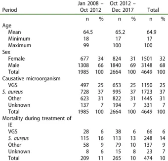
Cases of IE were identified using the Swedish national registry of IE (SRIE), with an estimated coverage of 88% of Swedish cases of IE. Detailed information of the causative organism was available in 92% of cases. Age and sex distribution in the SRIE is displayed in Table 1, with the in-hospital mortality and propor-tion causative microorganisms.
In 1995, the Swedish Society for Infectious Diseases introduced a Swedish national registry of IE (the SRIE). All 30 departments of infectious Table 1.Characteristics in the Swedish national register for infective endocarditis (IE).
Period Jan 2008– Oct 2012 Oct 2012– Dec 2017 Total n % n % n % Age Mean 64.5 65.2 64.9 Minimum 18 17 17 Maximum 99 100 100 Sex Female 677 34 824 31 1501 32 Male 1308 66 1840 69 3148 68 Total 1985 100 2664 100 4649 100 Causative microorganism VGS 497 25 653 25 1150 25 S. aureus 728 37 995 37 1723 37 Other 623 31 822 31 1445 31 Unknown 137 7 194 7 331 7 Total 1985 100 2664 100 4649 100 Mortality during treatment of
IE VGS 28 6 38 6 66 6 S. aureus 115 16 113 13 248 14 Other 58 9 79 10 137 9 Unknown 8 6 15 8 23 7 Total 209 11 265 10 474 10
The table shows the in-hospital mortality of IE caused by viridans group streptococci (VGS), Staphylococcus aureus, and other microorganisms (mainly enterococci and coagulase-negative staphylococci). Data are displayed separately for the period before and after the cessation of antibiotics in dentistry for the prevention of IE, in October 2012. 2 N. VÄHÄSARJA ET AL.
diseases (ID) in Sweden have participated in the registry since its inception. These ID departments have regional responsibility for the care of patients with severe infections, and patients requiring acute surgery for IE are, in most cases, treated in ID depart-ments during the pre- and/or postoperative period.
All cases are reported on a standardized naire at the time of discharge and a second question-naire after follow-up (mean: 3 months after treatment). Data regarding risk factors, presence of prosthetic valve, type of prosthetic valve and presence of other implantable cardiac devices is collected. The origin of the etiologic agent is verified using, for example, blood cultures, cultures from valves during surgery, and polymerase chain reaction (PCR) from tissue samples of valves. Information about antibiotic treatment, need for surgery, and treatment outcome is also included.
The registry has been used to present clinical data
regarding various pathogens [27–32] and has made
contributions to numerous studies within the
frame-work of the International Collaboration on
Endocarditis (ICE) with around 30 publications [3,6,33].
Children and young adults from the age of 0 to 16 years are treated at paediatric clinics not reporting to the register. However, the incidence of IE among children has been reported to be lower than among
adults in Sweden [7,34]. We studied data from
January 2008 to 2017 which covered an approximate period of 5-years before and 5-years after the recom-mendations to cease AP.
The incidence calculation was corrected for annual changes in the size of the Swedish population. The Swedish population, above 16 years of age, increased from 7 463 069 individuals in December (Dec) 2008, to 8 107 436 in Dec 2017. Data on population size was obtained from Statistics Sweden, the government agency responsible for developing, producing and disseminating official statistics and other government statistics in Sweden. Data are collected by Statistics
Sweden on the 31st of December each year, and is
public and readily available in the statistical database on their website [35].
Outcomes
The primary outcome measured in this study was the incidence of VGS-IE in Sweden. In addition to this, analyses were carried out on the total incidence of IE, as well as the incidence of IE caused by S. aureus.
Statistical analysis
The statistical analysis was carried out using Stata/IC 15 (StataCorp. 2017. Stata Statistical Software: Release 15. College Station, TX: StataCorp LLC). We used the
ITSA module (Ariel Linden, 2014.‘ITSA: Stata
mod-ule to perform interrupted time series analysis for single and multiple groups,’ Statistical Software Components S457793, Boston College Department of Economics, revised 8 December 2017) to perform a single group ITSA. The time of the intervention was
set to month number 59, corresponding to
October 2012, when the change in recommendations for AP in dentistry was published by the Swedish Medical Products Agency (MPA). Each point in the
ITSA represents the monthly incidence of
endocarditis.
Ethics
This study was approved by the Regional Ethical Review Board, Karolinska Institutet, Stockholm, Sweden (ref: 2015/1910-31/1) prior to onset of the study.
An ethical permit and approval for the study was also obtained from the register board of the SRIE, before the collection of data.
Informed consent was waived because the study does not pose any risk of physical harm to subjects, no identifying information is published as results are presented on group level only.
Results
Cases with known microbial aetiology not attributed to VGS or S. aureus, were mainly caused by
entero-cocci (11%) and coagulase-negative
staphylo-cocci (6%).
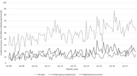
Figure 1 shows the incidence of IE, by causative microorganism across the study period. From January (Jan) 2008 to Dec 2017, the incidence of VGS-IE in Sweden varies between 5.2 and 22.0 cases per 10 million individuals per month (Figure 1).
IE caused by VGS
After October 2012 there is a statistically significant increase in the incidence of IE caused by VGS by 0.065 cases per 10 million individuals per month (95% CI = [0.017, 0.114]) (Figure 2).
Comparison with the pre-intervention trend yields
a decrease in the slope of the trend line by −0.007
cases per 10 million individuals per month (95%
CI = [−0.085, 0.082]) after October 2012. This
decrease is not statistically significant.
The starting level is estimated to 8.9 cases/10 million individuals/month in Jan 2008. The incidence appears to increase by 0.072 cases per 10 million individuals per month (95% CI = [−0.002, 0.146]) until October 2012. This trend is not statistically significant. Additional analyses were performed to account for lag in adherence to the new recommendations. Changing the time of intervention to 6, 12 or
18 months after October 2012 did not yield any statistically significant increase in the incidence of VGS-IE, compared to the pre-intervention trend.
The total incidence of IE
There is a statistically significant increase in the inci-dence of IE by 0.344 cases per 10 million individuals per month (95% CI = [0.187, 0.502]) before
October 2012 (Figure 3). After October 2012, there
is also a statistically significant increase in the inci-dence of IE, by 0.266 cases per 10 million individuals per month (95% CI = [0.115, 0.416]).
Comparison of the trends shows a decrease in the slope of the trend line after October 2012, relative to
the pre-intervention trend, by −0.079 cases per
10 million individuals per month (95%
CI = [−0.297, 0.139]). This decrease is not statistically significant. The starting level is estimated at 33.7 cases/10 million individuals/month in Jan 2008. Figure 1.Incidence of infective endocarditis (IE) in Sweden.
The incidence of all cases of infective endocarditis, cases caused by oral viridans group streptococci andStaphylococcus aureus in Sweden from January 2008 to December 2017.
Figure 2.Single-group ITSA with Newey–West standard errors and one lag.
ITSA of the monthly incidence of infective endocarditis caused by viridans group streptococci in Sweden from Jan 2008 to Dec 2017. Intervention is set to month 59, corresponding to October 2012. After October 2012 there is a decreasing trend, relative to the pre-intervention trend, in the incidence by−0.007 points (95% CI = [−0.085, 0.082]). This trend is not statistically significant.
IE caused by S. aureus
The incidence increases by 0.099 cases per 10 million individuals per month (95% CI = [0.022, 0.176]) until October 2012 (Figure 4). After October 2012, there is a decrease in the slope of the trend line, relative to the pre-intervention trend, by−0.012 cases per 10 million
individuals per month (95% CI = [−0.121, 0.096]). This decrease is not statistically significant. The start-ing level is estimated at 12.7 cases/10 million indivi-duals/month. Intervention is set to month 59, corresponding to October 2012. After October 2012 there is an increase in the incidence of IE caused by Figure 3.Single-group ITSA with Newey–West standard errors and one lag.
ITSA of the monthly total incidence of IE in Sweden from Jan 2008 to Dec 2017. Intervention is set to month 59, corresponding to October 2012. Comparison of the trends shows a decreasing trend after October 2012, relative to the pre-intervention trend, by −0.079 points (95% CI = [−0.297, 0.139]). This trend is not statistically significant.
Figure 4.Single-group ITSA with Newey–West standard errors and one lag.
ITSA of the monthly incidence of IE caused byStaphylococcus aureus in Sweden from Jan 2008 to Dec 2017. Intervention is set to month 59, corresponding to October 2012. After October 2012, there is a decreasing trend, relative to the pre-intervention trend, in the incidence by −0.012 points (95% CI = [−0.121, 0.096]). This trend is not statistically significant.
S. aureus by 0.087 cases per 10 million individuals per month (95% CI = [−0.011, 0.163]). This trend is not statistically significant.
Discussion
To our knowledge, this is the first study of the inci-dence of VGS-IE following the recommended cessa-tion of AP in Swedish dentistry for the prevencessa-tion of IE in October 2012. The results imply that there was no increase, over the predicted positive trend, in the incidence of VGS-IE.
The UK and Sweden are the only countries to have abandoned AP to individuals considered to be at high risk of developing IE. Although in the UK, it is now emphasised that the patient should make an informed decision [36]. In Sweden, AP is in general prescribed by the dentist, and the dentist is ultimately responsi-ble for the decision to administer or not to administer
AP before dental procedures [21]. Previously in
Sweden, 2 g of amoxicillin per os had been recom-mended 1 h before invasive dental procedures to patients with valve prosthesis, complicated heart valve disease, or previous endocarditis [21].
The number of prescriptions of amoxicillin by Swedish dentists decreased by 20% in 2013 compared to 2012 [37], and by 22.2% from 2013 to 2017 [38]. It has been suggested that the decrease may be due to
the new recommendations [38].
The results of the current study show a statistically significant increasing trend in the total incidence of IE in Sweden both before and after October 2012. An increasing incidence of IE across the general popula-tion is in accordance with earlier studies of the epi-demiology of IE in Sweden, USA, Italy, Taiwan and Canada [39, 40, 41, 4, Mackie et al.]. The increase may be due to increasing risk factors such as degen-erative valve disease, receiving haemodialysis and population ageing [4,39–41].
The results are consistent with those reported by Thornhill et al in 2011 [42]. They excluded any statis-tically significant increase in the incidence of IE in England, during the 25 months after a cessation of AP. In a later study however, Dayer et al. found an increased incidence over the projected trend [43] in England, and a 78.6% fall in AP prescribing. Although they did not have access to detailed data on the causative organism, and were unable to study the incidence of VGS-IE.
Current European and American guidelines have restricted the indications for AP in dentistry, stating that it should be administered only to individuals
with the highest risk of developing IE [2,20].
Thornhill et al. published a review of studies mon-itoring the impact of these restricted guidelines [44]. They found that several studies were limited metho-dological flaws such as difficulties in accurately
identifying IE caused by oral VGS and a lack of data on AP prescribing, and concluded that limiting AP use to those at highest risk seems pragmatic and appropriate.
Mackie et al. studied hospitalizations in Canada due to IE before and after the publication of the
2007 AHA recommendations [45]. They found an
increasing trend before and after 2007. Comparison showed no significant increase after 2007. They used ICD codes to identify cases of streptococcal IE, but were unable to identify cases caused by oral streptococci.
The incidence of IE has been reported to be 2–7
cases per 100 000 individuals per year [1,8], which is in accordance with our results based on the SRIE; 4 to 7 cases per 100 000 individuals per year. Ternhag et al. utilized the Swedish patient register to calculate the incidence of IE in Sweden from 1997 to 2007. We found a similar male predominance among IE-patients as in their study (40.8% females and 59.2% males), but their incidence of IE was higher, at 7.7 per 100 000 individuals per year. Notably, our study was not based on the patient registry, but on the SRIE.
It is possible that a calculation using the patient registry could overestimate the incidence of cases of IE in Sweden [4], while our calculation could under-estimate it. There are two reasons for this. The first is that the patient registry does not discriminate between definite, possible and rejected cases of IE according to the Duke criteria. For this reason, there is a risk of misclassification leading to the inclusion of rejected cases of IE. Secondly, the cover-age of the patient registry is higher than that of the SRIE.
The National Board of Health and Welfare founded the patient registry in 1964, and it reached full national coverage in 1987 [46]. The coverage of the SRIE has been estimated to 88%.
One advantage of the SRIE is that it contains detailed information on the microbiological aetiology of cases of IE. For this reason, we were able to calculate the incidence of VGS-IE, which is not pos-sible using the patient registry. Future research could integrate both registries to benefit from their respec-tive advantages.
The short follow-up time after the change in recommendations is one limitation, as the timeframe may not be long enough to detect subtle effects on the frequency of cases of VGS-IE. Despite this, ongoing data monitoring is imperative after the change in recommendations in Swedish dentistry.
Another limitation is that the microbial aetiology is unknown in 7.7% of cases in the register. Reasons for this are mainly that no blood culture was per-formed, or that the performed blood culture showed negative results. Additional analysis in the SRIE showed that in 43% of cases with unknown aetiology,
a blood culture was performed but the results were negative. Previous research concludes that in less than 50% of cases where the blood culture was nega-tive, this could be attributed to the administration of antibiotics prior to the drawing of blood for a blood
culture [7]. Some of these cases could have been
caused by VGS, thereby resulting in an underestima-tion of the incidence of IE with possible oral origin. On the other hand the effect on our analysis should be negligible, as it is unlikely that the microbial aetiology for unknown cases should differ before and after 2012.
A concern is that the study included all patients regardless of high or low risk to develop IE after invasive dental treatment. A study of a high-risk population, such as valve prosthesis bearers, is desirable.
The proportion of IE 1995–2012 caused by VGS, recorded in SRIE, decreased from 35 to 25% and the proportion of IE caused by S. aureus increased from 30 to 40%. These trends are in analogy with trends in other countries with developed health care systems and can make the results difficult to interpret.
Conclusions
The results suggest that the Swedish recommenda-tions for the cessation of antibiotics in dentistry pub-lished in October 2012 have not lead to an increase in the incidence of IE in Sweden. Caution should be taken when interpreting the results, due to a short follow-up period, no specific analysis of high-risk groups, such as patients with prosthetic valves, and a limited number of cases.
Authors’ contributions
The authors contributed to the conception and design of the study, and revised the manuscript. LO contributed to the acquisition and analysis of data. NV and ANA analysed and interpreted the patient data. All authors read and approved the final manuscript.
Availability of data and materials
The study was conducted using national Swedish registries. The data that support the findings of this study are avail-able from the SRIE, but restrictions apply to the availability of these data, which were used under license for the current study, and so are not publicly available. Data on population size used for the incidence calculations are readily available from Statistics Sweden.
Disclosure statement
The authors have no competing interests to declare.
Funding
Funding was provided by the board of doctoral education at Karolinska Institutet, the Public Health Agency of Sweden, Folktandvården Stockholm AB, Steering Group for Collaborative Odontological Research at Karolinska Institutet and Stockholm City County, and the Swedish Dental Association.
ORCID
Niko Vähäsarja http://orcid.org/0000-0002-4974-484X
References
[1] Que YA, Moreillon P. Infective endocarditis. Nat Rev Cardiol. 2011;8(6):322–336.
[2] Habib G, Hoen B, Tornos P, et al. Guidelines on the prevention, diagnosis, and treatment of infective endocarditis (new version 2009): the task force on the prevention, diagnosis, and treatment of infective endocarditis of the European society of cardiology (ESC). Endorsed by the European society of clinical microbiology and infectious diseases (ESCMID) and the international society of chemotherapy (ISC) for
infection and cancer. Eur Heart J. 2009;30
(19):2369–2413.
[3] Murdoch DR, Corey GR, Hoen B, et al. Clinical pre-sentation, etiology, and outcome of infective
endocar-ditis in the 21st century: the international
collaboration on endocarditis-prospective cohort
study. Arch Intern Med.2009;169(5):463–473. [4] Ternhag A, Cederström A, Törner A, et al.
A nationwide cohort study of mortality risk and long-term prognosis in infective endocarditis in Sweden. PLoS One.2013;8(7):e67519.
[5] Moreillon P, Que YA, Bayer AS. Pathogenesis of streptococcal and staphylococcal endocarditis. Infect Dis Clin North Am.2002;16(2):297–318.
[6] Wang A, Athan E, Pappas PA, et al. Contemporary clinical profile and outcome of prosthetic valve endocarditis. JAMA.2007;297(12):1354–1361.
[7] Hogevik H, Olaison L, Andersson R, et al.
Epidemiologic aspects of infective endocarditis in an urban population. A 5-year prospective study. Medicine (Baltimore).1995;74(6):324–339.
[8] Tleyjeh IM, Abdel-Latif A, Rahbi H, et al.
A systematic review of population-based studies of infective endocarditis. Chest.2007;132(3):1025–1035. [9] Castillo JC, Anguita MP, Torres F, et al. Comparison
of features of active infective endocarditis involving native cardiac valves in nonintravenous drug users with and without predisposing cardiac disease. Am J Cardiol.2002;90(11):1266–1269.
[10] Selton-Suty C, Hoen B, Delahaye F, et al. Comparison of infective endocarditis in patients with and without previously recognized heart disease. Am J Cardiol. 1996;77(12):1134–1137.
[11] van der Meer JT, Thompson J, Valkenburg HA, et al.
Epidemiology of bacterial endocarditis in the
Netherlands. I. Patient characteristics. Arch Intern Med.1992;152(9):1863–1868.
[12] Glenny AM, Oliver R, Roberts GJ, et al. Antibiotics for the prophylaxis of bacterial endocarditis in dentistry. Cochrane Database Syst Rev.2013;10:CD003813.
[13] Lockhart PB. The risk for endocarditis in dental practice. Periodontol 2000.2000;23:127–135.
[14] Lockhart PB, Brennan MT, Sasser HC, et al. Bacteremia associated with toothbrushing and dental extraction. Circulation. 2008;117(24):3118–3125. [15] Pallasch TJ, Slots J. Antibiotic prophylaxis and the
medically compromised patient. Periodontol 2000. 1996;10:107–138.
[16] Roberts GJ, Watts R, Longhurst P, et al. Bacteremia of dental origin and antimicrobial sensitivity following oral surgical procedures in children. Pediatr Dent. 1998;20(1):28–36.
[17] Schlein RA, Kudlick EM, Reindorf CA, et al. Toothbrushing and transient bacteremia in patients undergoing orthodontic treatment. Am J Orthod Dentofacial Orthop.1991;99(5):466–472.
[18] Habib G, Lancellotti P, Antunes MJ, et al. 2015 ESC guidelines for the management of infective endocardi-tis: the task force for the management of infective endocarditis of the european society of cardiology
(ESC). Endorsed by: European association for
cardio-thoracic surgery (EACTS), the European asso-ciation of nuclear medicine (EANM). Eur Heart J. 2015Nov 21;36(44):3075–3128.
[19] Nishimura RA, Otto CM, Bonow RO, et al. 2017 AHA/ACC focused update of the 2014 AHA/ACC guideline for the management of patients with valvu-lar heart disease: a report of the american college of cardiology/American heart association task force on clinical practice guidelines. J Am Coll Cardiol. 2017 Jul 11;70(2):252–289.
[20] Wilson W, Taubert KA, Gewitz M, et al. Prevention of infective endocarditis: guidelines from the American heart association: A guideline from the American heart association rheumatic fever, endocarditis, and kawasaki disease committee, council on cardiovascular disease in the young, and the council on clinical cardiology, coun-cil on cardiovascular surgery and anesthesia, and the quality of care and outcomes research interdisciplinary working group. Circulation.2007;116(15):1736–1754. [21] Läkemedelsverket SMPA, (MPA). Indikationer för
anti-biotikaprofylax i tandvården – ny rekommendation. Information från Läkemedelsverket;2012. p. 22–35. [22] Thornhill MH, Dayer MJ, Prendergast B, et al.
Incidence and nature of adverse reactions to antibio-tics used as endocarditis prophylaxis. J Antimicrob Chemother.2015;70(8):2382–2388.
[23] Khalil D, Hultin M, Rashid MU, et al. Oral microflora and selection of resistance after a single dose of amoxicillin. Clin Microbiol Infect.2016;22(11):949.e941-949.e944. [24] WHO. Antimicrobial resistance: Global report on
sur-veillance;2014.
[25] van de Sande-bruinsma N, Grundmann H, Verloo D, et al. Antimicrobial drug use and resistance in Europe. Emerg Infect Dis.2008;14(11):1722–1730.
[26] Li JS, Sexton DJ, Mick N, et al. Proposed modifica-tions to the Duke criteria for the diagnosis of infective endocarditis. Clin Infect Dis.2000;30(4):633–638. [27] Bläckberg A, Nilson B, Özenci V, et al. Infective
endocarditis due to Streptococcus dysgalactiae: clinical presentation and microbiological features. Eur J Clin Microbiol Infect Dis.2018;37(12):2261–2272.
[28] Lindell F, Söderquist B, Sundman K, et al. Prosthetic valve endocarditis caused by Propionibacterium spe-cies: A national registry-based study of 51 Swedish cases. Eur J Clin Microbiol Infect Dis. 2018;37 (4):765–771.
[29] Olaison L, Schadewitz K, Endocarditis S. Enterococcal endocarditis in Sweden, 1995-1999: can shorter ther-apy with aminoglycosides be used? Clin Infect Dis. 2002;34(2):159–166.
[30] Sunnerhagen T, Nilson B, Olaison L, et al. Clinical and microbiological features of infective endocarditis caused by aerococci. Infection.2016;44(2):167–173. [31] Van Vlasselaer A, Rasmussen M, Nilsson J, et al.
Native aortic versus mitral valve infective endocardi-tis: A nationwide registry study. Open Heart. 2019;6 (1):e000926.
[32] Werner M, Andersson R, Olaison L, et al. A clinical study of culture-negative endocarditis. Medicine (Baltimore).2003;82(4):263–273.
[33] Lalani T, Cabell CH, Benjamin DK, et al. Analysis of the impact of early surgery on in-hospital mortality of native valve endocarditis: use of propensity score and instrumen-tal variable methods to adjust for treatment-selection bias. Circulation.2010;121(8):1005–1013.
[34] Schollin J, Bjarke B, Wesström G. Infective endocar-ditis in Swedish children. I. Incidence, etiology, under-lying factors and port of entry of infection. Acta Paediatr Scand.1986;75(6):993–998.
[35] Population statistics. 2019 [cited 2019 Apr 30].
Av ailable f rom: https://www.scb.se/BE01
01-en#_TablesintheStatisticalDatabase.
[36] Thornhill M, Dayer M, Lockhart P, et al. Guidelines on prophylaxis to prevent infective endocarditis. Br Dent J.2016;220:51–56.
[37] Tandläkares antibiotikaförskrivning i öppenvård till och med mars 2019.2019 [cited 2019 Apr 30]. Available from:https://www.folkhalsomyndigheten.se/contentas
sets/3609491f2c594228b098329bd2f176a5/tandvard-antibiotikaforskrivning-mars2019.pdf.
[38] Swedres-Svarm. Consumption of antibiotics and occurrence of resistance in Sweden;2017.
[39] Correa de Sa DD, Tleyjeh IM, Anavekar NS, et al. Epidemiological trends of infective endocarditis: A
population-based study in Olmsted county,
Minnesota. Mayo Clin Proc.2010;85(5):422–426. [40] Fedeli U, Schievano E, Buonfrate D, et al. Increasing
incidence and mortality of infective endocarditis: A population-based study through a record-linkage system. BMC Infect Dis.2011;11:48.
[41] Lee CH, Tsai WC, Liu PY, et al. Epidemiologic fea-tures of infective endocarditis in Taiwanese adults involving native valves. Am J Cardiol. 2007;100 (8):1282–1285.
[42] Thornhill MH, Dayer MJ, Forde JM, et al. Impact of the NICE guideline recommending cessation of anti-biotic prophylaxis for prevention of infective endocar-ditis: before and after study. Bmj.2011;342:d2392. [43] Dayer MJ, Jones S, Prendergast B, et al. Incidence of
infective endocarditis in England, 2000-13: A secular trend, interrupted time-series analysis. Lancet. 2015;385(9974):1219–1228.
[44] Thornhill MH, Dayer M, Lockhart PB, et al. Antibiotic prophylaxis of infective endocarditis. Curr Infect Dis Rep.2017;19(2):9.
[45] Mackie AS, Liu W, Savu A, et al. Infective endocarditis hospitalizations before and after the 2007 American
heart association prophylaxis guidelines. Can
J Cardiol.2016;32(8):942–948.
[46] Ludvigsson JF, Andersson E, Ekbom A, et al. External review and validation of the Swedish national inpatient register. BMC Public Health. 2011;11:450.