is is an open-access article distributed under the terms of the Creative Commons Attribution-Non Commercial-Share Alike 4.0 License, which allows others to remix, tweak, and build upon the work non-commercially, as long as the author is credited and the new creations are licensed under the identical terms. Experts Corner
Orthodontic management of agenesis of mandibular
second premolars
Krister Bjerklin1,2
1Department of Orthodontics, e Institute for Postgraduate Dental Education, Jönköping, Sweden, 2Department of Orthodontics, Faculty of Odontology,
Malmö University, Malmö, Sweden.
*Corresponding author: Dr. Krister Bjerklin, Department of Orthodontics, e Institute for Postgraduate Dental Education, Jönköping, Sweden. krister.bjerklin@mau.se Received : 03 October 19 Accepted : 20 November 19 Published : 31 December 19 DOI 10.25259/APOS_122_2019 Quick Response Code:
INTRODUCTION
Except for the third molars, the mandibular second premolars are the most commonly missing teeth. In many populations, there has been reported a prevalence of 2.4–4.5%.[1-3] In most
children, the diagnosis can be made at the age of 9. In few subjects, however, the germ can be seen on a radiograph as late as at the ages of 11–12 years.
General dentists are responsible for detecting such conditions. Early diagnosis is important to plan the treatment and offer the patient the best solution.
In many cases, already at the ages of 10–11, the roots of the second primary molar can have severe root resorption, infraocclusion, or caries lesions and the tooth must be extracted. In some of these cases, the space closes spontaneously, but often a comprehensive orthodontic treatment is necessary later on. Furthermore, when severe crowding and space deficiency in the dental arch are diagnosed early, an extraction therapy can be started already in the mixed dentition. When one has the possibility to postpone decisions for treatment planning, there are a number of variables to take in consideration.
ese are for instance: Age of the patient, attitude to orthodontic treatment, from the patient and the parents, growth pattern, type of occlusion; molar relationship and incisor relationship, available space in the dental arch and condition of the primary molar; root resorption, infraocclusion, and caries – fillings.
ABSTRACT
Agenesis of mandibular second premolar is reported to be 2.4–4.5%. e diagnosis can be set on at average 9 years of age. Early treatment in the form of extraction of the second primary molar and in some cases also the remaining three second premolars and comprehensive orthodontic treatment are often a good treatment solution. In vertical deep bite cases, cases with spacing in the dental arch, mandibular posterior rotation and for extractions disadvantageous growth pattern, a treatment with retaining of the primary molar must be taken in consideration. When there is no or minor infraocclusion, root resorption less than half of the root length, and no caries or fillings at the age of 12–13 years, there is a good prognosis for longtime survival of the primary molar.
Keywords: Premolar agenesis, Primary molar, Infraocclusion, Root resorption, Orthodontic treatment
Extraction of the second primary molar and space closure with a good result can be expected in cases with crowding in the dental arch, normal or minimal overbite, Angle Class I occlusion with a normal growth pattern, and incisal inclination within normal range. In many of these extraction cases, it can be wise to also extract the remaining three second premolars. Non-extraction, if possible, is preferable in individuals with deep overbite, decreased lower facial height, retroclination of the lower incisors, low angle subjects, and no crowding in the mandible. In these cases, it is difficult to close spaces without adversely affecting the facial profile.
Treatment options
a. Exltraction of the primary molar and spontaneous space closure, sometimes in combination with the extraction of the second premolar on the same side in the upper jaw b. Extraction of the primary molar and comprehensive
orthodontic treatment, often in combination with the extraction of the second premolar on the same side in the upper jaw. In cases with risk for midline deviations also the extraction of the two second premolars on the opposite side
c. Autotransplantation d. Implant-supported crown e. Fixed prosthetic treatment f. Leave the primary molar in situ.
Extraction of the primary molar
Early extraction is often proposed. Joondeph and McNeill recommended early extraction of the primary molar to permit acceptable spontaneous space closure.[4] Lindqvist
pointed out the risk for tipping of teeth adjacent to the extraction site when the extraction was performed after completed root development of these teeth.[5] In a study
of 11 homogeneous subjects with normal occlusion, there were nine subjects with agenesis of one mandibular second premolar, two subjects with agenesis of both mandibular second premolars, extraction of the second primary molar, and the remaining second premolars were performed at the age of mean 11.8 years. After 4 years, there was a residual extraction space of mean 2 mm in the mandible. Both mesial movement of the first permanent molars and distal movement of the first premolars could be seen.[6] is indicates that some
orthodontic treatment with fixed appliances is required. When it is possible to decide when the extraction is due, the following has to be taken into consideration for a good result: • Soon after the age of 9
• The first premolar should have developed at least half of the root length.
Controlled slicing followed by hemi-sectioning of the second primary molar before extraction is recommended to produce
a bodily controlled mesial movement of the first permanent molar. To achieve the best result, it is recommended to start early, between the ages of 8 and 9 years when it is possible to stipulate the diagnosis agenesis already at the age of 8.[7]
Moreover, an option in non-crowded cases, as above, extraction space can be closed by mesial movement of the first permanent molar with the aid of temporary anchorage devices (TADs), screw, or plate. In these cases, further extractions are not necessary.
Autotransplantation
e width and the shape of a maxillary third molar fit very well to replace the second primary molar [Figure 1]. e best developmental stage for a tooth to be transplanted is when the root is developed at 50 - 75 %. is is normally between 18 and 20 years of age.[8] is treatment option must be
reserved for cases when late extraction of the primary molar is possible and where space closure is, for different reasons, judged difficult to perform and very time consuming.
Implant-supported crown
When the missing premolar is to be replaced with an implant-supported crown, one must know that this will impede the growth of the alveolar process. e implant should not be installed before the age of 20 on average.[9]
Like autotransplantation treatment, the treatment with an implant should be reserved for cases with late extraction of the primary molar, or if the treatment has to start with orthodontic appliances for space gaining.
Fixed prosthesis
Fixed prosthesis consists of either a permanent bridge with grinding of the adjacent teeth or a semi-permanent bridge bonded to one or both neighbor teeth. Both these solutions will, like an implant, impede the growth of the alveolar process.
Figure 1: Maxillary third molar transplanted to the region of the second premolar.
Leave the primary molar in situ
When extraction of the second primary molar is performed early, before the age of 12–13 years, spontaneous space closure and in some cases a comprehensive orthodontic treatment are expected.
In subjects, when the diagnosis agenesis of mandibular second premolar is established late or when the patient or the parents have refused extraction an option could be to leave the primary molar in situ. e best conditions for this is in non-crowded cases with Angle Class I occlusion, normal overbite and overjet with the primary molar in good condition.
In a study by Rune and Sarnäs, 77 subjects were followed to about 17 years of age. No primary molar was lost due to root resorption and only few were lost due to infraocclusion.[10] At
a follow-up about 15 years after normal exfoliation time, no second primary molar was lost due to root resorption, few cases showed increased infraocclusion and in three cases, extraction of primary molars had been performed due to caries.[11] Bjerklin and Bennett (2000) investigated the
long-term survival of lower second primary molars in subjects with agenesis of the premolars. e follow-up started at 12– 13 years of age to mean 20 years (SD 3.62). ey found, of 59 primary molars in 41 patients, that two primary molars were exfoliated, at age 13 years, 6 months, three were extracted due to progressing root resorption, at age 18 years.[12] Another
study had longer follow-up, the last registrations between 18 and 45 years, mean 24 years, 7 months.[13] In this study,
99 primary molars were followed. Eleven of these were lost. Four were lost because the patients had earlier been offered transplantation or implants when they reached the ages of 18 to 20. Of the remaining seven extracted primary molars, one was extracted at the age of 21 due to root resorption and infraocclusion, two at age 22 and 23 due to progressive infraocclusion, one at age 24 due to caries, two at age 33 due to root resorption, and one was extracted at the age of 35 due to caries. Mean values for infraocclusion and space loss were small except for some occasional cases, the seven extracted primary molars mentioned above. In almost 50% of the cases, the resorption level was not changed. On average, more than 90% of the primary molars can be expected a long-term survival.[13] Sletten et al. found 20 individuals
with 28 primary molars out of 6000 individuals, mean age 36.1 years (SD 12.9). At a follow-up 12.4 years (SD 7.7) later, four primary molars were lost due to caries or periodontal disease. Root resorption was very small.[14]
DISCUSSION
ere are many opinions about the best solution for individuals with agenesis of mandibular second premolars. Early treatment by extractions and later fixed orthodontic
appliances often leads to a good result when there is a favorable growth pattern, and patients and parents are motivated.
In crowded cases with mesial tipping of the mandibular first premolars, aligned incisors, and normal sagittal and vertical relations, it is possible to reach a good result with extractions and a limited fixed orthodontic treatment.
In countries or areas with good oral health, many 9–10 years old children have little or no experience of dental treatment. For these individuals, it may be difficult to start their odontology career with the extraction of teeth, especially in cases with no crowding, vertical deep bite, and flat mandibular plane angle. A long, time consuming, treatment with risk for adversely affecting the facial profile can be expected. With the aid of TADs, however, the space closure can be somewhat easier.
If there are primary molars in good condition, no crowding, and no favorable growth pattern for extractions, an option may be to leave the primary molars in situ.
ere is, however, a risk for increasing root resorption or infraocclusion.
Lee et al. proposed that the erupting pressure from the permanent tooth induces differentiation of hematopoietic cells into osteoclasts, which cause root resorption of the primary tooth.[15] However, root resorption of the primary
tooth could also be seen when the permanent tooth is missing. is may, in some part, be explained by the differences in the periodontal ligaments between the permanent and primary teeth.[16]
e follow-up studies have shown that root resorption of the primary molars increase very slow, but can, in single cases, have very advanced resorption early [Figure 2]. As well, infraocclusion can be very small and suddenly start to increase rapidly [Figure 3].
Hvaring et al. found that infraocclusion was a more critical factor for the prognosis of retained molars than root resorption. In this study, however, infraocclusion was set to 0.2 mm below the occlusion plane, instead of 1 mm which is the most often used level.[17]
Dental agenesis and associations with other dental disturbances such as peg-shaped maxillary lateral incisors, impacted maxillary canines, and infraocclusion of primary molars have been shown.[18-20] A clear reciprocal connection
between infraocclusion of primary molars and aplasia of the premolars has been confirmed.[18]
CONCLUSIONS
Early treatment with extraction of the second primary molar and the remaining three second premolars in combination
with fixed orthodontic appliances is often the best treatment solution in cases with normal occlusion and a favorable growth pattern.
Good cases for early extractions are as follows: • Class I occlusion
• Crowding within the dental arch • Normal overbite and overjet • Normal intermaxillary relations • Normal incisor inclination.
Good cases for the preservation of the primary molars are as follows:
• When extractions are not performed at the ages of 12–13 years • Spacing within the dental arch • Vertical deep bite cases • Cases with Class III tendency or with a plane mandibular angle • No or minor infraocclusion • Root resorption less than half of the root • No caries or filling.
If the above-mentioned criteria are fulfilled, there is 90% possibility for longtime survival of the second primary molars.
Declaration of patient consent
e authors certify that they have obtained all appropriate patient consent forms.
Financial support and sponsorship
Nil.
Conflicts of interest
ere are no conflicts of interest.
REFERENCES
1. ilander B, Myrberg N. e prevalence of malocclusion in Swedish school children. Scand J Dent Res 1973;81:12-21. 2. Wisth PJ, unold K, Böe OE. Frequency of hypodontia in
relation to tooth size and dental arch width. Acta Odontol Scand 1974;32:201-6.
3. Polder BJ, Van’t Hof MA, Van der Linden FP, Kuijpers-Jagtman AM. A meta-analysis of the prevalence of dental agenesis of permanent teeth. Community Dent Oral Epidemiol 2004;32:217-26.
4. Joondeph DR, McNeill RW. Congenitally absent second premolars: An interceptive approach. Am J Orthod 1971;59:50-66.
5. Lindqvist B. Extraction of the deciduous second molar in hypodontia. Eur J Orthod 1980;2:173-81.
6. Mamopoulou A, Hägg U, Schröder U, Hansen K. Agenesis of mandibular second premolars. Spontaneous space closure after extraction therapy: A 4-year follow-up. Eur J Orthod 1996;18:589-600.
7. Valencia R, Saadia M, Grinberg G. Controlled slicing in the management of congenitally missing second premolars. Am J Orthod Dentofacial Orthop 2004;125:537-43.
8. Kvint S, Lindsten R, Magnusson A, Nilsson P, Bjerklin K. Autotransplantation of teeth in 215 patients. A follow-up study. Angle Orthod 2010;80:446-51.
9. Ödman J, Gröndahl K, Lekholm U, ilander B. e effect of osseointegrated implants on the dentoalveolar development. A clinical and radiographic study in growing pigs. Eur J Orthod 1991;13:279-286.
10. Rune B, Sarnäs KV. Root resorption and submergence in retained deciduous second molars. A mixed-longitudinal study of 77 children with developmental absence of second premolars. Eur J Orthod 1984;6:123-31.
11. Ith-Hansen K, Kjaer I. Persistence of deciduous molars in subjects with agenesis of the second premolars. Eur J Orthod 2000;22:239-43.
12. Bjerklin K, Bennett J. e long-term survival of lower second primary molars in subjects with agenesis of the premolars. Eur J Orthod 2000;22:245-55.
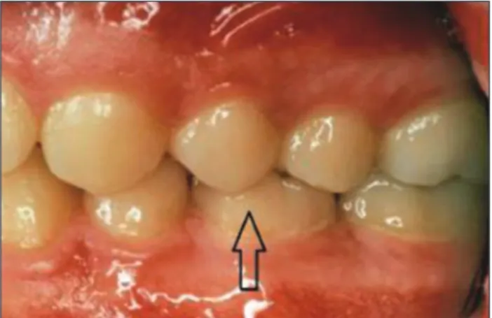
Figure 2: A 10-year-old boy with agenesis mandibular left second premolar. Note the severely resorbed primary molars.
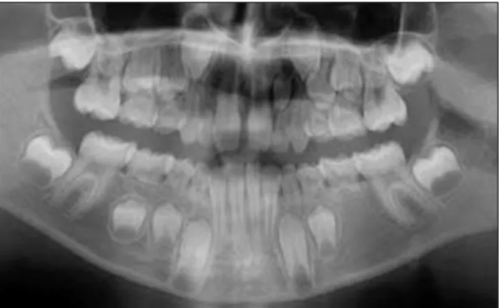
Figure 3: Agenesis of both mandibular second premolars. (a) Age 11.1 years minor infraocclusion, (b) 2 years later with severe infraocclusion, (c and d) 13 years of age.
d c
b a
13. Bjerklin K, Al-Najjar M, Kårestedt H, Andrén A. Agenesis of mandibular second premolars with retained primary molars: a longitudinal radiographic study of 99 subjects from 12 years of age to adulthood. Eur J Orthod 2008;30:254-61.
14. Sletten DW, Smith BM, Southard KA, Casko JS, Southard TE. Retained deciduous mandibular molars in adults: A radiographic study of long-term changes. Am J Orthod Dentofacial Orthop 2003;124:625-30.
15. Lee JE, Lee JH, Choi HJ, Kim SO, Song JS, Son HK, et al. Root resorption of primary teeth without permanent successors. J Korean Acad Pediatr Dent 2009;36:625-30.
16. Wu YM, Richards DW, Rowe DJ. Production of matrix-degrading enzymes and inhibition of osteoclast-like cell differentiation by fibroblast-like cells from the periodontal ligament of human primary teeth. J Dent Res 1999;78:681-9.
17. Hvaring CL, Øgaard B, Stenvik A, Birkeland K. e prognosis of retained primary molars without successors: Infraocclusion, root resorption and restorations in 111 patients. Eur J Orthod 2014;36:26-30.
18. Bjerklin K, Kurol J, Valentin J. Ectopic eruption of maxillary first permanent molars and association with other tooth and developmental disturbances. Eur J Orthod 1992;14:369-75. 19. Baccetti T. A controlled study of associated dental anomalies.
Angle Orthod 1998;68:267-74.
20. Garib DG, Peck S, Gomes SC. Increased occurrence of dental anomalies associated with second-premolar agenesis. Angle Orthod 2009;79:436-41.
How to cite this article: Bjerklin K. Orthodontic management of agenesis of mandibular second premolars. APOS Trends Orthod 2019;9(4):206-10.