R E S E A R C H
Open Access
Craniofacial height in relation to
cross-sectional maxillary and mandibular
morphology
Anna Klinge
1*, Karin Becktor
2, Christina Lindh
1and Jonas P Becktor
1Abstract
Background: In order to gain a better understanding of how growth of the alveolar bone is linked to the vertical development of the face, the purpose of this study was to investigate if there is an association between the cross-sectional morphology of the maxillary and mandibular bodies with the craniofacial height, using images from cone beam computed tomography (CBCT).
Methods: From 450 pre-treatment CBCT scans, 180 were selected to be included in the study. Lateral head images were generated from the CBCT scans and were used to categorise subjects into three groups based on their vertical craniofacial height. Cross-sectional images from CBCT volumes were reformatted of the maxillary and mandibular bodies at five locations in the maxilla and five in the mandible. Each image was measured at one height and two width measurements. Statistical analysis performed was the one-way analysis of variance with a Tukey post hoc test. A significance level of 5% was used in all comparisons.
Results: Patients with large vertical craniofacial height had a significantly higher cross-sectional area both in the maxilla and in the mandible. In the same group, the cross-sectional area was significantly thinner in the mandible compared with the other two groups, especially in the anterior region.
Conclusions: This study further highlights the close relationship between craniofacial height and alveolar bone dimensions and contributes with important knowledge for planning and follow-up of comprehensive dental- and orthodontic treatments.
Keywords: Craniofacial development, Treatment planning, Treatment timing, Cephalometrics, Biological basis of orthodontics
Background
Craniofacial growth is a complex process, and under-standing the factors involved is important in connection with orthodontic treatment of children and adolescents. The growth of the craniofacial complex can be divided into four different components: (1) growth of the cranial base, (2) growth of the maxilla, (3) growth of the man-dible and (4) growth of the dentoalveolar bone [1].
In the postnatal growth period, growth in the spheno-occipital synchondrosis cartilage and the cartilage of the condyle has a large influence on the vertical development
of the face, and adaptive growth of the maxillary sutures and growth of the dentoalveolar bone will fill out the face [2]. The link between the predominately genetically deter-mined growth of these two cartilages and growth of the dentoalveolar bone is also known as the dentoalveolar compensatory mechanism [3, 4]. The factors that are responsible for this dentoalveolar mechanism are not fully understood. However, it has been demonstrated that growth of the dentoalveolar bone is taking place in con-nection with tooth eruption and is accordingly dependent on a normal eruption process [4].
The vertical development of the face has a significant impact on the direction and magnitude of tooth eruption [5]. There is a noticeable variation in the eruption rate of the teeth, with a peak in eruption velocity at the time
* Correspondence:anna.klinge@mah.se
1Department of Oral and Maxillofacial Surgery and Oral Medicine, Faculty of
Odontology, Malmö University, SE-205 06 Malmö, Sweden Full list of author information is available at the end of the article
© The Author(s). 2017 Open Access This article is distributed under the terms of the Creative Commons Attribution 4.0 International License (http://creativecommons.org/licenses/by/4.0/), which permits unrestricted use, distribution, and reproduction in any medium, provided you give appropriate credit to the original author(s) and the source, provide a link to the Creative Commons license, and indicate if changes were made.
of the maximum pubertal rate of growth in body height [6]. Assuming that continued eruption is a compensa-tory mechanism for facial growth, it seems reasonable to conclude that eruption follows the general pattern of craniofacial growth [1, 4].
The growth of the alveolar bone is less genetically determined compared to the rest of the skeleton and can be influenced by functional conditions and orthodontic treatment [7]. Accordingly, knowledge of the expected growth pattern of the alveolar bone is important in rela-tion to orthodontic treatment of growing individuals. Furthermore, the morphology of the alveolar bone in three dimensions is important when it comes to the planning of orthodontic tooth movement as well as in connection with insertion of skeletal, temporary anchor-age devises (TADs) and dental implants for restorative purpose [8–13].
There are, however, only limited reports where the craniofacial morphology has been used as a guideline for evaluation of height and width of the alveolar bone. A comprehensive thesis on the various facial anatomical components and the relationship between them was pre-sented in 1966 by Solow [3]. In this thesis, a non-topographical positive correlation between height of the alveolar bone both in the maxilla and in the mandible, in relation to craniofacial height, was demonstrated in an adult sample. In more recent years, some studies have reported height and width of either maxillary or man-dibular alveolar bone in relation to craniofacial height in dry skulls [14, 15] or patients [16–18].
Since its introduction in the late 1990s, cone beam computed tomography (CBCT) has become a common imaging modality that provides a three-dimensional data set of the facial skeleton. To our knowledge, only one study investigated the relationship between facial height and alveolar bone morphology in the incisor and molar regions of the maxilla and the mandible [19]. The aim of this study was therefore, based on CBCT images of sub-jects, to explore the association between the cross-sectional morphology of the maxillary and mandibular bodies at several sites of the tooth-bearing regions and craniofacial height.
Methods
The study is conducted in accordance with the REport-ing of studies Conducted usREport-ing Observational Routinely-collected health Data (RECORD) guidelines [20].
Subjects
This study was conducted on pre-treatment CBCT scans collected from the archive at a private practice of ortho-dontics in Scandinavia. Images were obtained from pa-tients, females over 15 years and males over 16 years,
which were referred for specialist orthodontic and orthognathic treatment during 2008–2013.
CBCT scans from individuals with either missing per-manent teeth other than third molars, periodontal dis-ease visually detected on the radiographs, major asymmetries of the jaws or who had previously under-gone orthodontic treatment were excluded.
Radiography and categorisation of craniofacial height
Radiography was performed using an i-CAT CBCT (Im-aging Sciences International, Hatfield, Pennsylvania, USA). The patients were seated in an upright position, with the head in natural head position. Field of view (FOV) was 16 × 13 cm, acquisition time was 8.9 s with a voxel size of 0.3 mm and exposure was set at 120 kVp and 18.54 mAs with a total radiation dose of 458.6 mGy cm2. Calibration of the i-CAT CBCT was performed ac-cording to the manufacturer’s requirements twice a year. Lateral head images were generated from the CBCT scan using the i CAT software program. Cranio-facial height was determined cephalometrically using the Total Interactive Orthodontic Planning System (TIOPS) pro-gram [21].
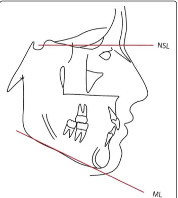
The measurement used was the inclination of the mandible in relation to the anterior cranial base. This is the angle formed between the nasion-sella line (NSL) and the mandibular line (ML), formed between menton and gonion (Fig. 1). The subjects were categorised in the three following groups: (i) a low angle (<27°), (ii) a nor-mal angle (27–37°) and (iii) a high angle (>37°).
Scans from 450 patients were available from CBCT ex-aminations performed during 2008–2013. After exclusion due to criteria previously stated, craniofacial height was cephalometrically determined for the rest of the scans. After identifying 60 scans from low-angle subjects, this number was set as the limit for the number of scans to be consecutively included in the normal- and high-angle groups for equal comparison. The number of individuals in each group was consequently set at 60 subjects.
By using iCATVision™ software [22], a fully reconstructed three-dimensional image with sagittal, coronal and axial slices was generated. When measuring in the maxilla, the nasion line (NL = a line from the anterior nasal spine con-tinuing posteriorly through the hard palate/nasal floor to the posterior nasal spine) was used as the horizontal plane and upper limit for vertical measurements in the maxilla. When measuring in the mandible, the lower border of the mandible was repositioned horizontally prior to measure-ments. All measurements were performed by one of the au-thors who only had access to the decoded CBCT scans and blinded to all other patient information. Measurement values were simultaneously recorded in a statistical spread sheet by one of the other authors.
Measurements of the cross-sectional maxillary and man-dibular height and width
Three-dimensional images were reconstructed by the iCATVision™ software (version 1.7.0.7, Imaging Sciences International) and saved in digital imaging [22]. Gener-ation of cross sections: A curved line was mapped to fit an axial CBCT section of the maxilla and mandible. Five cross sections (facial-lingual) were generated on each arch perpendicular to a tangent formed at these sites. Facial-lingual = width and vertical long axis = height.
The height and width of the maxillary and mandibular cross sections where measured between the dentition at ten locations, five in the maxilla and five in the man-dible. Each cross-sectional slide was measured at three sites, including one height and two width measurements (Fig. 2). The sites were named according to location in the following way: upper or lower jaw (UP or LO), molar (MOL), premolar (PRE) and midline (MID) and finally right (R) or left (L). Accordingly, UP-MOL-R stands for upper molar on the right side, height (H) (total length of the cross section), coronal width (W1) and apical width (W2) (Fig. 2).
The height was determined and measured by a line drawn through the long axis of the maxillary- or man-dibular cross-sectional area. When measuring in the maxilla, the line was drawn perpendicular from the al-veolar crest to the NL which was set as the upper limit. When measuring in the mandible, the line was drawn
perpendicular from the alveolar crest to the ML. The height measurements then represented a total height, which included both alveolar bone and basal bone. The width measurements were recorded perpendicular to the long axis line at two locations. One line was drawn at a distance of one third and the other at two thirds of the alveolar length. Fifteen measurements were performed in the maxilla and mandible, respectively, giving a total of 30 measurements (Fig. 2).
Measurements were performed by one observer. To calculate observer agreement, expressed as intra-class-correlation (ICC), the observer re-measured 20% of the sites in all the subjects after approximately 2 months. These sites were chosen by using random sampling.
Statistical analysis
All statistical analysis was performed using IBM SPSS software (version 22.0; IBM Corp Armonk, NY, USA). For all variables, the three groups (low, normal, high) were compared using a one-way analysis of variance with a Tukey post hoc test. A significance level of 5% was used in all comparisons.
Ethical approval
The study was conducted in accordance with the ethical principles of the World Medical Association Declaration of Helsinki (2008 version), approved by the Regional Ethical Review Board, Lund, Sweden (8 May 2014, Dnr 2014/288), the Danish Health and Medicines Authority, Denmark (20 July 2015, Sagsnr. 3-30-13-877/1/), and the
Fig. 2 Cross-sectional bone measurements of the maxilla and mandible. One height and two width measurements at each site were measured. The sites were named according to location to the neighbouring teeth. I.e UP-MOL R upper molar right (side). Fig. 1 Cephalometric analysis. Landmarks and reference lines used for
angular measurements in the lateral cephalograms, NSL nasion-sella line, ML mandibular line.
Danish Data Protection Agency, Denmark (7 July 2015, J.nr. 2015-41-4117).
Results
Height measurements
In both the maxilla and in the mandible, there was an increase in height from the molar region to the incisal region (midline) in all three groups. This increase was more pronounced in the high-angle group (displaying a steeper curve) and less pronounced in the low-angle group (a flatter curve) (Fig. 3a).
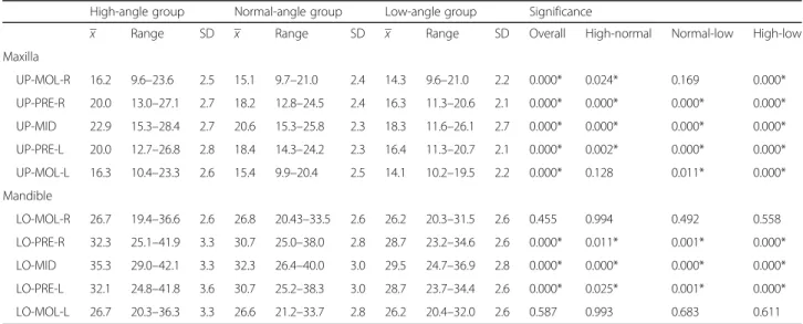
The midline area MID), the premolar area (UP-PRE) and the molar area (UP-MOL) of the maxillary cross section were significantly higher in the high-angle group compared to the normal- and the low-angle groups (Table 1).
The midline area (LO-MID) and the premolar area (LO-PRE) of the mandibular cross section were signifi-cantly higher in the high-angle group compared to the normal- and the low-angle group. However, in the molar region, there were no significant differences between the groups (Table 1).
The range in height of the bone in the midline area was between 11.6 mm (low angle) and 28.4 mm (high angle) in the maxilla (Table 1).
The range in height of the bone in the midline area was between 24.7 mm (low angle) and 42.1 mm (high angle) in the mandible (Table 1).
Width measurements
Concerning the cross-sectional coronal width measure-ments in the maxilla, Fig. 3b shows a V-shaped pattern, meaning that there was a decrease in width of the alveo-lar bone towards the midline area in all three groups. This decrease was more pronounced from the molar to premolar region for all three groups. Concerning the ap-ical width measurement, the figure showed that the cross section was narrower in the premolar region com-pared to the molar and midline area (Fig. 3b).
Concerning the cross-sectional coronal width mea-surements in the mandible, Fig. 3c showed a V-shaped pattern, meaning that there was a decrease in width to-wards the midline area in all three groups. With refer-ence to the apical width measurement, the figure showed that the cross section was significantly wider in the midline area compared to the premolar and molar area in all three groups (Fig. 3c).
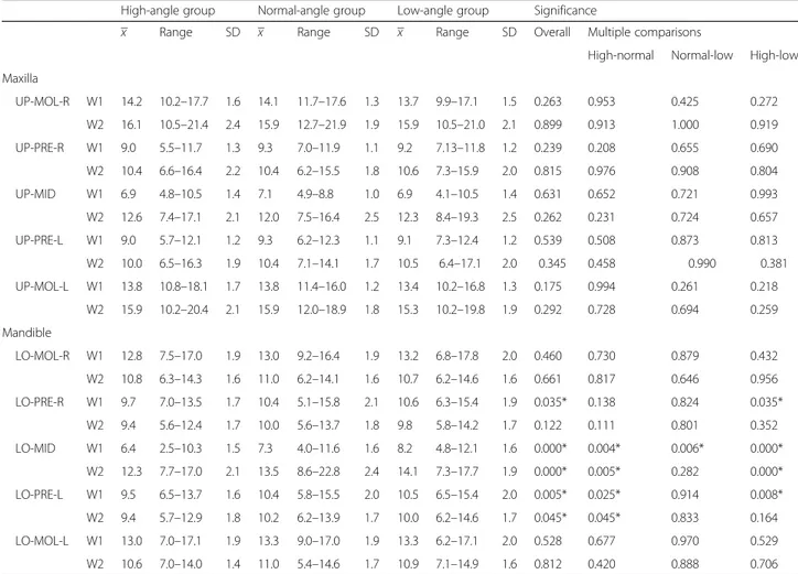
There were no statistically significant differences in width measurements in the maxilla when comparing the three groups, neither in the coronal nor in the apical width measurements (Table 2).
In the coronal and apical midline area in the mandible (LO-MID), the bone was significantly narrower in the high-angle group compared to the normal- and the
low-angle groups. In the molar area, there were no statistically significant differences in the coronal or in the apical width measurements between the three groups (Table 2).
The range in width, of the cross sections in the cor-onal midline area, was between 4.1 mm (low angle) and 10.5 mm (high angle/low angle) in the maxilla (Table 2).
The range in width of the coronal part of the cross sec-tion in the midline area was between 2.5 mm (high angle) and 12.1 mm (low angle) in the mandible (Table 2).
Gender differences
In a majority of the cross-sectional measurements (73%), significant differences were evident; male subjects tended to have wider and higher cross sections compared to fe-male subjects. Males displayed approximately 1 mm larger dimension of the maxillary and mandibular bone both in vertical and horizontal measurements (Table 3).
Intra-observer agreement
One thousand eighty (20%) out of 5400 measurements were randomly selected for re-measurement, and intra-class correlation (ICC) was calculated for intra-observer agreement. The correlation in height measures were be-tween 0.858 and 0.989. The correlation in width I mea-sures were between 0.762 and 0.944, and the correlation in width II measures were between 0.696 and 0.923.
Discussion
As CBCT is a rather new imaging modality, introduced in the late 1990s, there were no European guidelines on the use of CBCT for different dental- and maxillofacial conditions at the time when the majority of images used in this study were obtained. In 2012, European guide-lines for the use of CBCT were published [23] and there is a continuous interest in stating precise selection cri-teria following the principle of As Low As Reasonable Achievable (ALARA) for the use of X-rays [24]. The se-lection criteria for performing CBCT were craniofacial-and skeletal deviations, impacted teeth, patients planned for orthognathic surgery and trauma patients, none of which can be specifically disputed. It might be, however, if a study like this should be performed prospectively that selection criteria might be adjusted. Having access to these retrospectively collected images, from which a selection was used for this study, we considered it un-ethical not to obtain clinically valuable data. Ethical ap-proval was obtained for the use of these available CBCT images for research purpose.
Craniofacial height was determined cephalometrically using the Total Interactive Orthodontic Planning System (TIOPS) program [21] by one of the authors, a specialist in orthodontics. This analysis is a standard tool in ortho-dontic assessment and treatment planning. Observer re-liability of this technique has shown to be high, both in
conventional lateral radiographs and in CBCT scull im-ages [25, 26]. Height and width measurements of the maxillary and mandibular bodies was repeated at 20% of the sites showing a moderate to almost perfect agree-ment interpreted according to Landis and Koch [27].
Craniofacial morphogenesis evolves through an inter-action between the development of the facial tissue and that of the supporting skeletal framework. In the present study, focus has been on an aspect of craniofacial mor-phogenesis, which is known as the dentoalveolar com-pensatory mechanism. The present study indicates that in a high-angle craniofacial pattern, the bone in the mandibular midline is high and narrow, whereas in the low-angle morphology, the bone is shorter and wider.
Maxillary height
Gracco et al. showed no statistical difference in the midline area of the maxilla between high-, normal- and low-angle subjects on CBCT images. However, they investigated only the anterior region of the maxilla [16]. These results are not in agreement with our findings, which could be due to that growing individuals (12–40 years) were part of the study and some changes of the morphology of the maxil-lary bone must be anticipated to occur as the individuals
grow [16]. The differences could also be due to a slightly different measuring technique. In a recent investigation by Sadek et al. [19], a significant difference in height of the maxillary cross-sectional sites in the midline area was also reported. However, no significant differences were found in the molar region.
Mandibular height
The subjects with the high-angle morphology revealed a significant higher mandibular cross section in the midline and premolar region when compared to the other groups. These results are in accordance with Sadek et al. [19] and almost in accordance with the findings by Swasty et al. [18]. The only difference was that Swasty et al. demon-strated a negative correlation of the mandibular body in the molar region, where the high-angle group had a shorter body in the molar region compared to the low-angle group [18]. The same feature was also described by Kohakura et al. [28]. However, in the present investigation, we could not demonstrate a statistical difference in height in the molar region when comparing the groups. The dif-ference between the findings can be explained by the fact that individuals who were still growing were included in the Swasty study [18].
(See figure on previous page.)
Fig. 3 Cross-sectional height and width morphology measurements of the maxilla and mandible in the three groups (low-, normal- and high angle). UP-MOL-R (upper molar right), UP-PRE-R (upper premolar right), UP-MID (upper midline), UP-PRE-L (upper premolar left), UP-MOL-L (upper molar left), LO-MOL-R (lower molar right), LO-PRE-R (lower premolar right), LO-MID (lower midline), LO-PRE-L (lower premolar left), LO-MOL-L (lower molar left). Y-axis (mm). a Height (H) measurement at five cross-sectional sites in the maxilla and five in the mandible. b Width (W) measurement at five cross-sectional sites in the maxilla. W1 demonstrates the coronal width measurements, and W2 demonstrates the apical width measurements. c Width (W) measure-ment at five cross-sectional sites in the mandible. W1 demonstrates the coronal width measurements, and W2 demonstrates the apical width measure-ments. a Height in maxilla and mandible. b Coronal width. c Apical width
Table 1 Bone height measurements of the maxillary and mandibular body in millimeters
High-angle group Normal-angle group Low-angle group Significance
x Range SD x Range SD x Range SD Overall High-normal Normal-low High-low
Maxilla UP-MOL-R 16.2 9.6–23.6 2.5 15.1 9.7–21.0 2.4 14.3 9.6–21.0 2.2 0.000* 0.024* 0.169 0.000* UP-PRE-R 20.0 13.0–27.1 2.7 18.2 12.8–24.5 2.4 16.3 11.3–20.6 2.1 0.000* 0.000* 0.000* 0.000* UP-MID 22.9 15.3–28.4 2.7 20.6 15.3–25.8 2.3 18.3 11.6–26.1 2.7 0.000* 0.000* 0.000* 0.000* UP-PRE-L 20.0 12.7–26.8 2.8 18.4 14.3–24.2 2.3 16.4 11.3–20.7 2.1 0.000* 0.002* 0.000* 0.000* UP-MOL-L 16.3 10.4–23.3 2.6 15.4 9.9–20.4 2.5 14.1 10.2–19.5 2.2 0.000* 0.128 0.011* 0.000* Mandible LO-MOL-R 26.7 19.4–36.6 2.6 26.8 20.43–33.5 2.6 26.2 20.3–31.5 2.6 0.455 0.994 0.492 0.558 LO-PRE-R 32.3 25.1–41.9 3.3 30.7 25.0–38.0 2.8 28.7 23.2–34.6 2.6 0.000* 0.011* 0.001* 0.000* LO-MID 35.3 29.0–42.1 3.3 32.3 26.4–40.0 3.0 29.5 24.7–36.9 2.8 0.000* 0.000* 0.000* 0.000* LO-PRE-L 32.1 24.8–41.8 3.6 30.7 25.2–38.3 3.0 28.7 23.7–34.4 2.6 0.000* 0.025* 0.001* 0.000* LO-MOL-L 26.7 20.3–36.3 3.3 26.6 21.2–33.7 2.8 26.2 20.4–32.0 2.6 0.587 0.993 0.683 0.611
UP-MOL-R upper molar right, UP-PRE-R upper premolar right, UP-MID upper midline, UP-PRE-L upper premolar left, UP-MOL-L upper molar left, LO-MOL-R lower molar right, LO-PRE-R lower premolar right, LO-MID lower midline, LO-PRE-L lower premolar left, LO-MOL-L lower molar left, x mean value
*Statistically significant difference (p value < 0.05)
In our study as well as in Swasty et al. and Sadek et al. [18, 19], a pronounced difference in height from the molar to the midline region was described in the high-angle group and the least change in height proceeding from the molars to the midline was displayed in the low-angle group. Considering the growth pattern of the man-dible, these observations reflect the rotation of the mandible, where the incisors have to erupt signifi-cantly in order to compensate for the posterior rota-tions and the molars have to erupt less, whereas the incisors have to erupt less in subjects with an anterior rotating mandible [5].
Maxillary width
No significant difference was found in width measurements in the maxilla when comparing the three groups, neither in the coronal nor in the apical width measurements in the present study. This is not in accordance with the re-sults reported by Gracco et al. [16] who described a
negative correlation in width measurements in the midline region between subjects with different vertical craniofacial dimensions. In the study by Sadek et al. [19], significant difference between high- and low-angle individuals in width was reported in the midline area both in the coronal and apical region. The reasons for this difference could be the following: (1) they did not measure between the teeth, but across the incisors, and (2) the measurements were obtained at the mid-root level and at the apex making the measurements dependent more on the lengths of the root than on the length of the alveolar bone. Since the length of the bone seems to vary more than the length of the root, which is a more stable reference point, the cross-sectional measurements will not be equal.
Mandibular width
Figure 3c demonstrated a V-shaped pattern when pre-senting the measurements of the coronal region, as the width decreased along the dental arch towards the
Table 2 Bone width in millimeters of the maxillary and mandibular body
High-angle group Normal-angle group Low-angle group Significance
x Range SD x Range SD x Range SD Overall Multiple comparisons
High-normal Normal-low High-low Maxilla UP-MOL-R W1 14.2 10.2–17.7 1.6 14.1 11.7–17.6 1.3 13.7 9.9–17.1 1.5 0.263 0.953 0.425 0.272 W2 16.1 10.5–21.4 2.4 15.9 12.7–21.9 1.9 15.9 10.5–21.0 2.1 0.899 0.913 1.000 0.919 UP-PRE-R W1 9.0 5.5–11.7 1.3 9.3 7.0–11.9 1.1 9.2 7.13–11.8 1.2 0.239 0.208 0.655 0.690 W2 10.4 6.6–16.4 2.2 10.4 6.2–15.5 1.8 10.6 7.3–15.9 2.0 0.815 0.976 0.908 0.804 UP-MID W1 6.9 4.8–10.5 1.4 7.1 4.9–8.8 1.0 6.9 4.1–10.5 1.4 0.631 0.652 0.721 0.993 W2 12.6 7.4–17.1 2.1 12.0 7.5–16.4 2.5 12.3 8.4–19.3 2.5 0.262 0.231 0.724 0.657 UP-PRE-L W1 9.0 5.7–12.1 1.2 9.3 6.2–12.3 1.1 9.1 7.3–12.4 1.2 0.539 0.508 0.873 0.813 W2 10.0 6.5–16.3 1.9 10.4 7.1–14.1 1.7 10.5 6.4–17.1 2.0 0.345 0.458 0.990 0.381 UP-MOL-L W1 13.8 10.8–18.1 1.7 13.8 11.4–16.0 1.2 13.4 10.2–16.8 1.3 0.175 0.994 0.261 0.218 W2 15.9 10.2–20.4 2.1 15.9 12.0–18.9 1.8 15.3 10.2–19.8 1.9 0.292 0.728 0.694 0.259 Mandible LO-MOL-R W1 12.8 7.5–17.0 1.9 13.0 9.2–16.4 1.9 13.2 6.8–17.8 2.0 0.460 0.730 0.879 0.432 W2 10.8 6.3–14.3 1.6 11.0 6.2–14.1 1.6 10.7 6.2–14.6 1.6 0.661 0.817 0.646 0.956 LO-PRE-R W1 9.7 7.0–13.5 1.7 10.4 5.1–15.8 2.1 10.6 6.3–15.4 1.9 0.035* 0.138 0.824 0.035* W2 9.4 5.6–12.4 1.7 10.0 5.6–13.7 1.8 9.8 5.8–14.2 1.7 0.122 0.111 0.801 0.352 LO-MID W1 6.4 2.5–10.3 1.5 7.3 4.0–11.6 1.6 8.2 4.8–12.1 1.6 0.000* 0.004* 0.006* 0.000* W2 12.3 7.7–17.0 2.1 13.5 8.6–22.8 2.4 14.1 7.3–17.7 1.9 0.000* 0.005* 0.282 0.000* LO-PRE-L W1 9.5 6.5–13.7 1.6 10.4 5.8–15.5 2.0 10.5 6.5–15.4 2.0 0.005* 0.025* 0.914 0.008* W2 9.4 5.7–12.9 1.8 10.2 6.2–13.9 1.7 10.0 6.2–14.6 1.7 0.045* 0.045* 0.833 0.164 LO-MOL-L W1 13.0 7.0–17.1 1.9 13.3 9.0–17.0 1.9 13.3 6.2–17.1 2.0 0.528 0.677 0.970 0.529 W2 10.6 7.0–14.0 1.4 11.0 5.4–14.6 1.7 10.9 7.1–14.9 1.6 0.812 0.420 0.888 0.706
W1 demonstrates the coronal width measurements, and W2 demonstrates the apical width measurements. Y-axis (mm)
UP-MOL-R upper molar right, UP-PRE-R upper premolar right, UP-MID upper midline, UP-PRE-L upper premolar left, UP-MOL-L upper molar left, LO-MOL-R lower molar right, LO-PRE-R lower premolar right, LO-MID lower midline, LO-PRE-L lower premolar left, LO-MOL-L lower molar left, x mean value
midline. The same feature was described by Swasty et al. [18]. In the alveolar coronal region, the mandible was significantly thinner in the midline and premolar region in all three groups. This is in accordance with the results of both Swasty et al. and Sadek et al. [18–19]. Concern-ing the apical measurement, the cross section was sig-nificantly wider in the midline area compared to the premolar and molar area in all three groups (Fig. 3c). This could be due to the anatomy of the mental spine and the base of the mandible in the front region.
Gender differences
A general pattern was displayed, where the males pre-sented a greater height and width compared to the fe-males. Swasty et al. [18] described that there was no statistical differences in cortical bone thickness between the genders. However, they found a statistical difference in height of the cross sections between males and fe-males. Accordingly, it seems reasonable to conclude that
at least in height of the bone, there seems to be a gender-correlated difference.
Clinical implications
The results of this investigation may support the fact that the dentoalveolar compensatory mechanism aims to maintain a functional occlusion in connection with cra-niofacial growth. In the high-angle group, the alveolar bone had to grow more in order to compensate or partly compensate for the vertical craniofacial growth, and therefore, the bodies of the maxilla and mandible became higher. The fact that the arches became nar-rower in the midline of the mandible might be due to the difference in loading. It can be hypothesised that the alveolar bone in the high-angle subjects are less loaded than the bone in the low-angle group. In the low-angle group, the root resides in most of the arch, whereas in the high angle group, the root resides only in part of the body, which according to Wolff’s law will result in a less developed bone [29].
In orthodontic treatment, it is very important to understand how the development of the lower third of the face is closely linked to the dentoalveolar tory mechanism, because the dentoalveolar compensa-tory mechanism can be influenced in connection with treatment.
Also during active orthodontic treatment, knowledge of the morphology of the alveolar bone is important. In thin ridges, the buccal and lingual cortices can contact or be in close approximation with very little cancellous bone. The risk for root resorption increases if the roots are torqued into the cortex. In addition, there is a risk for moving teeth out of alveolar bone that has a narrow width [30].
In connection with insertions of skeletal TADs, the difference in the morphology of the cross section of the maxillary and mandibular bodies in different growth pat-terns is valuable information for therapy planning and adequate therapy. It has been proven that in many situa-tions, the palate is an ideal area for screw insertion where the screw is inserted at the third rugae with the tip in an anterior direction [9–11]. However, knowledge of the anatomy at the insertion site is important, and the present study has demonstrated that the alveolar bone is short in the mid-palatal area in the low-angle face com-pared to the normal- and high-angle cases, with less space superior to the apex of the incisors. Accordingly, an increased risk of root damage and insertion of the screw into the nasal cavity can be expected in low-angle patients.
Likewise, during treatment planning for growing patients with congenital missing teeth, the decision to replace missing teeth with implants or by orthodontic
Table 3 Difference between male and female when comparing measurements of jaw bone height and width in millimeters
Male Female
Maxilla
UP-MID Height 21.0 20.4
Width I 7.5* 6.7*
Width II 13.4* 11.7*
Right Left Right Left
UP-PRE Height 19.0* 19.1* 17.7* 17.8* Width I 9.7* 9.7* 8.9* 8.8* Width II 11.0* 10.9* 10.2* 9.9* UP-MOL Height 16.4* 16.5* 14.6* 14.6* Width I 14.7* 14.3* 13.6* 13.3* Width II 17.0* 16.4* 15.4* 15.1* Mandible LO-MID Height 34.6* 31.2* Width I 7.4 7.2 Width II 14.0* 12.9*
Right Left Right Left
LO-PRE Height 32.2* 32.4* 29.7* 29.5* Width I 10.6* 10.7* 10.0* 9.9* Width II 10.0 10.1 9.6 9.7 LO-MOL Height 28.3* 28.3* 25.6* 25.6* Width I 13.3 13.4 12.9 13.0 Width II 10.8 10.7 10.9 10.9
Right side, left side, midline (only one/jaw). Width I: The coronal width measurements. Width II: The apical width measurements
UP-MID upper midline, UP-PRE upper premolar, UP-MOL upper molar, LO-MID lower midline, LO-PRE lower premolar, LO-MOL lower molar
*Statistically significant difference (p < 0.05)
closure could be influenced by the expected development of the dentoalveolar compensatory mechanism. The risk of infra-occlusion of the implant supported crowns might be higher in the high-angle group, where the incisors seem to erupt more.
Vertical control is a common treatment approach in high-angle cases, and appliances such as skeletal anchor-age devices are described as very useful [31]. However, considering the A-shaped morphology (Fig. 3a) of the maxillary body in the vertical dimension, this treatment approach will actually accentuate the difference in height between the molar and incisor region. In orthognathic surgery, the aim is to increase the vertical dimension in the posterior region, which will flatten the same curve. How this difference in treatment strategies, aiming to solve the same problem, will influence final stability, air-ways and function should be investigated further.
In treatment with dental implants, a careful preopera-tive evaluation is important in order to achieve a pre-dictable, successful aesthetic outcome. The buccal bone wall thickness is of crucial importance when it comes to selection of appropriate treatment approach [32]. The overall width of the mandibular body in a high-angle subject tend to be thinner, which probably gives a thin-ner alveolar bone. In the present study, the range in width of the coronal part of the cross section in the mid-line area was between 2.5 mm (high angle) and 12.1 mm (low angle) in the mandible (Table 2). As suggested by Buser et al., to achieve an aesthetic and functional suc-cess in dental implant treatment, it is important to take into account the three-dimensional aspect of the alveolar bone [13]. It is recommended that the dental implant should be placed inside the biological envelope of the alveolar bone in order to obtain the best conditions for success [8].
Conclusions
The main findings of the present study were:
(1) The high-angle group had a significantly longer cross-sectional area of the maxillary and mandibular body compared to the other two groups.
(2) The high-angle group also had significantly nar-rower mandibular cross-sectional body compared to the other two groups, especially in the anterior region.
In this retrospective study, it has been demonstrated that there is a close link between vertical craniofacial de-velopment and the morphology of cross-sectional sites of the maxillary and mandibular bodies for patients referred for orthodontic and orthognathic treatment. Understand-ing this biological link is important in connection with many comprehensive dental- and orthodontic treatments.
Acknowledgements
There are no acknowledgements.
Authors’ contributions
JB and KB have made substantial contributions to the conception and design of the study. Acquisition of data and analysis and interpretation of data have been made by JB, KB and AK. JB, KB, CL and AK have been involved in drafting the manuscript or revising it critically for important intellectual content. JB and CL also contributed to the general supervision. All authors contributed significantly to this manuscript and have given final approval of the version to be published.
Ethics approval and consent to participate
The study was conducted in accordance with the ethical principles of the World Medical Association Declaration of Helsinki (2008 version), approved by the Regional Ethical Review Board, Lund, Sweden (8 May 2014, Dnr 2014/ 288), the Danish Health and Medicines Authority, Denmark (20 July 2015, Sagsnr. 3-30-13-877/1/), and the Danish Data Protection Agency, Denmark (7 July 2015, J.nr. 2015-41-4117).
Consent for publication
Not applicable. This manuscript has not been published elsewhere in part or in entirety and is not under consideration by another journal.
Competing interests
The authors declare that they have no competing interests.
Publisher’s Note
Springer Nature remains neutral with regard to jurisdictional claims in published maps and institutional affiliations.
Author details
1Department of Oral and Maxillofacial Surgery and Oral Medicine, Faculty of
Odontology, Malmö University, SE-205 06 Malmö, Sweden.2Clinic for Orthodontics and Oral Surgery, Strandvejen 116A, 2900 Hellerup, Copenhagen, Denmark.
Received: 2 May 2017 Accepted: 17 August 2017 References
1. Solow B. The dentoalveolar compensatory mechanism: background and clinical implications. Br J Orthod. 1980;7:145–61.
2. Coben SE. The spheno-occipital synchondrosis: the missing link between the profession’s concept of craniofacial growth and orthodontic treatment. Am J Orthod Dentofac Orthop. 1998;114:709–14.
3. Solow B. The pattern of craniofacial associations: a morphological and methodological correlation and factor analysis study on young male adults. PhD. Thesis, University of Copenhagen. Acta Odontol Scand; 1966;24(Suppl 46). pp. 174.
4. Björk A. The face in profile. An anthropological X-ray investigation on Swedish children and conscripts. PhD. Thesis, Uppsala University Svensk Tandläkare Tidskrift. 1947;40.
5. Björk A. Käkarnas tillväxt och utveckling i relation till kraniet i dess helhet. Holst JJ, Østby BN, Osvald O, editors. Nordisk Klinisk Odontologi. Copenhagen. Forlaget for faglitteratur; 1964. p. 1–44.
6. Siersbaek-Nielsen S. Rate of eruption of central incisors at puberty: an implant study on eight boys. Tandlaegebladet. 1971;75:1288–95. 7. Bjork A, Skieller V. Normal and abnormal growth of the mandible. A
synthesis of longitudinal cephalometric implant studies over a period of 25 years. Eur J Orthod. 1983;5:1–46.
8. Qahash M, Susin C, Polimeni G, Hall J, Wikesjo UM. Bone healing dynamics at buccal peri-implant sites. Clin Oral Implants Res. 2008;19:166–72. 9. Wehrbein H. Bone quality in the midpalate for temporary anchorage
devices. Clin Oral Implants Res. 2009;20:45–9.
10. Schatzle M, Mannchen R, Zwahlen M, Lang NP. Survival and failure rates of orthodontic temporary anchorage devices: a systematic review. Clin Oral Implants Res. 2009;20:1351–9.
11. Wilmes B, Nanda R, Nienkemper M, Ludwig B, Drescher D. Correction of upper-arch asymmetries using the Mesial-Distalslider. J Clin Orthod. 2013;47:648–55.
12. Feng X, Li J, Li Y, Zhao Z, Zhao S, Wang J. Effectiveness of TAD-anchored maxillary protraction in late mixed dentition. Angle Orthod. 2012;82:1107–14.
13. Buser D, Martin W, Belser UC. Optimizing esthetics for implant restorations in the anterior maxilla: anatomic and surgical considerations. Int J Oral & Maxillofac Implants. 2004;19:43–61.
14. Tsunori M, Mashita M, Kasai K. Relationship between facial types and tooth and bone characteristics of the mandible obtained by CT scanning. Angle Orthod. 1998;68:557–62.
15. Masumoto T, Hayashi I, Kawamura A, Tanaka K, Kasai K. Relationships among facial type, buccolingual molar inclination, and cortical bone thickness of the mandible. Eur J Orthod. 2001;23:15–23.
16. Gracco A, Lombardo L, Mancuso G, Gravina V, Siciliani G. Upper incisor position and bony support in untreated patients as seen on CBCT. Angle Orthod. 2009;79:692–702.
17. Gracco A, Luca L, Bongiorno MC, Siciliani G. Computed tomography evaluation of mandibular incisor bony support in untreated patients. Am J Orthod Dentofac Orthop. 2010;138:179–87.
18. Swasty D, Lee J, Huang JC, Maki K, Gansky SA, Hatcher D, et al. Cross-sectional human mandibular morphology as assessed in vivo by cone-beam computed tomography in patients with different vertical facial dimensions. Am J Orthod Dentofac Orthop. 2011;139:377–89.
19. Sadek MM, Sabet NE, Hassan IT. Alveolar bone mapping in subjects with different vertical facial dimensions. Eur J Orthod. 2015;37:194–201. 20. Benchimol EI, Smeeth L, Guttmann A, Harron K, Moher D, Petersen I,
Sørensen HT, von Elm E, Langan SM. The REporting of studies Conducted using Observational Routinely-collected health Data (RECORD) statement. PLoS Med. 2015:1–22.
21. Bjoern-Joergensen JB, Bjoern-Joergensen PB. Tiops. 2015. Available from: http://www.tiops.com/. Accessed 28 Feb 2017.
22. i-CAT Cone Beam 3D Imaging. 2015. Available from: http://www.i-cat.com/. Accessed 20 Nov 2016.
23. European Commission. Cone beam CT for dental and maxillofacial radiology (evidence-based guidelines). 2012. http://www.sedentexct.eu/files/radiation_ protection_172.pdf. Accessed 04 Apr 2017.
24. International Commission on Radiation Protection. The 2007
recommendations of the international commission on radiation protection. ICRP publication 103, 2007. Ann ICRP. 37:2–4.
25. AlBarakati SF, Kula KS, Ghoneima AA. The reliability and reproducibility of cephalometric measurements: a comparison of conventional and digital methods. Dentomaxillofacial Radiology. 2012;41:11–7.
26. Hariharan A, Diwakar NR, Jayanthi K, Hema HM, Deepukrishna S, Ghaste SR. The reliability of cephalometric measurements in oral and maxillofacial imaging: cone beam computed tomography versus two-dimensional digital cephalograms. Indian J Dent Res. 2016;27:370–7.
27. Landis JR, Koch GG. The measurement of observer agreement for categorical data. Biometrics. 1977;33:159–74.
28. Kohakura S, Kasai K, Ohno I, Kanazawa E. Relationship between maxillofacial morphology and morphological characteristics of vertical sections of the mandible obtained by CT scanning. J Nihon Univ Sch Dent. 1997;39:71–7. 29. Frost HM. Wolff’s law and bone’s structural adaptations to mechanical
usage: an overview for clinicians. Angle Orthod. 1994;64:175–88. 30. Handelman CS. The anterior alveolar: its importance in limiting orthodontic
treatment and its influence on the occurrence of iatrogenic sequelae. Angle Orthod. 1996;66:95–109.
31. Scheffler NR, Proffit WR, Phillips C. Outcomes and stability in patients with anterior open bite and long anterior face height treated with temporary anchorage devices and a maxillary intrusion splint. Am J Orthod Dentofac Orthop. 2014;146:594–602.
32. Braut V, Bornstein MM, Belser U, Buser D. Thickness of the anterior maxillary facial bone wall-a retrospective radiographic study using cone beam computed tomography. Int J Periodontics Restorative Dent. 2011;31:125–31.


