Impact of Surface Modification with Gold
Nanoparticles on the Bioelectrocatalytic
Parameters of Immobilized Bilirubin
Oxidase
D. V. Pankratov1,2, Y. S. Zeifman2, А. V. Dudareva3, G. K. Pankratova1, M. E. Khlupova1, Y. M. Parunova2, D. N. Zajtsev3, N. F. Bashirova3, V. O. Popov1,2, and S. V. Shleev1,2,3*
1 A.N. Bach Institute of Biochemistry of Russian Academy of Sciences, Leninsky Ave. 33, building 2,
119071 Moscow, Russia
2 National Research Center “Kurchatov Institute”, Akademika Kurchatova Sq. 1, 123182 Moscow,
Russia
3 I.G. Petrovsky Bryansk State University, Bezhitskaya St. 14, 241036 Bryansk, Russia *E-mail: shleev@inbi.ras.ru
Received 14.12.2013
Copyright © 2014 Park-media, Ltd. This is an open access article distributed under the Creative Commons Attribution License,which permits unrestricted use, distribution, and reproduction in any medium, provided the original work is properly cited.
abStract We unveil experimental evidence that put into question the widely held notion concerning the impact
of nanoparticles on the bioelectrocatalytic parameters of enzymatic electrodes. Comparative studies of the bio-electrocatalytic properties of fungal bilirubin oxidase from Myrothecium verrucaria adsorbed on gold electrodes, modified with gold nanoparticles of different diameters, clearly indicate that neither the direct electron transfer
rate (standard heterogeneous electron transfer rate constants were calculated to be 31±9 s-1) nor the biocatalytic
activity of the adsorbed enzyme (bioelectrocatalytic constants were calculated to be 34±11 s-1) depends on the
size of the nanoparticles, which had diameters close to or larger than those of the enzyme molecules.
KeyWordS gold nanoparticle; bilirubin oxidase; direct electron transfer; bioelectrocatalysis.
abbreViationS3D – three-dimensional; Areal – real/electrochemical surface area; CV – cyclic voltammogram;
ET – electron transfer; DET – direct electron transfer; k0– standard heterogeneous electron transfer rate
con-stant; kcatapp – apparent bioelectrocatalytic constant; MCO – blue multicopper oxidase; MvBOx – Myrothecium
verrucaria bilirubin oxidase; AuNPn – gold nanoparticles with diameter of n nm; AuNPn/Au – gold electrode
modified with AuNPn before and after cycling in sulfuric acid (m-AuNP/Au u-AuNP/Au, respectively); ThLc –
Trametes hirsuta laccase; PBS – phosphate buffered saline; NHE – normal hydrogen electrode; SEM – scanning
electron microscopy; Aspr – absorbance maximum; А450 – absorbance at the wavelength of 450 nm; Γ – enzyme
surface concentration; jmax – maximum current density.
introduction
numerous studies have reported on effective direct electron transfer (Det) of various enzymes (including blue multicopper oxidase – McO) immobilized on the surface of nanostructured electrodes with metal and carbon nanoparticles, carbon nanotubes, graphene, etc. [1–3]. An increase in the bioelectrocatalytic cur-rent when using nanostructured surfaces was regard-ed as the defining evidence behind the acceleration of the Det reaction in these studies; however, neither a quantitative comparative analysis of Det based on a voltammograms analysis, nor a calculation of the stand-ard constants of the heterogeneous electron transfer reaction (k0) has been performed. Moreover, there are serious discrepancies in the data even for a single en-zyme (in particular, laccase from the Trametes hirsuta
(ThLc) fungus immobilized on the gold surface). For in-stance, the use of AunP and nanoporous gold helped to increase Det [4], whereas an extremely low bioelectro-catalytic activity of the enzyme was observed for Det [5] in nano/microstructured silicon chips modified with gold with the enzyme immobilized on their surface. Since the use of bioelectrodes without a nano-modified surface leads to a very low heterogeneous transfer rate and sometimes to a complete absence of Det, the rou-tine explanation for enzyme “nanobinding” is the ori-entation of the enzyme on the nanostructured surface, which contributes to Det.
Despite the fact that a possible dependence of k0 on the size of the metal or carbon nanoparticles has yet to be studied and that the two opposite dependences of the bioelectrocatalytic oxygen reduction current
on the diameter of AunP on electrodes modified with McO were recently demonstrated (e.g., [6]), the belief remains that the size of AunP used for electrode na-nomodification is a very important factor that deter-mines et in the reactions between a redox enzyme and the electrode surface. this study offers experimental results that demonstrate the unlikeliness of this hy-pothesis.
eXPerimental
Materials and Methods
na2HPO4∙2H2O, naH2PO4∙H2O, nacl, HAucl4∙3H2O, H2O2, H2SO4, naBH4, and sodium citrate were pur-chased from Sigma-Aldrich GmbH (Germany) and used without further purification. Oxygen was ac-quired from AGA Gas AB (Sweden). Buffers and other solutions were prepared using deionized water (18 MΩ∙cm) produced using a PureLAB uHQ II sys-tem (eLGA Labwater, uK). All experiments were per-formed at room temperature in PBS (pH 7.4) consisting of a 50 mM HPO42-/H
2PO4- solution containing 150 mM
nacl.
MvBOx was a gift from Amano enzyme Inc.
(Ja-pan).
electrochemical measurements were performed us-ing a μAutolab type III/FrA2 potentiostat/galvano-stat (Metrohm Autolab BV, the netherlands) using a three-electrode circuit with a saturated calomel refer-ence electrode (242 mV vs. normal hydrogen electrode, nHe) and a platinum wire as an auxiliary electrode.
Sonication was performed using a ultrasonic clean-er XB2 bath (VWr Intclean-ernational Ltd., uK). SeM was performed on a FeI nova nanoLab 600 high-resolu-tion scanning electron microscope (the netherlands). Spectrophotometric studies were carried out using a PharmaSpec uV-1700 uV-visible spectrophotometer (china).
nanoparticles with a diameter of 5 to 60 nm were synthesized to study the impact of the AunP size on the biocatalytic properties of MvBOx.
Synthesis of gold nanoparticles with a preset particle size
AunP with the expected diameter of 5 nm (AunP5, Fig.
1A) were synthesized as described in [7]. 50 ml of a 250 μM HAucl4 solution was stirred for 1 min at room tem-perature, then 1111 μl of a 38.8 mM sodium citrate solu-tion was added to the initial solusolu-tion, and the mixture was stirred for another 1 min. next, 555 μl of a freshly prepared 0.075% (wt.) naBH4 solution in a 38.8 mM so-dium citrate solution was poured into the reaction mix-ture and the solution was stirred for another 5 min.
AunP with a diameter of 20–60 nm (Fig. 1A) were synthesized using sodium citrate as a reductant. 50 ml of a 250 μM HAucl4 solution was brought to boil under constant stirring; 750, 500, or 260 μl of a 1% (wt.) so-dium citrate solution was subsequently added to obtain
AunP with diameters of 20, 40, and 60 nm (AunP20,
AunP40 and AunP60), respectively. After adding sodi-um citrate, the mixture was incubated for 10 min under constant stirring without heating.
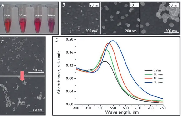
Fig. 1. A. Photos of the colloidal solutions of syn-thesized AuNPs; B. SEM images of AuNP/Au samples; C. SEM image of AuNP40/Au before (top) and after (bottom) 2 cycles in H2SO4: D. Absorbance spectra of AuNP suspensions with different diameters А B 5 nm 20 nm 40 nm 60 nm 20 nm 40 nm 60 nm 200 nm 200 nm 200 nm C 500 nm 500 nm D
Absorbance, rel. units
Wavelength, nm 5 nm 20 nm 40 nm 60 nm 400 450 500 550 600 650 700 750 0.20 0.16 0.12 0.08 0.04 0.00
the diameter of the resulting nanoparticles was evaluated spectrophotometrically in accordance with the procedure described in [7] using the wavelength of maximum absorbance (Aspr, Fig. 1D). the diameter of a AunP less than 35 nm in size was calculated using the
Aspr/A450 ratio.
nanoparticles 20-60 nm in size were further identi-fied using SeM. the samples for microscopy were pre-pared by applying a small amount of AunP obtained from diluted colloidal solutions over the flat surface of gold electrodes (Fig. 1B). It should be emphasized that the surface structure of the electrodes used in further studies was fundamentally different from that shown in Fig. 1 due to a significantly higher amount of applied AunP and surface changes resulting from treatment with H2SO4 (see below); thus, it can only be used to evaluate the diameters of the synthesized AunP.
the results of a size evaluation of the nanoparticles produced through independent methods are shown in table 1.
the data shown in Figs. 1B,D allow one to conclude that a direct determination of the AunP size using the SeM method provides the most accurate and consistent data suitable for a statistical evaluation of the particle size distribution. However, this method has sensitivity limitations; in particular, in this case it was impossible to estimate the size of nanoparticles smaller than 10 nm.
All the prepared AunP solutions, except for AunP with a diameter of 5 nm, were concentrated by centrifu-gation at 10,000 g for 30 min. 95% of the supernatant was removed, and the AunP precipitate was re-suspended using sonication. nanoparticles 5 nm in diameter could not be concentrated using the proposed method; thus, the diluted solution was used for further experiments.
Purification of gold electrodes and their modification with AuNP
Polycrystalline gold disc electrodes (Bioanalytical
Sys-tems, uSA) with a geometric surface area of 0.031 cm2
were mechanically cleaned through polishing with Microcloth paper (Buehler, uK) in an aluminum ox-ide suspension with a particle size of 0.1 μm (Struers, Denmark) to obtain a mirror surface. the electrodes were further washed with deionized water and elec-trochemically purified through cycling in 0.5 M H2SO4 using a range of potentials from –0.1 to +1.9 V vs. nHe for 20 cycles at a scan rate of 0.1 V∙s-1, then they were
washed with water and dried in an air stream.
Following this, 5 μl of the solution (for concentrat-ed suspensions) or 6 μl of the solution (for an AunP
suspension with a particle diameter of 5 nm (AunP5))
was applied to the cleaned gold electrode surface. the modified electrode was then dried at room tempera-ture. the AunP modification procedure was repeated twice for concentrated suspensions of nanoparticles
and 5 times for AunP5. the obtained electrodes were
cycled in 0.5 M H2SO4 with the potential ranging from 0.0 to +1.9 V vs. nHe. two cycles were performed in order to avoid desorption and/or agglomeration of na-noparticles on the surface (Fig. 1С) at a scan rate of 0.1 V∙s-1. the electrodes were then washed with water
and dried. the electrochemically active (real) surface area of the electrodes (Areal) was calculated accord-ing to [8], assumaccord-ing the level of the charge required for the reduction of gold oxide during electrochemi-cal cycling under specified conditions to be equal to 390 ± 10 μc∙cm-2 [9].
the results of the calculation of Areal presented in ta-ble 2 show no direct relationship between Areal and the size of the AunP used for surface modification. this fact indirectly confirms earlier results [10] on the for-mation of a disordered three-dimensional structure via repeated cycling of the AunP/Au electrode in 0.5 M
H2SO4. In connection to this, two types of AunP/Au
electrodes were used in further experiments: either
treated with H2SO4 (m-AunP/Au) or without cycling
(u-AunP/Au). Areal for u-AunP/Au electrodes was
as-sumed to be equal to Areal for m-AunP/Au samples.
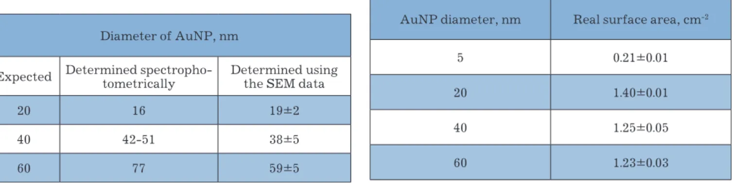
Table 1 | Comparative analysis of the diameters of synthe-sized AuNP as determined using different methods
Diameter of AunP, nm
expected Determined spectropho-tometrically Determined using the SeM data
20 16 19±2
40 42-51 38±5
60 77 59±5
Table 2 | Real surface area vs. nanoparticles size
AunP diameter, nm real surface area, cm-2
5 0.21±0.01
20 1.40±0.01
40 1.25±0.05
Biomodification of the surface of AunP/Au elec-trodes was carried out through direct adsorption of the enzyme for 20 min from a MvBOx solution with a pro-tein concentration of 0.25 mg∙ml-1. the surface
concen-tration of the enzyme was assumed to be 3.0 pmol∙cm-2.
the k0 and kcatapp values were calculated using the MathcAD 14 software package and the equation:
j j F RT E E k k RTF E T = + ⎡
(
−)
⎣ ⎢ ⎤ ⎦ ⎥+ − max ' exp exp 1 01 5 0 α EE T1 0'(
)
⎡ ⎣ ⎢ ⎤ ⎦ ⎥ ,k
k O
K
McatO
5 2 2=
+
[ ]
[ ]
.the kinetic scheme of enzyme functioning used to establish the equation was presented in [11].
reSultS and diScuSSion
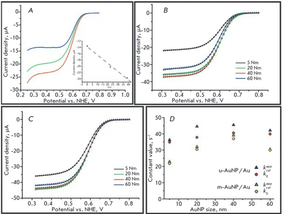
the bioelectrodes were placed in oxygenated PBS, fol-lowed by cV recording at an electrode rotation speed of 1,500 min-1 to eliminate possible diffusion limitations
(Fig. 2A). A pronounced bioelectrocatalytic response with an initial oxygen electrical reduction potential of about 0.75 V was registered for all the electrodes used. As illustrated in Fig. 2B,C, substantially similar jmax
values were obtained using the electrodes biomodified
with m-AunP/Au (31.4 ± 5.9 μA∙cm-2 ) and u-AunP/
Au (43.4 ± 5.6 μA∙cm-2) electrodes.
taking these data into account, the k0 (31 ± 9 s-1) and
kcatapp (34 ± 11 s-1) values were calculated. the results are
shown in Fig. 2D. the calculated k0 and kcatapp values are similar regardless of the AunP diameter and the elec-trode type used. the similarity of the constants for the electrodes based on m-AunP/Au and u-AunP/Au in-dicates that the assumption of identity of the Areal (de-spite different structures) for both types of samples does not add a critical error to the calculations. the overesti-mated constants for u-AunP/Au attest to the slightly higher surface area of those electrodes compared to m-AunP/Au, owing to the absence of AunP agglomerates (Fig. 1C), which also reduces the Areal. the pronounced bends of constant vs. AunP size profiles observed when using m-AunP/Au may be due to the surface modifi-cation because of the formation of different three-di-mensional (3D) structures during the treatment of elec-trodes in H2SO4. the behavior of the enzyme on these heterogeneous surfaces cannot be fully described by the single theory used in this study to calculate biocatalytic parameters without introducing additional corrections. Moreover, the formation of 3D agglomerates yields er-rors when a single value of the surface concentration of the enzyme (3.0 pmol∙cm-2) is used for all the m-AunP/
Fig. 2. А) Cyclic voltammo-grams (cathodic waves) of biomodified m-AuNP20/Au electrodes recorded at dif-ferent rotation rates, rpm: 0 (blue), 500 (green) and 1500 (red). Inset – current density at 0.35 V as a func-tion of ω1/2; B), C). Cyclic
voltammograms (cathodic waves) of MvBOx modi-fied m-AuNP/Au (B) and u-AuNP/Au (C) electrodes based on AuNPs of different diameters; D) Dependences of the calculated bioelectro-catalytic parameters on the size of AuNPs.
Conditions for all CVs: oxygen saturated PBS, scan rate – 20 mV s-1, second cycle А B Current density, µА Potential vs. NHE, V 0.2 0.3 0.4 0.5 0.6 0.7 0.8 0.9 1.0 0 -5 -10 -15 -20 -25 -30 0 -10 -20 -30 -40 0 -10 -20 -30 -40 -50 50 40 30 20 10 0 Current density, µА -5 0 5 10 15 20 25 30 35 40 -12 -14 -16 -18 -20 -22 -24 √ω Current density, µА Potential vs. NHE, V 0.3 0.4 0.5 0.6 0.7 0.8 5 Nm 20 Nm 40 Nm 60 Nm C D Current density, µА Potential vs. NHE, V 5 Nm 20 Nm 40 Nm 60 Nm 0.3 0.4 0.5 0.6 0.7 0.8 Constant value, s -1 AuNP size, nm 10 20 30 40 50 60 u-AuNP/Au m-AuNP/Au kcat app k0 kcatapp k0
Au-based bioelectrodes. this fact can also explain the shape of the curves. However, based on the experimen-tal data and simulation results, we can affirm that the bioelectrocatalytic properties of MvBOx immobilized on the AunP/Au surface show no dependence on the nano-particle diameter. the increased electrocatalytic cur-rent of the bioelectrodes modified with nanoparticles of different sizes found in a number of previous studies is most likely associated with an increase in the geometri-cal surface area rather than the acceleration of the Det reactions or an increase in the bioelectrocatalytic con-stants of the immobilized enzymes.
concluSionS
Our results have experimentally demonstrated no relationship between the bioelectrocatalytic
param-eters of MvBOx immobilized on the AunP/Au surface and the nanoparticle diameter. However, it should be noted that the results obtained in this study cannot be extrapolated to other nanobiomodified surfaces (e.g., other nanoparticles and redox enzymes). In particular, it is of particular interest to study the impact of nano-particles with a diameter lower than the size of the en-zyme that can promote electron transfer between the enzyme and the electrode surface. Such experiments will provide a more complete picture of the impact of nanoparticles on the bioelectrocatalytic parameters of oxidoreductases.
This study was supported by the Russian Foundation for Basic Research (grant № 12-04-33102-mol-a-ved).
reFerenceS
1. Murata K., Kajiya K., nakamura n., Ohno H. // energy & environmental Science. 2009. V. 2. № 12. P. 1280–1285. 2. Dagys M., Haberska K., Shleev S., Arnebrant t., Kulys J.,
ruzgas t. // electrochemistry communications. 2010. V. 12. № 7. P. 933–935.
3. Pankratov D. V., Zeifman Y. S., Morozova O. V., Shumako-vich G. P., Vasil’eva I. S., Shleev S., Popov V. O., Yaropolov A. I. // electroanalysis. 2013. V. 25. № 5. Р. 1143–1149. 4. Salaj-Kosla u, Poller S., Schuhmann W., Shleev S., Magner
e. // Bioelectrochemistry. 2013. V. 91. Р. 15–20.
5. ressine A., Vaz-Dominguez c., Fernandez V. M., De Lacey A. L., Laurell t., ruzgas, t., Shleev S. // Biosensors & Bio-electronics. 2010. V. 25. № 5. P. 1001–1007.
6. Gutierrez-Sanchez c., Pita M., Vaz-Dominguez c., Shleev
S., De Lacey A. L. // Journal of the American chemical Society. 2012. V. 134. № 41. P. 17212–17220.
7. Haiss W., thanh n. t. K., Aveyard J., Fernig D. G. // Ana-lytical chemistry. 2007. V. 79. № 11. P. 4215–4221.
8. Murata K., Kajiya K., nukaga M., Suga Y., Watanabe t., nakamura n., Ohno H. // electroanalysis. 2010. V. 22. № 2. P. 185–190.
9. trasatti S., Petrii O. A. // Pure and Applied chemistry. 1991. V. 63. № 5. P. 711–734.
10. Wang X., Falk M., Ortiz r., Matsumura H., Bobacka J., Ludwig r., Bergelin M., Gorton L., Shleev S. // Biosensors & Bioelectronics. 2012. V. 31. № 1. P. 219–225.
11. climent V., Zhang J. D., Friis e. P., Østergaard L. H., ulstrup, J. // the Journal of Physical chemistry c. 2012. V. 116. № 1. P. 1232–1243.