Full Terms & Conditions of access and use can be found at
http://www.tandfonline.com/action/journalInformation?journalCode=zjom20
Download by: [Malmö University] Date: 02 November 2017, At: 06:33
ISSN: (Print) 2000-2297 (Online) Journal homepage: http://www.tandfonline.com/loi/zjom20
Comorbidity of periodontal disease: two sides of
the same coin? An introduction for the clinician
Palle Holmstrup, Christian Damgaard, Ingar Olsen, Björn Klinge, Allan
Flyvbjerg, Claus Henrik Nielsen & Peter Riis Hansen
To cite this article: Palle Holmstrup, Christian Damgaard, Ingar Olsen, Björn Klinge, Allan Flyvbjerg, Claus Henrik Nielsen & Peter Riis Hansen (2017) Comorbidity of periodontal disease: two sides of the same coin? An introduction for the clinician, Journal of Oral Microbiology, 9:1, 1332710, DOI: 10.1080/20002297.2017.1332710
To link to this article: http://dx.doi.org/10.1080/20002297.2017.1332710
© 2017 The Author(s). Published by Informa UK Limited, trading as Taylor & Francis Group.
Published online: 14 Jun 2017.
Submit your article to this journal
Article views: 256
View related articles
REVIEW ARTICLE
Comorbidity of periodontal disease: two sides of the same coin? An
introduction for the clinician
Palle Holmstrupa, Christian Damgaarda,b, Ingar Olsenc, Björn Klinged,e, Allan Flyvbjergf, Claus Henrik Nielsena,b
and Peter Riis Hansena,g
aSection for Periodontology, Department of Odontology, Faculty of Health and Medical Sciences, University of Copenhagen,
Copenhagen, Denmark;bInstitute for Inflammation Research, Center for Rheumatology and Spine Diseases, Rigshospitalet, Copenhagen
University Hospital, Copenhagen, Denmark;cDepartment of Oral Biology, Faculty of Dentistry, University of Oslo, Oslo, Norway; dDepartment of Periodontology, Faculty of Odontology, Malmö University, Malmö, Sweden;eDivision of Periodontology, Department of
Dental Medicine, Karolinska Institutet, Stockholm, Sweden;fSteno Diabetes Center Copenhagen, Copenhagen, Denmark;gCardiology
Department, Herlev and Gentofte Hospital, Hellerup, Denmark
ABSTRACT
Increasing evidence has suggested an independent association between periodontitis and a range of comorbidities, for example cardiovascular disease, type 2 diabetes, rheumatoid arthritis, osteoporosis, Parkinson’s disease, Alzheimer’s disease, psoriasis, and respiratory infections. Shared inflammatory pathways are likely to contribute to this association, but distinct causal mechanisms remain to be defined. Some of these comorbid conditions may improve by periodontal treatment, and a bidirectional relationship may exist, where, for example, treatment of diabetes can improve periodontal status. The present article presents an overview of the evidence linking periodontitis with selected systemic diseases and calls for increased cooperation between dentists and medical doctors to provide optimal screening, treatment, and prevention of both periodontitis and its comorbidities.
ARTICLE HISTORY Received 27 October 2016 Accepted 7 January 2017 KEYWORDS Periodontitis; periodontal disease; low-grade inflammation; comorbidity; cardiovascular disease; type 2 diabetes; rheumatoid arthritis; osteoporosis; Parkinson’s disease; Alzheimer’s disease; psoriasis; pneumonia Introduction
Periodontitis (PDIS) is a common oral disease, the manifestations of which accumulate with increasing age. Often, one gets the impression that PDIS is generally regarded as a natural, almost inevitable physiological consequence of the aging process. It is important to change this outdated perception. The population and the overall health sector should understand that PDIS is an inflammatory disease linked to the individual’s oral microbiota and
immune system [1], and that the patients with
PDIS, independent of age, benefit from periodontal
treatment [2]. As outlined below, a number of other
common medical disorders have inflammatory back-grounds too, which may, at least in part, explain their comorbidity with PDIS.
The oral cavity harbors a large amount of bacteria. By using molecular methods, it is now possible to identify precisely and rapidly >700 bacterial species that com-prise the oral microbiome, and over the past 15 years, 68% of oral bacterial species in the mouth have been cultured [3]. Of further interest is that the oral micro-biome appears to be individualized, implying that it can vary quantitatively and qualitatively between
indivi-duals, although there are significant overlaps.
Moreover, there is significant variation in the microbiota at different sites of the oral cavity in one person [3,4]. It is also clear that the oral microbiota changes in relation to different diseases such as PDIS, caries, root canal infections, and mucositis [5]. For example, there is an abundance of anaerobic bacteria in the oral cavity, some of which commonly associates with PDIS. These include Porphyromonas gingivalis, Tannerella forsythia, and
Treponema denticola (the three members of the ‘red
complex’). Moreover, several bacterial species associated with PDIS have been suggested to be involved in the pathogenesis of a number of systemic diseases [6,7].
In the latest decades, there has been considerable scientific interest in the connection of PDIS with a number of medical diseases, several of which also have a high prevalence in the general population. Limited or no solid evidence exists for a direct causal relationship between PDIS and other inflammatory diseases, and documentation in the form of rando-mized clinical trials is, however, unlikely ever to be achieved. For example, randomized trials of effects of PDIS treatment on the incidence of medical comor-bidities, for example cardiovascular disease (CVD) or diabetes mellitus (DM), would require long-term fol-low-up of an enormous number of patients and be
CONTACTPalle Holmstrup pah@sund.ku.dk
VOL. 9, 1332710
https://doi.org/10.1080/20002297.2017.1332710
© 2017 The Author(s). Published by Informa UK Limited, trading as Taylor & Francis Group.
This is an Open Access article distributed under the terms of the Creative Commons Attribution License (http://creativecommons.org/licenses/by/4.0/), which permits unrestricted use, distribution, and reproduction in any medium, provided the original work is properly cited.
limited by treatment blinding issues and ethical reser-vations by leaving PDIS untreated in the placebo group. Therefore, health authorities must rely on an assessment of the accumulated plausibility of a causal relationship between PDIS and its comorbidities, which will probably forever remain based on a sum of indirect evidence.
PDIS and CVD
Atherosclerosis, the major cause of CVD, is an inflam-matory disease that develops in the large arteries, and is responsible for ischemic heart disease, stroke, and per-ipheral artery disease. Atheromatous plaques are usually asymptomatic until they become unstable with plaque rupture/erosion and thrombosis that are asso-ciated with increased inflammatory activity in both the arterial wall and systemically in the body [8]. As indi-cated above, the question of whether the relationship between PDIS and atherothrombosis is causal is diffi-cult to answer definitively. The present review will focus on clinical studies addressing this issue while results of numerous exciting experimental studies supporting the relationship are beyond the scope of the review.
In the recent Swedish‘PAROKRANK’ study
includ-ing 805 patients <75 years of age with first-time acute myocardial infarction (AMI) and 805 matched con-trols without AMI, clinical dental examination and panoramic X-rays were conducted on all participants [9]. PDIS, verified by radiographically rated bone loss, was more common in patients with AMI than it was in controls. There was an increased (+49%) risk of AMI among the PDIS patients. The risk remained signifi-cantly increased (+28%) after adjustment for co-vari-ables (smoking, DM, socioeconomic factors). These
findings from the largest and most well-conducted case-control study to date emphasize that there can be an independent association between PDIS and AMI [9], which was supported by another recent study from Scandinavia [10].
Explanatory models
Numerous other population studies, including different ethnicities, have shown a connection between PDIS and CVD, and there is increasing evidence for this connec-tion [11]. This link may be explained by several, not mutually exclusive, mechanisms (seeTable 1).
Significance of periodontal treatment
As indicated above, there are several options for PDIS to affect the development of atherosclerosis and its clinical manifestations. The question is whether per-iodontal treatment can influence this process. A recent systematic review with meta-analysis con-cluded that periodontal treatment improves a number of surrogate measures for atherosclerosis, including endothelial dysfunction and lipid parameters, gly-cated hemoglobin (HbA1c), and biomarkers such as high sensitive C-reactive protein and interleukin (IL)-6, especially among those who already suffer from
coronary heart disease and DM [21]. A longitudinal
study from the United States has also shown that improvement of the periodontal status with reduced clinical probing depth and diminished subgingival presence of bacteria associated with PDIS among 420 participants resulted in a reduced progression of carotid intima-media thickness (IMT) over 3 years, and the average progression of carotid IMT was
Table 1.Explanatory models for association of PDIS and CVD
Transfer of periodontal bacteria to atheromatous plaques
In subjects with periodontal inflammation, daily oral activities, including chewing and oral hygiene procedures, may result in transfer of periodontal bacteria from the inflamed pockets to the bloodstream [12]. A following infection of atherosclerotic arterial walls may result in instability of plaques with rupture and thrombus formation [13,14]. Studies have demonstrated the presence of DNA from oral bacteria in atheromatous plaques [15], and some studies using advanced cultivation techniques have also shown viable periodontal bacteria in the plaques [6,16].
Spillover of cytokines from periodontal tissues to the bloodstream
Inflammation in the periodontal tissues involves local production of proinflammatory cytokines [17]. Spillover of such cytokines and cytokine producing cells to the bloodstream may enhance inflammation in the atherosclerotic arterial walls and result in unstable plaques. Thus, elevated levels of some proinflammatory cytokines, including interleukin (IL)-6 and tumor necrosis factor alpha, have been found in the bloodstream of patients with PDIS, and PDIS may thus cause systemic low-grade inflammation [18–21] promoting, for example, endothelial dysfunction and development of insulin resistance.
Systemic production of cytokines Increased plasma level of cytokines in response to bacteremia after oral procedures is a well-described phenomenon. For example, increased IL-6 levels were found in the bloodstream as a result of bacteremia after scaling [22], and this cytokine is a known risk marker for CVD, including AMI [23,24]. Change of lipid metabolism as a result of
PDIS
In patients with PDIS, the lipid balance in the bloodstream, disturbance of which is associated with increased risk of atherosclerotic disease, shows an unfavorable shift with less high-density lipoprotein and more low-density lipoprotein cholesterol [25,26]. In addition, studies have indicated
improvement of lipid parameters after periodontal treatment [21].
Endothelial dysfunction PDIS is associated with endothelial dysfunction, which is considered the earliest marker of atherosclerosis [27]. Furthermore, periodontal treatment may improve endothelial function [28]. Shared genetic risk factors Obviously, there may be unknown shared risk factors of importance, including the genetic profile of
patients with the two diseases. For example, a shared variant in the IL-1 gene complex could be part of the background for the simultaneous occurrence of both diseases, and recent studies have identified a number of other shared genetic risk factors [29,30]. However, these factors do not appear to explain the observed association fully.
PDIS, periodontitis; CVD, cardiovascular disease; AMI, acute myocardial infarction.
inversely correlated with the improvement of period-ontal status [31]. According to the authors, the study emphasized the significance of periodontal treatment as a possible preventive health effort. As mentioned above, endothelial dysfunction and carotid IMT are surrogate measures for atherosclerosis. While the importance of periodontal treatment for reduction of clinical cardiovascular endpoints has been sug-gested in epidemiological studies, for ethical reasons, randomized trials are unlikely to be performed in this area of research.
A comprehensive longitudinal study in Taiwan with an average follow-up period of 7 years was based on a random sample of one million people
[32]. It was attended by 10,887 people, who had
received dental treatment during the study period. A total of 10,989 age-, sex-, and comorbidity-matched subjects who had not received dental treatment were also included. In the scaling group, a significantly lower incidence of AMI (1.6% vs. 2.2%; p < 0.001) and stroke (8.9% vs. 10%; p = 0.03) was seen. A multivariate analysis showed that scaling was inde-pendently associated with significantly reduced risk of AMI (hazard ratio [HR] = 0.69) and stroke (HR = 0.85). Furthermore, there was a dose-depen-dent correlation with increased frequency of scaling leading to greater reduction in the risk of AMI and stroke. A weakness of this study was, however, that correction for all known risk factors such as smoking
was not performed [32]. In a recent longitudinal
study, also from Taiwan, 13,573 patients were treated for mild PDIS in the period 2001–2010, and an equal number of matched patients were treated for severe PDIS [33]. Among the latter patients, those who were >60 years of age had more frequent cardiovascular events, suggesting that the severity of PDIS plays a role in these events.
Hypertension
Hypertension is associated with PDIS [34,35], and these conditions occur frequently in patients with greater attachment loss [36]. Since hypertension is a treatable risk factor for CVD, it is important to identify patients with hypertension. Therefore, it should be considered whether dentists can contribute to such screening, since patients usually visit dentists more frequently than they visit medical doctors for preventive healthcare mea-sures in the absence of known disease. Sublingual varices are associated with hypertension [37], and this oral manifestation may be used as indicator for screen-ing and referral of patients to their general physician. Moreover, for the dentist, it is also important to know if the patient has hypertension, which may contribute to increased bleeding during oral surgery. In addition, many patients with hypertension are treated with cal-cium antagonists that occasionally can cause gingival
hyperplasia, which again may result in increased pro-gression of PDIS [38]. Obviously, it is up to the dentist to disrupt this potentially vicious circle.
PDIS and type 2 diabetes
It is well known that there is a relationship between DM and PDIS. As a result of the obesity epidemic, there has been a significant growth in the number of
patients with type 2 diabetes (T2D) [39], and it is
expected that dentists will receive increasing numbers of such patients for diagnosis and treatment in the future. The relationship is bidirectional in that DM
predisposes for PDIS [40], and PDIS can worsen the
course of DM, as recently reviewed [41].
Explanatory models
There are several ways by which PDIS and T2D may interfere with each other. DM can affect the develop-ment of PDIS through a change in the oral micro-biota, although it is still uncertain whether such a change actually takes place [42]. The main factor for the increased propensity to develop PDIS among diabetics is probably the formation of advanced gly-cation end products (AGE) by glygly-cation of proteins and lipids [43]. At high blood-sugar levels, character-istic of poorly controlled DM, the formation of AGE is increased, and the receptors for AGE (RAGE) are also upregulated, which leads to increased production of proinflammatory cytokines and increased tissue degradation, including increased bone resorption and decreased bone formation [44,45]. In addition, there are studies suggesting that DM patients display altered function of neutrophils, which play a major role in the pathogenesis of PDIS [46]. It is important to emphasize that well-controlled DM patients are not at increased risk of PDIS. It is thus important for the dentist to have information about blood-sugar control in the individual DM patient. On the other hand, epidemiological studies have linked PDIS to insulin resistance, and PDIS appears to be an inde-pendent risk factor for T2D [47,48].
Undiagnosed diabetes
Not all patients with T2D are aware that they have the disease because the initial symptoms are mild, and probably almost half of these patients are undiagnosed. Because it is critical for prevention of DM complica-tions, including eye disease, kidney disease, neuropa-thy, and CVD, DM must be diagnosed as early as possible, and from an individual as well as societal and economic perspective, it is very unfortunate if diagnosis is delayed. It is also disadvantageous for dental treatment that the diabetic state is unknown. In the above-mentioned Swedish PAROKRANK study
[9], in which patients with a first AMI were compared to controls without ischemic heart disease, glucose metabolism was examined by oral glucose tolerance test, and 9.3% of patients with AMI and 5.2% of the control group had undiagnosed DM. Another recent study revealed that 3.1% of 291 patients without diag-nosed T2D who sought dental treatment at the Department of Odontology at the University of Copenhagen had HbA1c above the threshold for T2D, and similarly 27.1% had HbA1c above the threshold for pre-diabetes [49]. Pre-diabetes is a con-dition where blood-glucose levels are above the nor-mal, but still do not qualify for the T2D diagnosis. This condition, which is a precursor of manifest T2D, is also known as impaired glucose tolerance. Patients with PDIS more frequently had elevated HbA1c than the control group without PDIS did. It is easy and cheap to implement screening for elevated HbA1c, and since many patients visit the dentist more regularly than they do the medical doctor, irrespective of whether they feel healthy, there is a golden opportu-nity to implement HbA1c screening in selected risk patients in dental clinics, with referral to their general physician in the case of elevated values.
Significance of periodontal treatment
Studies on the significance of periodontal treatment for the course of T2D often carry considerable meth-odological limitations, for example missing sufficient confounder control and with incomplete information on the efficacy of the periodontal treatment. However, several meta-analyses have shown that non-surgical periodontal treatment reduces HbA1c levels significantly in the range of 0.31–0.65%
[50,51]. Even such small reductions in HbA1c can
be clinically important. Thus, a large British study demonstrated that every percentage point decrease in HbA1c may result in as much as 35% reduction of microvascular complications, and an average reduction in HbA1c of 0.2% was related to a 10% lower mortality rate [52]. Therefore, the reduction by 0.31–0.65%, which can be achieved by periodontal treatment, can have a great impact in terms of sys-temic health and societal economy.
PDIS and rheumatoid arthritis
Rheumatoid arthritis (RA) is an autoimmune disease
affecting 0.5–1% of the population in the Western
world. The disease involves inflammation of the joints, with cartilage degradation and joint deformity,
swelling, and pain [53]. Like PDIS, RA is a chronic
inflammatory condition, which leads to tissue degra-dation, and an association between the two diseases
has been demonstrated, as recently reviewed [54].
Despite a limited number of participants, the
available studies suggest that both younger and older patients with RA have an increased predisposi-tion to attachment loss [55–58]. This might argue for establishing periodontal prevention programs as part of routine treatment of patients with RA [55–58].
Explanatory models
PDIS and RA may associate bidirectionally. Both diseases display elevated circulating and target tissue levels of markers of inflammation and cytokine pro-files of‘tissue degrading’ nature, including increased production of IL-1 and tumor necrosis factor alpha (TNF-α) [55–59]. RA is furthermore characterized by the formation of autoantibodies, including rheuma-toid factors recognizing immunoglobulin G (IgG) and antibodies to citrullinated proteins (ACPAs)
[60]. The latter are found in approximately
three-quarters of RA patients, which also have a character-istic expression of major histocompability complex molecules capable of binding citrullinated peptides
[61]. Indeed, the subgroups of RA patients who
show immune responses to citrullinated proteins and those who do not are considered by many inves-tigators to be two distinct disease entities. Post-trans-lational conversion of the amino acid arginine to citrulline is catalyzed by enzymes of the peptidylargi-nine deiminase (PAD) family, and these are consid-ered important in disease progression, at least in
ACPA-positive RA [60].
Patients with antibodies against citrullinated pro-teins more frequently appear to have PDIS than patients with osteoarthritis do [62]. In addition, RA patients more frequently have antibodies against Porphyromonas gingivalis than healthy controls do
[63]. In search for a mechanistic link between PDIS
and RA, special attention has been drawn to P. gingi-valis, which is the only bacterium with capacity to produce a PAD (PPAD). Like the corresponding human enzyme, PPAD is capable of converting argi-nine to citrulline [64]. In theory, PPAD may therefore convert harmless host proteins into citrullinated autoantigens that become the target for autoantibo-dies and pathogenic T cells that drive RA. Smoking, which increases the risk of PDIS, is also the strongest life-style factor linked to the development of RA. Smoking is also believed to promote the secretion of PAD from leukocytes in the lungs and thus initiate citrullination [65].
Significance of periodontal treatment
Several studies have evaluated the effect of period-ontal treatment on RA and biomarkers of the disease
[58]. The available studies, however, are small and
with limited follow-up, but they suggest that non-surgical periodontal treatment may reduce clinical
symptoms and biomarkers of active RA. Major intervention studies in RA patients with PDIS are needed in order to draw firm conclusions on this matter.
PDIS and osteoporosis
A possible association between PDIS and osteoporosis was described already in 1968 [66]. Osteoporosis is a systemic skeletal disease characterized by reduced bone density. Clinically, osteoporosis is divided into (1) an idiopathic form that appears early in life and affects men and women with equal frequency, and (2) an involutional form, which is subdivided into two types, the first of which includes postmenopausal women, and the other being age-related and including both elder
men and women [67]. The most prevalent form is
postmenopausal osteoporosis, the possible association of which with PDIS has been examined in several clinical studies. The majority of these studies have been cross-sectional and with few participants, all post-menopausal women [68–75]. A recent cross-sectional study from Taiwan including 35,127 osteoporosis patients and 50,498 healthy controls showed that PDIS was associated with an increased risk of osteoporosis (odds ratio [OR] = 1.29) after adjustment for sex, age, and comorbidity, and that the risk increased with
increased degree of periodontal inflammation [76].
Furthermore, osteoporosis was associated with a sixfold increased risk of concurrent PDIS. These results are supported by a second cross-sectional study from South Korea, which showed a positive correlation between PDIS and osteoporosis (OR = 1.21) after adjustment for age [77]. However, longitudinal studies are missing to substantiate a causal relationship between PDIS and osteoporosis.
Explanatory models
Various systemic risk factors, such as genetics, age, sex, vitamin D deficiency, medical hormone therapy, diet, smoking, obesity, and physical activity, affects the development of osteoporosis [78,79], but several
of these are also risk factors for PDIS [80]. Bone
density changes throughout life, but after the meno-pause a decrease in estrogen production occurs, which seems to be associated with an increased risk of osteoporosis. Decreased bone density in the jaw bone in subjects with osteoporosis is obviously com-patible with this condition, leading to attachment loss in individuals with PDIS [72,73,81,82]. Besides being associated with decreased bone density, estrogen defi-ciency also affects the other periodontal tissues and the immune response against the periodontal biofilm in a proinflammatory direction [81].
Significance of periodontal treatment
As yet, no studies have evaluated the effect of period-ontal treatment on osteoporosis. Furthermore, it remains unclear whether bisphosphonate treatment of postmenopausal patients with osteoporosis wor-sens or improves periodontal parameters. One study, however, showed that bisphosphonate therapy did not reduce alveolar bone loss in osteoporosis patients with PDIS [82].
PDIS and Alzheimer’s disease
Alzheimer’s disease (AD) is a neurodegenerative
disease and the most common example of a group of diseases causing dementia. It is a progressive disease, with susceptibility genes working with lit-tle-understood environmental and behavioral
influ-ences [83]. AD is characterized by atrophy and
neuronal death, especially in the hippocampal
region of the brain [84]. There are two main
cate-gories of AD: the familial, early-onset form that targets individuals <65 years of age and accounts for about 2% of all cases of AD; and the late-onset form of AD that affects older (>65 years) subjects and accounts for approximately 98% of the cases. Late-onset AD has several genetic susceptibility
traits. Among these, the apolipoprotein APOE ɛ4
allele is considered to be the most important [85].
The disease is already a great economic burden for society, and there is currently no treatment. Late AD probably has several causes, while a genetic component is more essential for the early form. Characteristically, late AD includes inflammatory changes in the brain, which may be initiated by local or systemic infection [86].
Among the microorganisms most frequently asso-ciated with AD are bacteria such as spirochetes, P.
gingi-valis, Prevotella, fusobacteria, Actinomyces, and
Chlamydophila pneumoniae. Also, herpes virus
(Epstein–Barr virus and cytomegalovirus) and yeasts of
the genus Candida have been connected with AD [83].
With the exception of C. pneumoniae, all these micro-organisms can be present in the periodontal pockets.
There is emerging evidence of a link between PDIS and AD. The relationship has been shown in cross-sectional and longitudinal studies by examining the association of AD with clinical signs of PDIS and circulating levels of antibodies against bacteria asso-ciated with PDIS, respectively [84].
Explanatory models
Association of AD with periodontal bacteria
In addition to the red complex bacteria, Fusobacterium nucleatum and Prevotella intermedia, both known to be associated with PDIS, also associate with AD.
Indeed, in the National Health and Nutrition Examination Survey (NHANES), antibody levels to these organisms were significantly increased in serum from patients with AD compared to controls [87]. This result was significant after controlling for each sub-ject’s age, Mini-Mental State Examination score, and APOEԑ4 allele status. Unexpectedly, Noble et al. found that a high (>640 ng/mL) anti-Actinomyces naeslundii titer was present in 10% of subjects with increased risk of AD, suggesting that AD pathogenesis may involve a spectrum of bacteria [88].
In 14 studies, oral spirochetes that are neuro-trophic were demonstrated in the brain of AD patients. Seven different spirochetes were identified in 14/16 AD brains [89,90]. Spirochetes induced bio-logical and pathobio-logical characteristics of AD (plaque accumulations of beta-amyloid and neurofibrillary tangles) after exposure of neuronal and glial cells in organ cultures. Lipopolysaccharide (LPS) from P. gingivalis was also detected in human brains with
AD but not in control brains [91]. In a study based
on 2,355 people >60 years of age, a positive correla-tion between PDIS and cognitive impairment was found, and a negative correlation was observed between antibody titers to P. gingivalis and scores in cognitive tests [87,92].
The original inflammatory hypothesis of AD sug-gested that AD hallmark proteins, for example beta-amyloid, were the main contributors to central ner-vous system inflammation. This hypothesis has been expanded to include involvement of infections, and life-style, genetic, and environmental factors in AD pathogenesis. PDIS is a prototypical oral condition that encompasses all these factors, including patho-genic bacteria [93].
Microorganisms and inflammatory mediators may reach the brain
Oral microorganisms and inflammatory mediators can be transported from inflamed periodontal tissues to the brain via the bloodstream. An increased amount of cytokines, particularly the macrophage-secreted TNF-α, has been detected in the plasma of AD subjects [94]. Also, elderly people harbored a higher titer of
circulat-ing IgG against several periodontal pathogens [87].
Cestari et al. [95] found an association between
circu-lating IL-6 and TNF-α levels in patients with AD and
PDIS, implicating these proinflammatory cytokines in the overlapping pathogenic mechanisms between oral infections and AD [95].
As mentioned above, daily episodes of bacteremia follow from dental procedures, including toothbrush-ing and flosstoothbrush-ing, and from chewtoothbrush-ing, particularly in patients with PDIS. The bacteria involved can disse-minate into the brain by closely related anatomical pathways, that is, trigeminal and olfactorial nerves
[93]. The long-term effect of inflammatory mediators and pathogens and/or their virulence factors reaching the brain may over time prime the brain’s own microglia in individuals having inherent susceptibility traits. According to Singhrao et al., such susceptibil-ities could contribute to inadequate neutralization of invading agents reaching the brain and result in loss of cytoarchitectural integrity and vital neurons with subsequent deterioration of cognitive function [93].
Blood–brain barrier
The permeability of the blood–brain barrier (BBB) increases with age. Prolonged exposure to high con-centrations of TNF-α tends to weaken the protective role of the BBB, making it more permeable to
bac-teria or endotoxins [96]. The APOE ԑ4 gene is also
associated with increased BBB permeability, allowing microorganisms, their products, and inflammatory mediators such as TNF-α to penetrate into the brain
[97]. These microorganisms and substances can also
pass through circumventricular tissues and perivas-cular spaces of the brain because these regions lack a
BBB [98]. The olfactory nerve and the trigeminal
nerve also circumvent the BBB [99]. Indeed, olfactory cells may act as Trojan horses by which microorgan-isms can reach the brain [100].
Bacteria in the brain
Biofilm has been demonstrated in the brain of AD patients and was probably created by dental and Lyme spirochetes with accompanying local tissue activation of
the innate immune system [101]. Riviere et al. also
demonstrated the presence of seven different
Treponema species in 14/16 specimens from AD brains [102]. Microorganisms and their toxic products as well as microbial DNA have been reported in the brain tissue of AD patients and animal models [83]. Spirochetes induce a latent and slowly progressive infection by circumvent-ing host defense, and are able to induce beta-amyloid plaque formation in the brain [87,92]. Periodontal bac-teria, especially T. denticola, may contribute to AD pathology using a range of inflammatory mechanisms by which neurons would be attacked. This occurs despite the fact that these bacteria inhibit inflammasome activity [103,104]. Spirochetes possibly promote their own survi-val and proliferation by blocking the complement cas-cade [105]. Moreover, P. gingivalis has LPS with various lipid A structures and is capable of modifying the latter components, which may provide the bacteria with the capacity to disguise itself from recognition by the immune system via TLR4 [106].
Genetics, environmental factors, nutrition, and other factors
A very important risk factor for AD is the APOEԑ4
gene, which is associated with susceptibility for
infections and increases the expression of inflamma-tory mediators [107]. In total, 20 different genetic loci have been estimated to increase the susceptibility to
AD, including APOEԑ4. These include the genes for
IL-1β and TNF-α, which are also linked to the
devel-opment of PDIS [108]. The pathogenesis of AD
prob-ably includes an interaction between genes,
microorganisms/toxins, and environmental factors. Inadequate nutrient intake is common in the elderly and in people with dementia, and this can contribute to gradual loss of nerve synapses. In addition, neglect of or an inability to maintain oral hygiene in the elderly promotes inflammation in the periodontium, which may favor the transport of microorganisms and their products, as well as inflammatory media-tors, to the brain. Loss of teeth, which is often the result of PDIS, has been connected to a poor mem-ory [109].
PDIS and Parkinson’s disease
Parkinson’s disease is another chronic neurodegen-erative disease that results in selective loss of dopa-minergic neurons in the substantia nigra of the brain.
During the progression of Parkinson’s disease, there
is a gradual degeneration of the nigrostriatal com-pounds, leading to cognitive, motor, and psychiatric symptoms. There is still no solid evidence that PDIS
influences the pathogenesis of Parkinson’s disease
[110]. However, there are studies indicating that
PDIS is more common in patients with Parkinson’s
disease, although large longitudinal studies and ran-domized case-control or case-cohort studies are lack-ing to substantiate this association [111,112].
Explanatory models
Parkinson’s disease causes motor disability, which complicates the provision of simple daily oral proce-dures such as brushing the tooth, which will inevita-bly lead to the accumulation of plaque. In addition, the cognitive changes in patients with Parkinson’s disease may have an impact on the quality and fre-quency of the home dental-care habits (as well as the
dentists’ willingness to perform periodontal
treat-ment), which contributes to increased plaque accu-mulation and risk of PDIS. A number of studies also
indicate that systemic low-grade inflammation
induced by PDIS [17,19,22] contributes to neural dysfunction at early stages of Parkinson’s disease
[110]. Much evidence suggests that the pathogenesis
of Parkinson’s disease has an inflammatory compo-nent, for example elevated plasma IL-6 appears to increase the risk of the disease [113,114]. There are no published studies on the effect of periodontal treatment of patients with Parkinson’s disease.
PDIS and psoriasis
Psoriasis is a chronic inflammatory disease with a prevalence of up to 8.5% of the population in the Nordic countries. The disease is also characterized by extensive comorbidity in the form of, for example, CVD and T2D, probably on the basis of shared
inflammatory mechanisms [115–117]. An association
between psoriasis and chronic PDIS has been shown, and increased concentrations of proinflammatory
cytokines such as TNF-α and IL-1β have been
found in saliva from patients with psoriasis [118–
120]. The results from a large epidemiological study
from Taiwan also suggest that intensive treatment of chronic PDIS can reduce the risk of psoriasis [121]. Activated T-helper (Th)-17 cells producing IL-17 are key pathogenic players in psoriasis, and bacterial infection, including infection with P. gingivalis, can promote the polarization of naïve T-helper cells into Th-17 cells. Also, activated Th-17 cells have been found in periodontal lesions, and increased IL-17 levels have been demonstrated in crevicular fluid from patients with MP [122–124].
Lung diseases and oral hygiene
Colonization of the oral cavity with respiratory patho-gens related to a lack of oral hygiene and PDIS can be linked to the development of pneumonia. There is strong scientific evidence from randomized clinical trials that interventions aimed at improving oral hygiene may prevent pneumonia and reduce pneumo-nia-related deaths, particularly in elderly
care-depen-dent patients [125]. A Norwegian study also showed
that chronic PDIS occurs more frequently in patients with severe chronic obstructive pulmonary disease, even after adjusting for risk factors such as age, smok-ing, obesity, corticosteroid use, and decreased bone density [126].
Conclusion
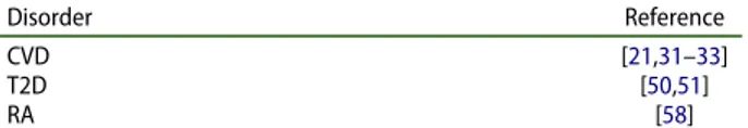
There are numerous studies showing a correlation between PDIS and a variety of medical disorders. This is not surprising, since the mouth, of course, is part of the human body. These medical conditions are particularly frequent in the elderly population. Important for the dental clinician, there is evidence to suggest that periodontal treatment may have ben-eficial effects on some of these conditions (Table 2). It is obvious that cooperation between medical doctors and dentists should be strengthened, and a major prerequisite for this is increased awareness and knowledge about the disease connections mentioned in this article. Importantly, low-grade inflammation is considered to be one of the most prominent
mechanistic links between PDIS and its medical comorbidities.
A considerable amount of knowledge has accumu-lated about the link between PDIS and CVD, with growing evidence for a causal relationship. There are plausible mechanistic data (including experimental results that are not discussed here), and it appears that periodontal treatment may reduce the risk of atherosclerotic disease.
There is also extensive evidence that poor blood-sugar control in patients with T2D leads to an increased risk of PDIS with increased severity and extension, and that PDIS may lead to increased risk of elevated HbA1c and T2D. The growing prevalence of T2D in the popu-lations will probably result in increased development of PDIS, which in turn can aggravate the course of T2D. Thus, the two diseases have a bidirectional relationship, presumably due to shared immunological reactions. Clinical studies also indicate that non-surgical period-ontal treatment can improve metabolic control, which may reduce the development of diabetic complications. There are ample reasons for establishing systematic examination, prevention, and therapy programs for PDIS in diabetic patients.
Evidence suggests that there is also a bidirectional link between PDIS and RA, with increased risk for PDIS in patients with RA, and non-surgical periodontal treat-ment may reduce clinical symptoms and biomarkers of active RA. The increased propensity for attachment loss in patients with RA can also speak for the establishment of a periodontal prevention program as part of routine treatment of these patients. Moreover, studies of patients with osteoporosis suggest that there is an increased propensity to develop attachment loss.
PDIS is also associated with certain neurological disorders. Parkinson’s disease involves motor impair-ment and cognitive changes, which may entail dete-rioration of home dental-care habits. Moreover, low-grade inflammation, for example as a result of PDIS, may contribute to neurological dysfunction in the early stages of Parkinson’s disease. AD has a complex and multifactorial etiology, and periodontal infection may be one of several risk factors for AD. Thus, the presence of periodontal bacteria and their products have been found in the brain of AD patients. Infection can occur decades before AD becomes apparent. Improved oral hygiene can be an important prophylactic measure, but unfortunately can be challenging because AD patients are not always cooperative.
Psoriasis is characterized by widespread comorbidity in the form of, for example, CVD, diabetes, and PDIS, which probably also has a background in shared inflam-matory mechanisms. Finally, there is scientific evidence that better oral health has a positive effect in the pre-vention of pneumonia, especially in elderly care-depen-dent patients.
Acknowledgments
A modified version of this article has also been published in the Scandinavian languages, in the journals of the respective national Nordic Dental Associations. Funding through the
European Commission (FP7-HEALTH-306029‘TRIGGER’)
and through grants from the Danish Dental Association, the Danish Foundation for Mutual Efforts in Dental Care, and the Simon Spies Foundation is kindly acknowledged.
Disclosure statement
No potential conflict of interest was reported by the authors.
Funding
This work was supported by the European Commission
[FP7-HEALTH-306029], Danish Dental Association,
Danish Foundation for Mutual Efforts in Dental Care, and Simon Spies Foundation.
Notes on contributors
Palle Holmstrup is professor and Section Head (Periodontology, Oral Microbiology, Surgery, Pathology, Physiology, Radiology and Community Dentistry) at School of Dentistry, Faculty of Health and Medical Sciences, University of Copenhagen. DDS from the Royal Dental College of Copenhagen in 1971. PhD in 1976 and Board Certification as specialist in Oral Surgery 1983. Dr. Odont. 1985. Honorary Doctor, Sahlgrenska Academy, University of Gothenburg in 2005. More than 400 guest lectures, seminars and courses in Scandinavia and abroad. Supervisor of 23 PhD students. Around 275 publications (Oral Medicine and Periodontology) in international and national scientific journals and book chapters.
Christian Damgaard is assistant professor at Section of Periodontology, Department of Odontology, Faculty of Health and Medical Sciences, University of Copenhagen. DDS from University of Copenhagen, Denmark in 2011. PhD in 2015 from University of Copenhagen and The Forsyth Institute, Cambridge, MA. Author of 13 publica-tions in international and national scientific peer-reviewed
journals. Board member of Danish Academy of
Periodontology.
Ingar Olsenis professor emeritus and guest researcher at Department of Oral Biology, Faculty of Dentistry,
University of Oslo. Senior Research Investigator,
Department of Molecular Genetics, Forsyth Institute,
Cambridge, MA. DDS from the Faculty of Dentistry,
University of Oslo in 1966. Dr. odont. in 1976. Professor in oral microbiology 1988. Dean for Research 2002-2008. Previously, main supervisor of more than 20 PhD students.
Table 2.Medical disorders with studies indicating that peri-odontal treatment has beneficial effect on course of disease or surrogate measures of disease
Disorder Reference CVD [21,31–33]
T2D [50,51]
RA [58]
T2D, type 2 diabetes; RA, rheumatoid arthritis.
Professor Björn Klinge received his DDS degree and his
PhD/Odont dr degree in Odontology from Lund
University in Malmö, Sweden. He is Senior Professor of Periodontology at the Faculty of Odontology, Malmö University and at the Department of Dental Medicine,
Division of Periodontology and Implant Dentistry,
Karolinska Institutet, Stockholm. His research interest and related publications (PubMed) has mainly focused on the oral-systemic link, peri-implantitis and computer guided implant surgery. He has lectured internationally in more than 30 countries.
Allan Flyvbjerggraduated from Aarhus University in 1986, defended his thesis (DMSc) in 1993, became a specialist in Endocrinology and Internal Medicine in 1999, Chief Physician in 2001, Professor in Experimental Medical Research in 2005, Chair in Endocrinology in 2009 and Dean of the Faculty of Health, Aarhus University in 2011. In 2016 he was appointed CEO of Steno Diabetes Center Copenhagen and Professor in Clinical Endocrinology at the University of Copenhagen. His research embraces preclini-cal and clinipreclini-cal research focused on the pathogenesis lead-ing to and treatment of diabetic angiopathy. He is author of approximately 600 scientific publications and is well-cited. Allan Flyvbjerg was President of the Danish Diabetes Association (which has approximately 80,000 members) from 2000–2011 and is member of several national and international boards and councils. He was a member of the editorial board of the Journal of the American Society of
Nephrology from 2001–2004, and Associate Editor of
Diabetologia from 2007–2013. He has received several international awards for his scientific contributions.
Claus Henrik Nielsen is a senior consultant and head of Institute for Inflammation Research at Copenhagen University Hospital Rigshospitalet and professor at the Section for Periodontology, Department of Odontology, Faculty of Health and Medical Sciences, Copenhagen University. MD in 1991, MSc in Experimental Biology in 1993, and PhD in 1998. Specialist in Clinical Immunology 2007. Author of around 140 publications in international and national scientific journals, and one textbook in Immunology. Currently main supervisor of 3 PhD students and 4 MSc students.
Peter Riis Hansenis a senior consultant invasive cardiolo-gist at the Department of Cardiology, Herlev and Gentofte Hospital, and professor at the Section for Periodontology, Department of Odontology, Faculty of Health and Medical Sciences, Copenhagen University, Copenhagen, Denmark. He received his PhD (1994) and DMSc (2000) based on experimental studies of inflammatory mechanisms in car-diovascular diseases. Prof. Hansen is a fellow of the European Society of Cardiology, cardiology specialist advi-sor to the Danish Medicines Agency, member of several working groups of the Danish Society of Cardiology, and current main supervisor of 6 PhD students.
References
[1] Bartold PM, Van Dyke TE. Periodontitis: a host-mediated disruption of microbial homeostasis.
Unlearning learned concepts. Periodontol. 2000
2013;62:203–217.
[2] Papapanou PN, Lindhe J, Sterrett JD, et al. Considerations on the contribution of ageing to
loss of periodontal tissue support. J Clin
Periodontol.1991;18:611–615.
[3] Dewhirst FE, Chen T, Izard J, et al. The human oral microbiome. J Bacteriol.2010;192:5002–5017. [4] Aas JA, Paster BJ, Stokes LN, et al. Defining the
normal bacterial flora of the oral cavity. J Clin Microbiol.2005;43:5721–5732.
[5] Belstrom D, Paster BJ, Fiehn NE, et al. Salivary bacterial fingerprints of established oral disease revealed by the Human Oral Microbe Identification using Next Generation Sequencing (HOMINGS) technique. J Oral Microbiol.2016;8:30170.
[6] Kozarov EV, Dorn BR, Shelburne CE, et al. Human
atherosclerotic plaque contains viable invasive
Actinobacillus actinomycetemcomitans and
Porphyromonas gingivalis. Arterioscler Thromb Vasc Biol.2005;25:e17–e18.
[7] Venkataramani A, Santo-Domingo NE, Main DM. Actinobacillus actinomycetemcomitans pneumonia
with possible septic embolization. Chest.
1994;105:645–646.
[8] Libby P, Hansson GK. Inflammation and immunity in diseases of the arterial tree: players and layers. Circ Res.2015;116:307–311.
[9] Ryden L, Buhlin K, Ekstrand E, et al. Periodontitis increases the risk of a first myocardial infarction: a report from the PAROKRANK study. Circulation. 2016;133:576–583.
[10] Hansen GM, Egeberg A, Holmstrup P, et al. Relation of periodontitis to risk of cardiovascular and all-cause mortality (from a danish nationwide cohort study). Am J Cardiol.2016;118:489–493.
[11] Stewart R, West M. Increasing evidence for an asso-ciation between periodontitis and cardiovascular dis-ease. Circulation.2016;133:549–551.
[12] Forner L, Larsen T, Kilian M, et al. Incidence of bacteremia after chewing, tooth brushing and scaling in individuals with periodontal inflammation. J Clin Periodontol.2006;33:401–407.
[13] Rosenfeld ME, Campbell LA. Pathogens and athero-sclerosis: update on the potential contribution of mul-tiple infectious organisms to the pathogenesis of atherosclerosis. Thromb Haemost.2011;106:858–867. [14] Aarabi G, Eberhard J, Reissmann DR, et al. Interaction between periodontal disease and
athero-sclerotic vascular disease–Fact or fiction?
Atherosclerosis.2015;241:555–560.
[15] Fiehn NE, Larsen T, Christiansen N, et al. Identification of periodontal pathogens in athero-sclerotic vessels. J Periodontol.2005;76:731–736. [16] Rafferty B, Jonsson D, Kalachikov S, et al. Impact of
monocytic cells on recovery of uncultivable bacteria
from atherosclerotic lesions. J Intern Med.
2011;270:273–280.
[17] Gamonal J, Acevedo A, Bascones A, et al. Levels of
interleukin-1 beta, −8, and −10 and RANTES in
gingival crevicular fluid and cell populations in adult periodontitis patients and the effect of period-ontal treatment. J Periodontol.2000;71:1535–1545. [18] Houcken W, Teeuw WJ, Bizzarro S, et al. Arterial
stiffness in periodontitis patients and controls. A case-control and pilot intervention study. J Hum Hypertens.2016;30:24–29.
[19] Teeuw WJ, Laine ML, Bizzarro S, et al. ANRIL poly-morphism is associated with elevated CRP levels in periodontitis: a pilot case-control study. Plos One. 2015;10:e0137335.
[20] Nicu EA, Van der Velden U, Nieuwland R, et al. Elevated platelet and leukocyte response to oral bacteria in periodontitis. J Thromb Haemost.2009;7:162–170. [21] Teeuw WJ, Slot DE, Susanto H, et al. Treatment of
periodontitis improves the atherosclerotic profile: a
systematic review and meta-analysis. J Clin
Periodontol.2014;41:70–79.
[22] Forner L, Nielsen CH, Bendtzen K, et al. Increased plasma levels of IL-6 in bacteremic periodontis
patients after scaling. J Clin Periodontol.
2006;33:724–729.
[23] Schenkein HA, Loos BG. Inflammatory mechanisms linking periodontal diseases to cardiovascular dis-eases. J Clin Periodontol.2013;40(Suppl 14):S51–S69. [24] Ridker PM, Rifai N, Stampfer MJ, et al. Plasma con-centration of interleukin-6 and the risk of future myocardial infarction among apparently healthy men. Circulation.2000;101:1767–1772.
[25] Nibali L, D’Aiuto F, Griffiths G, et al. Severe period-ontitis is associated with systemic inflammation and a dysmetabolic status: a case-control study. J Clin Periodontol.2007;34:931–937.
[26] Monteiro AM, Jardini MA, Alves S, et al.
Cardiovascular disease parameters in periodontitis. J Periodontol.2009;80:378–388.
[27] Amar S, Gokce N, Morgan S, et al. Periodontal dis-ease is associated with brachial artery endothelial
dysfunction and systemic inflammation.
Arterioscler Thromb Vasc Biol.2003;23:1245–1249. [28] Tonetti MS, D’Aiuto F, Nibali L, et al. Treatment of
periodontitis and endothelial function. N Engl J Med.2007;356:911–920.
[29] Geismar K, Enevold C, Sorensen LK, et al. Involvement of interleukin-1 genotypes in the asso-ciation of coronary heart disease with periodontitis. J Periodontol.2008;79:2322–2330.
[30] Schaefer AS, Bochenek G, Jochens A, et al. Genetic evidence for PLASMINOGEN as a shared genetic risk factor of coronary artery disease and periodon-titis. Circ Cardiovasc Genet.2015;8:159–167. [31] Desvarieux M, Demmer RT, Jacobs DR, et al. Changes
in clinical and microbiological periodontal profiles relate to progression of carotid intima-media thick-ness: the oral infections and vascular disease epide-miology study. J Am Heart Assoc.2013;2:e000254. [32] Chen ZY, Chiang CH, Huang CC, et al. The
associa-tion of tooth scaling and decreased cardiovascular disease: a nationwide population-based study. Am J Med.2012;125:568–575.
[33] Chou SH, Tung YC, Lin YS, et al. Major adverse cardiovascular events in treated periodontitis: a population-based follow-up study from Taiwan. Plos One.2015;10:e0130807.
[34] Desvarieux M, Demmer RT, Jacobs DR Jr., et al. Periodontal bacteria and hypertension: the oral infections and vascular disease epidemiology study (INVEST). J Hypertens.2010;28:1413–1421. [35] Darnaud C, Thomas F, Pannier B, et al. Oral health
and blood pressure: the IPC cohort. Am J Hypertens. 2015;28:1257–1261.
[36] Cortsen B. Sammenhæng mellem oral sundhed og generel sundhed, livsstil, medicinforbrug samt forbrug af tandplejeydelser. Resultater fra Tandundersøgelsen ved KRAM-undersøgelsen. København: Det Nationale Institut for Kommunernes og Regionernes Analyse og Forskning;2012.
[37] Hedstrom L, Albrektsson M, Bergh H. Is there a connection between sublingual varices and hyperten-sion? BMC Oral Health.2015;15:78.
[38] Livada R, Shiloah J. Calcium channel blocker-induced gingival enlargement. J Hum Hypertens. 2014;28:10–14.
[39] Carstensen B, Kristensen JK, Ottosen P, et al. Steering group of the national diabetes R. The Danish national diabetes register: trends in inci-dence, prevalence and mortality. Diabetologia. 2008;51:2187–2196.
[40] Casanova L, Hughes FJ, Preshaw PM. Diabetes and periodontal disease: a two-way relationship. Br Dent J.2014;217:433–437.
[41] Holmstrup P, Flyvbjerg A. Linkage between period-ontal disease and diabetes mellitus. In: Pedersen A,
editor. Oral infections and general health.
Switzerland: Springer International Publishing;
2016. p. 35–44.
[42] Taylor JJ, Preshaw PM, Lalla E. A review of the evidence for pathogenic mechanisms that may link periodontitis and diabetes. J Clin Periodontol. 2013;40(Suppl 14):S113–S134.
[43] Chapple IL, Genco R, working group 2 of the joint EFPAAPw. Diabetes and periodontal diseases: con-sensus report of the joint EFP/AAP Workshop on periodontitis and systemic diseases. J Periodontol. 2013;84:S106–S112.
[44] Brownlee M. Biochemistry and molecular cell biol-ogy of diabetic complications. Nature.2001;414:813– 820.
[45] Preshaw PM, Bissett SM. Periodontitis: oral compli-cation of diabetes. Endocrinol Metab Clin North Am.2013;42:849–867.
[46] Sima C, Rhourida K, Van Dyke TE, et al. Type 1 diabetes predisposes to enhanced gingival leukocyte margination and macromolecule extravasation in vivo. J Periodontal Res.2010;45:748–756.
[47] Demmer RT, Squillaro A, Papapanou PN, et al. Periodontal infection, systemic inflammation, and insulin resistance: results from the continuous National Health and Nutrition Examination Survey (NHANES) 1999-2004. Diabetes Care.2012;35:2235– 2242.
[48] Demmer RT, Jacobs DR Jr., Desvarieux M. Periodontal disease and incident type 2 diabetes: results from the first national health and nutrition examination survey and its epidemiologic follow-up study. Diabetes Care.2008;31:1373–1379.
[49] Holm NC, Belstrom D, Ostergaard JA, et al. Identification of individuals with undiagnosed dia-betes and pre-diadia-betes in a danish cohort attending dental treatment. J Periodontol.2016;87:395–402. [50] Corbella S, Francetti L, Taschieri S, et al. Effect of
periodontal treatment on glycemic control of patients with diabetes: A systematic review and meta-analysis. J Diabetes Investig.2013;4:502–509. [51] Sgolastra F, Severino M, Pietropaoli D, et al.
Effectiveness of periodontal treatment to improve metabolic control in patients with chronic period-ontitis and type 2 diabetes: a meta-analysis of rando-mized clinical trials. J Periodontol.2013;84:958–973. [52] Anonymous. Intensive blood-glucose control with sulphonylureas or insulin compared with conven-tional treatment and risk of complications in patients with type 2 diabetes (UKPDS 33). UK Prospective
Diabetes Study (UKPDS) Group. Lancet. 1998;352:837–853.
[53] Scott DL, Wolfe F, Huizinga TW. Rheumatoid arthritis. Lancet.2010;376:1094–1108.
[54] Holmstrup P, Nielsen CH. Linkage between period-ontal disease and rheumatoid arthritis. In: Pedersen A, editor. Oral infections and general health.
Switzerland: Springer International Publishing;
2016. p. 45–51.
[55] Havemose-Poulsen A, Westergaard J, Stoltze K, et al. Periodontal and hematological characteristics asso-ciated with aggressive periodontitis, juvenile
idio-pathic arthritis, and rheumatoid arthritis. J
Periodontol.2006;77:280–288.
[56] Kasser UR, Gleissner C, Dehne F, et al. Risk for periodontal disease in patients with longstanding
rheumatoid arthritis. Arthritis Rheum.
1997;40:2248–2251.
[57] Fuggle NR, Smith TO, Kaul A, et al. Hand to mouth: a systematic review and meta-analysis of the associa-tion between rheumatoid arthritis and periodontitis. Front Immunol.2016;7:80.
[58] Kaur S, Bright R, Proudman SM, et al. Does period-ontal treatment influence clinical and biochemical measures for rheumatoid arthritis? A systematic review and meta-analysis. Semin Arthritis Rheum. 2014;44:113–122.
[59] Kaur S, White S, Bartold PM. Periodontal disease and rheumatoid arthritis: a systematic review. J Dent Res.2013;92:399–408.
[60] Schellekens GA, de Jong BA, van den Hoogen FH, et al. Citrulline is an essential constituent of anti-genic determinants recognized by rheumatoid
arthri-tis-specific autoantibodies. J Clin Invest.
1998;101:273–281.
[61] Hill JA, Southwood S, Sette A, et al. Cutting edge: the conversion of arginine to citrulline allows for a high-affinity peptide interaction with the rheumatoid arthritis-associated HLA-DRB1*0401 MHC class II molecule. J Immunol.2003;171:538–541.
[62] Mikuls TR, Payne JB, Yu F, et al. Periodontitis and Porphyromonas gingivalis in patients with rheuma-toid arthritis. Arthritis Rheumatol. 2014;66:1090– 1100.
[63] Mikuls TR, Payne JB, Reinhardt RA, et al. Antibody responses to Porphyromonas gingivalis (P. gingiva-lis) in subjects with rheumatoid arthritis and period-ontitis. Int Immunopharmacol.2009;9:38–42.
[64] McGraw WT, Potempa J, Farley D, et al.
Purification, characterization, and sequence analysis of a potential virulence factor from Porphyromonas
gingivalis, peptidylarginine deiminase. Infect
Immun.1999;67:3248–3256.
[65] Catrina AI, Joshua V, Klareskog L, et al. Mechanisms involved in triggering rheumatoid arthritis. Immunol Rev.2016;269:162–174.
[66] Groen JJ, Menczel J, Shapiro S. Chronic destructive periodontal disease in patients with presenile osteo-porosis. J Periodontol.1968;39:19–23.
[67] Manolagas SC. From estrogen-centric to aging and oxidative stress: a revised perspective of the patho-genesis of osteoporosis. Endocr Rev. 2010;31:266– 300.
[68] Von Wowern N, Klausen B, Kollerup G.
Osteoporosis: a risk factor in periodontal disease. J Periodontol.1994;65:1134–1138.
[69] Mohammad AR, Brunsvold M, Bauer R. The strength of association between systemic postmeno-pausal osteoporosis and periodontal disease. Int J Prosthodont.1996;9:479–483.
[70] Mohammad AR, Bauer RL, Yeh CK. Spinal bone density and tooth loss in a cohort of postmenopausal women. Int J Prosthodont.1997;10:381–385. [71] Tezal M, Wactawski-Wende J, Grossi SG, et al. The
relationship between bone mineral density and per-iodontitis in postmenopausal women. J Periodontol. 2000;71:1492–1498.
[72] Singh A, Sharma RK, Siwach RC, et al. Association of bone mineral density with periodontal status in
post-menopausal women. J Investig Clin Dent.
2014;5:275–282.
[73] Hernandez-Vigueras S, Martinez-Garriga B, Sanchez MC, et al. Oral microbiota, periodontal status, and
osteoporosis in postmenopausal females. J
Periodontol.2016;87:124–133.
[74] Alves RC, Felix SA, Rodriguez-Archilla A, et al. Relationship between menopause and periodontal disease: a cross-sectional study in a Portuguese population. Int J Clin Exp Med.2015;8:11412–11419. [75] Passos JS, Vianna MI, Gomes-Filho IS, et al. Osteoporosis/osteopenia as an independent factor associated with periodontitis in postmenopausal women: a case-control study. Osteoporos Int. 2013;24:1275–1283.
[76] Huang YF, Chang CT, Liu SP, et al. The impact of oral hygiene maintenance on the association between periodontitis and osteoporosis: a nationwide
popula-tion-based cross sectional study. Medicine
(Baltimore).2016;95:e2348.
[77] Lee JH, Lee JS, Park JY, et al. Association of lifestyle-related comorbidities with periodontitis: a nation-wide cohort study in Korea. Medicine (Baltimore). 2015;94:e1567.
[78] Penoni DC, Torres SR, Farias ML, et al. Association of osteoporosis and bone medication with the peri-odontal condition in elderly women. Osteoporos Int. 2016;27:1887–1896.
[79] Cosman F, de Beur SJ, LeBoff MS, et al. Clinician’s guide to prevention and treatment of osteoporosis. Osteoporos Int.2014;25:2359–2381.
[80] Reynolds MA. Modifiable risk factors in periodonti-tis: at the intersection of aging and disease. Periodontol.20002014;64:7–19.
[81] Wactawski-Wende J. Periodontal diseases and
osteo-porosis: association and mechanisms. Ann
Periodontol.2001;6:197–208.
[82] Grgic O, Kovacev-Zavisic B, Veljovic T, et al. The influence of bone mineral density and bisphospho-nate therapy on the determinants of oral health and changes on dental panoramic radiographs in
post-menopausal women. Clin Oral Investig. 2016;
21:151–157.
[83] Olsen I, Singhrao SK. Can oral infection be a risk factor for Alzheimer’s disease? J Oral Microbiol. 2015;7:29143.
[84] Balin BJ, Hudson AP. Etiology and pathogenesis of late-onset Alzheimer’s disease. Curr Allergy Asthma Rep.2014;14:417.
[85] Corder EH, Saunders AM, Strittmatter WJ, et al. Gene dose of apolipoprotein E type 4 allele and the risk of Alzheimer’s disease in late onset families. Science.1993;261:921–923.
[86] Heneka MT, Carson MJ, El Khoury J, et al. Neuroinflammation in Alzheimer’s disease. Lancet Neurol.2015;14:388–405.
[87] Sparks Stein P, Steffen MJ, Smith C, et al. Serum antibodies to periodontal pathogens are a risk factor
for Alzheimer’s disease. Alzheimers Dement.
2012;8:196–203.
[88] Noble JM, Scarmeas N, Celenti RS, et al. Serum IgG antibody levels to periodontal microbiota are asso-ciated with incident Alzheimer disease. Plos One. 2014;9:e114959.
[89] Miklossy J. Emerging roles of pathogens in
Alzheimer disease. Expert Rev Mol Med.
2011;13:e30.
[90] Miklossy J. Alzheimer’s disease - a neurospirocheto-sis. Analysis of the evidence following Koch’s and Hill’s criteria. J Neuroinflammation.2011;8:90. [91] Poole S, Singhrao SK, Kesavalu L, et al. Determining
the presence of periodontopathic virulence factors in short-term postmortem Alzheimer’s disease brain tissue. J Alzheimers Dis.2013;36:665–677.
[92] Noble JM, Borrell LN, Papapanou PN, et al. Periodontitis is associated with cognitive impairment among older adults: analysis of NHANES-III. J Neurol Neurosurg Psychiatry.2009;80:1206–1211. [93] Singhrao SK, Harding A, Simmons T, et al. Oral
inflammation, tooth loss, risk factors, and association
with progression of Alzheimer’s disease. J
Alzheimers Dis.2014;42:723–737.
[94] Kamer AR, Craig RG, Pirraglia E, et al. TNF-alpha and antibodies to periodontal bacteria discriminate between Alzheimer’s disease patients and normal subjects. J Neuroimmunol.2009;216:92–97.
[95] Cestari JA, Fabri GM, Kalil J, et al. Oral infections and cytokine levels in patients with Alzheimer’s dis-ease and mild cognitive impairment compared with controls. J Alzheimers Dis.2016;54:845.
[96] Dickstein JB, Moldofsky H, Hay JB. Brain-blood permeability: TNF-alpha promotes escape of protein tracer from CSF to blood. Am J Physiol Regul Integr Comp Physiol.2000;279:R148–R151.
[97] Bell RD, Winkler EA, Singh I, et al. Apolipoprotein E controls cerebrovascular integrity via cyclophilin A. Nature.2012;485:512–516.
[98] Fry M, Ferguson AV. The sensory circumventricu-lar organs: brain targets for circulating signals
con-trolling ingestive behavior. Physiol Behav.
2007;91:413–423.
[99] Danielyan L, Schafer R, von Ameln-Mayerhofer A, et al. Intranasal delivery of cells to the brain. Eur J Cell Biol.2009;88:315–324.
[100] Leung JY, Chapman JA, Harris JA, et al. Olfactory ensheathing cells are attracted to, and can endocy-tose, bacteria. Cell Mol Life Sci.2008;65:2732–2739. [101] Allen HB, Morales D, Jones K, et al. Alzheimer’s
disease: a novel hypothesis for the development and
the subsequent role of beta amyloid. J
Neuroinfectious Dis.2016;7:211.
[102] Riviere GR, Riviere KH, Smith KS. Molecular and immunological evidence of oral Treponema in the
human brain and their association with
Alzheimer’s disease. Oral Microbiol Immunol.
2002;17:113–118.
[103] Olsen I, Yilmaz O. Modulation of inflammasome activity by Porphyromonas gingivalis in periodontitis and associated systemic diseases. J Oral Microbiol. 2016;8:30385.
[104] Olsen I, Singhrao SK. Inflammasome involvement in Alzheimer’s disease. J Alzheimers Dis. 2016;54: 45–53.
[105] Embers ME, Ramamoorthy R, Philipp MT.
Survival strategies of Borrelia burgdorferi, the etio-logic agent of Lyme disease. Microbes Infect. 2004;6:312–318.
[106] Coats SR, Jones JW, Do CT, et al. Human Toll-like receptor 4 responses to P. gingivalis are regulated by lipid A 1- and 4’-phosphatase activities. Cell Microbiol.2009;11:1587–1599.
[107] Sando SB, Melquist S, Cannon A, et al. APOE epsi-lon 4 lowers age at onset and is a high risk factor for Alzheimer’s disease; a case control study from cen-tral Norway. BMC Neurol.2008;8:9.
[108] Rosenberg RN, Lambracht-Washington D, Yu G, et al. Genomics of Alzheimer disease: a review. JAMA Neurol.2016;73:867–874.
[109] Stein PS, Kryscio RJ, Desrosiers M, et al. Tooth loss, apolipoprotein E, and decline in delayed word recall. J Dent Res.2010;89:473–477.
[110] Kaur T, Uppoor A, Naik D. Parkinson’s disease and
periodontitis - the missing link? A review.
Gerodontology.2016;33:434–438.
[111] Hanaoka A, Kashihara K. Increased frequencies of caries, periodontal disease and tooth loss in patients
with Parkinson’s disease. J Clin Neurosci.
2009;16:1279–1282.
[112] Einarsdottir ER, Gunnsteinsdottir H, Hallsdottir
MH, et al. Dental health of patients with
Parkinson’s disease in Iceland. Spec Care Dentist. 2009;29:123–127.
[113] Chao Y, Wong SC, Tan EK. Evidence of inflamma-tory system involvement in Parkinson’s disease. Biomed Res Int.2014;2014:308654.
[114] Chen H, O’Reilly EJ, Schwarzschild MA, et al.
Peripheral inflammatory biomarkers and risk of Parkinson’s disease. Am J Epidemiol.2008;167:90–95. [115] Parisi R, Symmons DP, Griffiths CE, et al. Global epidemiology of psoriasis: a systematic review of incidence and prevalence. J Invest Dermatol. 2013;133:377–385.
[116] Ahlehoff O, Gislason GH, Lindhardsen J, et al. Prognosis following first-time myocardial infarction in patients with psoriasis: a Danish nationwide cohort study. J Intern Med.2011;270:237–244. [117] Reich K. The concept of psoriasis as a systemic
inflam-mation: implications for disease management. J Eur Acad Dermatol Venereol.2012;26(Suppl 2):3–11. [118] Preus HR, Khanifam P, Kolltveit K, et al. Periodontitis
in psoriasis patients: a blinded, case-controlled study. Acta Odontol Scand.2010;68:165–170.
[119] Skudutyte-Rysstad R, Slevolden EM, Hansen BF, et al. Association between moderate to severe psor-iasis and periodontitis in a Scandinavian population. BMC Oral Health.2014;14:139.
[120] Ganzetti G, Campanati A, Santarelli A, et al. Involvement of the oral cavity in psoriasis: results
of a clinical study. Br J Dermatol. 2015;172:
282–285.
[121] Keller JJ, Lin HC. The effects of chronic periodontitis and its treatment on the subsequent risk of psoriasis. Br J Dermatol.2012;167:1338–1344.
[122] Moutsopoulos NM, Kling HM, Angelov N, et al. Porphyromonas gingivalis promotes Th17 inducing pathways in chronic periodontitis. J Autoimmun. 2012;39:294–303.
[123] Adibrad M, Deyhimi P, Ganjalikhani Hakemi M, et al. Signs of the presence of Th17 cells in chronic period-ontal disease. J Periodperiod-ontal Res.2012;47:525–531. [124] Shaker OG, Ghallab NA. IL-17 and IL-11 GCF levels
in aggressive and chronic periodontitis patients: rela-tion to PCR bacterial detecrela-tion. Mediators Inflamm. 2012;2012:174764.
[125] Linden GJ, Lyons A, Scannapieco FA. Periodontal systemic associations: review of the evidence. J Periodontol.2013;84:S8–S19.
[126] Leuckfeld I, Obregon-Whittle MV, Lund MB, et al. Severe chronic obstructive pulmonary disease: asso-ciation with marginal bone loss in periodontitis. Respir Med.2008;102:488–494.