Molecular mechanism(s) underlying
neurodegeneration in SCA7 disease
Role of NOX enzymes and oxidative stress
Abiodun Ajayi
iii
Molecular mechanism(s) underlying
neurodegeneration in SCA7 disease
Role of NOX enzymes and oxidative stress
iv
©Abiodun Ajayi, Stockholm 2015 ISBN: 978-91-7649-257-4
Printed in Sweden by Holmbergs, Malmö 2015
Distributor: Department of Neurochemistry, Stockholm University Part of this thesis are based on a previous licentiate thesis
v
vii
List of publications
The thesis is based on the following publications, which are referred to in the text by their indicated roman numerals:
Yu X, Ajayi A, Boga NR, Ström AL. (2012). Differential degradation of full-length and cleaved ataxin-7 fragments in a novel stable
inducible SCA7 model. J Mol Neurosci. 47:219-323.
Ajayi A, Yu X, Lindberg S, Langel U, Ström AL. (2012). Expanded
ataxin-7 cause toxicity by inducing ROS production from NADPH oxidase complexes in a stable inducible Spinocerebellar ataxia type 7 (SCA7) model. BMC Neurosci. 13:86.
Ajayi A, Yu X, Wahlo-Svedin C, Tsirigotaki G, Karlström V, Ström
AL. (2015). Altered p53 and NOX1 activity cause bioenergetic defects in a SCA7 polyglutamine disease model. Biochim Biophys Acta. 1847:418-428.
Ajayi A, Karlström V and Ström AL. NOX1 and p53 cross-talk in
viii
Other publications
Ajayi A, Yu X, Ström AL. (2012). The role of NADPH oxidase (NOX)
enzymes in neurodegenerative disease. Front. Biol. DOI 10.1007/s11515-012-1250-y
Yu X, Muñoz-Alarcón A, Ajayi A, Webling KE, Steinhof A, Langel Ü, Ström AL. (2013). Inhibition of autophagy via p53-mediated disruption of ULK1 in a SCA7 polyglutamine disease model. J Mol Neurosci. 50:586-599.
ix
Abstract
Spinocerebellar ataxia type 7 (SCA7) is an autosomal dominant neurodegenerative disorder caused by a CAG trinucleotide expansion in the SCA7 gene, resulting in progressive ataxia and retinal dystrophy. SCA7 belongs to a group of neurodegenerative disorders called polyglutamine (polyQ) diseases, that share the common feature of glutamine tract expansions within otherwise unrelated proteins. Common suggested mechanisms by which polyQ expanded proteins induce toxicity include aggregation and induction of oxidative stress.
In this work, we examined the connection between oxidative stress, aggregation and toxicity in SCA7 disease. We show that expression of the SCA7 disease protein, ataxin-7 (ATXN7), results in elevated levels of ROS and oxidative stress, which in turn lead to toxicity. Our results also revealed that the oxidative stress further contributes to mutant ATXN7 aggregation. Moreover, we show, for the first time, that the major source of the elevated ROS in mutant ATXN7 cells is the increase activation of NOX1 enzymes. Interestingly, our results further revealed that the increased level of NOX1 activity together with altered p53 function leads to a metabolic shift in mutant ATXN7 expressing cells. Treatments with antioxidants, a NOX1 specific inhibitor or NOX1 knock-down, all decreased the ROS level, restored the metabolic shift and ameliorated the mutant ATXN7 induced toxicity. Taken together, we conclude that mutant ATXN7 activate NOX1 enzymes which results in oxidative stress, increased mutant ATXN7 aggregation, metabolic dysfunction and toxicity. NOX1 specific inhibition could thus be a potential therapeutic strategy for SCA7.
x
Contents
1. Introduction ...1
1.1. Spinocerebellar ataxia type 7 (SCA7) ...1
1.2. SCA7 gene mutation ...1
1.3. ATXN7 protein and function ...2
1.4. SCA7 disease mechanisms ...4
1.4.1. Loss of function ...4
1.4.2. Gain of function ...4
1.5. PolyQ disease mechanisms ...6
1.5.1. PolyQ protein aggregation ...7
1.5.2. Aberrant UPS and autophagy activity ...8
1.5.3. Transcriptional dys-regulation ...9
1.5.4. PolyQ proteins and metabolic alterations ... 10
1.5.5. Oxidative stress and polyQ disease ... 11
1.6. The NOX family ... 12
1.6.1. NOX family expression in neurons ... 13
1.6.2. Regulation of NOX activity ... 13
1.6.3. NOX function in neurons ... 15
1.6.4. Role of NOX in neurodegenerative diseases ... 15
1.7. p53 and polyQ disease ... 16
1.8. SCA7 disease diagnosis and therapeutics ... 17
2. Aims of the thesis ... 19
3. Methodological consideration ... 20
3.1. Cell lines used ... 20
3.1.1. HEK 293T cells ... 20
3.1.2. HeLa cells ... 20
3.1.3. Stable inducible Tet-off PC12 cell lines ... 20
3.2. Cell treatments and transfection ... 20
3.3. Analysis of protein expression... 22
3.4. Analysis of mutant ATXN7 aggregation ... 23
3.5. Toxicity measurement ... 23
3.6. GSH assay ... 24
3.7. ROS measurement ... 24
xi
3.9. Semi-quantitative RT-PCR ... 25
3.10. Biotin labeling of oxidized p53 ... 26
3.11. Pulse Chase ... 27
4. Results and discussion ... 28
4.1. Mutant ATXN7 expression, aggregation and toxicity in a stable inducible SCA7 model (Paper I & II) ... 28
4.2. Differential degradation of full-length and mutant ATXN7 fragments (Paper I) ... 29
4.3. Mutant ATXN7 induced-oxidative stress enhances aggregation and toxicity (Paper I & II) ... 30
4.4. Increased NOX1 activity underlies the oxidative stress-induced toxicity (Paper II, III and IV) ... 31
4.5. Metabolic shift and energy reduction in mutant ATXN7 expressing cells (Paper III) ... 32
4.6. Altered p53 properties in mutant ATXN7 expressing cells (Paper III and IV) ... 33
5. Conclusions ... 35
6. Populärvetenskaplig sammanfattning på svenska ... 37
7. Acknowledgements ... 38
xii
Abbreviations
AD Alzheimer’s disease AIF Apoptosis-inducing factor ALS Amyotrophic lateral sclerosis ATP Adenosine Triphosphate ATXN Ataxin
BDNF Brain-derived neurotrophic factor cAMP Cyclic adenosine monophosphate CAT Catalase
CBP CREB-binding protein CDP CCAAT displacement protein
Cdx Caudal-related homeodomain proteins CNS Central nervous system
COX Cyclooxygenase
CRX Cone-rod homeobox protein CTCF CCCTC-binding factor DNA Deoxyribonucleic acid
DRPLA Dentatorubral and pallidoluysian atrophy DUB Deubiquitination
DUOX Dual oxidase
ETC Electron transport chain FAD Flavin adenine dinucleotide GATA GATA-binding factor GLUT Glucose transporter GPx Glutathione peroxidase GR Glutathione reductase GSH Glutathione
GST Glutathione-S.transferase GTP Guanosine-5'-triphosphate HAT Histone acetyltransferase HD Huntington’s disease
HNF-1α Hepatocyte nuclear factor-1α KDa Kilodalton
LOX Lipooxygenase LPS Lipopolysaccharide
MAPK Mitogen-activated protein kinase MHtt Mutant huntingtin
NADPH Nicotinamide adenine dinucleotide phosphate NES Nuclear export signal
NF-κB Nuclear factor kappa-light-chain-enhancer of activated B cells NLS Nuclear localization signal
NMDA N-methyl-D-aspartate NOX NADPH oxidase
xiii NOXA1 NOX activating protein 1
NOXO1 NOX organizing protein 1 OxPHOS Oxidative phosphorylation PC12 Pheochromocytoma PD Parkinson’s disease
PGC-1α Peroxisome proliferator-activated receptor (PPAR) gamma coactivator 1-alpha Phox Phagocyte oxidase
PKA Protein kinase A PKC Protein kinase C PolyQ Polyglutamine
PPAR Peroxisome proliferator-activated receptor Prx Peroxiredoxin
RORα RAR-related orphan receptor alpha ROS Reactive oxygen species
SAGA Spt/Ada/Gcn5 acetylase
SBMA spinal and bulbar muscular atrophy SCA Spinocerebellar ataxia
SCA7 Spinocerebellar ataxia type 7 SH3 SRC Homology 3 Domain SOD Superoxide dismutase SOR Superoxide reductase SP Specificity protein
STAGA SPT3-TAF(II)31-GCN5L acetylase TBP TATA-binding protein
TIGAR TP53-inducible glycolysis and apoptosis regulator Trx Thioredoxin
TrxR Thioredoxin reductase
UPS Ubiquitin–proteasome system USP Ubiquitin-specific protease Vit Vitanine
1
1. Introduction
1.1. Spinocerebellar ataxia type 7 (SCA7)
SCA7 is a progressive autosomal dominant inherited neurodegenerative disorder (Holmberg M. et al, 1998). It is the most common dominant spinocerebellar ataxia (SCA) in Sweden (Jonasson J. et al, 2000). Common neurological symptoms among SCA7 patients are ataxia, loss of vision as well as swallowing and speech difficulties (Johansson J. et al, 1998, David G. et al, 1997, 1998). Non-neurological symptoms like muscle stiffness, renal failure and congenital heart defects have also been reported in some infantile SCA7 patients (Benton
C. et al, 1998, Johansson J. et al, 1998, Donis KC. et al, 2015). Common
neuro-pathology in SCA7 patients includes atrophy of the cerebellum, brainstem and retina (Martin J. et al, 1994, Enevoldson T. et al, 1994, Gouw LG. et al,
1994). In the cerebellum, loss of Purkinje cells, granule cell and dentate nucleus
cells correlates with ataxia (Martin J. et al, 1994, Rüb U. et al, 2013, Seidel K.
et al, 2012). In the retina, degeneration of pigment epithelial cells, bipolar cells,
ganglion cells and both rod and cone photoreceptor cells result in the loss of vision (Martin J. et al, 1994). In the brain-stem, loss of the inferior olivary and pontine nuclei neurons results in swallowing and speech difficulties (Martin J.
et al, 1994, Berciano J. 1982). The mean onset age of SCA7 is about thirty
years and the loss of vision distinguishes the disease from other cerebellar ataxias (David G. et al, 1998).
1.2. SCA7 gene mutation
SCA7 is caused by mutation/expansion of a CAG repeat in the translated region of the SCA7 gene located on chromosome area 3p12-p21.1 (Holmberg M. et al,
1995, Benomar A. et al, 1995, Gouw LG. et al, 1995, Johansson J. et al, 1998, David G. et al, 1997). Since CAG codes for the amino acid glutamine, the
expansion of the CAG repeat results in an expanded polyglutamine (polyQ) domain in the N-terminal part of the ataxin-7 (ATXN7) protein (David G. et al,
1997). The CAG/polyQ repeat can expand to above 300 in SCA7 patients, while
healthy persons have about 10 to 17 CAG repeats (Michalik A. et al, 2004). The number of glutamine repeats observed in the majority of SCA7 patients ranges between 45 and 75 repeats with an average number of 51 (David G. et
al, 1998). The number of CAG repeats is inversely proportional to disease age
of onset (the longer the repeat the earlier the age of disease onset) (Johansson J.
2
inherited, SCA7 may appear with an early onset, rapidly progressive juvenile form, or even a very severe infantile death (Benton C. et al, 1998, Johansson J.
et al, 1998, David G. et al, 1997). A correlation between the number of CAG
repeats and initial disease symptom has also been reported (Johansson J. et al,
1998, Hernandez-Castillo R. et al, 2013). Visual impairment was the most
common initial symptom observed in patients with 59 repeats or more, while ataxia predominated in patients with less than 59 repeats (Johansson J. et al,
1998). Since the CAG repeat is highly unstable, SCA7 families usually
demonstrate a pattern of genetic anticipation, where subsequent generations inherit a longer repeat resulting in earlier onset. However, shorter repeats can also be inherited. The CAG instability occurs at the cellular level during genomic transmission or cell division and is greater in paternal than in maternal transmissions (David G. et al, 1998, Lee JM. et al, 2010). However, an infantile SCA7 case with a very long maternal CAG repeat expansion transmitted was recently reported (Trang H. et al 2015).
1.3. ATXN7 protein and function
As previously mentioned, the SCA7 gene encodes a protein called ATXN7 (Garden G. and La Spada R. 2008, David G. et al, 1997). There are two major isoforms of the ATXN7 protein, isoform a (892aa, 95 kDa) and b (945aa, 101 kDa) (sizes based on 10Qs) (Einum DD. et al, 2003, Ström AL. et al,
2005). The difference between these two isoforms is a small sequence at the C
terminal. Both isoforms are expressed in the central nervous system (CNS) (Einum DD. et al, 2003, Ström AL. et al, 2005, Lindenberg S. et al, 2000,
Cancel G. et al, 2000, Jonasson J. et al, 2002). However, isoform a is
predominantly located in the nucleus of the neurons, while isoform b is predominantly located in the cytoplasm (Einum DD. et al, 2003, Ström AL. et
al, 2005). The CCCTC-binding factor, also known as 11-zinc finger protein
(CTCF), is a highly conserved multi-functional transcription regulator that strictly controls the ATXN7 expression (Sopher BL. et al, 2011).
Both isoforms contain a proline rich region (PRR) that is capable of binding to proteins containing a SRC Homology 3 (SH3) domain, a nuclear export signal (NES), two confirmed functional nuclear localization signals (NLS) (aa 378-393, 704-709) and a third putative NLS (aa 834-839) (Taylor J. et al, 2006,
Mushegian A. et al, 2000, Chen S. et al, 2004, Kaytor M. et al, 1999).
Recently, other ATXN7-like protein homologues namely, ATXN7L1, ATXN7L2 and ATXN7L3 as well as the yeast homologue, Sgf73 was identified
3
and their genes localized at distinct loci (Helmlinger D. et al., 2004). Sequence comparison of ATXN7-like protein homologues with ATXN7 in vertebrates revealed two conserved blocks, block I (aa 126–176) and block II (aa 341–400) of which block II corresponds to a folded zinc-binding domain (ZBD), present in all homologues (Helmlinger D. et al, 2004, Bonnet J. et al, 2010). A block III (aa 508–565) conserved in all homologues except ATXN7L3 and Sgf73 was also identified (Helmlinger D. et al., 2004). The polyQ domain, located N-terminal to block I is present in ATXN7, but is absent in sgf73, ATXN7L1, ATXN7L2 and ATXN7L3 (Helmlinger D. et al. 2004). Studies indicate that human ATXN7, as well as yeast Sgf73, are integral components of the SPT3-TAF (II) 31-GCN5L acetylase (STAGA) complex, known as Spt/Ada/Gcn5 acetylase (SAGA) in yeast (Helmlinger D. et al., 2004, Sowa ME. et al, 2009,
Lang G. et al, 2011). STAGA/SAGA is a complex comprised of around 20
subunits and is a co-transcription activator complex with both histone deubiquitination (DUB) and acetylation (HAT) activity (Weake VM. and
Workman JL. 2008, Lee KK. et al, 2011, Lang G. et al, 2011).
STAGA/SAGA DUB activity is mediated via the ubiquitin specific protease 22 (USP22) and ubiquitin binding protein 8 (Ubp8) subunit in STAGA and SAGA respectively and has been shown to be required for replication and proliferation (McCormick MA. et al, 2014, Lang G. et al, 2011, Zhao Y. et al, 2008). The DUB removes ubiquitin modification of histones H2B and H2A (Zhang XY. et
al, 2008 , Atanassov BS. et al, 2009, Atanassov BS. and Dent SY. 2011, Lang G. et al, 2011, Zhao Y. et al, 2008). STAGA/SAGA HAT activity is, however,
mediated via the GCN5 subunit. GCN5 acetylates histones H3 or H4, as well as other non-histone substrates and has been shown to enhance transcriptional activity (Kuo MH. et al, 1996). The ATXN7 function in STAGA is not completely understood. However, ATXN7 has been shown to play essential role in the SAGA/STAGA complex activity and assembly (Helmlinger D. et al.,
2004, 2006, McMahon S. et al, 2005, Kohler A. et al, 2008, Lang G. et al, 2011). ATXN7 is known to link the deubiquitination module and facilitate the integration of the module into the SAGA/STAGA complex (Lee KK. et al,
2009, Samara NL. et al, 2010, Köhler A. et al, 2010). Depletion of ATXN7
was shown to cause dissociation of the deubiquitinase module from the SAGA complex, which resulted in alteration of the complex enzymatic activity (Köhler
A. et al, 2010, Samara NL. et al, 2010, Mohan RD. et al, 2014). As part of
STAGA, ATXN7 is believed to regulate many genes linked to neurodegeneration and cancer (Mohan RD. et al, 2014). ATXN7 has also been
4
shown to be required for proper differentiation of photoreceptor and cerebellar Purkinje and granule neurons (Yanicostas C. et al, 2012). In addition, ATXN7 was linked to the regulation of cytoskeleton stability (Nakamura Y. et al,
2012). Could alteration of these functions due to the polyQ expansion in
ATXN7 be the cause of SCA7?
1.4. SCA7 disease mechanisms
1.4.1. Loss of function
There are evidence suggesting that loss of normal ATXN7 function could contribute to SCA7 pathology. Studies have shown that ATXN7 deletion in Zebra fish and Drosophila leads to the loss of photoreceptor, cerebellar Purkinje and granule neurons (Yanicostas C. et al, 2012, Mohan RD. et al, 2014). Interestingly, the neurodegenerative symptoms displayed by the Drosophila ATXN7 knock-out are similar to those expressing expanded polyQ ATXN7
(Mohan RD. et al, 2014). The pathology in ATXN7 knock-out animals could
be due to altered STAGA/SAGA functions (Palhan VB. et al. 2005, McMahon
SJ. et al., 2005, Chen Y. et al, 2012, Burke TL. et al, 2013). Mutant ATXN7 is
incorporated into SAGA/STAGA and both STAGA/SAGA DUB and HAT activity are reported to be altered by the ATXN7 polyQ expansion (Köhler A. et
al, 2010, Samara NL. et al, 2010, Mohan RD. et al, 2014, Burke TL. et al, 2013). However, other studies have reported that polyQ expansions in ATXN7
does not affect the activity of the STAGA/SAGA deubiquitination or acetylation module in SAGA/STAGA, but rather sequester these modules away or recruits the modules towards the wrong substrates (Helmlinger D. et al, 2006,
McCullough SD and Grant PA. 2010, Duncan CE. et al, 2013, Lan X. et al, 2015, Yang H. et al, 2015). These results mean that loss of function of ATXN7
could explain the toxicity in SCA7.
1.4.2. Gain of function
Besides loss-of-function, gain-of-function mechanisms have also been suggested to cause SCA7. SCA7 belongs to the family of polyQ diseases, a group of neurodegenerative disorders that affect different brain region, but share the common feature of glutamine tract expansions within otherwise unrelated proteins (Cummings J. and Zoghbi Y. 2000). To date, nine polyQ diseases, including Huntington’s disease (HD), dentatorubral and pallidoluysian atrophy (DRPLA), spinobulbar muscular atrophy (SBMA) and six forms of
5
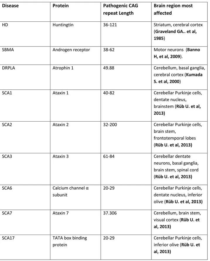
Spinocerebellar ataxias (SCA1-3, 6, 7 and 17) have been identified (Table 1) (Everett M. and Wood W. 2004, Orr T. and Zoghbi Y. 2007).
Disease Protein Pathogenic CAG repeat Length
Brain region most affected
HD Huntingtin 36-121 Striatum, cerebral cortex
(Graveland GA.. et al,
1985)
SBMA Androgen receptor 38-62 Motor neurons (Banno
H, et al, 2009).
DRPLA Atrophin 1 49.88 Cerebellum, basal ganglia,
cerebral cortex (Kumada
S. et al, 2000)
SCA1 Ataxin 1 40-82 Cerebellar Purkinje cells,
dentate nucleus, brainstem (Rüb U. et al,
2013)
SCA2 Ataxin 2 32-200 Cerebellar Purkinje cells,
brain stem,
frontotemporal lobes (Rüb U. et al, 2013)
SCA3 Ataxin 3 61-84 Cerebellar dentate
neurons, basal ganglia, brain stem, spinal cord (Rüb U. et al, 2013) SCA6 Calcium channel α
subunit
20-29 Cerebellar Purkinje cells, dentate nucleus, inferior olive (Rüb U. et al, 2013)
SCA7 Ataxin 7 37.306 Cerebellum, brain stem,
visual cortex (Rüb U. et
al, 2013)
SCA17 TATA box binding protein
20-29 Cerebellar Purkinje cells, inferior olive (Rüb U. et
al, 2013)
Table 1. Family of polyglutamine disorders
These disorders are all except SBMA, autosomal dominant progressive neurodegenerative diseases and share a common CAG repeat threshold for
6
disease onset (Orr T. and Zoghbi Y. 2007). Furthermore, all polyQ proteins, including ATXN7, share common features like misfolding, accumulation and aggregation (Michalik A. and Van Broeckhoven C. 2003). These abnormal properties are believed to confer new (gain of function) toxic properties on the mutant proteins. Most compelling evidence for gain of function of polyQ proteins are the early reports obtained from generation of hypoxanthine phosphoribosyl transferase (HPRT) polyQ mice. Insertion of a 146 CAG/polyglutamine repeat into the gene/protein of HPRT, which does not normally contain a CAG/polyglutamine repeat, resulted in neurodegeneration, meaning that an expanded polyQ domain by itself is toxic (Tallaksen-Greene
SJ. et al, 2003, Ordway JM. et al, 1997, Marsh JL. et al, 2000). In addition,
mice lacking ataxin-1 (SCA1-null mice) was found to not show evidence of ataxia or neurodegeneration, while knock-in models with expanded polyQ ataxin-1 stretch developed ataxia and Purkinje cell degeneration (Burright EN.
et al, 1995, Matilla A. et al, 1998). Moreover, early comparisons of knock-in
and knock-out HD models and the fact that huntingtin homozygous knock-out embryos die early, was considered as another proof of the gain-of-function (Wexler NS. et al, 1987, Duyao MP. et al, 1995, Nasir J. et al, 1995, Zeitlin
S. et al, 1995). During the last 20 years, several mechanisms through which
expanded polyQ protein gain of function could contribute to toxicity have then been identified and will be discussed below, see section 1.5
Most polyQ proteins are ubiquitously expressed (Ross CA et al, 1999). However, each disease has a distinct, specific pattern of degeneration (Table 1). The vulnerability of specific neurons in each polyglutamine disease is thus difficult to reconcile with a common polyQ domain-induced toxicity. It is thus likely that the surrounding amino acids in each polyQ protein and specific interacting partner proteins may play a role. For example, inactivation/interaction of the tissue-specific transcription factor cone-rod homeobox protein (CRX) or Purkinje cells expressed protein R85/ponsin by the mutant ATXN7 protein contribute to the pathology in SCA7 disease (La Spada
R. et al, 2001, Chen S. et al, 2004, Jiang YJ. et al, 2013). Taken together, it
could be summarized that, polyQ disease pathology may result from loss-of-function and/or from a toxic gain-of-loss-of-function.
1.5. PolyQ disease mechanisms
Many common polyQ disease mechanisms in neurons, as well as neighboring cells have been suggested. Evidence implicating involvement of multiple cell
7
types, including neighboring non-neuronal cells in polyQ toxicity is emerging
(Ilieva H. et al, 2009, Zoghbi HY. and Ort H. 2000). For example, in HD
models, it was shown that the disease mechanism is non-cell autonomous and it is based upon pathological cell–cell interactions (Zoghbi HY. and Ort H.
2000). The observation that Purkinje neurons die even when they do not express
mutant ATXN7 or, even more provocatively, when mutant ATXN7 is expressed only within the Bergmann glia, demonstrate that non–cell autonomous disease mechanisms is also involved in SCA7 (Custer SK. et al, 2006, Garden GA. et
al., 2002).
Ataxin-7 and many other polyQ proteins are subjected to proteolytic cleavage, mostly by caspases (Young JE. et al, 2007, 2009, Weber JJ. et al., 2014,
Guyenet SJ. et al, 2015, Garden GA. et al. 2002). The proteolytically cleaved
products are shorter and believed to form more aggregates and be more toxic. In fact, inhibition of cleavage is enough to ameliorate polyQ disease pathology in several models (Graham RK. et al. 2006, Guyenet SJ. et al, 2015, Davies SW.
et al, 1997, Sanchez I.. et al 2003, Skinner J. et al 1997). Proteolytic
fragments of mutant ATXN7 is predominantly detected in the nucleus and this may further enhance toxicity (Young JE. et al, 2007). Posttranslational modifications like phosphorylation, acetylation, ubiquitination, and sumoylation can influence polyQ protein cleavage (Takahashi T. et al, 2010, Mookerjee S.
et al 2009, Janer A. et al, 2010, Warby SC. et al, 2005). Inhibition or
enhancement of these modifications has been shown to ameliorate fragment formation and toxicity (Warby SC. et al, 2005, Terashima T. et al, 2002,
Janer A. et al, 2010).
1.5.1. PolyQ protein aggregation
Expanded polyQ proteins are believed to fold into aberrant β-sheet conformations and then aggregate into oligomers, aggregates and inclusion bodies (IB), both in the cytoplasm and the nucleus (Davies SW. et al, 1997,
Sanchez I. et al 2003, Skinner J. et al, 1997, Yang W. et al, 2002, Holmberg M. et al, 1998). The aggregates/IBs were early viewed as having a central role
in polyQ-mediated pathology (Sisodia S. 1998, Zoghbi Y. and Orr T. 2009,
Taroni F. and DiDonato S. 2004, Furrer SA. et al, 2013, Holmberg M. et al, 1995). Studies showed that aggregates or inclusion bodies could sequester
transcription factors (see section 1.5.3), as well as disrupt axonal transport in neurons leading to toxicity (Davies SW. et al, 1997, Morfini G. et al, 2005,
8
al, 2012). However, the primary role of the aggregates is still unclear as more
recent studies have argued that the aggregates might not be the main cause of polyQ toxicity and that the soluble misfolded monomers or smaller oligomers are likely the primary pathogenic agents (Yoo SY. et al, 2003, Arrasate M. et
al, 2004, Michalik A. and Van Broeckhoven C. 2003, Guyenet SJ. et al, 2015). Some studies even suggest that aggregates or IBs could in fact be a
protective measure by the cells (Arrasate M. et al. 2004, Michalik A. and
Van Broeckhoven C. 2003).
1.5.2. Aberrant UPS and autophagy activity
The Ubiquitin–proteasome system (UPS) and macroautophagy are the two main protein quality control pathways used to degrade unwanted or damaged proteins by most cells (Rubinsztein DC. 2006). UPS or autophagy should degrade misfolded proteins, like expanded polyQ proteins, that otherwise hamper cell survival (Smith SE. et al, 1996, Deter RL. et al, 1967). UPS degrades small or short-lived proteins and occurs in both the cytoplasm and the nucleus, while macroautophagy (hereafter referred to as autophagy) degrades dysfunctional organelles, as well as proteins, but only occurs in the cytoplasm (Smith SE. et
al, 1996, Deter RL. et al, 1967.
For a protein to be degraded by UPS, the protein is first tagged for degradation by attachment of a small protein called ubiquitin by ubiquitin ligases (Rubinsztein DC. 2006). Several additional ubiquitin proteins are then added, resulting in a polyubiquitin chain that is recognized by the proteasome, which contains proteases that degrades the substrate (Rubinsztein DC. 2006). PolyQ proteins, including ATXN7, have been reported to be resistant to proteasomal degradation and disrupt UPS activity (Jana NR. et al, 2001, Bennett EJ. et al,
2007, Tydlacka S. et al, 2008, Wang HL. et al., 2007, Ortega Z. et al, 2010).
However, other studies have suggested that UPS is capable of degrading polyQ proteins (Dantuma NP. and Bott LC. 2014).
For a protein to be degraded by autophagy, the protein is isolated from the rest of the cell within a autophagosome, a double membrane vesicle. The autophagosome then fuses with a lysosome and the content is degraded by lysosomal hydrolases (Majeski AE. and Dice JF. 2004). Impairment of autophagy has been shown to result in accumulation of neuronal protein inclusions and neurodegeneration, meaning that autophagy protein quality control is crucial for neuronal cell survival (Hara T. et al. 2006, Komatsu M.
9
et al. 2007). Expanded polyQ proteins including ATXN7, have been shown to
be degraded by autophagy as well as be resistant to autophagic degradation (Qin
Q. et al, 2006, Ravikumar B. et al, 2002, Yu X. et al 2012, 2013, Young JE. et al, 2007, Mookerjee S. et al 2009). In fact, some polyQ expanded proteins
have been shown to disrupt autophagy. For example, cells expressing mutant ATXN7 display decreased autophagy induction and autophagic capacity (Yu X.
et al, 2013, Young JE et al, 2007). Treatments which enhance autophagy and
promote degradation of mutant polyQ proteins has been shown to reduce mutant polyQ induced toxicity (Yu X. et al, 2012, 2013, Wang HL. et al, 2013, Nisoli
I. et al, 2010). Taken together, mutant polyQ proteins tend to display disruption
of normal cellular autophagy and/or UPS activity. This could lead to toxicity by disrupting the cellular homeostasis.
1.5.3. Transcriptional dys-regulation
Transcriptional dys-regulation is another common proposed mechanism through which polyQ proteins have been suggested to induce toxicity (Cohen-Carmon
D. and Meshorer E. 2012). Gene expression could be altered when mutant
polyQ proteins disrupt the function of transcription factors. Several transcription factors like CREB-binding protein (CBP) and steroid receptor coactivator1 (SRC1) are sequestered into expanded polyQ protein aggregates (Steffan JS. et
al., 2000, Nucifora FC. Jr et al, 2001, Stenoien DL. et al 1999). Furthermore,
mutant huntingtin, ataxin-3, (ATXN3), ATXN7 and atrophin-1 all interact or colocalize with specificity protein 1 (SP1), TAFII130 and TATA-binding protein (TBP), and thus represses transcription of some neuronal genes (Yvert
G. et al. 2001, Shimohata T. et al, 2000, Dunah AW. et al, 2002, Perez MK. et al, 1998). Moreover, mutant ATXN7 was shown to suppress the activity of
the transcription factor nuclear receptor RAR-related orphan receptor alpha (RORα) (Ström AL. et al, 2005). Other studies also reported that the normal function of ATXN7 to interact or activate the transcription factor CRX and other transcription machinery components was disturbed by mutant ATXN7 and this contribute to cone-rod dystrophy, cerebellar dysfunction and ataxia (Yvert
G. et al. 2001, La Spada R. et al, 2001, Chen S. et al, 2004, Chou AH et al, 2010). Taken together, expanded polyQ protein seems to acquire additional
function that allows them to interact with transcription factors and alter transcriptional regulation resulting in neurodegeneration (McCullough SD. et
10
1.5.4. PolyQ proteins and metabolic alterations
Energy in the form of ATP is generated in most cells through glycolysis and mitochondrial respiration. However, neurons are highly dependent on ATP generated from the mitochondria and do not appear to display increased glycolytic rate, like many other cells, upon mitochondrial inhibition (Bolanos
JP. et al., 2008, 2010). The high reliance on mitochondria makes neurons
vulnerable to metabolic dysfunction (Kann O. and Kovács R. 2007, Nehlig A.
and Coles JA. 2007).
Mitochondria play a central role in cellular metabolism by generating ATP during oxidative phophorylation (OxPHOS), which in turn, is coupled to the electron transport chain (ETC) (Warburg 1956, Weir EK. and Archer S.
2010). The ETC contains five protein complexes (complex I, II, III, IV and V)
sitting in the inner mitochondrial membrane. The transfer of electrons through ETC complexes I–IV provides the energy to drive protons against their concentration gradient across the inner mitochondrial membrane (out of the mitochondrial cytoplasm and into the mitochondria intramembrane space) (Perry SW. et al, 2011). This results in a net accumulation of H+ in the intramembrane space and generation of a mitochondrial membrane potential (Δψm, a charge or electrical gradient). When protons then flow back into the mitochondria cytoplasm through the complex V, ATP is produced (Perry SW.
et al, 2011). Malfunctioning of the ETC complexes has been shown to result in
mitochondrial dysfunction (Davey GP. et al., 1998, Hroudová J. et at, 2011,
Milakovic T. and Johnson GV. 2005).
Several studies have linked altered glycolytic activity and/or mitochondrial complex dysfunction to polyQ, as well as other neurodegenerative diseases (Damiano M. et al, 2010, Milakovic T. and Johnson GV. 2005, Power WJ. et
al, 2007, Li X.J. et al, 2010, Gu M. et al, 1996, Bae BI. et al, 2005, Silva AC. et al, 2013). For example, mitochondrial dysfunction was implicated in the
pathogenesis of HD (Gu M. et al, 1996), SBMA (Beauchemin AM. et al,
2001), DRPLA (Lodi R. t al., 2000), SCA1 (Kish SJ. et al., 1999), SCA3
(Laco et MN. et al., 2012) and SCA7 (Forsgren L .et al 1996). In fact, mutant huntingtin (mHtt) and ataxin-3 has been shown to interact with the mitochondria (Shirendeb UP. et al, 2012, Laco MN. et, al., 2012). Recently, in vivo neurometabolic profiling in patients with SCA 1, 2, 3 and 7 revealed metabolic alterations (Adanyeguh IM. et al, 2015).
11
1.5.5. Oxidative stress and polyQ disease
Reactive oxygen species (ROS) are low-molecular-weight oxygen free radicals with an unpaired electron and in order to gain stability, they “steal” electrons from neighboring atoms or molecules (Santos CX. et al, 2009, Jones DP.
2008). Nucleic acids, proteins and lipids can be modified by ROS and hence
become damaged (Jones DP. 2008, Heales SJ. and Bolanos JP. 2002). A low level of ROS is involved in signal transduction and defense against harmful invasion by modification of specific proteins (Heales SJ. and Bolanos JP.
2002). However, an elevated level of ROS, above the capacity of the
anti-oxidant system, results in oxidative stress and excessive damage to cellular molecules (Sies H. 1985, Sayre LM. et al, 2008). Neurons are highly sensitive to oxidative stress due to an abundance of oxidative-sensitive lipids, low anti-oxidant defense capacity, a restricted renewal and regenerative capacity, as well as high usage of mitochondrialrespiration (Facecchia K. et al, 2011, Gandhi S.
and Abramov A. 2012). Oxidative stress has been linked to polyQ and other
neurodegenerative disorders, including Alzheimer’s disease (AD), Parkinson’s disease (PD) and amyotrophic lateral sclerosis (ALS) (Uttara B. et al, 2009,
Greenamyre J. et al, 2004, Grimm S. et al, 2011, Tabrizi SJ. et al, 1999). The
oxidative stress in neurodegenerative disease could be caused by a decrease of antioxidant enzymes and/or an increased production of ROS (Jones DP. 2006,
Sayre LM. et al, 2008).
Antioxidants are molecules that are capable of preventing the oxidation of other molecules (Ionov M. et al, 2011, Coyle JT. and Puttfarcken P. 1993). Cellular antioxidants include superoxide dismutases (SODs), superoxide reductases (SOR), catalases (CAT), peroxiredoxins (Prx), glutathione (GSH), glutathione peroxidases (GPx), glutathione reductase (GR), glutathione-S transferase (GST), thioredoxins (Trx) and thioredoxin reductase (TrxR). Low molecular weight antioxidants include vitamin C (Vit C) and alpha-tocopherol (also known as vitamin E (Vit E)) (Fatokun A. et al, 2008). Altered levels of various endogenous antioxidants have been reported in polyglutamine disorders. For example, increased levels of antioxidants like SODs, Gpx, CAT and Trx2 has been reported in several polyQ models and could be a response to the oxidative stress induced by polyQ proteins (Reijonen S. et al, 2010, Mason P. et al,
2013). However, reduced levels of various antioxidants has also been reported in
several polyQ disease models (St-Pierre J. et al. 2006, Mason P. et al, 2013,
Browne S. et al, 1997, Yu YC. et al, 2009). The reduction of these molecules
12
disease. The exact role of antioxidants and how their variations contribute to polyQ disease pathology is thus not completely understood. It is possible that mutant polyQ proteins could differentially trigger the induction or repression of the activity/expression levels of specific antioxidants.
ROS is normally the main product in many reactions, for instance by OxPHOS in mitochondria, xanthine and NADPH oxidase (XO and NOX, respectively), cyclooxygenase (COX) and lipooxygenase (LOX) (Fatokun A. et al, 2008). So far, four mechanisms by which polyQ proteins could result in increased generation of ROS have been reported. First, increased mitochondria ROS production due to mitochondria or mitochondria ETC dysfunction has been identified (Browne S. et al, 1997, Maksimović I. et al, 2001, Ribeiro M. et al,
2013). Studies have shown that mitochondria complex functions are altered by
mutant polyQ proteins and this result in oxidative damage/stress and toxicity (Browne S. et al, 1997, Maksimović I. et al, 2001, Ribeiro M. et al, 2013). Second, aberrant transcriptional regulation of the peroxisome proliferator-activated receptor (PPAR) gamma co-activator 1-alpha (PGC-1α) has been shown to occur in HD models (Cui L. et al, 2006). PGC-1α is a transcriptional co-activator that regulates genes involved in energy metabolism and antioxidant defense (St-Pierre et al. 2006). The study showed that mutant huntingtin mediated repression of PGC-1α transcriptional activity leads to the downregulation of ROS defense genes resulting in increased oxidative damage and neuronal death (St-Pierre et al. 2006). Third, inclusion bodies formed by polyQ proteins are shown to act as centers for ROS generation (Firdaus W. et
al, 2006). Fourth, polyQ proteins were shown to activate NADPH oxidase
(NOX) enzymes leading to increased ROS generation (Bertoni A. et al, 2011,
Valencia A. et al, 2013). NOX enzymes, which are the most relevant to this
thesis, will be discussed further below.
1.6. The NOX family
The NOX family of enzymes transfers electrons across biological membranes to oxygen in the cell exterior or in intracellular compartments thereby generating ROS (Katsuyama M. et al, 2012, Ago T. et al, 2010, Bedard K. and Krause
K. 2007). There are seven members in the NOX family namely: NOX1-5 and
dual oxidase 1 and 2 (DUOX1 and DUOX2) (Lambeth JD. and Neish AS.
2014, Katsuyama M. et al, 2012, Bedard K. and Krause K. 2007, Ago T. et al, 2010). All NOX enzymes contain at least six transmembrane domains plus a
13
phosphate (NADPH) binding domain in the cytosolic C-terminal (Katsuyama
M. et al, 2012, Ago T et al, 2010, Bedard K. and Krause K. 2007). The
electron is transferred from the substrate NADPH, through FAD and two heme groups coordinated by the transmembrane helixes, to oxygen (Bedard K. and
Krause K. 2007).
1.6.1. NOX family expression in neurons
NOX enzyme activity and expression have been reported in the brain, including in neurons, astrocytes and microglia (Hernandes MS. and Britto LR. 2012,
Nayernia Z. et al, 2014). In neurons, the most abundant isoforms reported are
NOX1, NOX2, NOX3, and NOX4. NOX2 is expressed in all regions of the forebrain, midbrain and hindbrain, with particularly high levels of NOX2 in neurons in the hippocampus (CA1 and CA3 areas), cortex, amygdala, striatum and thalamus (Noh KM. and Koh JY. 2000, Serrano F. et al. 2003,
Tejada-Simon MV. et al., 2005). NOX1, NOX2, NOX3, as well as NOX4 have been
reported in cerebellum and photoreceptor cells, the most affected neurons in SCA7 (Mizuki K, et al,1998, Coyoy A. et al., 2008, Bhatt L. et al., 2010,
Nayernia Z. et al, 2014). NOX5, DUOX1 and DUOX2 expression have also
been reported in the brain (Hernandes MS. and Britto LR. 2012).
1.6.2. Regulation of NOX activity
The NOX members differ in their dependency on cytosolic components for their enzymatic activity (Chuong Nguyen MV. et al, 2015). NOX1 and NOX3 uses the NOX organizing protein 1 (NOXO1), the NOX activating protein 1 (NOXA1) and the Guanosine-5'-triphosphate (GTP) binding protein, Rac1, while NOX2 (also known as gp91phox ) uses p47phox (NOXO1 homologue), p67phox (NOXA1 homologue), p40phox and Rac1 or 2 (Katsuyama M. et al,
2012, Ago T. et al, 2010, Banfi B. et al, 2001). NOX4, NOX5, DUOX1 and
DUOX2 requires no additional cytosolic subunits (Katsuyama M. et al, 2012,
Ago T. et al, 2010, Banfi B. et al, 2001).
NOX family enzyme activity is regulated by the protein expression of the NOX enzyme itself, as well as by the expression and the assembly of the regulatory subunits (Lambeth J. et al, 2007). The NOX2 enzyme has been most extensively studied and is believed to be regulated in a complex manner. Transcription factors like nuclear factor kappa-light-chain-enhancer of activated B cells (NF-κB) and CCAAT displacement protein (CDP) have been identified as a regulator of NOX2 expression (Skalnik DG. et al, 1991, Anrather J. et al,
14
2006). However, for full activity of NOX2, the assembly of a multi-subunit
complex consisting of NOX2, p22phox, p47phox, p67phox, p40phox and Rac1/2 must take place (Katsuyama M. et al, 2012, Ago T. et al, 2010). Upon cell stimulation, Rac and p47phox are independently redistributed from the cytosol to the plasma membrane and recruited to the NOX2 complex. Phosphorylation of p47phox on several serine residues induces a conformational change and triggers the translocation to the membrane allowing p47phox to interact with p22phox. The movement of p47phox brings with it the other subunits, p67phox and p40phox. Upon recruitment, the p67phox binds to NOX2 and induces the enzymatic activity. It is believed that p40phox modification is involved in the modulation of NOX2 activity by facilitating the membrane translocation of p47phox and p67phox (Katsuyama M. et al, 2012, Ago T. et
al, 2010, Lopes LR. et al, 2004). NOX1 expression has been reported to be
regulated by transcription factors GATA-binding factor 6 (GATA-6), hepatocyte nuclear factor-1α (HNF-1α), and caudal-related homeodomain proteins (Cdx) (Valente A. et al, 2008, Brewer A. et al, 2006). NOX1 can be modulated in different cells by different stimuli including growth factors and growth-related agonists like angiotensin II in vascular smooth muscle cells, lipopolysaccharide (LPS) in gastric mucosal cells , oxidative stress in vascular smooth muscle cells, hypoxia in pulmonary epithelial cells and Interferon-γ in colon cancer cells (Geiszt M. et al, 2003, Kawahara T. et al, 2005, Goyal P. et al, 2004,
Wingler K. et al, 2001, Stanic B. et al, 2010). Full activation of NOX1
requires the assembly of its cytosolic sub-units, NOXO1, NOXA1 and Rac through mechanisms yet to be fully established (Hernandes MS. and Britto
LR. 2012, Geiszt M. et al 2003, Hordijk P. 2006). However, the activity of
NOX1 is positively and negatively controlled by NOXO1 and NOXA1 phosphorylation respectively (Hordijk P. 2006, Kroviarski Y. et al, 2010,
Yamamoto A. et al, 2013). For example, phosphorylation of NOXO1 by
protein kinase C (PKC) or protein kinase A (PKA) positively regulate NOX1-catalyzed ROS production (Yamamoto A. et al, 2013), while phosphorylation of NOXA1 by mitogen-activated protein kinase (MAPK), PKC or PKA negatively regulate NOX1-catalyzed ROS production (Kroviarski Y. et al,
2010, Oh H. et al, 2010). At present, no or few reports on the transcriptional
regulation of NOX3-5 and DUOX1-2 are known. However, transcriptional regulation of NOX4 and NOX5 by NF-κB has been reported (Manea A. et al,
15
NOX1, while NOX4 is constitutively active (Hernandes MS. and Britto LR.
2012, Brown DI. and Griendling KK. 2009). NOX5, DUOX1 and DUOX2 all
have extended N-terminal Ca2+ binding EF-hand motifs and their activity is Ca2+ dependent (Brown DI. and Griendling KK. 2009, Hernandes MS. and
Britto LR. 2012) However, reports that expression of DUOX1/2 enzymes are
controlled by cyclic adenosine monophosphate (cAMP) remains debated (Pachucki J. et al, 2004).
1.6.3. NOX function in neurons
ROS produced by NOX has been implicated in the regulation of neuronal differentiation, regulation of angiotensin II effects, and N-methyl-D-aspartate (NMDA)-mediated synaptic plasticity (Katsuyama M. et al, 2012, Gao HM. et
al, 2012). For example, it was shown that NOX2-mediated ROS is important to
stimulate normal growth and proliferation of hippocampal neural stem cells (Dickinson B. et al, 2011). In addition, mice that lack functional NOX2 are deficient in synaptic plasticity, learning and memory (Pao M. et al, 2004,
Kishida K. et al, 2006). NOX-dependent ROS was as well reported to stimulate
retinal ganglion cell survival (Mackey A. et al, 2008, Groeger G. et al, 2009). In addition, the physiological level of superoxide produced from NOX2 was suggested to be essential for proper survival signalling in retinal cell lines (Groeger G. et al, 2009). However, much remains to be discovered about the roles of NOX-dependent ROS production in healthy neurons.
1.6.4. Role of NOX in neurodegenerative diseases
Involvement of NOX enzymes in neurodegeneration has been reported, although the mechanism is not completely understood (Hernandes MS. and Britto LR.
2012, Angeloni C. et al, 2015, Cristóvão AC et al, 2012, Qin L. et al, 2013).
NOX1, NOX2 and NOX4 has been linked to neuronal oxidative stress, cerebrovascular dysfunctions and behavioral deficits in different neurodegenerative disease models (Park L. et al., 2008, Wu D. et al., 2006,
Marden JJ. et al. 2007, Choi DH. et al, 2012, Cristóvão AC et al, 2012).
Studies have also implicated NOX enzymes in polyQ diseases (Maldonado PD.
et al, 2010, Valencia A. et al, 2013). Studies of the role of NOX in SCA7 was
one main aim of this thesis, see result section.
1.7. p53 and polyQ disease
The p53 tumor suppressor protein regulates many cellular functions including cell cycle progression, apoptosis, neuronal differentiation, autophagy and
16
metabolism (Bargonetti J. et al., 2002, Blum D. et al. 1997, de la Monte SM.
et al, 1998, Duan W. et al, 2002, Kaya SS. et al., 1999). For this thesis, the role
of p53 in metabolic regulation is the most relevant. Many metabolic related genes are transcriptionally regulated by p53, including apoptosis-inducing factor (AIF), TP53-inducible glycolysis and apoptosis regulator (TIGAR) and glucose transporters (GLUTs) (Liang Y. et al, 2013, Berkers CR. et al, 2013). One way that p53 function is to slow down glycolysis and promote oxidative phosphorylation thus balancing the use of glycolysis and OxPHOS.
The p53 protein mainly functions as a transcription factor and is regulated by post-translational modifications. Examples of these modifications include phosphorylation, oxidation, ubiquitination and acetylation (Gu W. et al., 1997,
Ito A. et al, 2001, Vousden KH. 2002, Woods DB. et al, 2001, Wu HH. et al, 2000, Marchenko ND. et al, 2007). These modifications are associated with
changes in p53's activity and localization (Ito A. et al, 2001, Liang SH. et al.,
2001, Illuzzi J. et al, 2011). Acetylation and/or phosphorylation promote p53
nuclear localization and transcriptional activity, while oxidation and mono-ubiquitination promotes cytoplasmic localization and impedes transcriptional activity (Gu W. et al, 1997, Shieh SY. et al, 1997, Wu HH et al, 2000). Other molecules have also been shown to modulate p53 DNA specificity and functions (McLure KG. et al, 2004). For example, NAD+ binds to p53, induces a conformational change and inhibits p53's DNA binding function (McLure KG.
et al, 2004). Interestingly, NAD+ which is a known product of NOX enzymes is
also known to induce the expression and activity of SIRT1, a deacetylase enzyme that control the level of p53 acetylation (Hayashida S. et al, 2010,
Puca R. et al, 2010).
Change in subcellular localization of p53 has been reported in polyQ diseases (Yu X. et al, 2013, Illuzzi J. et al, 2011, Guo X. et al, 2013). A shift in p53 from the nucleus to the cytoplasm or the mitochondria have been observed in several polyQ disease models and supports the argument that p53 localization is altered in polyQ disease (Guo X. et al, 2013, Yu X. et al., 2013, Tsoi H. et al,
2012). Increased mitochondria and cytoplasmic p53 levels have been shown to
promote apoptosis and inhibit autophagy, respectively (Marchenko ND. et al,
2007, Yu X. et al, 2013). Moreover, different polyQ aggregates were found to
contain p53 (Nucifora FC. Jr et al., 2001, Steffan JS. et al, 2000, 2001, Yu X.
et al, 2013). For instance, mutant Htt was shown to interact with p53 and
17
aggregates in a SCA7 models was shown to result in reduced p53 transcription activity (Yu X. et al, 2013). However, other studies have reported increased levels of p53 regulated apoptotic genes, like Bcl-2-associated X protein (BAX), in SCA7 models (Wang HL. et al 2005). Moreover, deletion of p53 was shown to suppress mHtt and ataxin-1 (ATXN1)-induced degeneration in Drosophila and mice, respectively (Steffan JS. et al, 2000, Bae BI. et al, 2005, Ryan AB.
et al, 2006). The role of p53 in polyQ disease is thus complex.
1.8. SCA7 disease diagnosis and therapeutics
Presently, there is no cure for SCA7 or the other polyQ disorders. Proper diagnosis and supportive therapy to manage the symptoms are the only available treatment. Diagnosis of SCA7 is done based on neurological examination e.g neuroimaging, assessment of visual acuity, visual fields and color vision and genetic testing. Supportive therapies that help to improve symptoms include the use of speech and communication therapy devices, which can help to manage cerebellar ataxia symptoms (http://www.ncbi). Use of sunglasses, limitation to UV exposure or optical aids for individuals with peripheral visual loss is also in use (http://www.ncbi). For patients with swallowing difficulties, video esophagrams is used to identify the food that is likely to trigger aspiration complication (http://www.ncbi).
Several potential therapeutic strategies are currently investigated for SCA7 and other polyQ diseases. First, efforts into testing allele-specific RNA silencing/interference strategies are ongoing (Scholefield J. et al, 2009,
Ramachandran PS. et al, 2014). Non-allele specific silencing of ATXN7 has
been shown to improve the disease phenotype in a mouse model of SCA7 (Ramachandran PS. et al, 2014). However, if the disease is caused by loss of function, a non-allele specific approach might not be that beneficial. Other strategies that aims at enhancing protein quality control systems, reducing oxidative stress through antioxidant stimulation, or increasing transcriptional activity have also been proposed based on the positive results obtained from various studies (Cortes CJ. and La Spada AR. 2014, Kamat CD. et al, 2008,
Katsuno M. et al, 2005, Ferrante RJ. et al, 2013). Treatments aimed at
increasing the level of brain-derived neurotrophic factor (BDNF) has been show to prevent the degeneration of mouse striatal neurons in HD models (Pineda JR.
et al. 2007, Silva A. et al, 2015). At present, transplantation of stem cells,
capable of secreting BDNF, is tested in 20 clinically diagnosed SCA1 patients in a phase II clinical study (https://www.clinicaltrials). In addition,
over-18
expression of hepatocyte growth factor (HGF), a pleiotrophic growth factor with highly potent neurotrophic activities on cerebellar neurons, was shown to attenuates the degeneration of Purkinje cells and Bergmann glia in a knock-in mouse model of spinocerebellar ataxia type 7 (Noma S. et al, 2012).
19
2. Aims of the thesis
The overall aim of this thesis was to investigate the molecular mechanisms by which polyQ expanded ATXN7 induce toxicity and whether modulating these mechanisms could ameliorate toxicity. Specific aims of each paper are listed below:
Paper I.
The aim of paper I was to establish and characterize a new stable inducible SCA7 model and to use this model to investigate how different ATXN7 species are degraded.
Paper II.
Paper II aimed to investigate the role and source of oxidative stress in mutant ATXN7 expressing cells. We also aimed to investigate whether ameliorating oxidative stress could constitute a potential therapeutic strategy in SCA7 disease.
Paper III.
The aim of paper III was to investigate the molecular mechanisms by which mutant ATXN7 cause metabolic dysfunction.
Paper IV.
The aim of paper IV was to further investigate the cross-talk between NOX1 and p53 in mutant ATXN7-induced metabolic dysfunction and toxicity.
20
3. Methodological consideration
The methods used in this thesis are described in detail in the contributing papers. This part will summarize the main methods utilized with some theoretical aspects. The statements below are valid for all papers when nothing else is stated.
3.1. Cell lines used
HEK 293T, HeLa and stable neuronal-like PC12 cells expressing wild-type or mutant ATXN7 were used.
3.1.1. HEK 293T cells
HEK 293T are transformed cancer cells originally derived from human embryonic kidney cells (Graham FL. et al, 1977). This cell line is good for research because it is of human origin and is easy to transfect. In addition, HEK cells express endogenous ATXN7.
3.1.2. HeLa cells
HeLa are transformed cancer cells originally derived from human cervical cells (Scherer WF. et al, 1953). Like HEK 293T cells, they are easy to transfect and express endogenous ATXN7 making them suitable for our research study.
3.1.3. Stable inducible Tet-off PC12 cell lines
PC12 is a neuronal-like cell line derived from a pheochromocytoma of the rat adrenal medulla (Greene LA. and Tischler A. 1976). Generation of stable inducible PC12 cell lines expressing N-terminal FLAG- and C-terminal GFP-tagged ATXN7 with 10 (FLQ10 line) or 65 (FLQ65 line) glutamines were made by transfecting a commercial PC12 Tet-off cell lines with FLQ10-pTRE-tight or FLQ65-pTRE-tight plasmid construct, respectively. In these cell lines, expression of the corresponding protein namely: ATXN7Q10-GFP or ATXN7Q65-GFP is induced upon removal of doxycline (Dox) from the media. Our choice of the Tet-off system over the Tet-on system was to avoid any possible interference by doxycycline on our readouts when ATXN7 is expressed (Ermak G. et al, 2003). Colonies with GFP tagged ATXN7 expression levels similar to that of endogenous ATXN7 were selected to avoid over-expression effects.
3.2. Cell treatments and transfection
To study the role of UPS and autophagy in paper I, inhibitors or activators of UPS and/or autophagy was used. Rapamycin and trehalose were used as
21
autophagy activators (Guertin DA. and Sabatini DM. 2007, Sarkar S. et at,
2007). Rapamycin inhibits the mammalian target of rapamycin (mTOR), a
negative regulator of autophagy, while the exact mechanism of trehalose is not known, but is believed to be mTOR independent (Guertin DA. and Sabatini
DM. 2007, Sarkar S. et at, 2007). NH4Cl or 3-MA was used to inhibit
autophagy (Hart PD. et al, 1983, Gordon AH. et al, 1980, Seglen PO. and
Gordon PB. 1982). NH4Cl inhibit autophagosome-lysosome fussion, while
3-MA inhibit the formation of autophagosomes (Gordon AH. et al, 1980, Seglen
PO. and Gordon PB. 1982 ). Epoxomycin was used as inhibitor of proteasome
catalytic function (Meng L. et al, 1999).
To study the role of oxidative stress in paper II and III, antioxidants or pro-oxidants were used. N-acetyl cysteine (NAC) or Vitamin E (Vit E) was used as antioxidants (Aruoma I. et al, 1989, Burton G. and Traber M. 1990). NAC act as an antioxidant by regulating GSH metabolism, while vit E acts as a chain-breaking antioxidant that prevents the propagation of free radical reactions (Aruoma I. et al, 1989, Burton G. and Traber M. 1990). Hydrogen peroxide (H2O2) or buthionine sulphoximine (BSO) was used as pro-oxidants (Kato H.
et al, 1997, Skapek S. et al, 1988). H2O2 directly generate reactive oxygen
species, while BSO deplete GSH (Kato H. et al, 1997, Skapek S. et al, 1988). To study the activity and role of NOX complexes in oxidative stress, general or specific inhibitors were used in paper II, III and IV. Apocynin and gp91ds-TAT (NOX inhibitor peptide conjugated to a cell penetrating peptide named TAT) was used as more general NOX inhibitors (Vejrazka M. et al, 2006, Rey F. et
al, 2001). Apocynin inhibits the p47phox subunit of the NADPH oxidase, while
gp91ds-TAT inhibits the interaction of NOX2 with p47phox thereby inhibiting NOX enzyme assembly and activation. Based on sequence homology, apocynin and gp91ds-tat may also affect NOX1 assembly (Tejada-Simon MV. et al.,
2005). However, an antioxidant effect of apocynin has also been reported
(Heumüller S. et al, 2010). For more specific inhibition, NOX1 siRNA and ML171 was therefore also used (Gianni D. et al, 2010). ML171 inhibits the assembly of NOX1 complexes and the selectivity for NOX1 (IC50 of 0.25 μM) over other NADPH oxidases (IC50 > 3 μM) has been shown (Gianni D. et al,
2010).
To study mitochondrial activity/function in paper III, inhibitors of mitochondria complexes I-V were used. Rotenone, 3-nitropropionic acid (3-NP), antimycin A,
22
sodium azide (NaN3) or oligomycin was used as inhibitors of mitochondria complex I, II, III, IV and V, respectively (Forkink M. et al, 2014,
Cillero-Pastor B. et al, 2013).
To study the role of p53 proper in papers III and IV, a p53 inducer, Nutlin-3, was used (Vassilev LT. et al, 2004). Nutlin-3 binds to the binding site of p53 on MDM2 thereby inhibiting degradation of p53 (Vassilev LT. et al, 2004). To study p53 oxidation in paper IV, a pro-oxidant to sulfhydryls groups, diamide was used (Leichert LI. and Jakob U. 2006). Diamide forms a reversible disulphide bond with sulfhydryls group on proteins (Leichert LI. and Jakob U.
2006).
To study protein half-lives in paper IV, a protein synthesis inhibitor, Cycloheximide (CHX), was used (Poehlsgaard J. and Douthwaite S. 2005). Cells were transfected using polyethyleneimine (PEI), lipofectamine2000 (Invitrogen) or lipofectamine RNAiMAX.
3.3. Analysis of protein expression
Western blot was used to analyze the expression levels of specific proteins. Total or subcellular protein fractions were obtained from cells and boiled together with sodium dodecyl sulphate (SDS) to denature the proteins and gives them a negative net charge in proportion to their size (Weber K and Osborn
M. 1969). This difference in charge was then used to separate proteins according
to size on a polyacrylamide gel that is run in an electric field. The separated proteins were further transferred to a membrane with the help of an electric current. Specific proteins were then probed by using specific antibodies directed against the target protein of interest. A horseradish peroxidase (HRP) coupled secondary antibody, which binds to the primary antibody, was then added in order to visualize the protein by enhanced chemiluminescence (ECL) (Veitch
NC. 2004, Mruk DD. and Cheng CY, 2011). The ECL contains luminol and
hydrogen peroxide (H2O2) (Mruk DD and Cheng CY, 2011). On addition of the ECL, HRP will catalyze the oxidation of luminol generating a sensitized intermediate reagent near the molecule of interest. The intermediate product is further oxidized by H2O2 to produce a triplet-excited carbonyl, which emits light when it decays to the singlet carbonyl (Mruk DD and Cheng CY, 2011). This signal was captured on a charge-coupled device (CCD) camera. The relative abundance of the target protein was quantified by densiometric analysis using Image lab software. Both the specificity and sensitivity of this technique
23
depends on the antibody. If there is cross reactivity of the antibody, the result might be difficult to interpret and the specificity of the antibody should be confirmed using either over-expression or knock-down/knock-out system of the target protein.
3.4. Analysis of mutant ATXN7 aggregation
Analysis of mutant ATXN7 aggregation was done using filter trap assay and microscopy. For filter trap assay, cells were lysed, samples centrifuged and the pellet fractions containing aggregated materials filtered through a nitrocellulose membrane with a pore size of 0.2 µm in diameter using vacuum aspiration. Under these conditions, aggregated materials larger than 0.2 µm in diameter will be trapped on the membrane. Probing of the membrane was then performed using primary ATXN7 antibody and a corresponding horseradish peroxidase (HRP) coupled-secondary antibody. Visualization was performed using enhanced chemiluminescence (ECL). The benefit of this method is that quantification of the result is easy and objective.
For microscopy, cells grown on glass cover-slips were washed with phosphate-buffered saline (PBS) and fixed with paraformaldehyde to stop all life processes and to prevent disintegration of the cellular structure. Cells were then permeabilized with Triton X-100 to create holes in the plasma membrane and create access to intracellular or intraorganellar antigens. Cells were then incubated with primary antibodies and with fluorescent secondary antibodies. Slides were washed and mounted using Vectashield-mounting medium. Fluorescence microscopy was carried out using a Leica DM IRBE epifluorescence microscope with a ×100 objective. The advantage of microscopy analysis is that specific structures and location of aggregates can be studied.
3.5. Toxicity measurement
Toxicity was measured with WST-1 and membrane integrity assay. The WST-1 assay is based on the enzymatic cleavage of the tetrazolium salt WST-1 to formazan by cellular mitochondrial dehydrogenases present in viable cells. This assay can be used to measure cell proliferation or toxicity (https://www.caymanchem.com/pdfs). To measure toxicity and not proliferation, we normalized the absorbance value with the protein concentration in each sample. By doing this, we can rule out readouts that correspond to changes in proliferation. Advantage of using the WST-1 salt is that the salt yields a water-soluble cleavage product, which can be measured directly without
24
any further solubilization. The assay requires no washing step making it easy and fast to perform. In addition, WST-1 is more stable in contrast to MTT, XTT or MTS.
Membrane integrity as a measure of toxicity was determined by analyzing leakage of lactate dehydrogenase (LDH) from the cytoplasm of damaged cells. Measurement of leaked LDH from the cytoplasm into the surrounding culture medium has been widely accepted as a valid method to estimate toxicity when normalized with the total LDH value or protein concentration (Decker T. and
Lohmann-Matthes ML, 1988). The assay is based on LDH dependent
enzymatic conversion of resazurin to resorufin whose fluorescens can be measured. The advantage of this assay is that it is easy to handle, require little time and is non-destructive (cells can be further analyzed by other method) since it can be performed on culture media.
3.6. GSH assay
The total level of reduced glutathione was measured using the Glutathione assay kit (GSH.GloTM). In cells, GSH is oxidized to glutathione disulphide (GSSH) by reactive oxygen species and GSSH can then be converted back to GSH by glutathione reductase (GSR). Measuring the total level of reduced glutathione (GSH) provides information about ROS mediated depletion of GSH. The assay is based on the conversion of a luciferin derivative into luciferin in the presence of glutathione, catalyzed by glutathione S-transferase (GST). The signal generated is coupled to firefly luciferase. Light from the luciferase is dependent on the amount of luciferin formed, which is in turn dependent on the amount of GSH present. The kit provides a stable signal with a half-life greater than 2 hours thereby eliminating the need for strictly timed luminescent detection. In addition, only a small sample is required because of the enhanced sensitivity.
3.7. ROS measurement
Total ROS was analyzed using the oxidative-sensitive dye, dichloro-fluorescein-diacetate (DCF-DA). DCF-DA is colorless and non-fluorescent and can passively diffuse into the cell (Zulueta JJ. et al, 1997). On entering the cell, the acetate groups is cleaved by intracellular esterases to yield a non-fluorescent compound which is later oxidised by ROS into 2', 7’ –dichlorofluorescein (DCF) , a highly fluorescent compound whose fluorescens can be measured. Advantage of DCF-DA over other dyes is that it is easy to use and extremely sensitive to changes in the redox state of a cell.