Can oral contrast enhance image quality at
MRCP?
A literature review
Author: Mohassad Al-Atia
Supervisor: Thomas Strand
1
ABSTRACT
Background: Examinations conducted with magnetic resonance imaging (MRI) with the
purpose of imaging the cholangiopancreatography can sometimes be challenging regarding the acquisition of images with good quality to ensure the planning of surgical procedures. Different methods and ingredients can be useful in combinations when patients go through an MRCP (Magnetic Resonance CholangioPancreatography). Negative oral contrast can
theoretically be used to improve the image quality by suppressing the signal from tissues surrounding the biliary tract.
Aim: The overall aim of the study was to investigate the effects on image quality of oral
contrast when examining the biliary tract with MR.
Method: A structured literature review was conducted with the aim of analyzing the results
from latest scientific studies about the use of oral contrast in conjunction with cholangiopancreatography.
Results: The results showed that different negative oral contrast can be used to improve the
image quality at MRCP. They also showed to be effective in suppressing signals from the adjacent background.
Conclusion: This literature review gives knowledge in a somewhat unknown area within
radiology. Radiographers can use this literature review to insert implementation in respective workplace to increase the quality of MRCP examinations.
2
TABLE OF CONTENTS
Abstract ... 1 RESEARCH QUESTION ... 3 BACKGROUND ... 3 MRI Modality ... 3Negative & positive oral contrast ... 4
Patient experience... 4
Preparations before an MRI ... 5
Perspective and presumptions ... 7
AIM ... 8 METHOD ... 8 Data Collection ... 10 Data analysis ... 10 Ethical Considerations ... 11 RESULTS ... 11
Juices & Date syrup ... 12
Black tea ... 13
DISCUSSION ... 17
Discussion of chosen method ... 17
Discussion of the main findings of the results ... 18
Conclusions & Clinical implementations ... 20
REFERENCES ... 22 ATTACHMENT 1
3
RESEARCH QUESTION
Magnetic Resonance can be used in medical imaging for visualization of anatomy and the physiological processes of the body (Westbrook, 2011). Magnetic Resonance
CholangioPancreatography (MRCP) is an examination that provide the ability to visualize fluids in motion in the biliary tract, primary with the help of T2-weighted images. Bile and pancreatic juice become naturally hyper intense. These hyper intense structures can also be visualised in organs such as duodenum and the gastric. By letting the patient apply the concept of fasting before the examination, it is possible to eliminate most of the interferences in the images that are acquired. It is vital to collect images of good quality to ensure that they can be used in planning of surgical procedures. A negative oral contrast can be used to
prevent other signals from the gastric to appear in the MRI-image (Sakamoto, 2016). There is an interest to delve into the prospect of using an oral contrast such as date syrup, black tea or different juices to examine the effects on the images when examining the biliary tract. If interferences or artefacts appear in the images than it would disturb the image quality and the consequence might be that they are assessed as images with no diagnostic value (Westbrook, 2011). This could hamper the time frame of planning eventual surgical procedures (Sakamoto et al, 2016). It is therefore of high interest to examine through this literature review how to acquire images of good quality and to ensure if usage of oral contrast can increase image quality or not (Westbrook, 2011 & Sakamoto, 2016).
BACKGROUND
MRI Modality
With the technique of magnetic resonance imaging (MRI) it is possible to produce high quality images of the human body. This is primary because of the presence of a magnetic field and radio waves. More specifically there are certain factors that has to be fulfilled. There needs to be a magnet for nuclear alignment, a source for radio frequency (RF excitation), a magnetic field, a computer setup, a processor for images so that signals can be converted into images. Magnetism is defined by how different materials behave in a presence of an external magnetic field. This is also called magnetic susceptibility (Westbrook, 2011).
4
Negative & positive oral contrast
There are both positive and negative contrast in MRI but also in CT and conventional radiology. In CT the positive contrast is based on iodide and is injected intravenously, with this technique it is possible to compare the contrast of the images in organs or vessels that have absorbed the contrast. It is possible to diagnose different tumours by assessing if the tumours have absorbed the contrast or not. However negative contrast in CT could be CO2 (carbon dioxide), this is used when examining the bowels. The contrast is visualised as gas is filled in the bowels, this is also a method that is used so that the radiologists have more material when they diagnose for example constipations. In MRI the positive contrast
(gadolinium) is also injected intravenously. What is an important aspect is that in MR images the image contrast is determined by the relaxation mechanisms. Tissues who have long relaxation times appear in a different way to tissues with short relaxation times. Contrast media is based on the fact of the agents ability to affect on the existing magnetic field and the relaxation times of T1 as well as T2. In MRI there are other contrast agents that can be useful for the examinations. These are negative contrast agents. Elements like manganese or iron oxide can be used to create alterations in T1 respectively T2 relaxation times. Manganese can be used for liver imaging. The effects they have is that they shorten T1 relaxation time and appear very bright on T1 images. Manganese is absorbed in the kupffer cells in the liver, it means that in the image the lesions will be visualised as even darker (Westbrook, 2011).
A negative oral contrast is sometimes used in medical concerns. When going through
different examinations with MRI, it is possible that in combination with the examination that the patient needs some kind of negative oral contrast. There are different negative oral contrast, such as for example pineapple juice. A negative contrast, such as pineapple juice is used as a drink when conducting MRCP examinations. The patient drinks a certain amount of the pineapple juice and within a specific time frame that the radiographer decides (Duarte et al, 2012)
Patient experience
Examinations with this modality is in principle associated with causing no pain, however with higher magnetic fields (3 tesla and above) some sequences can be rather uncomfortable for the patient. The diffusion sequence can be apprehended as a bit frightening and in some cases
5 on the individual level some people feel pain when going through this particular sequence (diffusion) (McRobbie, 2007). When going through an MRCP examination the patient should in practical not feel any pain associated with the examination. The patient only need to lay still and hold the breath at certain times during the whole procedure (Hjartarson, 2016) (McRobbie, 2007 & Hjartarson, 2016).
Depending on the examination and the question formulation it is sometimes needed to inject contrast media through the blood vein. This is because the radiologist wants to compare the contrast in the image with and without the contrast media or to see if specific structures load contrast media or not. This is the ultimate way of detecting specific diseases or pathological structures. The procedure to contain images through MRI is based on the patient being placed on the examination table and then slowly moving into the tunnel. The radiographer positions as a routine in isocenter and over the area that is to be examined. The tunnel that is a part of the magnetic camera and is located in a room with a magnetic field with a certain tesla value is open at both ends. It means that there is an open space at the patients’ headboard as well as the feet. The radiographer is able to talk to the patient during the examination by using a microphone and the patient through a headset. This is important so that the patient isn’t feeling left out during the procedure. MRI scans take usually between 20-120 minutes so therefore it is important for the radiographer to establish a relationship with the patient. Some patients experience claustrophobia in contention with going through an MRI examination. It is vital to handle these people delicately and make sure to not underestimate their fear of being felt like they’re locked in (Westbrook, 2011 & McRobbie, 2007). It is sometimes possible to inject a tranquilizer (stesolid) that affect the central nervous system of the patient and as a consequence the ability of awareness is decreased and the patients feel somewhat calmer during the examination (Hunt, 2011). (Westbrook, 2011, Mcrobbie, 2007 & Hunt et al, 2011).
Preparations before an MRI
There are certain questions that the radiographer shall always ask the patient before they go through the examination. There are also some preparations that has to be done before the patient can go through with the examination. These routines are to be done properly to minimize the risk of something occurring during the examination. It is of high importance to conclude if the patient has a pacemaker or not. It could be dangerous for a patient with a
6 pacemaker to enter a room with a high magnetic field. The pacemaker might interact with the magnetic field in the room and adjust differently because they interfere with each other and the consequences might be disastrous for the patient as it can lead to the pacemaker not working properly anymore. Different hospitals have different opinions and routines as well as guidelines on how to handle pacemaker patients when calling for an MRI examination. But it is important to with take actions on a preparation level when letting a patient with a
pacemaker enter the room. There should at all times be someone watching them and make sure that it has not been manipulated by the high magnetic field. The same preparations and safety measures should be applied with patients who have a shunt. The patient must be asked by the radiographer if they have a shunt or a pacemaker before a decision is made on whether the examination will take place or not. Other aspects or questions that might be asked or thought of by the radiographer is if the patient have specific diseases that might be considered as contra indicated when examining patients who needs a contrast media injection. If the patient has kidney disease or diabetes and is being treated it indicates that the patient might not handle contrast media optimally. The radiographer than has to calculate the glomerular filtration rate (GFR) to see if it is possible or not for the patient to receive contrast media as well to decide the amount of contrast media. If the patient’s kidney function is to low (low GFR value) then there is a possibility that the patient is suffering from some kind of kidney disease and this means that it is not proper to administrate contrast media. When these patients arrive the radiologist has to make a decision about which protocol is proper to be used. This decision is made according to the question contained in the referral. Other questions of importance might be if the patient has myasthenia gravis, tattoos, any metals, implants. Females are asked to answer if they’re pregnant or not (Sammet et al, 2016).
7
Perspective and presumptions
The main focus of a radiographer is radiography and within it there are certain areas of responsibility. The responsibility areas that a radiographer shall possess are nurturance, image- and function medicine, radiation physics as well as medicine. The aforementioned implicates that a radiographer is characterised by his work under different ethical principles. They are:
“the principle of respect and empowerment” “the principle not to hurt”
“the principle to do good”
“the principle of justice” (competence description for radiographers by Swedish union, 2012, p. 7).
In the “competence description for radiographers by Swedish union” a model exists for radiographers to follow. In this constructed model there are certain work assignments that a radiographer should have the ability of as well as performing them. These work assignments are the theory and practice of radiography, research, development, education as well as leadership. A radiographer need to obtain an ethical approach through these competence areas. It means that a radiographer need to possess knowledge in different areas. Producing images with good quality is one of these areas. The image shall always be acquired and then worked through. The quality of the images that are required is dependent on what the
radiologist is looking for in the images and the radiographer applies the right techniques in reconstructing after the radiologists demands. A conclusion of the image quality is a priority to make, are the images of diagnostic relevance or not is a way of thinking for every
radiographer. The radiographer shall therefore be able to know methods or techniques to directly influence the image quality if necessary. The radiographer has to be aware of
handling the information of the patient with sensitivity and confidentiality. Patient safety can be obtained in different way, a secure one is by proceeding through guidelines, routines and constitutions (competence description for radiographers by Swedish union, 2012)
According to the patient safety law (SFS 2010: 659) it is mentioned that “Personnel that works in health care has to take required actions to prevent suffering for patients” (chapter 3, Paragraph 2). This can be implemented by giving tranquilizers for those who suffer of claustrophobia in contention with an MRI scan. “The radiographer need to document the
8 distribution of work throughout the handling of the patients’ information” By obtaining this law it is preferred because it gives the radiographers at the workplace an overview of who is responsible for what and whom (chapter 3, paragraph 9). It is also mentioned that
_”radiographers shall possess a contributing attitude of increasing patient safety” (chapter 6, paragraph 4). Focusing on patient safety is of high importance and it is a guideline to be followed. (SFS 2010:659).
AIM
The overall aim of the study was to investigate the effects on image quality of oral contrast when examining the biliary tract with MRI.
METHOD
Scientific knowledge is obtainable with proven established methods. Extracting information from a certain area of research is possible. This study was based as a literature review.
Articles with scientific and academic requirements were selected. The articles were chosen in the area of radiography and were analysed systematically (Friberg, 2012).
An overview of knowledge is optimally obtained by literature reviews, it also illuminates specific knowledge regions within a selected topic. “To contribute to evidence based nursing care with basics in analysis of quantative research” is a model to follow when writing
scientific studies with basics in literature. Friberg (2012) tells us that when conducting literature reviews it is optimal to establish perception and analytical methods of the studies that are being published and included. The biggest source for a literature review is scientific articles (Friberg, 2012)
Research can be made with qualitative and quantitative methods. When analysing a literature study, research can be found using one or two of these methods. What is favourable with a literature review is that it frequently contributes to an update of scientific knowledge and provides effectiveness in the work organisation if given the chance of implementation (Friberg, 2012).
9
Selection
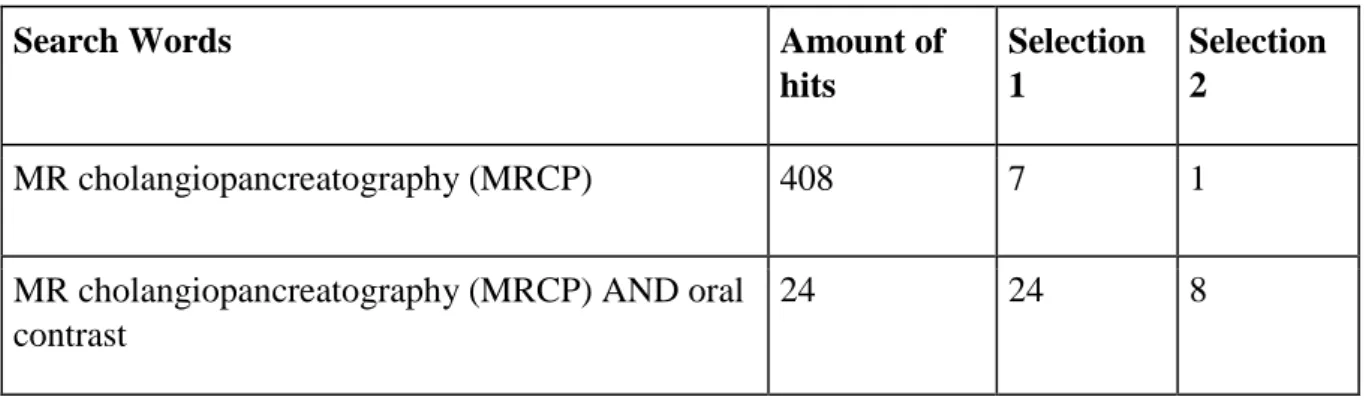
There are different data bases that can be used in a systematically way to choose and extract articles with scientifically and academically character. PubMed is one of those data bases and has been used to look for scientific articles. Cinahl is another data base that were included when searching for articles, though it was not used in this current study because ultimately the same articles were found on cinahl as were on PubMed, but a broader variation of articles could be chosen from PubMed, hence the exclusion of Cinahl. There is a possibility to add limitations, inclusions as well as exclusions to simplify the aspect of searching for articles. PubMed is a resource in a literature review in this current study. Both patient and phantom studies were included in the current study. The inclusion of both variants was to ensure that no patients were harmed in any way of the oral contrast that were used in the different studies. When choosing the articles there was no prioritising in terms of age nor gender, not even in phantom size. The chosen articles has been published in the last 10 years to ensure that the research is up to date. One article from 2005 were chosen because it strengthened the results. This was an exception from all of the other chosen articles. All of the articles were written in English except from two that were in a foreign language, though at first hand they were read in English but they couldn’t be located. This was also an inclusion criteria at first hand but wasn’t fulfilled as two were only available in a foreign language. All of the studies were reviewed for ethical approach and if answering to the purpose of the study or not. A specific exclusion criteria was all review articles. A table#1 has been inserted below to illustrate the search words when looking for the articles. A selection table can be seen below. Selection was based as a two-sided method. At first hand a selection 1 was made after reading abstracts and then the articles and at selection 2 the articles were chosen to be included in this current study.
Table 1. Searching schedule on the data base PubMed
Search Words Amount of hits Selection 1 Selection 2 MR cholangiopancreatography (MRCP) 408 7 1
MR cholangiopancreatography (MRCP) AND oral contrast
10
Data Collection
To be able to have a good orientation in a current chosen area of radiography certain aspects must be fulfilled. There needs to be inclusion criteria’s as well as exclusion criteria’s. Another aspect is the usage of Boolean operators such as AND, which means that the articles need to include the search word that has been written both before and after “AND”. There are also other Boolean operators such as “OR” and “NOT”, but these were not used in the searches in the current study. When first searching for the articles an overall review was made by the author which included reading the purpose, method and results of the article. The second selection was conducted to filtrate those articles that fitted to the current study. Each chosen article was analysed and a quality evaluation was performed by using a reviewing template (Willman, Stoltz & Bahtsevani, 2006). To ensure the scientifically character of the articles a categorization was made by placing each article in a specific quality level. A folder (see attachment 1) was used as a resource to ensure the quality of each article. A specific system was used to evaluate the quality, through answering different questions. All of the articles reached 100 % quality with the exception of one article from 2005 that only received 90 % quality. The calculation was conducted through a specific point system and then calculated in per cent. In all cases the articles proved it was of the highest possible quality. The numbers 1 and 2 were chosen to indicate which quality level that the scientific article fulfilled. Number 1 indicated highest possible quality, meanwhile number 2 indicated of second highest quality.
Data analysis
All of the selected studies in this current literature review reached scientifically and
academically character in accordance with mentioned criterias in Friberg (2012). The author assessed all of the involved articles in this study. The relevance of every article has been studied by the author. Aspects such as resemblances and differences in the articles were looked at by the author to ensure repeatedly the quality for usage in this study. In the analysis of the results section, a categorization was made by the author that seemed to fit in with the extracted results. The topics were “Juices & Date syrup, Black Tea and Mengan chloride tetra hydrate”. The author categorised by emphasising on the relation from one article to the other by looking at differences and resemblances. In every article that was used in the results section the essential results were located and placed beneath respective correct subheading
11 (Kristensson, 2014 & Friberg, 2012).
Concluding the results in this literature review was possible through integrated analysis. Each article was presented in relation to one another and this enabled comprehending the results in a qualitative way (Friberg, 2012).
Ethical Considerations
Each article that was included in this literature review has been approached for ethical views. Certain requirements were fulfilled, such as, no participants were harmed in any way. An ethical approval on this aspect has therefore been fulfilled. Only articles that answers to the purpose of this literature review have been mentioned or included. The author of this literature review has only included results of interest and in terms of what to achieve specifically with the results. Some of the results might indicate that one or several of the methods might be more applicable or optimal than others (Beauchamp & Childress, 2001). The significance of mentioning these explanations is to ensure that none of the methods were harmful for the patient. In all cases, in each article, the author of this literature review confirmed that the patients had given their approval for participation in each article/study.
RESULTS
In this current study results were based on 9 scientific articles (Govindarajan et al, 2014., Ghanaati et al., 2011., Tang et al, 2013., Bittman & Callahan, 2014., Sanchez et al, 2009., Cooppens et al, 2005., Watanabe et al, 2016., Marugami et al, 2013 & Tajima et al, 2013). In all of these studies, the effect of a negative oral contrast was assessed when patients and/or in some cases a phantom went through an MRCP examination. In all cases it resulted in better visualisation and better image quality. The results were based on a combination of
suppressing signal intensity of adjacent background organs such as gastric content that are not of interest in a MRCP examination as well as an increase of signal intensity in organs such as the common bile duct or the pancreatic duct. The results were based on different negative oral contrast. One of the studies assessed date syrup (Govindarajan et al, 2014), four studies assessed different juices (Govindarajan et al, 2014., Bittman & Callahan, 2014., Sanchez et al,
12 2009 & Cooppens et al, 2005). Two of the studies analysed the positive effects of black tea on MRCP examinations (Ghanaati et al, 2011 & Tang et al, 2013). Three of the studies assessed the effect of Manganese chloride tetra hydrate (MCT) on MRCP examinations (Watanabe et al, 2016., Marugami et al, 2013 & Tajima et al, 2013). In three of the studies (Govindarajan et al, 2014., Sanchez et al, 2009 & Cooppens et al, 2005) a further specific result was that in combination with the negative oral contrast the image quality was enhanced especially in the T2 sequences.
Juices & Date syrup
According to the study of (Govindarajan et al, 2014) a phantom study containing different negative oral contrasts was conducted. Date syrup, ferumoxsil, pineapple juice and water were tested with a 1,5 T MRI and T2 sequences were acquired in an MRCP examination. The results showed that when obtaining images using date syrup it enhanced the improvement of image quality. It also suppressed the signal in the gastrointestinal tract. What was also observed was that the visibility in the common bile duct, cystic duct and the pancreatic duct increased. The patients meanwhile were not affected in any way, either positively nor negatively.
In the study of (Bittman & Callahan, 2014) juices were examined as oral negative contrast when conducting MRCP examinations. Three different juices were tested, Acai, blueberry and pineapple juice. All of the three juices resulted in improvement of image quality when
conducting MRCP examinations. The patients preferred the acai juice better than blueberry and pineapple though none of the mentioned juices seemed to have any effect on the patients.
In another study made by (Sanchez et al, 2009) the juice of Acai was analysed as a negative oral contrast for MRCP examinations. Two radiologists compared obtained images with and without the usage of Acai to determine which method that was the most optimal. Neither one of the radiologists knew which images that were acquired with the negative oral contrast as well as without the contrast. Both the radiologists agreed that with the usage of Acai it was possible to eliminate overlapping signals over the pancreatic biliary tract. Results showed an overall improvement of image quality specifically in the T2 sequence. More specifically it was the depiction of the gallbladder, common bile duct and the pancreatic duct that improved
13 distinctly. The ingestion of Acai also resulted in suppression of adjacent background organs that are not of interest at a MRCP examination such as gastric content or bowel loops. None of the patient were affected in any negative way nor harmed at all by the Acai juice.
In a fourth study (Cooppens et al, 2005) the authors analysed the effects of juice as a negative oral contrast. Three different pineapple juices and a minimum concentration of gadolinium were tested as negative oral contrast when acquiring T2 sequences. Signal intensity was analysed in the gastroduodenal lumens and the pancreas biliary ducts, furthermore, the quality of the images were analysed if they had improved with the negative oral contrast or not. The results showed that the signal intensity in the gastroduodenal lumens was eliminated
efficiently as well as the signal intensity increase in the pancreas biliary ducts. This resulted in improvement of visualisation in the pancreas biliary ducts. The patients did not take any harm from the contrast and it was ingested easily in all cases.
Black tea
In a study by (Ghanaati et al., 2011) 35 patients went through an MRCP examination. The results were based on acquired sequences before any oral contrast, 5 minutes and 15 minutes after consumption. The results showed that when using black tea, the image quality increased significantly in areas such as the distal part of the common bile duct and the upper part of the pancreatic duct. The images that were obtained 5 respectively 15 minutes with black tea consumption were significantly of better image quality than the images that were obtained without using any negative oral contrast. The difference between the images that were obtained after 5 respectively 15 minutes were not noteworthy. The usage of different brands of black tea did not matter for the outcome of the quality in the obtained images. The black tea did not in any case cause the patient nausea, diarrhoea, stomach pain nor vomiting.
In a study by (Tang et al, 2013) images were also acquired with a negative oral contrast (black tea) to examine whether image quality improved in combination with going through an
MRCP examination. The results were based on patients at first going through the examination without any black tea and then 5, 10 as well as 15 minutes after with only completing T1 and T2 sequences. What was specifically analysed was the signal intensity, the signal to noise ratio of the stomach, duodenum, liver parenchyma, common bile duct, pancreatic duct and gall bladder. In extent of this, the loss of signal to the stomach and duodenum as well as the
14 overall image quality was assessed. The results showed that Lipton black tea (a brand of tea) was the optimal drink. The results also indicated that there were no significant differences in MRCP between the 3 different time delays. When assessing the signal intensity of the different organs and the signal to noise ratio as well as the signal loss in the stomach and duodenum the results showed significant improvements in image quality after tea
consumption.
Manganese chloride tetra hydrate
In the study of (Watanabe et al, 2016) the usage of negative oral contrast were used for the purpose of improving the image quality at MRCP. Manganese chloride tetra hydrate (MCT) was used as a negative oral contrast. The results visualised a significant improvement of image quality when using low temperature of MCT at MRCP examinations on a 1,5 T MR. The results were based on different concentrations of MCT-doped water, 30 %, 50%, 70% and 90%. The tested temperatures were 10°C, 15°C, 23°C, 35°C and 40°C. With higher temperatures it also resulted in larger values of T1 and T2 sequences. At the low temperatures (23°C and less) the contrast between the common bile duct and the adjacent background were optimal. The low temperature of MCT were also easier to drink. The results showed only improvement when using lower temperatures of MCT, thus any results on improvements could not be established on the different concentrations.
In another study conducted by (Marugami et al, 2013) MCT was also tested as an oral negative contrast at MRCP examinations. The study was based on a phantom as well as on volunteered patients. Different MCT concentrations were tested immediately, 30 minutes, 60 minutes, 120 minutes and 180 minutes after oral contrast ingestion of MCT to see the effect at MRCP examinations, more specifically the MRI signal. The usage of MCT in a phantom showed that at higher concentrations the MRI signal to the common bile duct increased. In the patient study, similar results were achieved. The image quality improved significantly after ingestion of MCT. The patients did not feel any different after ingestion of MCT, suggesting that they were not harmed or affected in any way. The results only showed improvement in image quality, no artefacts nor any other unwanted signals were seen in the obtained images.
15 examinations for epigastric pain with different modalities. The results showed that there was no possibility to conclude a diagnosis for the patient with CT (computed tomography) nor EGD (Esophagogastroduodenoscopy). However the patient went through an MRCP and drank MCT. When assessing the MRCP examinations the results showed improvement in signal intensity compared to the other conducted examinations from the other modalities. The
MRCP also showed loss of signal in the adjacent background, which ultimately paved the way for the radiologist to be able to set a diagnosis. MRCP with a negative oral contrast such as
MCT was according to the results the ultimate way of examining this patient.
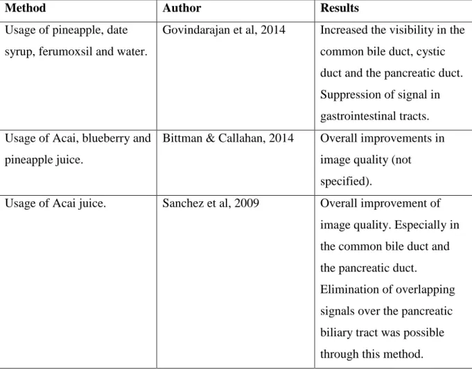
In table#2 the essential results are illustrated.
Table 2. Concluded results from the article.
Method Author Results
Usage of pineapple, date syrup, ferumoxsil and water.
Govindarajan et al, 2014 Increased the visibility in the
common bile duct, cystic duct and the pancreatic duct. Suppression of signal in gastrointestinal tracts. Usage of Acai, blueberry and
pineapple juice.
Bittman & Callahan, 2014 Overall improvements in
image quality (not specified).
Usage of Acai juice. Sanchez et al, 2009 Overall improvement of
image quality. Especially in the common bile duct and the pancreatic duct.
Elimination of overlapping signals over the pancreatic biliary tract was possible through this method.
16 Three different pineapple
juices.
Cooppens et al, 2005 Elimination of signal
intensity in the
gastroduodenal lumens. Increase in signal intensity in the pancreas biliary ducts.
Usage of black tea Ghanaati et al., 2011 Improved image quality in
the distal part of the common bile duct and the upper part of the pancreatic duct.
Usage of black tea Tang et al, 2013 Signal intensity improved in
wanted areas and loss of signal intensity in unwanted areas. Overall improvement on image quality.
Usage of Manganese chloride tetra hydrate juice
Watanabe et al, 2016 Improvement of image
quality at lower temperatures of MCT. Contrast relation between common bile duct and the adjacent background was optimal.
Usage of Manganese chloride tetra hydrate juice
Marugami et al, 2013 Increased MRI signals to the
common bile duct. Image quality improved
significantly.
Usage of Manganese chloride tetra hydrate juice
Tajima et al, 2013 Loss of signal in the adjacent
background. Improved the overall image quality.
17
DISCUSSION
Discussion of chosen method
A literature review has been created for the purpose of collecting information about specific methods that can improve image quality at MRCP. According to Evans (2002) this method is appropriate when the goal is to identify, evaluate and summarise the best existing research in a specific field. The demonstration of results in this literature review were established through articles with scientifically and academically character. The chosen method meant that a filtration of extracting high quality scientifically articles were possible and this enabled finding articles with evidence based knowledge. The possibility of implementation at the workplace is made possible through this literature review.
In this literature review, an integrated analysis has been made. This is shown by the author who related articles to one another and mentioned the resemblances as well as the differences. The essential parts of the discussion is the establishment of results that supports the purpose, and hence increasing the value of this literature review. In an integrated analysis this is also helpful for categorization of sub headings.
Certain aspects are mentioned in the background section to explain why it was of interest that a literature review was conducted. This literature review constitutes of several scientific articles, the analysation of these articles in the discussion aim to filtrate quality assured methods that can be implemented in a work place to improve image quality at MRCP.
The selection of all included scientific studies went through a working process for inclusion. Main requirements from each article were secured to insure the quality was enough for this literature review. Two essential requirements that were analysed in each article was the reliability and the validity of the article. All of the articles included on how to improve image quality at MRCP through a negative oral contrast, though different negative oral contrasts were used. They were black tea Acai, date syrup, juices and manganese chloride tetra hydrate. Nine articles in these areas were chosen to represent the results section and most thoroughly answered to the purpose of this literature review. There were more high quality articles that could be extracted from PubMed, though they did not strengthen the results of this literature
18 review, in a larger study though, it could be up for discussion to include more scientific articles in the results section. The author only had ambitions of choosing articles that were published in the last 10 years, however one included article did not fulfil this requirement (Cooppens et al, 2005), but was found to be interesting for the section of results and answered to the purpose of this current study. The search words were: MRI, MRCP, Image quality, Improve and Oral contrast. All of the search words were included in a systematically way of searching when conducting literature reviews. This means that at first hand a search was made, than either by adding or removing a word or words to narrow down the search to the expected required area so that extraction of articles could be simplified. The words on PubMed that were used to ensure that no articles of high quality were not missed was (MR, cholangiopancreatography (MRCP), Improvement, image, negative, oral contrast).
This literature review gives a comprehension in an area that seems to be rather unknown in MRI. The decision to make this literature review was with consideration of influencing concerned staff members that work with MRI to enhance a competence area that is not fully developed in MRI. To create a more specific way of working and implementing new methods to ensure the best image quality at MRCP.
To establish a precise way of interpreting the results a common approach is by narrative discussions. This means that a general description of the main findings is conducted. Moreover what is profitable with a narrative summary is that it provides an overview of the main findings, the major themes as well as issues of importance (Evans, D, 2002).
Discussion of the main findings of the results
In this literature review it was intended to investigate how the image quality could improve at MRCP. The results showed that it was possible to improve the image quality significantly. All of the articles (Govindarajan et al, 2014., Ghanaati et al., 2011., Tang et al, 2013., Bittman & Callahan, 2014., Sanchez et al, 2009., Cooppens et al, 2005., Watanabe et al, 2016.,
Marugami et al, 2013 & Tajima et al, 2013) showed an improvement in image quality at MRCP examinations. However a categorization of different negative oral contrast were used to visualise the achieved results. One of the articles (Govindarajan et al, 2014) showed it by using date syrup, this could be because date syrup contains a lot of iron which according to
19 the literature of Westbrook (2011) is a good method of usage when examining the liver at MRI. In four of the studies (Govindarajan et al, 2014., Bittman & Callahan, 2014., Sanchez et al, 2009 & Cooppens et al, 2005) the image quality improvement could be visualised by using different juices, this is also because of that these specific juices contain iron and is well emphasized in liver exams at MRI. In two of the studies (Ghanaati et al, 2011 & Tang et al, 2013) the improvement in image quality was established by using black tea as negative oral contrast, this might also because black tea contains a lot of iron. Furthermore, three studies (Watanabe et al, 2016., Marugami et al, 2013 & Tajima et al, 2013) evidently showed the improved effects when using manganese chloride tetra hydrate, this has been established in the literature of Westbrook (2011) as a valid method for exams in the liver, such as MRCP. Additionally there were three studies (Govindarajan et al, 2014., Sanchez et al, 2009 & Cooppens et al, 2005) that concluded the enhancement in image quality in T2 images with the combination of a negative oral contrast. This is indicated on the contrary to what is said in the literature of Westbrook (2011), there it seems that T1 images appear brighter as a
consequence of using manganese as a negative oral contrast and an enhancement for the radiologist to diagnose.
Of all mentioned negative oral contrast there was not a specific one that featured as more optimal than the other. None of the articles mentioned improvement in terms of quantity, which makes it more difficult to compare the different negative oral contrasts that were used. There was however another interesting aspect that was found. The feasibility of using one rather than the other oral contrast. It was indicated that it felt (by the patients) was somewhat more difficult to drink manganese chloride tetra hydrate of higher temperatures (Watanabe et al, 2016). This could be a factor when deciding which negative oral contrast to use in purpose of improving the image quality.
The decision to give patients negative oral contrast in purpose of improving image quality at MRCP is further proved by the study of Petersein et al (2000). It’s mentioned that negative oral contrast material can be used before an MRCP examinations. It provides higher quality images and visualisation of the bile and pancreatic ducts. There is also no prove of any
negative effects on the patient. In this literature study, in all of the studies (Govindarajan et al, 2014., Ghanaati et al., 2011., Tang et al, 2013., Bittman & Callahan, 2014., Sanchez et al, 2009., Cooppens et al, 2005., Watanabe et al, 2016., Marugami et al, 2013 & Tajima et al, 2013) the results have shown improvement in image quality on specific organs such as the
20 common bile or pancreatic duct, as well as suppressing MRI signals in the adjacent
background, such as in the gastrointestinal tract.
The reason why the enhancement in image quality is assured when using a negative oral contrast is a combination of two aspects. The negative oral contrast contain ingredients that might be emphasised with MRI when enhancing the MRI signal in the common bile duct or the pancreatic duct. The other aspect is that the negative oral contrast is able to suppress MRI signals from adjacent background which indirectly improves the image quality. In three articles (Govindarajan et al, 2014., Sanchez et al, 2009 & Cooppens et al, 2005) it was
established that T2 images in combination with the negative oral contrast was also regarded as an improvement. This is interesting because it is not mentioned in any articles why taking these specific sequences result in better image quality in combination with the negative oral contrast. An important aspect on this literature review is the possibility to increase higher quality at planning surgical procedures based on the improvements of image quality at MRCP.
Conclusions & Clinical implementations
This literature review has established a variety of methods that were applicable to improve the overall image quality at MRCP. Through this study, the results showed that the enhancement in image quality might simplify the planning of surgical procedures. Without using the negative oral contrast at MRCP it was indicated through the results that too many artefacts were caused from the adjacent background. What was written in the literature by Westbrook (2011) also confirmed the results from the scientific articles (Watanabe et al, 2016.,
Marugami et al, 2013 & Tajima et al, 2013), that manganese can be useful as a negative oral contrast when examining the liver, like MRCP. If the radiographers don’t apply one or several of the presented methods to improve the image quality at MRCP, the planning of a surgical procedure might be jeopardised. For the purpose of promoting higher levels of knowledge, one of the radiographer’s main assignments should be to always have research and education in mind. With frequent search for scientific knowledge and possible
implementations, the radiographer evolves within his/her profession. This is applied so that the radiographer is possible to influence or directly apply methods that can be benefited from
21 the patients and in the work place. The ultimate technique was not mentioned in any of the studied articles, though one of the mentioned methods were supported from the literature of Westbrook (2011). The method of using manganese as a negative oral contrast were
confirmed as a method that could improve the overall image quality, furthermore it was specified as a great method for usage in liver examinations in the literature of Westbrook (2011). The methods of using date syrup, different juices as well as black tea were also established as methods that improved the image quality. An important aspect to consider is the economical part. In all cases these negative contrast are not expensive and the possibility to use one over the other is also supported and a possibility for those who dislikes a certain juice or perhaps syrup.
22
REFERENCES
Bittman, ME & Callahan, MJ (2014). The effective use of acai juice, blueberry juice and pineapple juice as negative contrast agents for magnetic resonance cholangiopancreatography in children. Journal of pediatric radiology. Volume 44, Issue 7, pp 883-887
Coopens, E., Metens T., Winant, C & Matos, C (2005). Pineapple Juice labelled with gadolinium: a convenient oral contrast for magnetic resonance cholangiopancreatography.
Journal of European Radiology. Volume 15, Issue 10, pp 2122-2129
Evans, D (2002). Systematic reviews of interpretive research: Interpretive Data synthesis of processed data. 2002-2003 Volume 20 Number 2
Duarte, JA., Furtado, AP & Marroni, CA (2012). Use of pineapple juice with gadopentetate dimeglumine as a negative oral contrast for magnetic resonance cholangiopancreatography: a
multicentric study. Journal of abdominal imaging. Volume 37, Issue 3, pp 447-456
Friberg, F. (red.) (2012). Dags för uppsats: vägledning för litteraturbaserade
examensarbeten. (2., [rev.] uppl.) Lund: Studentlitteratur.
Ghanaati, H., Rokni-Yazdi, H., Jalali, AH., Abahashemi, F., Shakiba, M & Firouznia, K (2011). Improvement of MR cholangiopancreatography (MRCP) images after black tea consumptions. Journal of European Radiology. Volume 21, Issue 12, pp 2551-2557
Govindarajan, A., Lakshmanan, PM., Sarawagi, R & Prabhakaran, V (2014). Evaluation of date syrup as an oral negative contrast agent for MRCP. American Journal of roentgenology. Volume 203, issue 5.
Hjartarson, JH., Hannesson, P., Sverrisson, J., Bjöndal, S., Ivarsson, B & Björnsson, ES. (2016). The value of magnetic resonance cholangiopancreatography for the exclusion of choledocholithiasis. Scandinavian journal of gastroenterology. Volume 95, issue 8, pp 34-41.
Hunt, CH., Wood, CP., Lane, JI., Bolster, BD., Bernstein, MA & Witte, RJ (2011). Wide, short bore magnetic resonance at 1,5t: reducing the failure rate in claustrophobic patients.
Jorunal of clinical neuroradiology. Volume 27, issue 3, pp 97-112
Kristensson, J. (2014). Handbok i uppsatsskrivande och forskningsmetodik för studenter inom
hälso- och vårdvetenskap. (1. utg.) Stockholm: Natur & Kultur.
Marugami, N., Takewa, M., Iwaki, Y., Hazeyama, Y., Iwato, K., Takahama, J., Marugami, A., Okuaki, T & Kichikawa, K (2013). MR signal changes on hepatobiliary imaging after oral ingestion of manganese chloride tetrahydrate: preliminary examination. Japanese journal of
radiology. Volume 31, Issue 11, pp 713-723
McRobbie, D.W. (2007). MRI: from picture to proton. (2. Ed.) Cambridge: Cambridge University Press.
23 Peterstein, J., Reisinger, W., Mutze, S & Hamm, B. (2000). Value of negative oral contrast media in MR cholangiopancreatography (MRCP). Volume 172, issue 1, pp 55-60.
Sakamoto, K., Shinagawa, Y., Inoue, K., Morita, A., Urakwa, H., Fujimitsu, R., Takano, K & Yoshimitsu, K (2016). Obliteration of the Biliary system after administration of an oral contrast medium is probably due to regurgitation: A pitfall on MRCP. Journal of Magnetic
resonance in medical sciences. Volume 15, issue 1, p.137-143.
Sammet, S. (2016). Magnetic resonance safety. Journal of abdominal radiology. doi: 10.1007/s00261-016-0680-4. Volume 18, issue 9, p. 15-29
Sanchez, TA., Elias, J., Colnago, LA., De Almeida Troncon, LE., De Oliveira, RB., Baffa, O & De Araujo, DB (2009). Clinical feasibility of Acai pulp as an oral contrast agent for magnetic resonance cholangiopancreatography. Journal of computer assisted tomography. Volume 33, issue 5, pp 666-671
Tajima, N., Utano, K., Kijima, S., Kawai, A., Fujita, A., Sakuma, K., Sugimoto, H & Fujii, H (2013). Intraductal papillary mucinous neoplasm penetrating to the stomach, duodenum, and jejunum, demonstrated on MR cholangiopancreatography with an oral negative contrast agent. Journal of magnetic resonance imaging. Volume 38, issue 1, pp 206-215.
Tang, HH., Song, B., Huang, ZR & Yao, H (2013). (Application of black tea as a negative oral contrast in magnetic resonance cholangiopancreatography (MRCP)). Volume 44, issue 3, pp 476-482.
Watanabe, K., Ishimori, Y., Sakurai, H., Iwai, Y., Miida, K & Kurita, K. A low-temperature Manganese chloride Tetrahydrate improves the image quality of Magnetic resonance
Cholangiopancreatography. Volume 72, issue 4, pp 311-318.
Westbrook, c., Kaut-Roth, C & Talbot, J (2011). MRI in practice. (4. Ed.) Chichester, West Sussex: Wiley-Blackwell.
ATTACHMENT 1
Author Year
Title Aim Method Participants Results Quality
Govindarajan et al, 2014
Evaluation of date syrup as an oral negative contrast agent for MRCP. Examine the effects of date syrup, ferumoxsil and pineapple juice at MRCP The patient drank different negative oral contrast before an MRCP.
Patient study Increased the visibility in the common
bile duct, cystic duct and the pancreatic duct. Suppression of signal in
gastrointestinal tracts.
1
Bittman & Callahan, 2014
The effective use of acai juice, blueberry juice and pineapple juice as negative contrast agents for
magnetic resonance cholangiopancreatography in children. Examine different juices as negative oral contrast at MRCP. The patient drank different negative oral contrast before an MRCP.
Patient study Overall improvements in image quality
(not specified).
1
Sanchez et al, 2009
Clinical feasibility of Acai pulp as an oral contrast agent for magnetic resonance cholangiopancreatography To examine the effect of Acai as a negative oral contrast at MRCP. The patients was examined (MRCP) with and without Acai.
Patient study Overall improvement of image quality.
Especially in the common bile duct and the pancreatic duct. Elimination of overlapping signals over the pancreatic biliary tract was possible through this method.
Cooppens et al, 2005
Pineapple Juice labelled with gadolinium: a convenient oral contrast for magnetic resonance cholangiopancreatography. Examine different pineapple juices as negative oral contrast at MRCP. Three differed pinapple juices were tested.
Patient study Elimination of signal intensity in the
gastroduodenal lumens. Increase in signal intensity in the pancreas biliary ducts.
2 Ghanaati et al., 2011 Improvement of MR cholangiopancreatography (MRCP) images after black tea consumptions.
Examine with and without black tea as a negative oral contrast at MRCP. Patients went through MRCP with none contrast, than black tea 5 and 15 minutes after
consumption.
Patient study Improved image quality in the distal part
of the common bile duct and the upper part of the pancreatic duct.
1
Tang et al, 2013
Application of black tea as a negative oral contrast in magnetic resonance cholangiopancreatography
Examine with and without black tea as a negative oral contrast at MRCP. Patients went through MRCP with none contrast, than black tea 5, 10 and 15 minutes after consumption.
Patient study Signal intensity improved in wanted areas
and loss of signal intensity in unwanted areas. Overall improvement on image quality.
Watanabe et al, 2016
A low-temperature Manganese chloride Tetrahydrate improves the image quality of Magnetic resonance Cholangiopancreatography . To examine the effects of manganese chloride tetra hydrate as a negative oral contrast at MRCP. Different temperatures and concentrations of manganese were tested and the patients drank them.
Patient study Improvement of image quality at lower
temperatures of MCT. Contrast relation between common bile duct and the adjacent background was optimal.
1
Marugami et al, 2013
MR signal changes on hepatobiliary imaging after oral ingestion of manganese chloride tetrahydrate: preliminary examination. manganese chloride tetra hydrate as a negative oral contrast at MRCP Manganese was used as a negative oral contrast. The patients tested immediately, 30 min, 60, 120 and 180 minutes after consumption. Patient & Phantom study
Increased MRI signals to the common bile duct. Image quality improved significantly. 1 Tajima et al, 2013 Intraductal papillary mucinous neoplasm
penetrating to the stomach, duodenum, and jejunum, demonstrated on MR cholangiopancreatography with an oral negative contrast agent. manganese chloride tetra hydrate as a negative oral contrast at MRCP A 65 year old patient went through an MRCP and drunk MCT to see the effect on image quality at MRCP.
Patient study Loss of signal in the adjacent
background. Improved the overall image qulity.