I N V I T R O A N D I N V I V O S T U D I E S O F S A L I V A R Y F I L M S A T S O L I D / L I Q U I D I N T E R F A C E S
Malmö University
Health and Society Doctoral Dissertations 2009:2
© Ida E. Svendsen 2009 ISBN (Malmö) 978-91-7104-227-9 ISSN (Malmö) 1653-5383 Holmbergs, Malmö 2009
Malmö University, 2009
Faculty of Health and Society
IDA E. SVENDSEN
IN VITRO
AND
IN VIVO
STUDIES OF SALIVARY FILMS
AT SOLID/LIQUID INTERFACES
I am among those who think that science has great beauty. A scientist in his laboratory is not only a technician: he is also a child placed before
natural phenomena which impress him like a fairy tale. Marie Curie
CONTENTS
ABBREVIATIONS AND SYMBOLS ... 9
ABSTRACT ... 11
LIST OF PAPERS ... 13
INTRODUCTION ... 15
AIMS ... 17
BACKGROUND ... 18
Protein – surface interactions ...18
Natural and artificial surfaces in the oral cavity...20
The salivary film formation...22
The pellicle composition and function...23
Dynamics of salivary films ...24
Pellicle proteins of interest ...27
MATERIALS AND METHODS... 28
Surfaces ...28
Saliva and salivary proteins...30
General ...34
In situ null ellipsometry ...35
Atomic force microscopy (AFM) ...39
Total internal reflectance fluorescence (TIRF)...40
Experimental outline ...41
Pellicle protein composition ...42
Gel electrophoresis and staining ...43
Protein content analysis...44
RESULTS AND DISCUSSION... 46
Single component systems (papers I and II) ...47
Complex systems (papers V and VI) ...58
CONCLUSIONS AND FUTURE OUTLOOK ... 63
POPULÄRVETENSKAPLIG SAMMANFATTNING ... 65
ACKNOWLEDGEMENTS ... 67
9
ABBREVIATIONS AND SYMBOLS
αx Angle of incidence or angle of refraction
∆ Ellipsometric angle related to the relative phase shift of the light components
δ Thickness of unstirred layer closest to the solid/liquid interface Γ Adsorbed amount per unit area
κ-1 Debye screening length
λ Wavelength
ζ-potential Zeta potential
ν Partial specific volume of adsorbate
Ψ Ellipsometric angle related to the relative amplitude change of the light components
1-DE One dimensional gel electrophoresis 2-DE Two dimensional gel electrophoresis AFM Atomic force microscopy
aPRPs Acidic proline rich proteins BSM Bovine submaxillary mucin cac Critical association concentration cmc Critical micelle concentration CTAB Cetyl trimethylammonium bromide D Diffusion coefficient
dn/dc refractive index increment
dx Ellipsometric thickness, subscript indicates film thickness (df) or
silicone oxide thickness (d1)
EDTA Ethylenediaminetetraacetic acid FITC Fluorescein-5-isothiocyanate
G Gibbs free energy
10
HA Hydroxyapatite
HPS Human parotid saliva
HSMSLS Human submandibular and sublingual saliva HWS Human whole saliva
IC50 Concentration at which growth or activity is inhibited by 50 %
IEF Isoelectric focusing gel electrophoresis IPG Immobilized pH gradient
M/A Ratio of molar weight to molar refractivity of adsorbate MMA methyl methacrylate
MUC5B Human high molecular weight salivary glycoprotein
Mw Molecular weight
N Complex refractive index
nx Refractive index (real part), subscript indicates silicon oxide
(n1), film (nf) or bulk (nb)
PBS Phosphate Buffered Saline. In this work PBS= 10 mM phosphate buffer supplemented with 50 mM NaCl, pH 7.0 PEM Polyelectrolyte multilayer
pI Isoelectric point
PRP-1 Human acidic proline rich protein 1 PMMA Poly (methyl methacrylate)
pzc Point of zero charge Rg Radius of gyration
S Entropy
SDS Sodium dodecyl sulphate
SDS-PAGE Sodium dodecyl sulphate polyacrylamide gel electrophoresis
T Temperature
Ti Titanium
11
ABSTRACT
A conditioning film, the pellicle, of which many salivary proteins are important constituents, covers the surfaces present in the mouth. The pellicle forms in a selective adsorption process, and it has protective and lubricating functions as well as an influence on the adherence of oral microbes that ultimately leads to the development of dental plaque. Understanding the interactions responsible for the selective pellicle formation would make it possible to strive at creating a pellicle that serves its protective and lubricating functions and also promotes a healthy biofilm for the benefit of the individual. The aim of this research was to characterize the adsorption of salivary proteins to different types of substrates, to evaluate the influence of different protein-surface interactions on the adsorption process, and also to assess substrate dependent differences in film composition. Furthermore, as it is well known that complexes form between different salivary proteins and mucins (large glycoproteins) in bulk saliva, the aim was also to study interactions between mucins and other pellicle constituents at the solid/liquid interface. Additionally, the effects of a surfactant, sodium dodecyl sulphate (SDS), on the protein films were investigated, to evaluate the stability of the films and also the possibilities to completely remove the in vivo formed pellicle.
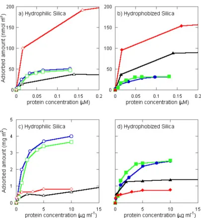
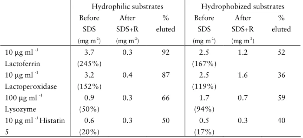
The adsorption experiments were performed in vitro using null ellipsometry, by which parameters such as adsorbed amount per unit area and average layer thickness can be obtained. Hydrophilic and hydrophobized silica were used as model substrates. The adsorption behaviour of the cationic, antimicrobial proteins lactoferrin, lactoperoxidase, lysozyme, and histatin 5 indicated that the adsorption on hydrophilic substrates was mainly driven by electrostatics, while on hydrophobized substrates hydrophobic interactions also influenced the adsorption process. Furthermore, it was shown that
12
sequential alternating adsorption of the anionic salivary mucin MUC5B and lactoperoxidase resulted in the build-up of multilayered structures on the surface. The rate of build-up was influenced by the surface characteristics. Sequential adsorption studies showed that neither MUC5B nor human whole saliva (HWS) was able to exchange substantial amounts of the pre-adsorbed anionic pellicle proteins acidic proline-rich protein 1 (PRP-1) or statherin, respectively. The resistance of the adsorbed mixed protein films to SDS elution depended on surface properties as well as on the number of layers adsorbed and adsorbed components. Pre-adsorbed PRP-1 was to some extent protected from SDS elution by the sequential adsorption of MUC5B to the PRP-1 film.
Pellicles formed on natural tooth enamel were collected in vivo and investigated using two-dimensional gel electrophoresis (2-DE). Mechanically-assisted SDS elution was used to collect the in vivo formed pellicle. The effectiveness of the collection procedure was validated in vitro by means of mechanical removal in combination with HCl treatment. The results indicated that rubbing the tooth surfaces with fibre pellets soaked in 0.5 % (w/v) SDS was sufficient to completely remove the pellicle from human enamel. In addition, 2-DE analysis of pellicles formed in vitro on human enamel and the dental materials titanium and poly (methyl methacrylate) (PMMA) showed differences in composition when compared to each other, revealing that the pellicle is influenced by the substrate properties.
13
LIST OF PAPERS
This thesis is based on the following papers, which will be referred to in the main text by their Roman numerals. The papers are appended at the end of the thesis.
I. Adsorption behaviour and surfactant elution of cationic salivary pro-teins at solid/liquid interfaces, studied by in situ ellipsometry.
Ida E. Svendsen Ida E. Svendsen Ida E. Svendsen
Ida E. Svendsen, Liselott Lindh and Thomas Arnebrant
Colloids and Surfaces B: Biointerfaces 2006, 53, 157-66.
II. Lactoperoxidase and Histatin 5 – their adsorption behaviour on silica and hydrophobized silica surfaces, and implications on their role in the initial salivary film formation.
Ida. E. Svendsen, Ida. E. Svendsen, Ida. E. Svendsen,
Ida. E. Svendsen, Liselott Lindh and Thomas Arnebrant
Zeitschrift für Physicalische Chemie 2007, 221, 65-73.
III. The salivary mucin MUC5B and lactoperoxidase can be used for layer-by-layer film formation.
Liselott Lindh, Ida E. SvendsenIda E. SvendsenIda E. SvendsenIda E. Svendsen, Olof Svensson, Marité Cárdenas and Thomas Arnebrant
Journal of Colloid and Interface Science 2007, 310, 74-82.
IV. Studies on the exchange of early pellicle proteins by mucin and whole saliva.
Ida E. Svendsen Ida E. Svendsen Ida E. Svendsen
Ida E. Svendsen, Liselott Lindh, Ulla Elofsson and Thomas Arnebrant
14
V. Validation of mechanically-assisted sodium dodecyl-sulphate elution as a technique to remove pellicle protein components from human enamel.
Ida E. Svendsen Ida E. Svendsen Ida E. Svendsen
Ida E. Svendsen, Thomas Arnebrant and Liselott Lindh
Biofouling 2008, 24, 227-33.
VI. The composition of enamel salivary films is different from the ones formed on dental materials.
Ida E. Svendsen Ida E. Svendsen Ida E. Svendsen
Ida E. Svendsen and Liselott Lindh
Biofouling 2009, 25, 255-61.
Reprint permissions have been granted by Elsevier Inc. (papers I, III and IV), Oldenbourg Wissenschaftsvelag (paper II) and Taylor and Francis Ltd. (papers V and VI).
Contributions by the respondent
In papers I, II, V and VI, I performed most of the planning and all the experimental work, including analysing the data. I was also the main contributor to writing the manuscripts. In paper III, I did most of the experimental work, and did also participate in analysing data and in discussions concerning the manuscript and writing minor parts of the paper. In paper IV, I did most of the experimental work and was also the main contributor to writing the manuscript.
Other papers not included in the thesis:
Human palatal saliva: adsorption behaviour and the role of low-molecular weight proteins.
Ida E. Svendsen Ida E. Svendsen Ida E. Svendsen
Ida E. Svendsen, Thomas Arnebrant and Liselott Lindh
15
INTRODUCTION
Human whole saliva (HWS) is a mixture of secretions from both major and minor salivary glands, and does also contain crevicular exudate and bacterial and cellular components1
. The salivary secretion consists to 99 % of water, and contains approximately 2 g of proteins/L fluid1
. Many important every-day functions occurring in the mouth, that we take for granted, can be attributed to saliva, such as lubrication to facilitate mastication, swallowing, deglutination and speaking. Furthermore, saliva maintains a steady state between tooth demineralisation and remineralisation, thereby preserving the integrity of the teeth. In addition, saliva also has an influence on the microbial growth in the mouth. All of these functions can to a large extent be assigned to the proteins of this watery secretion1
.
Due to the surface activity that is a general feature of proteins, salivary proteins adsorb to the different types of surfaces present intra-orally and form a conditioning film, often referred to as the pellicle2. This film is believed to form by selective protein adsorption3
and studies have confirmed this hypothesis by the identification of particular proteins in the pellicle, whereas others were not found3-5. The composition of the pellicle is known to influence the subsequent attachment of oral microbes, of which saliva contains approximately 109
/ml fluid1
. The microbial biofilm, frequently referred to as plaque, may in later stages progress into plaque related diseases such as caries and periodontitis. Understanding which components that are key participants in the initially formed pellicle, and also how the adsorbed components interact with each other, would make it possible to aim at enhancing the protective functions of the salivary film so that the microbial biofilm that develops would be of a healthy, protective nature only.
Studies focused on identifying pellicle constituents have reported many different types of proteins, of both anionic and cationic character4-6
16
proteolytically derived proteinaceous fragments7, 8
. It can be hypothesized that cationic salivary proteins may interact with anionic pellicle components and thereby increase the thickness and also the cohesiveness of the film. As numerous studies have shown that functions, such as antimicrobial activities, of several cationic salivary proteins can be retained after adsorption9-14
, the incorporation of these proteins may contribute to the defence function of the pellicle. Several studies have identified protein complex formation in bulk saliva, consisting of cationic proteins such as lactoferrin and lysozyme, combined with the large, anionic glycoproteins referred to as mucins15-17
. Also complexes between anionic salivary constituents have been identified in bulk saliva18-20. As many of these complexes consist of pellicle proteins, such complexes may be expected to adsorb onto, and/or form on oral surfaces.
17
AIMS
The specific objectives of this thesis have been:
To characterize the adsorption behaviour of single protein systems, specifically the cationic salivary proteins lactoferrin, lactoperoxidase, lysozyme and histatin 5, with respect to the influence of the underlying substrate characteristics and film susceptibility to SDS elution.
To investigate systems containing several components regarding the dynamics of the adsorption process. More specifically, sequential adsorption of relevant cationic and anionic salivary proteins and possibilities for multilayer construction should be investigated. Influence of substrate characteristics as well as the film stability with respect to SDS elution should also be addressed.
To devise a method for complete removal of in vivo and in vitro formed pellicles on human enamel surfaces by mechanically assisted SDS elution. A further aim was to investigate compositional differences between in vitro
pellicles formed on different dental materials as well as on human enamel under otherwise identical conditions.
18
BACKGROUND
Protein – surface interactions
The polypeptide chain of proteins is composed by, in general, 20 different amino acids linked together by peptide bonds. Depending on the side chain properties of the amino acids, they are considered as hydrophobic, polar and/or charged. The wide variety of combinations of these amino acids as well as the varying length of the peptide backbones result in very different compositions and folding of proteins, and hence properties. Proteins usually contain hydrophobic amino acids as well as polar and charged ones. This results in both an amphiphilic and an ampholytic character that will be of decisive importance when proteins in solution are adsorbed at an interface.
Protein adsorption only takes place when the Gibbs free energy (G) of the complete system decreases:
∆adsG = ∆adsH-T∆adsS <0
Generally, the characteristics of the protein, the surface and the surroundings will determine the sign and magnitude of ∆adsG. The interplay
between protein, surface and solution depends on several different interactions that are briefly summarized below. See Norde21, 22
for more detailed descriptions.
Hydrophobic interactions
Hydrophobic interactions are considered to be one of the main driving forces for protein adsorption. The basis of this interaction is that water molecules strive to avoid non-polar components present in their vicinity. Water molecules close to a hydrophobic surface are more ordered than those
19 in solution and thus have lower entropy. Upon adsorption of e.g. a protein from solution, ordered water will be released from the hydrophobic substrate, as well as from hydrophobic groups of the protein, which will result in increased entropy, and hence a driving force for adsorption.
Electrostatic interactions
Electrostatic interactions will depend on the charges of the protein and the substrate, i.e. repulsion between similar charges and attraction between opposite charges. The structural rigidity of the proteins is also of relevance. Flexible (“soft”) proteins are generally able to structurally adapt to minimize charge repulsion with the surface, and adsorb with those amino acids that are electrostatically attracted to the surface. Hence a net negatively charged flexible protein may adsorb to a negatively charged substrate (see also effects of conformational entropy below).
Conformational entropy
Conformational entropy is associated with the three-dimensional structure of proteins, and is a factor that counteracts the folding of the polypeptide chain. Studies have indicated structural rearrangements in proteins upon adsorption, which both may increase (i.e. more unordered protein structure) or decrease (e.g. increased secondary structure at apolar surfaces) the conformational entropy. Generally, changes in conformational entropy may be important for adsorption of larger, structurally less stable proteins.
van der Waals interactions
van der Waals interactions are based on interactions between permanent and/or induced dipoles. These types of interactions usually favour protein adsorption, however, they are short ranged and rather weak when compared with hydrophobic and electrostatic interactions.
Hydrogen bonds
Hydrogen bonds occur between an electronegative atom and hydrogen atoms usually bound to oxygen or nitrogen. The strength of these interactions with respect to protein adsorption is usually regarded to be in the same range as van der Waals interactions.
Also the composition of the ambient solution is of importance for protein adsorption. The concentration of ions as well as their valency, which both
20
affect the Debye screening length (κ-1
), may to a large extent determine the adsorption process, as it will affect the range of the electrostatic interactions between the protein and the surface, as well as other intra- and inter-molecular interactions. The solution pH is also of relevance as it controls the ionisation of many amino acid residues.
Natural and artificial surfaces in the oral cavity
Different types of surfaces of both hard and soft character, and composed of both natural and artificial materials, may be present in the oral cavity. Large differences in chemical composition, charge, microstructure and surface free energy are observed for these surfaces. The following section focuses on human enamel, as well as on the dental materials titanium and poly (methyl methacrylate) [PMMA] that were used in studies included in this thesis.
Human enamel
Dental enamel is the outer layer of the tooth crown, constituting the hardest tissue present in the human body. It is highly mineralised, and the main mineral component of enamel (approximately 95 %) is hydroxyapatite (HA), Ca10(PO4)6(OH)2
23
. The calcium ions have a stronger tendency to dissolve in aqueous solutions than the phosphate ions, which results in excess of the phosphate ions (HPO4
and H2PO4
-) at the interface, giving the enamel surface a net negative charge. Counter-ions will then “coat” this surface, which results in an electric double layer (Figure 1).
An organic matrix is present between the HA prisms of the enamel. This matrix (constituting ∼1 % of the enamel), consists of specific proteins, mostly amelogenins, but also e.g. ameloblastin, enamelin and enamel proteases24. In addition, the dental enamel also carries small amounts of water (∼4 %).
Reported values of surface free energy of pure enamel are 77 ±10 to 87 ±6 mJ×m-2 25, 26
, indicating complete water wetting, which also has been reported for HA27. The point of zero charge (pzc) of enamel has been reported to be 4.4-5.023
. HA particles have been reported to have pzc values between 6.5-7.328
21
Figure 1. Schematic drawing of the enamel surface in aqueous solution at
approximately pH 7. Adapted from23. Not drawn according to scale.
Titanium
Titanium (Ti) is a frequently used biomaterial, not only for dental reconstructions and implants but also as implants in orthopaedic applications, such as hip joint replacements29. When titanium is exposed to air or aqueous solutions, an oxide layer is spontaneously formed on the surface, and TiO2 is
the most common oxide of titanium. A typical Ti dental implant is formed to resemble a single tooth root and has a roughened or smooth surface. Modification of the surface properties of titanium, such as surface roughness and surface free energy, is known to influence osseointegration30-32
. Titanium has many properties that make it an excellent biomaterial; it is e.g. nontoxic and mechanically resistant. It has a pI at approximately 6 and a high surface free energy (water contact angle <8°)29. Titanium covered with TiO2 has been
investigated in detail regarding biocompatibility with respect to interactions with blood33-35
, but few studies have been carried out on saliva interactions.
PMMA
Poly(methyl methacrylate) (PMMA; (
C
5O
2H
8)n, Figure 2) is a polymer usedin e.g. dentures and bone cements36. It is highly biocompatible and low-priced36. PMMA has a low surface free energy (water contact angle: 75°)37
Ca2+ Ca2+ Ca2+ Ca2+ Ca2+ HPO42- HPO42- HPO42- H2PO4- H2PO4-
22
and, considering its molecular structure, it is uncharged provided that the ester functionalities are intact.
Although PMMA is biocompatible, the toxic monomer of PMMA, methyl methacrylate (MMA), may remain after the initial polymerization. However, after completion of the polymerization process, only small amounts of monomer are released during the lifetime of the prosthesis, and the toxic effects are therefore almost negligible36.
Figure 2. Drawing of the molecular structure of a PMMA subunit. n=number of subunits.
The salivary film formation
Numerous in vitro studies have been performed to elucidate the physico-chemical phenomena in the pellicle formation (see e.g.38-41
); e.g. protein adsorption from HWS and salivary fractions, as well as studies of adsorption of single salivary proteins to model surfaces,. It is generally accepted that after approximately 1-2 hours of film formation, plateau values regarding adsorbed amount and thickness of the salivary film has been reached, depending somewhat on experimental conditions4, 38
. A relatively wide variation in adsorption behaviour exists among different salivary fractions and purified salivary proteins38
. However, a general feature is that larger amounts are usually adsorbed on hydrophobized surfaces compared to hydrophilic surfaces38, 40-42. AFM studies of adsorbed films from HWS have shown that both hydrophobized and hydrophilic silica are completely covered by a salivary film, however, the size and shape of adsorbed components, as well as the density of the film, are substrate dependent40
.
From investigations on the adsorption of individual salivary protein, fundamental data concerning driving forces for adsorption as well as substrate affinity and specific protein-substrate interactions may be elucidated. Such information is essential for the analysis of data obtained from adsorption
23 studies of solutions containing two or more components. Since statherin and acidic proline-rich proteins (aPRPs) are known components of the initial pellicle, the adsorption behaviour of statherin, aPRPs, and also salivary mucins have attracted quite a bit of attention (see e.g.40, 41, 43-48). The salivary mucin MG1, which consists primarily of MUC5B49
, is also known as a pellicle constituent, and was recently shown to have potential as a biomaterial coating, suppressing neutrophil adhesion and activation50. Furthermore, by means of adsorption experiments and streaming potential studies, it has been shown that the in vitro formed pellicle contains a significant fraction of positively charged proteins51, 52, which merits the investigation of the adsorption of individual cationic proteins present in saliva. The cationic proteins lysozyme and lactoferrin have been studied extensively as model proteins for adsorption on different types of surfaces53-57
, and adsorption studies of lactoperoxidase and histatin 5 have also been performed9, 14, 58-60
. Most studies have concluded that the pellicle to a large extent is irreversibly adsorbed. Adsorption experiments performed in vitro often include a final step of buffer rinsing or surfactant addition, to get an estimate of the binding properties of adsorbed components. In the case of adsorption from complex solutions like saliva, the differences in elutable fractions on different surfaces could indicate compositional differences or multiple states of binding.
The pellicle composition and function
Collection of the pellicle has been the objective in numerous studies6, 61-65, and complete pellicle elution has been shown to be a difficult task. However, many different types of proteins, as well as carbohydrates and lipids, have been identified as pellicle constituents4, 5
. Proteins such as lysozyme, aPRPs, amylase, albumin, histatins, statherin, lactoferrin, mucins, cystatins and immunoglobulins are among the most frequently identified proteins in the pellicle on enamel4, 6
, and some of these have also been found in pellicles on different dental materials66-70. The physiological roles of these proteins include maintaining calcium homeostasis, antimicrobial activity and lubrication. In addition, the pellicle has been shown to act as a diffusion barrier to acids71
, a function not attributed to specific components but rather to the whole film.
Microbial adherence to oral surfaces, leading to the formation of plaque or a microbial biofilm, is in its initial stage influenced by the pellicle composition, due to specific (receptor-donor) and non-specific interactions72-74
. Factors such as dental hygiene and frequency and type of food intake will influence the
24
pellicle composition, as well as the survival and proliferation of the biofilm community.
Dynamics of salivary films
Competitive adsorption and sequential exchange
The pellicle formation is initiated by the adsorption of specific small proteins, known as pellicle precursors, e.g. statherin, histatins and aPRPs4. A continuous adsorption then takes place, which presumably involves interactions between components in saliva and the precursor proteins, and to patches on the surface that are still uncovered.
Studies on sequential salivary adsorption processes are rather scarce. However, as both saliva and blood are examples of complex fluids containing a wide range of e.g. proteins and ions, it may be anticipated that a process analogous to “the Vroman effect” (adsorption and exchange of components in blood dependent on e.g. concentration and molecular weight75) takes place on surfaces in the oral cavity. It has been shown that the high molecular weight salivary mucin MUC5B adsorbs rather slowly43
, indicating that it does not play an important role during the initial pellicle formation, but it nevertheless appears in the film at later stages76. MUC5B has a higher adsorption affinity for HA compared to smaller pellicle precursor proteins such as aPRPs and statherin77
, indicating its potential to exchange these components at the interface. Furthermore, a recent study indicated that α-amylase has the potential to replace lysozyme on fluoroapatite particles, and albumin on silica surfaces78
.
Multilayer formation
Outside the oral field, the interest in polyelectrolyte multilayer (PEM) films is large, as such structures have the potential to be used as e.g. biomaterial coatings, thus creating more biocompatible surfaces79
. In general, oppositely charged polyelectrolytes are used for the layer-by-layer deposition process originally developed by Decher et al.80
. The driving force for the build-up of these films is the charge reversal that occurs after each adsorption step (alternating between a positive and a negative charge) resulting in an electrostatic attraction between each layer80
. A film with arbitrary thickness can be constructed simply by varying the number of adsorption cycles80
25 Factors such as pH and ionic strength will also influence the build-up behaviour79
.
Several publications using proteins and polypeptides in PEMs have been reported, and with respect to oral significance, lysozyme and mucins, respectively, have been used to build multilayered films successfully with oppositely charged polyeletrolytes81-83, and enzymatically active lactoperoxidase has been incorporated into PEM films in so-called enzyme nanoreactors84. Also, antimicrobial peptides have been inserted in PEM films to protect surfaces from bacterial colonization85
, a function relevant for the oral cavity. Embedding components with specific functions in multilayered films could increase the depot of these active constituents and hence promote specific functions of the film.
Action of surfactants
The interactions between an adsorbed protein film and a surfactant can be used as a measure of the strength of the protein–surface interaction86
. The degree of removal depends on the surfactant type as well as on its concentration. Also, the protein and substrate properties are of importance, as is the adsorption time87
. Generally, sodium dodecyl sulphate (SDS), which is a negatively charged surfactant, is more effective in the removal of protein and salivary films, compared to cationic surfactants (e.g. CTAB) and other elution agents (e.g. EDTA)61-63, 86, 88.
Surfactants are introduced e.g. in toothpastes, thereby combining the cleaning effect of the surfactant with the mechanical cleansing action of the toothbrush. Although the mechanical action has been indicated to be of main importance for salivary film removal62, the type of surfactant employed is also of significance61-63
. In general, at concentrations close to the critical micelle concentration, cmc, of the surfactant (in reality the critical association concentration, cac, of the surfactant-protein system), the elutability reaches its maximum. An example is SDS elution of HWS films on HA in vitro, where a SDS concentration above cmc had no further effect on the desorption89
. It is often observed that the surfactant removes parts of the adsorbed protein films, indicating a complex film composition and/or multiple states of binding90. Variations in the desorbable fraction of proteins and saliva have been observed on hydrophilic and hydrophobized substrates40, 42, 86, 88
. For saliva, binding to hydrophobic surfaces typically is stronger and may involve exposure of hydrophobic domains of the protein to the surface. Such
26
conformational changes may result in a decreased removable fraction22
, however, also the film composition is of relevance91
.
SDS has been used extensively as a component in e.g. toothpastes, usually in concentrations up to 2%92. Studies have shown that SDS may be harmful to mucosal surfaces92, 93
, and that SDS treatment reduces the lubrication properties of salivary films94, 95. On today’s market, many different types of surfactants are used in oral care products, such as sodium lauryl sarcosinate and polyehtylene glycol esters that generally are less irritating compared to SDS. In the studies presented here, SDS was used as a model surfactant to investigate the stability of the adsorbed films.
Two models have been suggested to be the main modes of protein elution by surfactants96
:
Figure 3. Schematic illustration of the formation of soluble complexes (a) and
the replacement mechanism (b). Adapted from96
. Not drawn according to scale. =protein, = surfactant micelle, = surfactant monomer.
Formation of soluble complexes (Figure 3a)
The surfactant interacts with the adsorbed proteins, generating complexes that may be electrostatically repelled from the surface (in case of similarly charged surfactant and substrate). This is typical for SDS, which is known to interact strongly with proteins86. Conformational changes and/or higher solubility of the formed complexes may also explain the removal. The surfactant itself does not adsorb to the surface, cf. SDS on a negatively charged surface.
surfactant
rinse
a
rinse
surfactant
b
27
Protein replacement (Figure 3b)
The surfactant has a higher affinity for the surface compared to the adsorbed proteins, which results in an exchange of components on the surface. As water soluble surfactants, such as SDS, are expected to be reversibly adsorbed on e.g. hydrophobic surfaces, a subsequent rinse removes the surfactant from the surface. The surfactant does not interact with the adsorbed proteins.
In many systems, a combination of the two models described above may be operational, e.g. when the surfactant interacts with both the substrate and the adsorbed proteins.
Pellicle proteins of interest
Some specific salivary proteins have been selected for adsorption studies in the present thesis. The selection was based primarily on the following criteria: (i) their presence in the pellicle (ii) important functions of the proteins (iii) suitability as model proteins in adsorption studies. Short descriptions of the proteins chosen are given in the materials and methods section below.
28
MATERIALS AND METHODS
Surfaces
Enamel
Human teeth, extracted due to orthodontic treatment, were used as enamel samples. Previous to use, the teeth were pumiced using a rubber cup and fine grade pumice, followed by extensive rinsing in water and drying in a stream of filtered air. The surfaces were used directly after cleaning.
Titanium
Titanium surfaces were obtained from Nobel Biocare AB (Göteborg, Sweden). The surfaces were cleaned by extensive rinsing in water, ethanol and water, and dried with filtered air, after which the surfaces were plasma cleaned in low pressure air for 5 minutes in a glow-discharge Harrick Plasma cleaner (Model PDC-32G, Harrick Scientific Corporation, Ossining, NY, USA). The surfaces were used immediately after plasma cleaning.
PMMA
PMMA (Microdent, Type 1/class 1 according to ISO 1567:1988 (E)) was obtained from Forshaga Dental depå AB (Forshaga, Sweden). The surfaces were cast and treated according to the instructions from the manufacturer. Before use, the surfaces were cleaned by rinsing in water, followed by rinsing in ethanol and finally water, after which they were dried with filtered air.
29
Silica surfaces
Silicon slides, used for the ellipsometry and AFM experiments, were obtained from Okmetic OY (Espoo, Finland). The surfaces, which had an oxide thickness of approximately 300 Å97
, were plasma cleaned for 5 min as described for the titanium surfaces. Subsequently, the surfaces were made hydrophilic by 5 minutes of gentle boiling in a solution of NH4OH
(25%):H2O2 (30%):H2O (1:1:5 v/v), followed by water rinsing and another 5
minutes of gentle boiling in a solution of HCl (37%):H2O2 (30%):H2O (1:1:5
v/v). Finally the surfaces were rinsed in water and ethanol and stored in ethanol. Just prior to use, the surfaces were rinsed in water, ethanol and water, dried in a stream of nitrogen gas followed by 5 minutes of plasma cleaning. Hydrophobized silica surfaces were prepared from the hydrophilic substrates by means of liquid phase silanization. The surfaces were rinsed in trichloroethylene before immersion in a 0.05% (v/v) dichlorodimethylsilane solution in trichloroethylene for 1 h. Subsequently the surfaces were rinsed in trichloroethylene followed by ethanol, and stored in ethanol. Before experiments, the hydrophobized surfaces were rinsed in water, ethanol and water and then dried with nitrogen gas. Both types of surfaces have a reported ζ potential of -45 ±5 mV in 1 mM NaCl pH 7.098
and water contact angles of <10° for hydrophilic silica and 103° for hydrophobized substrates (advancing contact angles)99.
Techniques aimed at investigating interactions taking place at solid/liquid interfaces usually require surfaces that have well-defined features, such as low surface roughness and specific optical or mechanical properties, to be able to record e.g. protein adsorption and thus accurately interpret adsorption mechanisms (see e.g.100). Dental enamel is highly heterogeneous in terms of surface composition and surface structure. These features make enamel difficult to apply to the techniques used here (ellipsometry, AFM and TIRF, see below) without treatments that might change the surface characteristics. The wide range in wettability spanned by the model silica surfaces include surface wettabilities of many types of surfaces that may be found in the oral cavity, which justifies the choice of model substrates.
Glass surfaces
Glass slides (Gold Seal Rite-on, Microslides, Clay Adams, NY, USA) were used for the TIRF experiments (see below). They were cleaned and modified as described for the silica surfaces to obtain hydrophilic and hydrophobic
30
characteristics, respectively. Reported water contact angles are <10° for hydrophilic glass and 103° for hydrophobized glass (advancing contact angles)99.
Saliva and salivary proteins
HWS
Unstimulated HWS was collected by drooling into ice-chilled tubes for approximately 10 min, as described by Dawes101
. The donors were considered to be in good general and oral health. The collection was performed in the morning, at least 2 hours after food intake and tooth brushing. The collection was a standardized procedure and has been shown to yield reproducible results40, 91, 102
. All collections of HWS and pellicle (see below) were approved by the Committee for research ethics at Lund University (approval No LU518-02).
For the adsorption experiments (paper IV), saliva was diluted to 10%. It has previously been shown that the major part of the adsorbed film had been formed at this concentration103
, and experiments performed at this concentration also avoided problems due to light scattering. No significant differences in total adsorbed amounts and film thicknesses were observed between different donors at identical saliva concentrations102, 104
, however, qualitative differences have been indicated102
.
Proteins
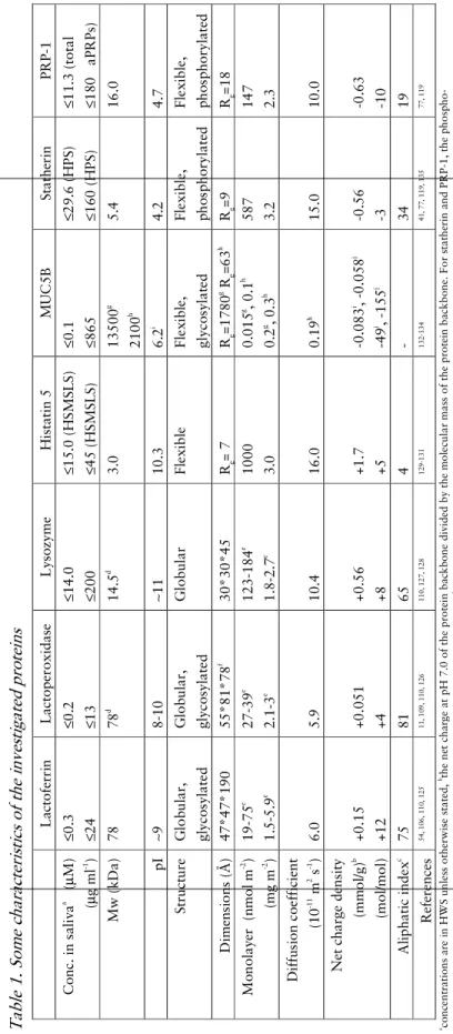
Background information of the investigated proteins is given below, with some characteristics summarized in Table 1. The commercially available proteins were used as received.
The adsorption behaviour of the cationic proteins lactoferrin, lactoperoxidase, lysozyme and histatin 5 was investigated using concentration limits reported for saliva (Table 1). Although not all of the proteins used were of human origin, their characteristics were similar11, 105, and therefore their adsorption behaviour were assumed to resemble that of the respective components in saliva.
Lactoferrin
Lactoferrin is a non-enzymatic glycoprotein, with iron-chelating properties, thereby depriving microbes of this essential nutrient10
31 anti-inflammatory, anti-cancer and antiviral features106
, as well as being present in the pellicle107 108
. Human milk lactoferrin (L0520) was obtained from Sigma-Aldrich Sweden AB (Stockholm Sweden).
Peroxidase
Salivary peroxidase is a glycoprotein which catalyses the oxidation of specific molecules (generally thiocyanate ions), resulting in the formation of highly reactive derivatives, such as hypothiocyanite/hypothiocyanous acid, which are toxic to several types of microorganisms11, 109-112
. Peroxidase has been identified in the pellicle12, 63, and retains its activity after adsorption12. Bovine milk lactoperoxidase (L8257) was obtained from Sigma-Aldrich Sweden AB.
Lysozyme
Lysozyme exerts its antimicrobial functions by e.g. inducing bacterial lysis leading to cell death110, 111. Lysozyme has been identified in pellicles formed on both natural and artificial dental surfaces13, 63, 66, 108, 113
, and studies have further shown that lysozyme remains active when incorporated in the pellicle13
. Lysozyme from chicken egg white (Sigma-Aldrich Sweden AB (L-6876)) was used in the adsorption experiments.
Histatin 5
Histatins are a family of histidine-rich proteins that are known to have antifungal activities114. Histatin 5 is mostly known to inhibit the growth of
Candida albicans, the main fungi responsible for candidiasis114
. Histatin 5 has shown to adsorb to different materials9, 58, 60
. Synthetic histatin 5 was purchased from American Peptide Company Inc. (Sunnyvale, CA, USA).
MUC5B
MUC5B is one of the major mucins present in saliva, and known primarily for its lubricating functions115. It has been identified in the 2 h old pellicle6, 116. It is a large oligomeric molecule, where the monomers are joined by disulphide bridges. MUC5B has been reported to be composed of approximately 80% carbohydrates, of which approximately 55% are neutral and 45% are acidic (sulphated or sialylated)117 resulting in an expected low pI of MUC5B.
MUC5B was purified from HWS, as previously described43, 118. Studies on the initial adsorption rate indicated that the MUC5B preparation consisted of both intact mucin macromolecules as well as subunits and T-domains43.
32
Statherin
Statherin is a small protein, known mainly for its involvement in maintaining calcium homeostasis119. In addition, statherin has been shown to have lubricating abilities45, 120 and is also recognized to mediate microbial colonization on HA surfaces121
. It has been reported in several studies to be present in the pellicle63, 108, 122.
Proline-rich protein 1 (PRP-1)
PRP-1 belongs to the family of acidic proline-rich proteins and is known to be involved in maintaining the calcium homeostasis of saliva, e.g. inhibiting crystal growth119, and also in lubrication45. PRP-1 is known to adsorb in the initial phase of pellicle formation4
and has been identified in the pellicle61
. Statherin and PRP-1 was purified from human parotid saliva (HPS) as described in Li et al.123 and Lindh et al.44. The identities and purities of both proteins were confirmed by gel electrophoresis, NH2-terminal amino acid
sequencing and bacterial binding properties123
.
Estimations of diffusion coefficients
As no diffusion coefficients of histatin 5, statherin and PRP-1 could be obtained in the literature they were estimated from a graph of known diffusion coefficients vs. molecular weights of a series of well defined proteins, as previously described44, 124. As the proteins comprised in this graph are of globular structure, the diffusion coefficient of histatin 5, statherin and PRP-1 may be overestimated.
3 3 T ab le 1 . So m e ch ar ac te ri st ic s o f th e in ve st ig at ed p ro te in s aco n ce n tr at io n s ar e in H W S u n le ss o th er w is e st a te d , bth e n et c h ar g e at p H 7 .0 o f th e p ro te in b ac k b o n e d iv id ed b y th e m o le cu la r m a ss o f th e p ro te in b ac k b o n e. F o r st a th er in a n d P R P -1 , th e p h o sp h o -se ri n es w er e al so t ak en i n to a cc o u n t, cth e re la ti ve v o lu m e o cc u p ie d b y al ip h at ic s id e ch ai n s, dth e m o le cu la r w ei gh t o f th e co m p o n en t u se d i n t h e p re se n t st u d ie s (o b ta in ed f ro m l it er at u re o r Sw is s-P ro t d at ab as e p ro vi d ed b y T h e S w is s In st it u te o f B io in fo rm a ti cs ), a lt h o u gh c lo se ly r es em b li n g th e tr u e m o le cu la r w ei gh t o f th e sa li va ry c o u n te rp ar ts , esi d e o n – e n d o n , fR C SB P ro te in D at a B an k 2 G J1 gw h o le m u ci n 1 3 4 a n d hsu b u n it 1 3 4, ith eo re ti ca l v al u e o f th e am in o a ci d s eq u en ce o n ly , jg ly co sy la te d d o m ai n L ac to fe rr in L a ct o p er o x id a se L ys o zy m e H is ta ti n 5 M UC 5 B S ta th er in P R P -1 Co n c. i n s a li va a ( µ M ) (µ g m l -1 ) ≤ 0 .3 ≤ 2 4 ≤ 0 .2 ≤ 1 3 ≤ 1 4 .0 ≤ 2 0 0 ≤ 1 5 .0 ( H S M S L S ) ≤ 4 5 ( H S M S L S ) ≤ 0 .1 ≤ 8 6 5 ≤ 2 9 .6 ( H P S ) ≤ 1 6 0 ( H P S ) ≤ 1 1 .3 ( to ta l ≤ 1 8 0 a P R P s) M w ( k D a ) 7 8 7 8 d 1 4 .5 d 3 .0 1 3 5 0 0 g 2 1 0 0 h 5 .4 1 6 .0 p I ∼ 9 8 -1 0 ∼ 1 1 1 0 .3 6 .2 i 4 .2 4 .7 S tr u ct u re G lo b u la r, g ly co sy la te d G lo b u la r, g ly co sy la te d G lo b u la r F le x ib le F le x ib le , g ly co sy la te d F le x ib le , p h o sp h o ry la te d F le x ib le , p h o sp h o ry la te d D im en si o n s (Å ) 4 7 * 4 7 * 1 9 0 5 5 * 8 1 * 7 8 f 3 0 * 3 0 * 4 5 Rg = 7 Rg = 1 7 8 0 g R g = 6 3 h Rg = 9 Rg = 1 8 M o n o la ye r ( n m o l m -2 ) (m g m -2 ) 1 9 -7 5 e 1 .5 -5 .9 e 2 7 -3 9 e 2 .1 -3 e 1 2 3 -1 8 4 e 1 .8 -2 .7 e 1 0 0 0 3 .0 0 .0 1 5 g , 0 .1 h 0 .2 g , 0 .3 h 5 8 7 3 .2 1 4 7 2 .3 D if fu si o n c o ef fi ci en t (1 0 -1 1 m 2 s -1 ) 6 .0 5 .9 1 0 .4 1 6 .0 0 .1 9 h 1 5 .0 1 0 .0 N et c h a rg e d en si ty (m m o l/ g ) b (m o l/ m o l) + 0 .1 5 + 1 2 + 0 .0 5 1 + 4 + 0 .5 6 + 8 + 1 .7 + 5 -0 .0 8 3 i , -0 .0 5 8 j -4 9 i , -1 5 5 j -0 .5 6 -3 -0 .6 3 -1 0 A li p h a ti c in d ex c 7 5 8 1 6 5 4 - 3 4 1 9 Re fe re n ce s 5 4 , 1 0 6 , 1 1 0 , 1 2 5 1 1 , 1 0 9 , 1 1 0 , 1 2 6 1 1 0 , 1 2 7 , 1 2 8 1 2 9 -1 3 1 1 3 2 -1 3 4 4 1 , 7 7 , 1 1 9 , 1 3 5 7 7 , 1 1 9
34
Amounts corresponding to monolayer coverage
For all investigated proteins, the adsorbed amounts corresponding to monolayer coverage, was calculated from the formula:
where Γ is the adsorbed amount per unit area (mol×m-2
), A is the area per molecule (m2
) and Na is Avogadro’s constant (6.022×10 23
×mol-1
). Multiplying Eq. 1 with the molecular weight of the protein changes the unit of Γ tomass per unit area. Dimensions of the globular proteins lactoferrin, lactoperoxidase and lysozyme are known from the literature (Table 1).
The dimensions of histatin 5, statherin and PRP-1 are to the author’s knowledge unknown. As these proteins can be considered as flexible, fairly unstructured molecules, amounts corresponding to monolayers of these proteins were estimated by assuming hexagonally close packed spheres with a cross sectional area of πRg
2
. the same assumption was applied to monolayer calculations for MUC5B based on its reported radius of gyration (Rg). Rg was
calculated for histatin 5, statherin and PRP-1 (assuming theta solvent) using the following formula:
where n is the number of amino acids and l is the length of each amino acid backbone (approximately 3.5 Å136). The Rg was used to obtain a first
approximation of the dimensions. The hydrodynamic radius, i.e. the radius of the hydrated molecule, would be a more accurate estimation of the molecular dimensions and expected to be slightly larger than Rg.
General
All water used was processed in an Elgastat UHQ II unit (Elga Ltd, High Wycombe, Bucks, England) (Papers I-III, and V-VI), or in a MilliQ unit (Millipore, Bedford, MA, USA, including an ion exchange active carbon adsorption and reverse osmosis) (Paper IV). All ethanol used was of at least
a
AN
1
=
Γ
Eq. 16
2 2nl
R
g=
Eq. 235 96% purity. All chemicals were of at least analytical grade (VWR International, Stockholm, Sweden; Primalco, Helsinki, Finland; Amersham Biosciences, Uppsala, Sweden; Bio-Rad, Sundbyberg, Sweden; Sigma-Aldrich Sweden AB, Stockholm, Sweden). The buffer solution used for experiments described in papers I-IV, PBS, was a 10 mM phosphate buffer supplemented with 50 mM NaCl, with a pH of 7.0. The buffer composition had a calculated ionic strength of approximately 80 mM89. SDS was purchased from Sigma-Aldrich Sweden AB (L6026), and was used as received. It was dissolved in PBS to a concentration of 17 mM for experiments performed with ellipsometry, AFM and TIRF. The cmc of SDS in PBS has been estimated to be 1.95 mM89
. For pellicle collections, SDS was diluted in UHQ. The cmc of SDS in water is known to be 8.3 mM (i.e. 0.25% (w/v)).
In situ
null ellipsometry
Null ellipsometry is an optical technique based on analysing changes in the polarisation of light upon its reflection at an interface. Ellipsometry can be used to gain detailed information on adsorbed films with respect to e.g. adsorbed mass per unit area and average film thickness with a resolution down to approximately 0.05 mg×m-2
and a few Å, respectively.
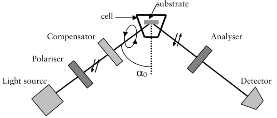
Figure 4. Ellipsometer set-up.
This non-destructive technique is time-resolved, allowing information on the kinetics of the adsorption to be registered without interfering with the adsorption process. The measurements are done in a temperature controlled liquid cell, with continuous stirring, and it is also possible to exchange the
Polariser
Compensator Analyser
α
0Light source Detector
cell
36
solution in the cell by continuous rinsing, so that e.g. reversibility of adsorption may be monitored.
The instrument set-up is shown in Figure 4. Light from a light source (in our case a xenon arc-lamp, filtered to λ= 4429 Å, for experiments described in paper IV: λ= 4015 Å) first passes a polariser which results in linearly polarised light. The plane polarised light then passes the compensator (a quarter-wave plate), which induces a relative phase shift in the light vectors (light oscillating parallel and perpendicular to the fast and slow axis), rendering it elliptically polarised. The reflection at the solid/liquid interface further changes the polarisation, resulting in an additional relative phase shift and amplitude change. The polariser can be adjusted in such a way that the ellipticity prior to reflection results in linearly polarised light after reflection, and can be “extinguished” by the second polariser, called the analyser. The light intensity after the analyser is recorded by the detector. During a measurement, the optical components (usually the polariser and analyser, while the compensator azimuth is kept constant at ±45°) are adjusted by stepping motors to minimize the light reaching the detector. There are four sets of positions for the polariser, analyser and compensator that all result in light minima. These four settings are known as the four zones, and by averaging the azimuths obtained in all zones, most optical imperfections, such as misalignment of the sample and tilted optical components, can be corrected for137
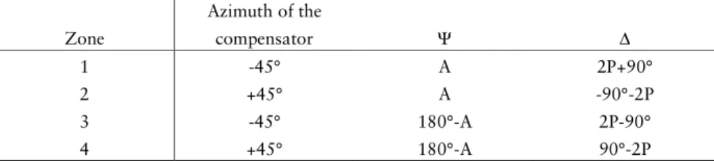
. Averaging over two zones, keeping the compensator azimuth fixed, corrects for the major optical imperfections. From the azimuths of the polariser and analyser obtained at the nulling settings, the ellipsometric angles Ψ and ∆ can be calculated. The relative amplitude ratio (Ψ) is obtained from the azimuth of the analyser, while the relative phase shift (∆) is obtained from the azimuth of the polariser. Four different sets of Ψ and ∆ can be obtained according to Table 2:
Table 2. Conversion from azimuth of the analyser (A) and polariser (P) into
the ellipsometric angles Ψ and ∆ for the different zones137
. Zone Azimuth of the compensator Ψ ∆ 1 -45° A 2P+90° 2 +45° A -90°-2P 3 -45° 180°-A 2P-90° 4 +45° 180°-A 90°-2P
37 The instrument used was a Rudolph thin film ellipsometer type 43603-200E (Rudolph Research, Fairfield, NJ, USA), automated according to Cuypers138
. For each experiment, a two-zone (or a four-zone, paper IV) surface calibration, was carried out in two ambient media (air and PBS). A three-layer optical model (ambient, surface oxide layer and bulk surface) was used to determine all unknowns in the clean system, i.e. the complex refractive index (N0 = n0 – ik0) of the silicon and the real part of the refractive index (n1) and
thickness (d1) of the silicon oxide layer (the oxide layer was assumed to be
transparent, i.e. k1=0). For the calculations, the dimethylsilane layer on the
hydrophobized silica was assumed to be included in the oxide layer. This simplification will result only in a minor error due to the minor thickness of the dimethylsilyl layer (a few Å)139.
After surface characterization, the sample was added to the cell, and Ψ and ∆ were recorded in situ in real time. When the optical properties of the substrate and the ambient media is known, the average layer thickness (df) and
refractive index (nf) of the protein film can be solved by numerical iteration
from the changes in Ψ and ∆140
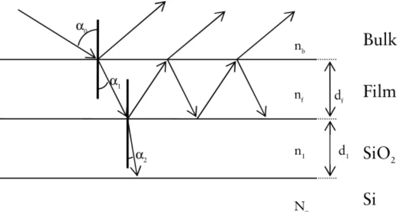
, assuming a homogeneous film. The evaluation of the adsorbed protein film was performed by using a four-layer optical model, with isotropic media and planar interfaces (Figure 5).
Figure 5. Schematic illustration of the 4-layer model. αx= angle of incidence or
angle of refraction.
If the film is inhomogeneous, or if the level of adsorption is low, resulting in only minor changes in Ψ and ∆, the refractive index (nf) and average thickness
(df) estimations of the film are unreliable 141
. The relative errors in nf and df are
relatively high for low adsorbed amounts (Γ<0.5 mg m-2), however, for Γ>1
α0 n1 α2 α1 nb df nf No
Film
Bulk
Si
d1SiO
238 mg m-2
, the error is only 5-10%142
. Errors in nf and df co-vary, resulting in a
much higher accuracy for the calculated adsorbed mass, even at small changes in Ψ and ∆ (an error of approximately 15% at Γ= 0.1 mg m-2
and less than 1% for Γ= 2 mg m-2)142.
From the film thickness and the refractive index, the adsorbed amount can be calculated according to the model by Cuypers et al.141
(Papers I-II and IV):
where
Cuypers’ model is based on the difference in refractivity of different functional groups. This is described by the ratio of the molar weight to the molar refractivity (M/A) of the adsorbate and its partial specific volume (ν). Values of M/A and ν used (papers I-II and IV) were 4.10 g ml-1
and 0.75 ml g -1
, respectively. These values are averages for proteins, and have been used previously in studies of adsorption from saliva and salivary components42, 89, 102,
103
.
The adsorbed amounts can also be calculated according to the de Feijter formula (paper III)143
:
An assumption of linearity of the increment in refractive index with concentration is the basis of this equation. A dn/dc value of 0.16 ml/g20, 82, 83
was used, based on the characteristics of the mucin.
Comparison between the models of Cuypers and de Feijter, respectively, has shown quantitatively the same results144.
A drawback when using ellipsometry is that no information on the composition of the adsorbed film is obtained. Nevertheless, for single protein solutions the results can be interpreted in terms of e.g. orientation of adsorbed molecules. It should be kept in mind that the thickness obtained from
[
(
1
)
/(
2
)
]
(
)
)
/
(
)
(
3
.
0
2 2 f b b b fn
n
n
n
M
A
n
df
−
+
−
−
=
Γ
ν
dc
dn
n
n
d
f f b/
−
=
Γ
Eq. 3 Eq. 5)
2
)(
2
(
)
(
2 2+
+
+
=
b f b f fn
n
n
n
n
f
Eq. 439 ellipsometry measurements assumes a homogeneous film with a mean refractive index. This results in a film thickness that is averaged over the measured area of the film, which may not necessarily describe the real situation. Further, surfaces need to be planar, smooth and reflecting, and the solution has to be transparent to avoid too much light scattering, which otherwise decreases the sensitivity of the measurement.
Mass transport analysis
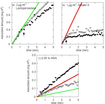
The initial adsorption kinetics of the cationic proteins (papers I and II) was investigated to elucidate if the adsorption was mass transport controlled. To this end, the model by Trurnit145 was employed. The model relies on a stirred system where the mass transport to the solid/liquid interface is determined by diffusion over an unstirred layer closest to the surface. The thickness of this unstirred layer was in the present investigations estimated to be 20 µm146
. Trurnit’s model further assumes an irreversibly adsorbed layer where the time to create a constant concentration gradient in the unstirred layer is negligible. Conditional on the above-mentioned criteria, the initial adsorbed amounts versus time, for a diffusion controlled system are:
where Γ is the adsorbed amount (mg×m-2
), D the diffusion coefficient (m2
×s-1
), C0 the bulk concentration (mg×m
-3
), δ the thickness of the unstirred layer (m) and t is the time (s).
Atomic force microscopy (AFM)
AFM was employed to investigate the structure of the adsorbed protein films, as described in paper III. For these experiments, AFM imaging was performed to acquire images at different stages in the multilayer formation. Values on surface roughness, size, shape, as well as distribution of adsorbed components can be obtained, with a resolution in the z-direction down to the nanometre or Ångström scale147.
Briefly, AFM imaging is based on scanning a surface using a cantilever with a very sharp tip. Interactions between the sample/surface and the tip will cause the cantilever to bend. A laser beam is reflected on the cantilever and will
δ
t
DC
0=
40
register changes in the cantilever position. These changes are then recorded by the photodetector and converted to a 3D reconstruction of the sample topography. In contact mode, which was used here, the interaction between the tip and surface is kept constant by moving the sample in the z-direction during scanning.
AFM is a powerful technique and has the advantage, in comparison to other similar techniques such as transmission electron microscopy, that it only requires minimal sample preparation and images can be acquired in aqueous solution at ambient temperatures, simulating physiological conditions. However, adsorbed protein films, which are of a soft nature, can be deformed during scanning, resulting in altered structures that do not resemble the actual film morphology. Therefore, in the experiments presented here, the surface load was fixed to balance the electrostatic repulsive barrier148
, to minimize such alterations. The experiments were performed in a liquid cell, and the images were obtained after rinsing the liquid cell with buffer, to reduce interference due to e.g. viscoelasticity from components in the ambient solution.
The instrument used was a scanning probe microscope from Veeco (Picoforce multimode SPM with a Nanoscope IV control unit). Silicon nitride tips with a cantilever spring constant of ≤0.32 N/m from Veeco, type DNP, were used.
Total internal reflectance fluorescence (TIRF)
TIRF is a technique that detects fluorescence from components adsorbed at, or close to, an interface. The fluorescence may be emitted from intrinsically fluorescent amino acids or, as described in paper IV, from fluorescent groups covalently bound to a protein.
The basic principle of TIRF is the excitation of fluorescent molecules by an evanescent wave that forms in the liquid phase close to the interface. The evanescent wave is a result from superposition of incident and reflected beams upon total reflection at the interface between two media with different optical properties. The evanescent wave decays exponentially normal to the interface on the far side of the optically rarer medium (the protein solution), thereby confining the excitation to the interfacial region. In the experimental set-up used here, the penetration depth, which is the distance over which the evanescent wave amplitude decreases to e-1 of its initial value, was estimated to be 200 nm. A detailed description of the instrument used is given by Lassen
41 and Malmsten149
. Briefly, it consist of a 488 nm argon ion laser (model 161 B, Spectra physics, USA) as a light source, a flow cell, and the detection part of the instrument consisted of a monochromator (Model H20 UV, Jobin Yvon, France), a photo-multiplier tube (Model R 298, Hamamatsu, Japan) and a photocounter (Model SR 400, Stanford Research System, USA).
TIRF is a technique that records adsorption in a time-resolved manner, similar to ellipsometry. It is a good complement to ellipsometry, as both the total adsorbed amount as well as the fraction of specific components in the adsorbed film can be elucidated by these techniques. Due to factors such as quenching, uneven probe distribution across the adsorbed layer, and local pH fluctuations, quantification of the adsorbed amounts is not straightforward, and therefore the results obtained by TIRF (paper IV) was analysed based on relative changes in fluorescence.
As for ellipsometry, TIRF requires a transparent solution, and the surfaces used for TIRF needs to be smooth, planar and transparent, to allow total reflection at the interface between the surface and the protein solution.
For the TIRF experiments, PRP-1 and statherin were fluorescently labelled with fluorescein-5-isothiocyanate (FITC). The average molar ratio of FITC to protein was <0.15 mol FITC/mol protein. The low labelling densities minimizes the effect of the fluorophore on the protein structure and decreases risks of quenching or changes in adsorption behaviour. However, the signal to noise ratio of the measurements may be lower.
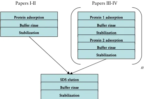
Experimental outline
Figure 6 shows the general experimental outline for the experiments described in papers I-IV. Time intervals are given in the respective papers.
42
Figure 6. Experimental outline. n symbolizes number of times the procedure was repeated (n=1-4, papers III and IV).
Pellicle protein composition
In vivo
pellicle collection and
in vitro
validation
The in vivo pellicle collections were performed by rubbing tooth surfaces
with fibre pellets (Quick-sticks, Dentonova AB, Huddinge, Sweden) soaked in different concentrations of SDS (2%, 0.5%, 0.25% and 0.1%(w/v), respectively). The collection, as described in detail in paper V, was based on a previously reported procedure61
, and was performed from one donor considered to be in good oral and general health. The teeth were isolated during pumicing, cleaning and collection; contact with gingiva as well as the crevical third and approximal areas of the teeth was avoided. After the collection with 3 fibre pellets soaked in SDS solution, one dry fibre pellet was also used.
For the in vitro validation, HWS was collected as described above, from the same donor as the in vivo pellicle. Extracted teeth were used for the validation trial. Each tooth was treated separately during the validation test, except
Papers III-IV Papers I-II
SDS elut SDS elut SDS elut SDS elutionionionion
Buffer rinse Buffer rinse Buffer rinse Buffer rinse Stabilization Stabilization Stabilization Stabilization n Protein adsorption Protein adsorptionProtein adsorption Protein adsorption
Buffer rinse Buffer rinseBuffer rinse Buffer rinse Stabilization Stabilization Stabilization Stabilization Protein 1 adsorption Protein 1 adsorption Protein 1 adsorption Protein 1 adsorption Buffer rinse Buffer rinse Buffer rinse Buffer rinse Stabilization Stabilization Stabilization Stabilization Protein 2 adsorption Protein 2 adsorption Protein 2 adsorption Protein 2 adsorption Buffer rinse Buffer rinse Buffer rinse Buffer rinse Stabilization Stabilization Stabilization Stabilization
43 during the in vitro pellicle formation. A detailed description of the validation is given in paper V. The in vitro pellicle collection was performed by using three fibre pellets, soaked in 0.5% SDS (w/v) and one dry fibre pellet per 3 teeth (both buccal and palatal/lingual sides were collected).
Investigation of
in vitro
pellicles from different oral surfaces
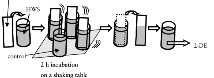
Figure 7 shows the procedure of the in vitro pellicle formation on human enamel, titanium and PMMA, respectively, which is described in detail in paper VI.
Figure 7. Procedure for in vitro pellicle investigation.
The experiments were performed at room temperature. The substrates in Figure 7 symbolises the respective type of material, while the empty container symbolizes the HWS control with no substrate submerged. HWS remaining after in vitro pellicle formation was stored at -20 °C until subjected to 2-DE.
Gel electrophoresis and staining
Gel electrophoresis is a technique that separates proteins on account of some specific characteristics, usually the molecular weight or charge. The proteins move at different velocities in an electric field, based on e.g. their molecular mass. SDS-polyacrylamide gel electrophoresis (SDS-PAGE) gels are frequently used, where SDS applies negative charge to the proteins, yielding an almost uniform charge to mass ratio for all proteins. As a result, the migration in the gel is assumed to only depend on the size of the proteins.
Combining isoelectric focusing (IEF) with SDS-PAGE results in two-dimensional gel electrophoresis (2-DE). IEF, which is performed prior to SDS-PAGE, separates proteins based on their isoelectric point (pI); proteins migrate
2-DE 2 h incubation on a shaking table 2 h incubation on a shaking table substrate HWS control