This is an author produced version of a paper published in Journal of
Controlled Release. This paper has been peer-reviewed but does not include
the final publisher proof-corrections or journal pagination.
Citation for the published paper:
Hernández, Aura Rocio; Boutonnet, Marine; Svensson, Birgitta; Butler, Eile;
Lood, Rolf; Blom, Kristina; Vallejo, Bibiana; Anderson, Chris; Engblom,
Johan; Ruzgas, Tautgirdas; Björklund, Sebastian. (2019). New concepts for
transdermal delivery of oxygen based on catalase biochemical reactions
studied by oxygen electrode amperometry. Journal of Controlled Release,
vol. 306, p. 121-129
URL: https://doi.org/10.1016/j.jconrel.2019.06.001
Publisher: Elsevier
This document has been downloaded from MUEP (https://muep.mah.se) /
DIVA (https://mau.diva-portal.org).
1
New concepts for transdermal delivery of oxygen based on catalase
1biochemical reactions studied by oxygen electrode amperometry
23
Aura Rocio Hernández1,2,3, Marine Boutonnet1,2, Birgitta Svensson4, Eile Butler5, Rolf Lood6, 4
Kristina Blom7, Bibiana Vallejo3, Chris Anderson8, Johan Engblom1,2, Tautgirdas Ruzgas1,2, and 5
Sebastian Björklund1,2,* 6
7
1Department of Biomedical Science, Malmö University, SE-205 06, Malmö, Sweden.
8
2Biofilms - Research Center for Biointerfaces, Malmö University, SE-205 06, Malmö, Sweden
9
3Department of Pharmacy,Universidad Nacional de Colombia, Bogota 1101, Colombia.
10
4Bioglan AB, SE-202 13, Malmö, Sweden
11
5Biogaia AB, SE-223 62, Lund, Sweden
12
6Department of Clinical Science, Lund University, SE-221 84, Lund, Sweden
13
7Medibiome AB, SE-435 43, Pixbo, Sweden
14
8Department of Clinical and Experimental Medicine, Linköping University, SE-581 83 Linköping,
15
Sweden 16
17
*Corresponding author: Sebastian.bjorklund@mau.se 18
2
Abstract
1
The development of formulation concepts for improved skin tissue oxygenation, including 2
methods for measuring oxygen (O2) transport across biological barriers, are important research
3
topics with respect to all processes that are affected by the O2 concentration, such as radiation
4
therapy in oncology treatments, wound healing, and the general health status of skin. In this 5
work we approach this topic by a novel strategy based on the antioxidative enzyme catalase, 6
which is naturally present in the skin organ where it enables conversion of the reactive oxygen 7
species hydrogen peroxide (H2O2) into O2. We introduce various applications of the skin covered
8
oxygen electrode (SCOE) as an in-vitro tool for studies of catalase activity and function. The SCOE 9
is constructed by placing an excised skin membrane directly on an O2 electrode and the
10
methodology is based on measurements of the electrical current generated by reduction of O2
11
as a function of time (i.e. chronoamperometry). The results confirm that a high amount of native 12
catalase is present in the skin organ, even in the outermost stratum corneum (SC) barrier, and 13
we conclude that excised pig skin (irrespective of freeze-thaw treatment) represents a valid 14
model for ex vivo human skin for studying catalase function by the SCOE setup. The activity of 15
native catalase in skin is sufficient to generate considerable amounts of O2 by conversion from
16
H2O2 and proof-of-concept is presented for catalase-based transdermal O2 delivery from topical
17
formulations containing H2O2. In addition, we show that this concept can be further improved
18
by topical application of external catalase on the skin surface, which enables transdermal O2
19
delivery from 50 times lower concentrations of H2O2. These important results are promising for
20
development of novel topical or transdermal formulations containing low and safe 21
concentrations of H2O2 for skin tissue oxygenation. Further, our results indicate that the O2
22
production by catalase, derived from topically applied S. epidermidis (a simple model for skin 23
microbiota) is relatively low as compared to the O2 produced by the catalase naturally present
24
in skin. Still, the catalase activity derived from S. epidermidis is measurable. Taken together, this 25
work illustrates the benefits and versatility of the SCOE as an in vitro skin research tool and 26
introduces new and promising strategies and formulation concepts for transdermal oxygen 27
delivery, and simultaneous detoxification of H2O2, based on native or topically applied catalase.
28 29
Key words: skin tissue oxygenation; topical and transdermal oxygen delivery; epidermis; 30
stratum corneum; catalase; skin microbiota; hydrogen peroxide; oxygen electrode 31
3
1. Introduction
1
The development of concepts related to improved oxygenation of skin and other tissues, 2
including the development of methods for measuring oxygen (O2) transport across biological
3
barriers, are important research topics for all processes that are affected by the O2
4
concentration. For example, in oncology treatments, involving radiation therapy or 5
photodynamic therapy, the level of O2 is crucial for suppressed development of tumors after
6
ionizing radiation and generation of reactive oxygen species (ROS) [1, 2]. Other biologically 7
relevant processes, where the O2 concentration is an important factor, are related to wound
8
healing and the overall health status of the skin barrier. The skin is the only organ, except for 9
the lungs, that is in direct contact with external atmospheric O2 and it has been shown that the
10
upper skin layers are almost exclusively supplied by external O2 [3, 4]. Considering this, it is likely
11
that some superficial skin defects may be related to insufficient skin oxygenation from the 12
atmosphere, rather than by a malfunction in the capillary O2 transport, which has been
13
suggested [3]. 14
The fact that skin is exposed to atmospheric O2 also means that this organ is highly exposed to
15
oxidative stress from generation of ROS. Therefore, it is perhaps not surprising that the skin 16
organ comprises a robust antioxidative system consisting of both molecular antioxidants and 17
antioxidative enzymes such as catalase, superoxide dismutase, glutathione peroxidase, 18
peroxiredoxin, and heme oxygenase [5]. In particular, catalase is highly expressed in the skin 19
organ and its presence increases towards the O2 rich atmosphere. In fact, the presence of
20
catalase in skin is nearly one order of magnitude higher in epidermis as compared to the 21
underlying dermis [6]. Further, it should be noted that catalase in skin is present not only in the 22
viable dermis and epidermis, but also in the most superficial part of the skin, the stratum 23
corneum (SC), which is often considered as being a dead tissue [6]. In other words, there is a 24
good correlation between the expression of catalase, as a function of skin depth, and the 25
concentration of O2, derived from the external atmosphere [3, 6].
26
The main catalase reaction is conversion of hydrogen peroxide (H2O2) into water (H2O) and O2
27
according to 𝐻2𝑂2+ 𝐻2𝑂2
𝐶𝑎𝑡𝑎𝑙𝑎𝑠𝑒
→ 2𝐻2𝑂 + 𝑂2, which may be seen as a detoxification process. 28
In line with this, reduced expression of catalase in skin has been associated to skin diseases, such 29
as vitiligo, and to compensate for this loss and treat some skin disorders, topical application of 30
exogenous and artificial catalase has been proposed [7]. Moreover, recognizing that catalase 31
reaction generates O2, the application of topical formulations containing H2O2 and catalase has
32
been attempted as a solution for topical delivery of O2 into wounds or ischemic skin tissue [8].
33
Catalase can also catalyze peroxidase-type reactions by oxidizing suitable hydrogen donors, such 34
as polyphenols or ethanol, with production of acetaldehyde according to 𝐻2𝑂2+ 𝐶𝐻3𝐶𝐻2𝑂𝐻
35
𝐶𝑎𝑡𝑎𝑙𝑎𝑠𝑒
→ 2𝐻2𝑂 + 𝐶𝐻3𝐶𝐻𝑂 [9]. Here, it should be noted that catalase is the only enzyme of the 36
antioxidative system that produce O2 after exposure to H2O2. Further, it is relevant to point out
37
that no O2 is produced in the case for other substrates, such as alcohols or polyphenols. These
38
facts are taken advantage of in this work where we use an electrochemical experimental setup 39
that measures O2 and is therefore specific towards catalase activity after exposure to H2O2.
40
Taken together, there is a considerable need for monitoring and understanding catalase 41
function in skin to exploiting this enzyme for improved skin health and development of concepts 42
related to enhanced oxygenation of the skin tissue. To approach this topic, it is crucial to have 43
methods for measuring O2 transport across the skin barrier and how the concentration of O2
44
changes in the skin tissue. A substantial knowledge about catalase reactions in the skin organ 45
and transdermal O2 delivery can be gained by using relevant in-vitro tools, which minimizes the
46
need for human or animal studies. In this work, we demonstrate that the skin covered oxygen 47
electrode (SCOE) is a useful in-vitro tool to monitor the function of catalase in skin. In this 48
4 context, it should be mentioned, that utilization of the SCOE setups for studies of transdermal 1
delivery have been introduced by us in 2015 [10, 11]. In 2017 Nocchi et al. illustrated that the 2
SCOE can be used to monitor reactions that involve native epidermal catalase [12]. In this work 3
we extend the use the SCOE setup and introduce several applications of this in vitro tool to 4
characterize transdermal delivery of O2 from H2O2 solutions and show that catalase is present
5
both in SC and in the viable epidermis where it can oxygenate the skin tissue. In addition, we 6
show that topically applied catalase, including catalase derived from Staphylococcus (S.) 7
epidermidis (as a primitive model of skin microbiota) can be used as a source for increased skin
8
oxygenation. 9
2. Materials and methods
102. 1. Materials
11
Hydrogen peroxide (H2O2, 35 %), phosphate buffer saline (PBS, pH 7.4) in tablets, tannic acid,
12
catalase from bovine liver (2000-5000 units/mg), sodium azide (NaN3), 3-amino-1,2,4-triazole
13
(3AT), and polyethylenimine were purchased from Sigma-Aldrich (Darmstadt, Germany). Fresh 14
Staphylococcus epidermidis (S. epidermidis) cultures, with colony-forming units of 8x10⁸ cfu/mL,
15
were provided by Biogaia AB (Lund, Sweden). The oxygen electrode consisted of a 5 μm thick 16
Teflon membrane, a 250 μm diameter platinum (Pt) electrode melted in glass, and an internal 17
Ag/AgCl reference electrode; purchased from Optronika UAB (Vilnius, Lithuania). All solutions 18
were prepared by using ultrapure water with a resistivity of 18.2 Ωcm. 19
2.2. Preparation of split-thickness skin and stratum corneum (SC) membranes
20
Fresh pig ears were obtained from a local abattoir and stored at -80 °C until use. To prepare 21
skin membranes the ears were thawed and cleaned under flow of cold tap water. Cleaned ears 22
were cut into strips with a scalpel and shaved. Pieces of approximately 500 μm thick skin 23
membranes were sliced with a dermatome. The resulting skin stripes were punched out to 24
make circular membranes with 16 mm diameter. These membranes were kept frozen (-20 °C) 25
until use, usually not longer than four weeks. Before use, the membrane was thawed by 26
placing them on a filter paper, soaked with PBS, and kept for 1-2 hours at room temperature 27
(22oC).
28
Human breast skin, which is regarded as discarded tissue, was obtained from an anonymous 29
female donor of Caucasian origin and provided by Medibiome AB (no ethical approval is 30
necessary for unidentified residual tissue). Freshly obtained human skin were used within three 31
days and stored in the fridge soaked in saline (0.9 % NaCl). The human skin samples were about 32
3 mm thick and included the adipose tissue, which is not optimal for the present SCOE setup. 33
Normally, the adipose tissue is easily removed by using a dermatome or scalpel. However, due 34
to the relatively small area of the human skin samples this was a challenging task. Therefore, 35
human skin was only investigated in the form of SC membranes, which are conveniently 36
prepared by trypsin treatment. 37
SC membranes (approximately 10-30 µm thick) from pig and human skin were prepared by 38
soaking full thickness or split-thickness skin membranes in 0.1 % trypsin solution in PBS for 24h 39
at 4oC. After that, the SC layer was easily removed by forceps, washed with PBS and cleaned
40
with cotton tipped applicators from residual tissue. The SC membranes were immediately 41
mounted on oxygen electrodes for SCOE measurements. 42
With regards to enzyme viability and storage protocol, in general, it is expected that the enzyme 43
activity is better preserved inside intact tissue samples, or crude extracts, etc., as compared to 44
purified samples where removal of important matrix components may lead to poorer enzyme 45
activity. Considering that the experiments in this work were conducted with relatively intact skin 46
tissue samples, in combination with the fact that relatively high catalase activity was observed 47
5 in these experiments, we conclude that the viability, in terms of catalase activity, was fully 1
satisfactory in all samples studied herein (even after freeze-thaw treatment). Further, there are 2
studies in support of this conclusion where similar storage conditions as used here were 3
investigated [13, 14]. 4
2.3. Preparation of skin covered oxygen electrode (SCOE)
5
The SCOE was prepared as described previously [12]. Briefly, the surface of the Pt cathode of 6
the oxygen electrode was polished using an alumina suspension (1 μm alumina particles, 7
Buehler, Lake Bluff, IL) and rinsed with deionized water. The body of the electrode was filled 8
with saturated KCl solution and covered with a 5 μm Teflon membrane. Next, the electrode was 9
covered with either a split-thickness skin or SC membrane, directly on top of the Teflon 10
membrane, resulting in the SCOE (see Fig. 1A for a schematic representation). 11
2.4. Topical catalase treatment of the skin covered oxygen electrode (SCOE)
12
In order to attach catalase on the outer skin surface, the tip of the assembled SCOE was first 13
immersed into a solution of tannic acid (1 mg/mL in PBS) for 3 minutes. Tannic acid is a 14
recognized agent for agglutination and mediator for increased protein adsorption [15, 16]. In 15
other words, the reason for this procedure was to increase the adsorption of catalase on the 16
surface of the skin membrane. Next, the SCOE was washed in PBS for 1 minute, followed by 17
immersion into a catalase solution (10 mg/mL in PBS) for 3 minutes. Finally, the SCOE, with 18
topically adsorbed catalase, was washed in PBS to minimize the presence of any loosely bound 19
catalase. Initially, it was concluded that the catalase-doped SCOE was significantly more 20
sensitive to exposure to H2O2, as compared to normal (untreated) SCOE. To optimize the
21
protocol, we investigated if the results were improved by repeating the described protocol 22
several times. For this, the catalase adsorption steps were repeated so that the total times of 23
immersion into the catalase solution were 3, 6, 9, or 12. From these experiments it was 24
concluded that 3 times was sufficient to achieve a significant increase in the sensitivity, in terms 25
of O2 production after H2O2 exposure, as compared to the normal SCOE. However, the results
26
improved in terms of reproducibility when the catalase adsorption protocol was repeated at 27
least 6 times, without any further benefits of 9 and 12 repeats. Thus, the described protocol was 28
repeated 6 times (at least). 29
2.5. Topical treatment of the skin covered oxygen electrode (SCOE) with Staphylococcus (S.)
30
epidermidis culture
31
To investigate if bacteria from a S. epidermidis culture could adsorb on the skin surface of the 32
SCOE, the tip of the SCOE was immersed into a suspension of S. epidermidis at room 33
temperature for 24h. The bacterial suspension was agitated with magnetic stirrer at 50 rpm. 34
Before measurements, the electrode was washed with abundant PBS solution, after which it 35
was placed into the electrochemical cell. 36
2.6. Immobilization of catalase and Staphylococcus epidermidis on the Teflon membrane of
37
the oxygen electrode
38
To attach catalase directly on the Teflon membrane of the oxygen electrode, the electrode was 39
immersed into a solution of tannic acid (1 mg/mL in PBS) for 3 minutes and then washed in PBS 40
for 1 minute. As stated above, tannic acid was used to increase the adsorption of catalase on 41
the surface of the Teflon membrane [15, 16]. Next, the electrode was immersed into a catalase 42
solution (10 mg/mL in PBS) for 3 minutes and finally washed in PBS to remove any loosely bound 43
catalase. 44
To evaluate the catalase activity in S. epidermidis by the oxygen electrode, the bacteria were 45
attached to the Teflon membrane. For this, the oxygen electrode was immersed into a 46
polyethylenimine solution (1 mg/mL in water) for 3 minutes, followed by washing with water 47
6 for 1 minute. Finally, the electrode was immersed into a suspension of S. epidermidis for 3 1
minutes. The positively charged polyethylenimine is a recognized attachment factor for various 2
cell lines [17] and thus used here to increase the immobilization of the net negatively surface 3
charged S. epidermidis. 4
2.7. Amperometric monitoring of catalase reactions using oxygen electrode
5
The different types of electrodes, i.e. the SCOE and the oxygen electrode modified with either 6
catalase or S. epidermidis, were immersed into an electrochemical cell filled with 10 mL PBS (pH 7
7.4). The current of the electrode was recorded by using a CompactStat potentiostat from IVIUM 8
Technologies (Eindhoven, The Netherlands). The oxygen electrode was connected to the 9
potentiostat in a two-electrode configuration and the amperometric measurement was 10
conducted by applying -0.7 V vs Ag/AgCl/KCl (sat) on a Pt cathode of the oxygen electrode. After 11
a baseline current was established, a defined amount of H2O2 was pipetted into the
12
electrochemical cell to obtain a known concentration. In the presence of active catalase, the 13
reduction current of the oxygen electrode increased due to O2 generation (see Eq. 1). This is true
14
for active catalase either in the form of native catalase inside the skin membrane, externally 15
adsorbed catalase, or catalase derived from adsorbed S. epidermidis at the outer skin surface. 16
In all experiments, the solution surrounding the oxygen electrode was continuously mixed with 17
a magnetic stirrer at 250 rpm and all measurements were conducted at room temperature 18
(22°C). 19
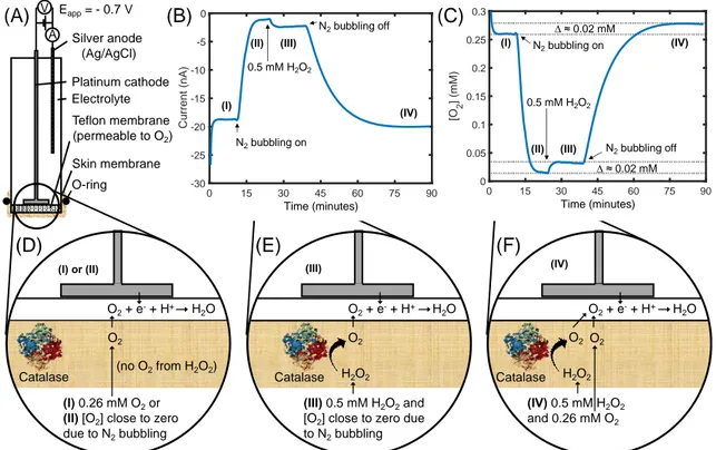
3. Results and discussion
20The general aim of this work was to investigate O2 generation by the enzyme catalase by in vitro
21
measurements with a skin covered oxygen electrode (SCOE). The general setup of the SCOE and 22
working principle is illustrated in Fig. 1. Fig. 1A shows the construction of the oxygen electrode 23
with an excised skin membrane mounted on top of the Teflon membrane and sealed by an O-24
ring. A proof-of-concept is presented in Fig. 1B where raw data from a chronoamperometric 25
measurement of the following four experimental conditions is investigated: 26
I. SCOE in neat PBS without H2O2 ([O2] = 0.26 mM, no N2 bubbling)
27
II. SCOE in neat PBS without H2O2 and with N2 bubbling ([O2] ≈ 0 mM)
28
III. SCOE in PBS with 0.5 mM H2O2 and with N2 bubbling (i.e. O2 production only according
29
to Eq. 1) 30
IV. SCOE in PBS with 0.5 mM H2O2 (no N2 bubbling, i.e. [O2] = 0.26 mM plus O2 production
31
according to Eq. 1) 32
These four experimental conditions are schematically illustrated in Fig. 1D (I and II), E (III), and F 33
(IV), together with the particular mechanism of O2 generation in each case. In Fig. 1C, the O2
34
concentration corresponding to the raw data in Fig. 1B is presented. To enable conversion from 35
current into O2 concentration we calibrate each individual SCOE setup by first recording a stable
36
baseline (I) in PBS buffer with known O2 concentration (0.26 mM or 8.3 mg/L at T=22 °C and 1
37
atmosphere). By this one-point calibration we avoid the variability of individual SCOE setups, 38
which is mainly due to the combined biological variance of O2 permeability and activity of the
39
native catalase in individual skin membranes. Returning to Fig. 1C, the signal corresponding to 40
condition (II) is obtained by bubbling N2 gas through the PBS solution to eliminate dissolved O2.
41
Then, H2O2 is added to generate a defined concentration of 0.5 mM in the PBS solution (III),
42
which clearly results in an increase of the O2 concentration, corresponding to around 0.02 mM,
43
due to conversion of H2O2 into O2 by catalase. Finally, in the last case (IV), the N2 bubbling is
44
turned off and the O2 concentration comes back to the baseline level. In fact, the final O2
45
concentration is 0.28 mM, which is in perfect agreement with the combined contributions of 46
dissolved O2 in PBS (0.26 mM), in addition to the O2 that was generated from H2O2 by catalase
47
(0.02 mM). For simplicity, all further measurements were performed without N2 bubbling.
7 1
Figure 1. (A) Schematic illustration of the skin covered oxygen electrode (SCOE) and its working 2
principle under different experimental conditions. (B) The change in O2 concentration is
3
registered by a change in the cathodic current. Upon immersion, between approximately 0-10 4
min, the O2 concentration is 0.26 mM in PBS solution of the electrochemical measuring
5
compartment. At around 10 min, N2 is bubbled through the solution, which effectively minimizes
6
the reducing current. Next, around 25 min, 0.5 mM H2O2 is added to the solution, which results
7
in an increase of reducing current in proportion to the generated O2. Finally, after about 40 min
8
the N2 bubbling is turned off and the signal returns to a level slightly below the baseline current
9
due to the extra O2 generated by catalase from the added H2O2. (C) The corresponding O2
10
concentration from the experimental data in (B). (D, E, and F) Schematic representations of the 11
mechanism(s) responsible for the measured O2. Case I: baseline current corresponding to PBS
12
saturated with O2. II: minimal baseline current due to N2 bubbling. III: minimal baseline current
13
due to N2 bubbling and O2 produced by catalase from H2O2. IV: baseline current corresponding
14
to PBS saturated with O2, plus O2 produced by catalase from H2O2.
15
3.1. Activity of native catalase in epidermis and stratum corneum (SC) membranes
16
Based on the proof-of-concept presented in Fig. 1, we continue this work by illustrating the 17
versatility of the SCOE setup for investigating the function of native catalase residing in excised 18
skin membranes in vitro. For this, the oxygen electrode was covered with either pig split-19
thickness skin membranes, pig SC membranes, or human SC membranes. By included 20
measurements with human skin we aim at illustrating that the SCOE in vitro tool with pig skin, 21
even after freeze-thaw treatment, is a valid model for ex vivo human skin. Representative 22
measurements from these experiments are presented in Fig. 2. 23 O2+ e-+ H+ H2O (IV) 0.5 mM H2O2 and 0.26 mM O2 Silver anode (Ag/AgCl) A V Eapp= - 0.7 V (I) 0.26 mM O2or (II) [O2] close to zero due to N2bubbling Catalase O-ring Platinum cathode Electrolyte Skin membrane Teflon membrane (permeable to O2) (III) 0.5 mM H2O2and [O2] close to zero due to N2bubbling O2+ e-+ H+ H2O Catalase O2 H2O2 O2+ e-+ H+ H2O O2 O2 O2 Catalase H2O2 (no O2from H2O2) 0 15 30 45 60 75 90 Time (minutes) -30 -25 -20 -15 -10 -5 0 C u rr e n t (n A ) 0 15 30 45 60 75 90 Time (minutes) 0 0.05 0.1 0.15 0.2 0.25 0.3 [O 2 ] (m M ) (A)
(I) or (II) (III) (IV)
(F) (E) N2bubbling on 0.5 mM H2O2 N2bubbling off (I) (III) (II) (IV) (I) (III) (II) (IV) N2bubbling on 0.5 mM H2O2 N2bubbling off (B) (C) (D) ∆ ≈ 0.02 mM ∆ ≈ 0.02 mM
8 1
Figure 2. Change in O2 concentration measured with the SCOE after stepwise addition of H2O2.
2
Representative results from (A) pig split-thickness skin, (B) pig SC, (C) human SC, and (D) 3
compilation of the change of O2 concentration (∆O2), normalized by the change of H2O2
4
concentration (∆H2O2), from several measurements of the different types of membranes (A=pig
5
split-thickness, B=pig SC, C=human SC). The error bars represent the standard error of the mean. 6
After two additions of H2O2, NaN3 is added to inhibit the catalase present in the skin/SC
7
membrane, after which the [O2] value returns to baseline level.
8
Fig. 2A illustrates how the concentration of O2 is changed after addition of H2O2 from a
9
measurement with pig split-thickness skin membranes (i.e. the membrane contains SC, 10
epidermis, and parts of dermis). After establishment of a stable baseline, [H2O2] is first changed
11
from 0 to 0.5 mM, which results in ∆[O2] ≈ 0.05 mM. Next, [H2O2] is increased from 0.5 to 1.5
12
mM, which results in ∆[O2] ≈ 0.10 mM. In other word, ∆[O2] is approximately proportional to
13
∆[H2O2]. These results confirm that H2O2 penetrates the skin membrane where it is enzymatically
14
converted into O2 by native catalase, after which the O2 is transported the oxygen electrode for
15
detection (see Fig. 1). This conclusion is confirmed by the fact that addition of NaN3, which is a
16
well-known catalase inhibitor, results in a decrease of [O2] back to the baseline level [12]. It
17
should be noted that in some cases, when a stable baseline or a stable reading after H2O2
18
addition was not fully achieved, the value after roughly 30 minutes was approximated as 19
endpoint (e.g. Fig. 2A). In general, about 10-30 minutes is required to obtain a stable baseline, 20
corresponding to [O2] = 0.26 mM, and the time variation is most likely due to biological
21
differences between individual skin membranes. In addition, equilibration of the skin membrane 22
after immersion into the buffer solution, is a complex process, which may involve, for example, 23
hydration-induced changes of the molecular properties of the protein and lipid components, 24
swelling of the corneocytes, and ion redistribution between the membrane and the buffer [18-25 20]. 26 0 30 60 90 120 150 Time (minutes) 0.2 0.3 0.4 0.5 [O 2 ] (m M ) 0 10 20 30 40 50 Time (minutes) 0.2 0.3 0.4 0.5 [O 2 ] (m M ) 0 6 12 18 24 30 Time (minutes) 0.2 0.3 0.4 0.5 [O 2 ] (m M )
(A)
NaN3(B)
(C)
∆[O2](D)
0.5 mM H2O2 NaN3 NaN3 1.5 mM H2O2 0.5 mM H2O2 1.0 mM H2O2 0.5 mM H2O2 1.0 mM H2O29 A natural continuation from the studies employing split-thickness membranes is to investigate 1
if catalase is present in an active form in the outermost skin barrier. For this, the electrode was 2
covered with SC membranes, which were separated from the underlying epidermis by trypsin 3
treatment. In these experiments, we included both pig SC (Fig. 2B) and human SC (Fig. 2C) for a 4
more complete characterization and to investigate if the pig skin model is a valid model for 5
human skin ex vivo. In both cases, the responses of the SC covered electrodes to H2O2 were, in
6
principle, similar as compared to the response of the electrode covered with split-thickness skin 7
membrane (Fig. 2A), i.e. stepwise changes of [O2] after H2O2 addition. This proves that catalase
8
is present in an active form, and able to convert H2O2 to O2, inside the SC barrier of both pig and
9
human skin. This is an intriguing result considering the rather solid-like environment of the SC, 10
where a majority of the proteins and lipids are in a rigid molecular state [18, 19]; even though 11
the SC membrane is fully hydrated as in the present experiments. 12
The results in Fig. 2 show that generation of O2 as a response to addition of H2O2 is, clearly, more
13
rapid in the case of only SC (Fig. 2B and C), as compared to the split-thickness membrane (Fig. 14
2A). Similarly, the response to the catalase inhibitor (NaN3) is also significantly faster in the case
15
of only SC (Fig. 2B and C) as compared to the split-thickness membrane (Fig. 2A). The diffusional 16
pathway from the solution to the oxygen electrode, in the case of only SC, is much shorter (total 17
thickness of SC is about 10-30 µm), as compared to the thicker skin membranes (total thickness 18
about 500 µm). This indicates that the thickness, and perhaps the hydrophilicity of the viable 19
epidermis, in combination, act to decrease the transport of the relatively hydrophobic O2
20
molecule across the membrane. However, it cannot be excluded that the SC membrane contains 21
macroscopic barrier defects, as a result from the separation of SC from the underlying epidermis, 22
which could make it easier for H2O2 to reach catalase in the SC membrane and/or make it easier
23
for the produced O2 to diffuse to the electrode via defective regions of the SC membrane.
24
The experiments with SC from human skin resulted in significantly higher generation of O2 after
25
addition of identical amounts of H2O2, as compared to SC from pig skin (i.e. higher value of ∆[O2]/
26
∆[H2O2], see Fig. 4D, p-value = 0.001, 2-tailed t-test with 2-sample unequal variance). In fact, the
27
SC from human skin gave a similar response as compared to pig split-thickness skin membranes, 28
i.e. no statistically significant difference (p-value = 0.638). This should be compared to the 29
observed difference of the change of [O2] from the experiments with pig SC and the pig
split-30
thickness, which is statistically different at a weak significance level (p-value = 0.018). Taken 31
together, these results indicate that the catalase activity in ex vivo human skin is higher as 32
compared to pig skin. However, it is appropriate to issue certain caveats here; the pig skin 33
samples were exposed to freeze-thaw treatment and originated from ears while the human skin 34
samples were not freeze-thawed and collected from breast. Therefore, the comparison 35
presented in Fig. 2D should be considered as a qualitative proof-of-principle showing that the 36
different SCOE setups, corresponding to the results in Fig. 2A, B, C, all successfully work 37
according to the principle illustrated in Fig. 1. In other words, a key conclusion is that the present 38
results illustrate that excised pig skin, even after freeze-thaw treatment, is a valid in vitro model 39
for human skin ex vivo for studying native skin catalase function. In general, it is well established 40
that pig skin is a relevant model to human skin in terms of anatomy [21], permeability [22-26], 41
and electrical properties [26, 27]. Following these studies, it would be interesting to perform 42
further investigations, for example with pig and human skin samples harvested from the 43
corresponding skin sites and treated with identical protocols. 44
3.2. New strategies for transdermal delivery of oxygen
45
From the results presented above it can be concluded that the SCOE setup enables studies of 46
the catalase reaction in skin. The reaction, of course, must involve addition of H2O2 and
47
subsequent O2 generation. Keeping this in mind, we will now illustrate the versatility of the SCOE
48
setup as an in-vitro tool to study transdermal delivery of O2 from solutions containing H2O2 and
10 to examine if the methodology can be extended to study the catalase activity in topically 1
attached S. epidermidis as a simple, but relevant, mimic for skin microbiota. In all cases, the O2
2
is generated from catalase. However, the catalase is either provided topically, as such, or derived 3
from topically adsorbed microbiota. Finally, we also include experiments where the catalase or 4
microbiota is immobilized directly on the Teflon membrane of the oxygen electrode, to illustrate 5
the concept of catalase-based O2 delivery in a clear and simple model system.
6
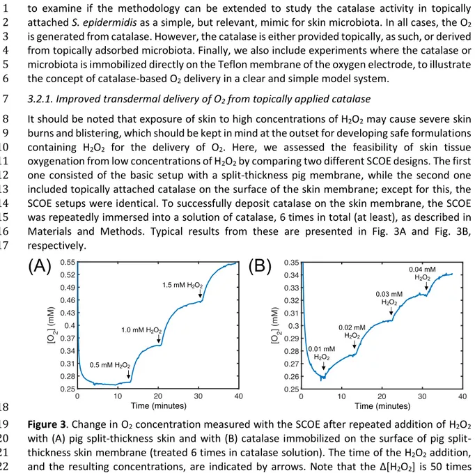
3.2.1. Improved transdermal delivery of O2 from topically applied catalase
7
It should be noted that exposure of skin to high concentrations of H2O2 may cause severe skin
8
burns and blistering, which should be kept in mind at the outset for developing safe formulations 9
containing H2O2 for the delivery of O2. Here, we assessed the feasibility of skin tissue
10
oxygenation from low concentrations of H2O2 by comparing two different SCOE designs. The first
11
one consisted of the basic setup with a split-thickness pig membrane, while the second one 12
included topically attached catalase on the surface of the skin membrane; except for this, the 13
SCOE setups were identical. To successfully deposit catalase on the skin membrane, the SCOE 14
was repeatedly immersed into a solution of catalase, 6 times in total (at least), as described in 15
Materials and Methods. Typical results from these are presented in Fig. 3A and Fig. 3B, 16
respectively. 17
18
Figure 3. Change in O2 concentration measured with the SCOE after repeated addition of H2O2
19
with (A) pig thickness skin and with (B) catalase immobilized on the surface of pig split-20
thickness skin membrane (treated 6 times in catalase solution). The time of the H2O2 addition,
21
and the resulting concentrations, are indicated by arrows. Note that the ∆[H2O2] is 50 times
22
lower in (B), as compared to (A). 23
In general, when comparing the results in Fig. 3, it is immediately clear that the SCOE with 24
immobilized catalase on the skin surface responds to significantly lower concentrations of H2O2,
25
as compared to the basic SCOE design. The basic SCOE, employing pig split-thickness skin, 26
requires concentrations of approximately 0.5 mM H2O2 for adequate measurements, while no
27
notable response is achieved for H2O2 concentrations in the range of 0.01-0.04 mM. In other
28
words, the increase in O2 concentration shown in Fig. 3B is primarily due to O2 produced by
29
topically applied catalase at the skin surface; and subsequent diffusion of O2 from the skin
30
surface across of the membrane to the electrode. This is different as compared to the basic SCOE 31
setup, where H2O2 diffuses into the skin membrane to the site of the native catalase where it is
32
converted into O2, which then diffuses to the electrode surface. The ∆[H2O2] is 50 times higher
33
in the experiment with the normal SCOE (Fig. 3A), as compared to the catalase-doped SCOE (Fig. 34
3B), at the same time as the corresponding values of ∆[O2] only differs by a factor of 5; i.e.
35
0.10±0.01 and 0.020±0.005, respectively. This implies that the limiting factor in these 36
experiments is the flux of H2O2 into the skin membrane to the site of catalase, while the
37
conversion into O2 and the subsequent flux of O2 are relatively fast processes. Thus, in the case
38
of topically applied catalase, O2 is converted at the surface of the skin, generating a significant
11 transdermal flux of O2 across the skin tissue to the electrode. This is a striking finding that
1
illustrates the potential of combining low and safe concentrations of H2O2 in topical formulations
2
together with topically applied catalase for transdermal delivery of O2.
3
3.2.2. Inhibition of native skin catalase to enable detection of oxygen derived from catalase in
4
Staphylococcus epidermidis
5
When considering enzymes of the antioxidative system of skin in general, including the 6
contribution from native catalase, one should not ascribe all antioxidative activity to the 7
enzymes located inside the skin organ. On the contrary, a substantial part of the antioxidant 8
activity could be attributed to external skin microbiota, which therefore could play a relevant 9
role for maintaining the redox homeostasis of the skin organ. This hypothesis is supported by a 10
recent study demonstrating that Propionibacterium acnes on skin produce the antioxidant 11
enzyme radical oxygenase, which thus increases the antioxidant capacity of the skin [28]. To 12
approach this topic, we investigated if the SCOE in-vitro setup could be adopted to detect 13
catalase on skin membrane derived from microbiota. In particular, we wanted to investigate if 14
the catalase activity derived from skin microbiota can produce sufficient amounts of O2 to be
15
detected by the SCOE, in a similar manner as demonstrated above for native skin catalase and 16
topically applied catalase. For this, we selected S. epidermidis, which is a main component of the 17
commensal skin microbiota [29], as a simple model for skin microbiota. In brief, the basic SCOE 18
was immersed in a culture of S. epidermidis for 24h, after which it was thoroughly washed before 19
measurements. Initial measurements indicated that the catalase activity from external S. 20
epidermidis was relatively low, but detectable. To achieve better sensitivity, and to scrutinize
21
between O2 generated by S. epidermidis or by native skin catalase, it was decided to irreversibly
22
inhibit the native skin catalase. The fact that the SCOE, after inhibition with NaN3 and thorough
23
washing in fresh PBS, continued to give a considerable response after addition of H2O2 allowed
24
us to conclude that NaN3 is a reversible inhibitor of catalase. Therefore, instead of using NaN3,
25
we used 3AT, which has been reported to be an irreversible inhibitor of catalase [6]. In short, 26
the SCOE was kept in H2O2 and a solution containing 40 mM 3AT for approximately 3h. To
27
evaluate this concept, we performed the following experiments. First, the SCOE was immersed 28
in PBS for about 1h to reach a stable baseline, after which H2O2 was added to obtain a
29
concentration of 1 mM, followed by inhibition with 3AT (curve 1 in Fig. 4A). Next, the SCOE was 30
rinsed in PBS for 10 minutes and immediately exposed to 1 mM H2O2 again (curve 2 in Fig. 4A).
31
After this, the SCOE was kept in either PBS for 24h (curve 3a in Fig. 4A) or in a suspension of S. 32
epidermidis for 24h (curve 3b in Fig. 4A), after which the SCOE was evaluated once more by
33
addition of H2O2.
34
35
Figure 4. O2 production by catalase derived from topically applied S. epidermidis. The
36
experimental protocol is illustrated in (A), while the results from several experiments (n=6-3) 37
are summarized in (B) with error bars representing the standard error of the mean. In (A), the 38
basic SCOE, with pig split-thickness skin, was first exposed to 1.0 mM H2O2, to ensure that the
12 SCOE setup functioned normally, followed by inhibition with 3AT for approximately 3h (protocol 1
1: 3AT inhibition). Next, the catalase-inhibited SCOE was rinsed in PBS, followed by repeated 2
exposure in 1.0 mM H2O2 (protocol 2: 10 min in PBS). In the following step, three replicates were
3
treated in neat PBS buffer for 24h (protocol 3a: 24h in PBS), whereas three replicates were 4
treated in a suspension of microbiota culture for 24h (protocol 3b: 24h in S. epidermidis). After 5
these treatments (3a and 3b), the electrodes were again exposed to 1.0 mM H2O2. In (A), all
6
curves are shifted in time so that the addition of H2O2 to the electrochemical cell occurs at the
7
same point, as indicated by the arrow. It is not possible to conclude that the observed increase 8
in O2 generation, after treating the catalase-inhibited SCOE in the S. epidermidis culture, in fact
9
is higher as compared to the non-microbiota treated catalase-inhibited SCOE (p-level 0.343). 10
In summary, the results in Fig. 4 show that 3AT significantly suppresses the O2 production and
11
that catalase remains inhibited despite of washing the skin membrane in PBS for 10 min or 24h 12
(curve 2 and 3a, Fig. 4A). These results conclude that catalase inhibition by 3AT can be 13
considered as irreversible, in contrast to inhibition with NaN3. Still, it should be pointed out that
14
there is a minor residual O2 generation even after 3AT inhibition (approximately 10 %). In fact,
15
a number of experimental efforts, such as repeated conditioning of the SCOE in H2O2/3AT
16
solution, were performed to completely remove this small trace of O2 production without any
17
success. This is perhaps a surprising observation considering that catalase is the only enzyme 18
that generates O2 from the substrate H2O2. However, it is possible that an unidentified
catalase-19
like (i.e. O2 releasing) enzymatic or biochemical reaction can explain this residual oxygen trace.
20
For example, it has been shown that some peroxidases can generate O2 from the substrate H2O2
21
via the radical anion superoxide O2•− [5, 30]. Nonetheless, the residual trace of O2 that remained
22
after 3AT inhibition was accepted as it still allowed for evaluation of the amount of O2 produced
23
by catalase originating from topical S. epidermidis, which is shown by curve 3b in Fig. 4A. In 24
particular, a notable difference is observed when comparing the change of [O2] between curves
25
3a and 3b (Fig. 4A). The described experimental cycle was repeated with multiple individual 26
SCOE setups, with (n=3) and without (n=3) modification with S. epidermidis, and the results are 27
summarized in Fig. 4B. The mean value of case 3b in Fig. 4B is associated with high standard 28
deviation and the difference between the change of [O2] from the different SCOEs, with and
29
without S. epidermidis, is not fully conclusive (3a and 3b in Fig. 4B). In other words, it is not 30
possible to conclude that the observed increase in O2 generation, after treating the
catalase-31
inhibited SCOE in the S. epidermidis culture, in fact is higher as compared to the non-microbiota 32
treated catalase-inhibited SCOE (p-level 0.343). To address this point, we performed similar 33
experiments with only the Teflon membrane as alternative to the more complex situation of 34
catalase-inhibited skin membrane. 35
3.2.3. Detection of topical catalase and catalase derived from topical Staphylococcus epidermidis
36
Topical application of catalase has been proposed to compensate for reduced expression of this 37
enzyme in some skin diseases, such as vitiligo [7]. In addition, production of O2 by catalase after
38
application of topical formulations containing H2O2 is a promising concept for topical delivery of
39
O2 into wounds or ischemic skin tissue [8]. If it would be possible to introduce, or promote,
40
commensal skin bacteria containing catalase, with the aim to contribute to the removal of H2O2
41
from the skin surface and/or to supply O2 to the skin tissue; this would be novel applications for
42
either transdermal O2 delivery or detoxification of H2O2. Therefore, to approach these questions,
43
and in particular to prove that catalase originating from S. epidermidis can provide measurable 44
amounts of O2 from H2O2, we immobilized catalase or S. epidermidis directly on the Teflon
45
membrane of the oxygen electrode (instead of the skin membrane). The results from these 46
experiments are shown in Fig. 5A and B, respectively. The change of [O2] after addition of H2O2
47
are obvious. In particular, the sensitivity of the electrode with adsorbed catalase is significantly 48
higher as compared to the electrode with topically attached S. epidermidis. Still, it is promising 49
to conclude that topical application of S. epidermidis, which dominates the skin microbiota, can 50
13 contribute with catalase activity on the skin. However, the combined results presented in Fig. 4 1
and 5B illustrate that the procedure of topical application of external microbiota needs to be 2
further optimized to allow for improved transdermal oxygen delivery and/or increased 3
detoxification of H2O2.
4
5
Figure 5. O2 production by (A) catalase (as such) and (B) catalase derived from S. epidermidis
6
immobilized directly on the Teflon membrane of the oxygen electrode. Note that the ∆[H2O2] is
7
0.01 mM in (A), while the corresponding situation in (B) is ∆[H2O2] = 0.1 and 0.9 mM (i.e. “low”
8
and “high” concentrations). 9
4. Conclusions
10A common perception is that skin receives its O2 supply from the internal circulation. However,
11
recent investigations have shown that a significant amount of O2 may enter skin from the
12
external atmospheric O2 and it has been shown that the upper skin layers are almost exclusively
13
supplied by external O2 [3, 4]. Considering this, it is likely that maintenance of the general skin
14
health and successful wound healing are strongly dependent on adequate skin oxygenation [4]. 15
The ability to deliver topical and transdermal O2 to defective skin, such as wounds or ischemia
16
tissue, may allow the clinician to support the metabolically active wounded tissue for improved 17
healing. Several O2 delivery systems have been developed, such as supersaturated O2 emulsions
18
capable of incorporating high levels of O2 [31], topically applied gaseous O2 [32], and sustained
19
transdermal delivery of O2 via silicone tubing channeled subcutaneously [33]. This study reports
20
on a novel proof-of-concept for catalase-based transdermal O2 delivery by conversion of H2O2
21
as substrate. We introduce several new applications of the skin covered oxygen electrode (SCOE) 22
as an in-vitro tool for studies of native or externally applied catalase. The SCOE is made by 23
placing split-thickness skin or stratum corneum (SC) membranes directly on the O2 electrode
24
(Fig. 1). We demonstrate that excised skin membranes have a high amount of native catalase, 25
even in the outermost SC barrier, and conclude that pig skin (irrespective of freeze-thaw 26
treatment) represents is a valid model for ex vivo human skin for studying catalase function with 27
the SCOE setup (Fig. 2). The activity of native catalase in the skin barrier is high enough to 28
generate a considerable amount of O2 by conversion from H2O2, which enables successful skin
29
tissue oxygenation. We show that this concept can be further improved by topical application 30
of catalase on the skin surface, which enables transdermal O2 delivery from 50 times lower
31
concentrations of H2O2 (Fig. 3). This is an important and promising finding that opens up for
32
development of topical or transdermal formulations containing low and safe concentrations of 33
H2O2 for transdermal O2 delivery.
34
Taken together, this work illustrate that it is possible to develop novel catalase-based 35
transdermal formulations with the aim to deliver O2 and detoxify H2O2 for accelerated wound
36
healing and strengthening the overall health status of the skin organ. Further, future research 37
efforts should focus on, for example, localization of native catalase in the complex 38
macromolecular matrix of the skin barrier, and how its activity can be regulated, e. g. by 39
(A)
(B)
0.01 mM H2O2 0.02 mM H2O2 0.03 mM H2O2 0.1 mM H2O2 1.0 mM H2O214 hydration [20], excipients [34], humectants [35], penetration enhancers [36, 37], UV radiation 1
[38], or various biogenic stressors of the complex neuro-endocrine system [39]. 2
Acknowledgements
3Financial support from The Crafoord Foundation (SB: grant number 20180740), Malmö 4
University (SB: LED 1.3-2017/498), The Knowledge Foundation (JE, TR, SB: grant number 5
20170058), and The Gustaf Th. Ohlsson Foundation (JE, TR) are greatly acknowledged. ARH is 6
grateful to the Colombian Science Foundation for granting her participation in the PhD 7
exchange program. 8
References
9[1] M. Kržič, M. Šentjurc, J. Kristl, Improved skin oxygenation after benzyl nicotinate
10application in different carriers as measured by EPR oximetry in vivo, Journal of
11Controlled Release, 70 (2001) 203-211.
12[2] Y. Sheng, H. Nesbitt, B. Callan, M.A. Taylor, M. Love, A.P. McHale, J.F. Callan,
13Oxygen generating nanoparticles for improved photodynamic therapy of hypoxic
14tumours, Journal of Controlled Release, 264 (2017) 333-340.
15[3] M. Stücker, A. Struk, P. Altmeyer, M. Herde, H. Baumgärtl, D.W. Lübbers, The
16cutaneous uptake of atmospheric oxygen contributes significantly to the oxygen
17supply of human dermis and epidermis, The Journal of Physiology, 538 (2002)
985-18994.
19[4] D.F. Roe, B.L. Gibbins, D.A. Ladizinsky, Topical dissolved oxygen penetrates skin:
20model and method, Journal of Surgical Research, 159 (2010) E29-E36.
21[5] F.A.D.T.G. Wagener, C.E. Carels, D.M.S. Lundvig, Targeting the redox balance in
22inflammatory skin conditions, International Journal of Molecular Sciences, 14
23(2013) 9126-9167.
24[6] L. Hellemans, H. Corstjens, A. Neven, L. Declercq, D. Maes, Antioxidant enzyme
25activity in human stratum corneum shows seasonal variation with an
age-26dependent recovery, Journal of Investigative Dermatology, 120 (2003) 434-439.
27[7] K.U. Schallreuter, J. Moore, J.M. Wood, W.D. Beazley, D.C. Gaze, D.J. Tobin, H.S.
28Marshall, A. Panske, E. Panzig, N.A. Hibberts, In vivo and in vitro evidence for
29hydrogen peroxide (H2O2) accumulation in the epidermis of patients with vitiligo
30and its successful removal by a UVB-activated pseudocatalase, Journal of
31Investigative Dermatology Symposium Proceedings, 4 (1999) 91-96.
32[8] H.M. Abdel-Mageed, H.M. El-Laithy, L.G. Mahran, A.S. Fahmy, K. Mader, S.A.
33Mohamed, Development of novel flexible sugar ester vesicles as carrier systems for
34the antioxidant enzyme catalase for wound healing applications, Process
35Biochemistry, 47 (2012) 1155-1162.
36[9] H.N. Kirkman, G.F. Gaetani, Mammalian catalase: a venerable enzyme with new
37mysteries, Trends in Biochemical Sciences, 32 (2007) 44-50.
38[10] H. Gari, J. Rembiesa, I. Masilionis, N. Vreva, B. Svensson, T. Sund, H. Hansson,
39A.K. Morén, M. Sjöö, M. Wahlgren, J. Engblom, T. Ruzgas, Amperometric in vitro
40monitoring of penetration through skin membrane, Electroanalysis, 27 (2015) 111
41- 117.
4215
[11] J. Rembiesa, H. Gari, J. Engblom, T. Ruzgas, Amperometric monitoring of
1quercetin permeation through skin membranes, International Journal of
2Pharmaceutics, 496 (2015) 636-643.
3[12] S. Nocchi, S. Björklund, B. Svensson, J. Engblom, T. Ruzgas, Electrochemical
4monitoring of native catalase activity in skin using skin covered oxygen electrode,
5Biosensors and Bioelectronics, 93 (2017) 9-13.
6[13] D. Bravo, T.H. Rigley, N. Gibran, D.M. Strong, H. Newman-Gage, Effect of storage
7and preservation methods on viability in transplantable human skin allografts,
8Burns, 26 (2000) 367-378.
9[14] L.P. Ge, L.H. Sun, J.Y. Chen, X.L. Mao, Y. Kong, F. Xiong, J. Wu, H. Wei, The viability
10change of pigskin in vitro, Burns, 36 (2010) 533-538.
11[15] I. Chibata, T. Tosa, T. Mori, T. Watanabe, N. Sakata, Immobilized tannin - a novel
12adsorbent for protein and metal ion, Enzyme and Microbial Technology, 8 (1986)
13130-136.
14[16] S.V. Boyden, The adsorption of proteins on erythrocytes treated with tannic
15acid and subsequent hemagglutination by antiprotein sera, Journal of Experimental
16Medicine, 93 (1951) 107-120.
17[17] A.R. Vancha, S. Govindaraju, K.V.L. Parsa, M. Jasti, M. Gonzalez-Garcia, R.P.
18Ballestero, Use of polyethyleneimine polymer in cell culture as attachment factor
19and lipofection enhancer, Bmc Biotechnology, 4 (2004).
20[18] S. Björklund, T. Ruzgas, A. Nowacka, I. Dahi, D. Topgaard, E. Sparr, J. Engblom,
21Skin membrane electrical impedance properties under the influence of a varying
22water gradient, Biophysical Journal, 104 (2013) 2639–2650.
23[19] S. Björklund, A. Nowacka, J.A. Bouwstra, E. Sparr, D. Topgaard, Characterization
24of stratum corneum molecular dynamics by natural-abundance 13C solid-state
25NMR, PLoS ONE, 8 (2013) e61889.
26[20] S. Björklund, J. Engblom, K. Thuresson, E. Sparr, A water gradient can be used
27to regulate drug transport across skin, Journal of Controlled Release, 143 (2010)
28191-200.
29[21] U. Jacobi, M. Kaiser, R. Toll, S. Mangelsdorf, H. Audring, N. Otberg, W. Sterry, J.
30Lademann, Porcine ear skin: an in vitro model for human skin, Skin Research and
31Technology, 13 (2007) 19-24.
32[22] R.L. Bronaugh, R.F. Stewart, E.R. Congdon, Methods for in vitro percutaneous
33absorption studies. 2. Animal models for human skin, Toxicology and Applied
34Pharmacology, 62 (1982) 481-488.
35[23] I.P. Dick, R.C. Scott, Pig ear skin as an in vitro model for human skin
36permeability, Journal of Pharmacy and Pharmacology, 44 (1992) 640-645.
37[24] W.G. Reifenrath, E.M. Chellquist, E.A. Shipwash, W.W. Jederberg, Evaluation of
38animal models for predicting skin penetration in man, Fundamental and Applied
39Toxicology, 4 (1984) S224-S230.
40[25] V. Vallet, C. Cruz, D. Josse, A. Bazire, G. Lallement, I. Boudry, In vitro
41percutaneous penetration of organophosphorus compounds using full-thickness
42and split-thickness pig and human skin, Toxicology in Vitro, 21 (2007) 1182-1190.
4316
[26] N. Sekkat, Y.N. Kalia, R.H. Guy, Biophysical study of porcine ear skin in vitro and
1its comparison to human skin in vivo, Journal of Pharmaceutical Sciences, 91 (2002)
22376-2381.
3[27] D. Marro, R.H. Guy, M.B. Delgado-Charro, Characterization of the iontophoretic
4permselectivity properties of human and pig skin, Journal of Controlled Release, 70
5(2001) 213-217.
6[28] M. Allhorn, S. Arve, H. Bruggemann, R. Lood, A novel enzyme with antioxidant
7capacity produced by the ubiquitous skin colonizer Propionibacterium acnes,
8Scientific Reports, 6 (2016).
9[29] A.L. Cogen, V. Nizet, R.L. Gallo, Skin microbiota: a source of disease or defence?,
10The British journal of dermatology, 158 (2008) 442-455.
11[30] A.N.P. Hiner, J. Hernandez-Ruiz, G.A. Williams, M.B. Arnao, F. Garcia-Canovas, M.
12Acosta, Catalase-like oxygen production by horseradish peroxidase must
13predominantly be an enzyme-catalyzed reaction, Archives of Biochemistry and
14Biophysics, 392 (2001) 295-302.
15[31] S.C. Davis, A.L. Cazzaniga, C. Ricotti, P. Zalesky, L.C. Hsu, J. Creech, W.H. Eaglstein,
16P.M. Mertz, Topical oxygen emulsion - A novel wound therapy, Archives of
17Dermatology, 143 (2007) 1252-1256.
18[32] G.M. Gordillo, S. Roy, S. Khanna, R. Schlanger, S. Khandelwal, G. Phillips, C.K. Sen,
19Topical oxygen therapy induces vascular endothelial growth factor expression and
20improves closure of clinically presented chronic wounds, Clinical and Experimental
21Pharmacology and Physiology, 35 (2008) 957-964.
22[33] H.K. Said, J. Hijjawi, N. Roy, J. Mogford, T. Mustoe, Transdermal
sustained-23delivery oxygen improves epithelial healing in a rabbit ear wound model, Archives
24of Surgery, 140 (2005) 998-1004.
25[34] S. Björklund, Q.D. Pham, L.B. Jensen, N.O. Knudsen, L.D. Nielsen, K. Ekelund, T.
26Ruzgas, J. Engblom, E. Sparr, The effects of polar excipients transcutol and
27dexpanthenol on molecular mobility, permeability, and electrical impedance of the
28skin barrier, Journal of Colloid and Interface Science, 479 (2016) 207-220.
29[35] S. Björklund, J. Engblom, K. Thuresson, E. Sparr, Glycerol and urea can be used
30to increase skin permeability in reduced hydration conditions, European Journal of
31Pharmaceutical Sciences, 50 (2013) 638–645.
32[36] Q.D. Pham, S. Björklund, J. Engblom, D. Topgaard, E. Sparr, Chemical penetration
33enhancers in stratum corneum — Relation between molecular effects and barrier
34function, Journal of Controlled Release, 232 (2016) 175-187.
35[37] S. Björklund, J.M. Andersson, Q.D. Pham, A. Nowacka, D. Topgaard, E. Sparr,
36Stratum corneum molecular mobility in the presence of natural moisturizers, Soft
37Matter, 10 (2014) 4535-4546.
38[38] A.T. Slominski, M.A. Zmijewski, P.M. Plonka, J.P. Szaflarski, R. Paus, How UV light
39touches the brain and endocrine system through skin, and why, Endocrinology, 159
40(2018) 1992-2007.
41[39] A.T. Slominski, M.A. Zmijewski, C. Skobowiat, B. Zbytek, R.M. Slominski, J.D.
42Steketee, Sensing the environment: regulation of local and global homeostasis by the
4317