Örebro University School of Medical Sciences Degree project, 15 ECTS
June 2020
Retinopathy of prematurity (ROP) in Örebro : a 10-year
perspective
Version
2
Author: Natasha Abedi
Supervisor: Pia Lundgren MD,PhD Ögonmottagningen, Örebro University Hospital Örebro, Sweden
Abstract:
Background: The survival rate of premature infants has increased in Sweden over the past decade. Preterm infants run the risk of developing a potentially blinding disease known as retinopathy of prematurity (ROP). A recent Swedish national study showed that the frequency of ROP has increased over the past years and there are regional differences across the country. Aim: Our aim was to evaluate the frequency of ROP in Örebro region (Örebro) and compare with the rest of Sweden over a 10-year period.
Methods: A retrospective cohort study was conducted on all premature infants born before gestational week 31, screened for ROP in Örebro, from 2008 to 2017. Data such as number of infants, birth weight (BW), gestational age (GA) and ROP-outcome was retrieved from a national quality register; SWEDROP. Comparisons were made with national data during the same time-period.
Results: The study included 200 infants with a median GA of 28.4 weeks and BW 1144 grams. Of the screened infants 99 (49.5%) developed ROP and 20 (10%) were treated during the study period. During the study period, mild ROP decreased (p=0.024), severe ROP increased (p=0.032), however there was no change in ROP-treated infants (p=0.159). The percentage of ROP-treated infants was higher in Örebro than the rest of Sweden (p=0.024).
Conclusion: Our study showed that the frequency of mild ROP decreased in Örebro whilst severe ROP increased during the 10-year period. The frequency of infants treated for ROP was significantly higher in Örebro compared to the rest of Sweden.
List of abbreviations
BW Birth weight
GA Gestational age
IGF-1 Insulin-like growth factor-1
PMA Post-menstrual age
ROP Retinopathy of prematurity
Table of Contents
1. Introduction ... 1
1.1 Premature Birth in Sweden ... 1
1.2 ROP ... 1
1.2.1 Normal Vascularization ... 1
1.2.2 Pathogenesis of ROP ... 1
1.2.3 Classification of ROP ... 2
1.2.4 ROP Treatment ... 3
1.2.5 Risk Factors for ROP ... 4
1.2.6 ROP Screening ... 4
2. Aim ... 5
2.1 Research Question ... 5
3. Methods and Material ... 5
3.1 Study Population ... 5 3.2 Method ... 5 3.3 Statistical Analyses ... 5 3.4 Ethical Aspects ... 6 4. Results ... 6 4.1 Background Data ... 6 4.2 ROP Frequency ... 9 4.3 Comparison to Sweden ... 9 5. Discussion ... 10 6. Conclusion ... 12 7. Acknowledgments ... 13 References ... 14
1. Introduction
1.1 Premature Birth in Sweden
A full-term pregnancy lasts between 39-40 weeks of gestation. According to WHO, infants born alive before week 37 of gestation are considered premature [1]. In Sweden, around 110 000 infants are born every year. Approximately 5 % of these births are considered preterm [2]. Due to improved neonatal care, the survival rate of premature infants has increased over the past decade in Sweden. Today, infants as young as 22 weeks of gestation can survive with neonatal-intensive care [3]. However, premature birth can have negative consequences on the normal development of the heart, brain, lung and the eye. Several complications such as patent ductus arteriosus, cerebral palsy, infant respiratory distress syndrome and retinopathy of prematurity (ROP), may affect the preterm infant. ROP is a potentially sight-threatening disease that occurs if the retinal vasculature of the eye does not develop properly [4].
1.2 ROP
1.2.1 Normal Vascularization
The vascularization of the retina begins in the second trimester of gestation and is completed at the end of the third trimester [5]. As the neuroretina develops, the metabolic demand exceeds the oxygen supply delivered by the retinal blood vessels. This results in an up-regulation of vascular growth factors, such as vascular endothelial growth factor (VEGF) and insulin-like growth factor-1 (IGF-1) which leads to the formation of new blood vessels [6]. The retinal blood vessels grow from the head of the optic nerve and progress to the peripheral edge of the retina towards the ora serrata [7].
1.2.2 Pathogenesis of ROP
ROP is an eye condition caused by abnormal vascular growth of the retina. Infants born prematurely are exposed to high concentrations of oxygen after birth. Room air by itself causes hyperoxia, as oxygen pressure is much lower in the intrauterine environment (less than 50 mmHg) compared to room air. In addition to this, premature infants often require oxygen supplementation in order to breathe. As the retina becomes hyperoxic, vascular growth factors are suppressed and retinal vessel growth ceases [6]. This stage of impaired vessel growth is known as phase I of ROP. These attenuated blood vessels cannot provide enough oxygen to the developing neuroretina, resulting in retinal hypoxia. Hypoxia stimulates an increase in VEGF and IGF-1, causing uncontrolled vessel growth and proliferation known as phase II of ROP.
2 These pathological leaky vessels may grow into the vitreous and cause retinal detachment and blindness, if not treated in time. Not all infants diagnosed with ROP require treatment, as the condition can regress spontaneously [4]. The transition between phase I and phase II in ROP disease occurs at approximately 30 to 32 weeks post-menstrual age (PMA) [8]. Figure 1 illustrates the pathogenesis of ROP.
Figure 1: Illustrates the normal fetal vascularization of the eye (left), and the two phases of
retinopathy of prematurity (right) after preterm birth.
1.2.3 Classification of ROP
1.2.3.1 Stages
Classification of ROP is used to determine the severity of ROP and whether treatment is required. According to the International Classification of Retinopathy of Prematurity, ROP is graded by the location of the disease and the stage of ROP [9].
1.2.3.2 Retinal Zones
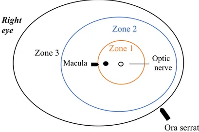
The retina can be divided into three different zones where ROP can occur. Each retinal zone surrounds the optic disc, (Figure 2).
Zone 1 is the innermost zone including the optic nerve and the macula. Zone 2 extends from zone 1 to the ora serrata on the nasal side.
Zone 3 is the remaining crescent-zone surrounding zone 2 on the temporal side.
ROP found in zone 2 and 3 are considered to be less severe than ROP found in zone 1 [10].
VEGF IGF-1
VEGF IGF-1 Retinopathyof Prematurity Normal fetal vasculature
Preterm Birth
Metabolism increases
Phase 1 (Hyperoxia)
Blood vessels stop growing
Phase 2 (Hypoxia)
Uncontrolled blood vessel growth Normal blood vessel
growth in the retina
Figure 2: ROP is classified into three retinal zones where the disease may occur. Retinal
vascularization develops from the optic nerve head in zone 1(innermost zone) and grows out towards the retinal periphery until reaching zone 3(outermost zone). Zone 2 is the intermediate zone surrounding zone 1 and extending to the ora serrata on the nasal side. The figure represents the right eye.
ROP itself can be divided into 5 different stages based on the severity of the disease. ROP stage 1: Demarcation line
A thin white line that separates the avascular retina from the vascularized retina. ROP stage 2: Ridge
A ridge arises in the periphery of the demarcation line, with both height and width, extending above the plane of the retina.
ROP stage 3: Extraretinal vascular proliferation
Proliferation and neovascularization of blood vessels develops on the border of the ridge and grows into the vitreous.
ROP stage 4: Partial retinal detachment ROP stage 5: Total retinal detachment [9].
1.2.4 ROP Treatment
ROP stage 3 is considered to be severe ROP. Although spontaneous regression may occur at this stage, signs of progression to retinal detachment, requires treatment [9]. Laser treatment destroys areas of the hypoxic retina permanently, to save central vision. In recent years, intravitreal anti-VEGF injections have been used for treating ROP. Anti-VEGF treatment aims to reduce the levels of VEGF, while limiting the amount of tissue damage. The dose of anti-VEGF must be determined carefully since the anti-anti-VEGF molecule can leak into the blood
Zone 3
Zone 2 Zone 1
Macula Optic nerve
Ora serrata Right
4 circulation, hindering systemic vascular growth in many organs. This could potentially prevent the normal growth of the brain and other organs in premature infants [11]. If retinal detachment occurs (ROP stage 4-5), surgical procedures such as vitrectomy is required [12].
1.2.5 Risk Factors for ROP
Genetic factors may contribute to the development of ROP. The disease is more common in males and in multiple birth pregnancies [13]. Low gestational age (GA), low birth weight (BW) and high oxygen supply are known risk factors for ROP [14]. Neonatal morbidities such as pulmonary diseases, intraventricular hemorrhage and sepsis have also been associated with ROP [15]. Furthermore, low levels of serum IGF-1 have been linked to poor postnatal growth and ROP development [4].
1.2.6 ROP Screening
1.2.6.1 Screening of Premature Infants
In Sweden, infants born before gestational week 31 are included in the ROP screening program. Weekly ROP-screening examinations are performed until approximately week 40 PMA, which is usually when the retina is completely vascularized. Depending on when the infant is born, screening begins week 5 postnatal age or PMA 31 weeks [16].
1.2.6.2 SWEDROP Register
The results of the ROP screening program, are registered in a national quality-register known as SWEDROP [17]. SWEDROP is part of the Swedish perinatal quality register, SNQ [18]. SWEDROP contains data on premature infants who have completed ROP screening in Sweden. The SWEDROP register started in 2006 and it has a national coverage of 96% [17]. The SWEDROP register contains information regarding BW, GA, birth clinic, ROP stage and treatment and number of eye examinations amongst others.
1.2.6.3 Previous Studies
The SWEDROP register has been an important tool in previous studies on ROP. It has facilitated the identification of risk factors, which has led to modifications of national guidelines regarding screening [19]. According to a recent study by Holmström et al. [16], there has been an increase in the frequency of ROP over the past 10 years in Sweden. Holmström et al. points out several reasons for this, one of them being increased survival rates due to improved neonatal care. Additionally, in 2014, the target level of oxygen supply for preterm infants was raised from 85-89% to 91-95% in order to increase the survival rate of premature infants. Holmström et al. also reported regional differences in incidence and treatment of ROP across the country.
However, the ROP trend in Örebro region (Örebro) during the past 10-years has not been studied and it is not known how Örebro compares to the rest of Sweden.
2. Aim
2.1 Research Question
The purpose of this study was to evaluate the frequency of infants screened for ROP and the severity of ROP in Örebro over the past 10 years. More specifically this study aimed to answer the following questions:
1.) Has the number, BW and GA of infants screened for ROP in Örebro changed over the past 10 years (2008 to 2017)?
2.) Has the frequency of ROP in Örebro changed over the past 10 years (2008 to 2017)? 3.) Are there any differences between the frequency of ROP in Örebro compared to Sweden
as a whole?
3. Methods and Material
3.1 Study Population
The present study was a retrospective cohort study including premature infants born between 1st of January 2008 to 31st of December 2017 in the region of Örebro. Örebro region includes Örebro, Karlskoga and Lindesberg. Data regarding ROP screened infants, was retrieved from SWEDROP. The information gathered from SWEDROP included, GA, BW, number of eye examinations, ROP stage and treatment. Information regarding BW, that was not registered in SWEDROP, was retrieved from patient medical records.
3.2 Method
All the data was collected and sorted in Microsoft Excel. Data from the study conducted by Holmström et al. [19] was used to compare ROP frequency in Örebro and the rest of Sweden between 2008 to 2017.
3.3 Statistical Analyses
The study population was presented descriptively, with continuous variables described as median and range and categorical variables described as number and percentage. The Mantel-Haenszel Chi-square trend test was used to examine the linear association between birth year during the study period and ROP stage. Due to the small number of infants included in the study, two-year intervals were analyzed (2008-2009, 2010-2011, 2012-2013, 2014-2015 and 2016-2017). The Fisher exact test was performed to determine statistical significance when
6 comparing frequency of ROP in Örebro and the rest of Sweden. All statistical analyses were performed using SPSS software.
3.4 Ethical Aspects
The infants included in this study were registered in SWEDROP. Consent to use personal information for research purposes, was given by the parents upon registration. Approval for medical record access was made by the head of the department; Petra Hedlund, with identification number 20RS1046, in order to complement SWEDROP data.
In order to access medical records, the patient security numbers were obtained from SWEDROP and transformed into a temporary password protected excel-file. After data was collected, the infants’ security numbers were deleted and replaced by an identification number and the final study did not contain any personal data. All excel files were deleted at the end of the study. Furthermore, to ensure conformity with data protection laws, this study was registered in the personal data processing register. There is a potential risk of patient data being spread, however, considering the measures taken above, we determine this risk to be relatively small.
This study will provide a better understanding of the characteristics of the infants screened for ROP and the degree of disease in Örebro. The advantages of this study outweigh any potential risks that may occur.
4. Results
4.1 Background Data
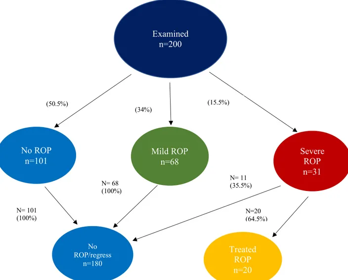
Between 2008 to 2017, a total of 200 infants with a GA of less than 31 weeks were screened for ROP in Örebro. In total, 99 infants (49.5%), developed some stage of ROP. Of these, 20 (10%) were treated for ROP. Regression in ROP disease, occurred in 11 (35.5%) of the infants with severe ROP, (Figure 3).
During the study period the number of ROP screened infants remained similar ranging from 8-25 infants per year. Out of the 200 infants, 88 (44%) were female and 46 (23%) of the infants were multiple-birth pregnancies (data not shown).
Figure 3: Flow chart showing, examined infants and course of retinopathy of prematurity (ROP), during
the study period (2008 to 2017).
Examined n=200 Mild ROP n=68 Severe ROP n=31 Treated ROP n=20 No ROP n=101 No ROP/regress n=180 N= 101 (100%) N= 68 (100%) N= 11 (35.5%) N=20 (64.5%) (50.5%) (34%) (15.5%)
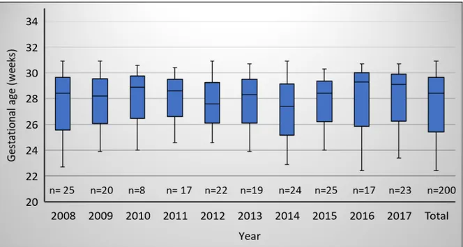
8 The median GA of the entire cohort was 28.4 weeks (range 22.4-30.9 weeks), (Figure 4). The median GA of the cohort remained similar during the study period.
The median BW of the study group was 1144 grams (range 440-1990 grams). The lowest BW was 440 grams recorded in 2014 (Figure 5). The median BW of the cohort remained similar during the study period.
Figure 5: The median birth weight (grams) of premature infants born with gestational age ≤ 31
weeks between 2008 to 2017 (n= total number of infants) in Örebro.
Figure 4: The median gestational age (weeks) of premature infants born between 2008 to 2017 in
4.2 ROP Frequency
In Table 1, the frequency of infants with ROP each year is presented in relation to ROP severity and treatment during the study period. When estimating the trend of infants without any ROP “No ROP”, the frequency remained similar throughout the study period (p=0.560). Mild ROP decreased (p=0.024) and severe ROP increased (p=0.032) between 2008 to 2017. There was no significant change in the frequency of ROP treatment in Örebro throughout the study period (p=0.159).
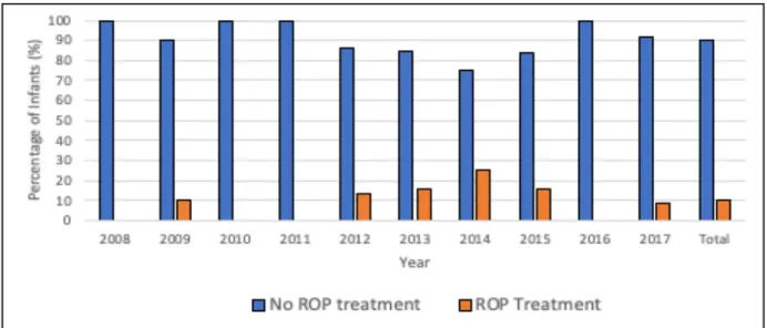
The frequency of ROP-treatment between the years 2008 to 2017 is presented in Figure 6. The number of infants treated for ROP was the highest during 2014 with a total of 6 (25%) infants treated.
4.3 Comparison to Sweden
In Table 2, the frequency and severity of ROP in Örebro and the rest of Sweden between 2008 to 2017 are presented. During the study period, there were significantly more infants with “No
Table 1: Frequency and severity of retinopathy of prematurity (ROP), every year from 2008 to 2017 Year 2008 n=25 2009 n=20 2010 n=8 2011 n=17 2012 n=22 2013 n=19 2014 n=24 2015 n=25 2016 n=17 2017 n=23 Total n=200 ROP level, n (%) No ROP 11 (44.0) 10 (50.0) 5(62.5) 9 (52.9) 12 (54.5) 8(42.1) 10(41.7) 13(52.0) 11(64.7) 12(52.2) 101(50.5) Mild ROP 13 (52.0) 7(35.0) 2(25.0) 8(47.1) 7(31.8) 7(36.8) 7 (29.2) 8(32.0) 4(23.5) 5(21.7) 68 (34.0) Severe ROP 1(4.0) 3(15.0) 1(12.5) 3(13.6) 4(21.1) 7 (29.2) 4 (16.0) 2(11.8) 6(26.1) 31(15.5)
Figure 6: Frequency of infants ( gestational age ≤ 31 weeks) with and without ROP-treatment, from
10 ROP” in Sweden compared to Örebro (p<0.001). The percentage of mild ROP was significantly higher in Örebro than the rest of Sweden, (p<0.001). Örebro had a higher percentage of treated ROP (p=0.024) compared to the rest of Sweden. The frequency of severe ROP in Örebro was not significantly higher compared to Sweden (p=0.053), (see Table 2).
GA= Gestational age, BW= Birth weight P-values are in parentheses
5. Discussion
The aim of this study was to evaluate the frequency of ROP-screened infants and the severity of ROP in Örebro region during a 10-year period. Our study shows that the number of premature infants screened for ROP remained similar over time. Likewise, the BW and GA of premature infants remained unchanged. Thus, we could conclude that neither the birth characteristics nor the number of infants screened for ROP in Örebro changed over the 10-year study period. When comparing the premature infants in Örebro and the rest of Sweden, the median GA and BW appeared similar. However, Holmström et al. [19] reported that the median GA and BW of treated infants in Sweden, decreased during the same study period. It would be interesting to investigate whether the median GA and BW of treated infants has changed in Örebro. This would give us a better understanding of whether ROP infants in Örebro have the same attributes as the rest of Sweden.
In our cohort, the frequency of mild ROP decreased, but severe ROP increased during the study period. A possible explanation to this, is improved neonatal care, resulting in more fragile premature infants surviving. As these fragile “survivors” are more likely to develop a severe
Table 2. Frequency and severity of retinopathy of prematurity (ROP) in Örebro and the rest of Sweden from 2008 to 2017
Örebro n=200 Sweden n=7049 Median GA (weeks), (min,max) 28.4 (22.4: 30.9) 28.3 (21.6:30.6) Median BW (grams), (min, max) 1100
(440:1990) 1109.5 (307:3245) ROP stage, n (%) No ROP, n (%) 101 (50.5) (p<0.001) 4939 (68.1) Mild ROP, n (%) 68 (34) (p<0.001) 1500 (20.7) Severe ROP, n (%) 31 (15.5) (p=0.053) 810(11.2) Treated ROP, n (%) 20 (10) (p=0.024) 440 (6.1)
form of ROP, we see a decrease in the frequency of mild ROP. This is in line with a study from Norman et al.[3] showing that the survival of premature infants (GA 22-26 weeks), increased in 2014-2016 compared to 2004-2007. If this pattern continues, there is a risk that severe ROP will proceed to increase.
Our results show that the frequency of treated ROP did not significantly increase during the 10-year period. However, our results have to be interpreted with caution as the number of infants in our study cohort is small. Further studies have to be carried out over a longer period of time, with a larger cohort, to conclude whether ROP treatment is not increasing in Örebro, before investigating what factors contribute to this finding.
In 2014, there was a peak of premature infants treated for ROP in Örebro. When we investigated the possible reasons for this, we found that in 2014, 4 out of the 6 treated infants, did not have Örebro as their registered birth clinic. A rise of transferred infants to Örebro could explain the high incidence of treated infants during this year. In Sweden, ROP treatment is only performed in University hospitals. In the region of Örebro, ROP screening and treatment is performed in Örebro University hospital. Örebro University hospital is recognized to be in the frontline when it comes to retinal surgery in Sweden. Due to this, many smaller hospitals and other University hospitals, transfer their ROP infants to Örebro for treatment. These premature infants are born in another region, but are registered in Örebro in SWEDROP once they are treated there. Additionally, in 2014, out of the 6 treated infants, there were 4 males and 2 infants of multiple birth pregnancies. Male gender and multiple gestations are possible risk factors associated with ROP. This could also explain the peak of ROP treated infants in 2014. Furthermore, in 2014 the target oxygen range for premature infants gradually changed from 85-89% to 91-95% to reduce mortality. According to Holmström et al. [16], there was an increase in treatment-requiring ROP after changes in oxygen target range in Sweden. Change in oxygen range could also have impacted the frequency of ROP-treatment in Örebro during this time. The factors mentioned above are all possible explanations for the peak of ROP treated infants in 2014. However, as there were only 6 infants, it is difficult to conclude what the main cause may be.
In comparison to Sweden, our study shows that there are more premature infants with “No ROP” in the rest of Sweden than in Örebro. Conversely, there are significantly more infants
12 receives many transferred ROP infants from other regions that may not have the same screening and treatment options. This could be the reason why Örebro has had a higher frequency of ROP treated infants compared to Sweden. Similarly, there is a higher frequency of “ No ROP” in the rest of Sweden as the larger and healthier infants born in smaller hospitals remain in their birth region. Although not statistically significant, our results also showed a tendency of more infants with severe ROP in Örebro than the rest of Sweden. It would have been interesting to investigate how many infants have been transferred to Örebro over the past 10 years to determine whether the differences between Örebro and the rest of Sweden could be explained by this.
When comparing ROP trends in Sweden and Örebro, adherence and compliance to screening programs must be considered. Holmström et al. [19] suggests that non-adherence to screening program could explain the regional differences of ROP frequency in Sweden. Moreover, Norman et al. [20] reported that 65% of visual disability caused by ROP, was avoidable if compliance with best practice was followed. Failure to follow best practice was mostly related to untimely or suboptimal detection or treatment of ROP, as well as variations in infrastructure. Further studies are ongoing, investigating compliance to best practice in Örebro and the rest of Sweden.
A strength with this study is that the SWEDROP register itself has a high coverage of about 96%. All infants with a GA less than 31 weeks born between 2008 to 2017 and registered in SWEDROP, were included in the study.
A limitation of this study is that the study cohort is small making it difficult to draw definite conclusions despite finding statistical significance. Future studies would have to take this into consideration and base the study on a larger sample size, to confirm statistical significance and improve the accuracy of the study. Another limitation is the transfer of infants to Örebro for screening and ROP treatment. Therefore, our results might not accurately describe ROP frequency in Örebro.
6. Conclusion
The present study showed that the frequency of mild ROP decreased in Örebro whilst severe ROP increased throughout the study period. No significant increase in the frequency of treatment requiring ROP occurred between 2008 to 2017. The percentage of infants treated for
ROP was significantly higher in Örebro compared to the rest of Sweden. The transfer of infants requiring ROP treatment from other regions could explain this result.
7. Acknowledgments
A special thanks to my supervisor, Pia Lundgren for her meticulous and excellent supervising throughout the writing process, and for being available at all hours of the day. Furthermore, a special thanks to Aldina Pivodic for her excellent statistical support.
14
References
1. WHO. Preterm birth [Internet]. [cited 2020 Jan 14]; Available from: https://www.who.int/news-room/fact-sheets/detail/preterm-birth
2. Socialstyrelsen. Fakta och statistik [internet] [Internet]. 2017 [cited 2020 May 4]; Available from: https://www.socialstyrelsen.se/meranbaramamma/statistik/
3. Norman M, Hallberg B, Abrahamsson T, Björklund LJ, Domellöf M, Farooqi A, et al. Association Between Year of Birth and 1-Year Survival Among Extremely Preterm Infants in Sweden During 2004-2007 and 2014-2016. JAMA 2019; 321:1188–99. 4. Smith L, Hellström A, Liegl R. Retinopathy of prematurity: the need for prevention. Eye
and Brain 2016; :91.
5. Saint-Geniez M, D’amore PA. Development and pathology of the hyaloid, choroidal and retinal vasculature. Int J Dev Biol 2004; 48:1045–58.
6. Hellström A, Smith LE, Dammann O. Retinopathy of prematurity. The Lancet 2013; 382:1445–57.
7. Sun Y, Smith LEH. Retinal Vasculature in Development and Diseases. Annu Rev Vis Sci 2018; 4:101–22.
8. Eckert GU, Fortes Filho JB, Maia M, Procianoy RS. A predictive score for retinopathy of prematurity in very low birth weight preterm infants. Eye 2012; 26:400–6.
9. International Committee for the Classification of Retinopathy of Prematurity*. The International Classification of Retinopathy of Prematurity Revisited. Archives of Ophthalmology 2005; 123:991–9.
10. Molinari A, Weaver D, Jalali S. Classifying retinopathy of prematurity. Community Eye Health 2017; 30:55–6.
11. Mutlu FM, Sarici SU. Treatment of retinopathy of prematurity: a review of conventional and promising new therapeutic options. Int J Ophthalmol 2013; 6:228–36.
12. Roohipoor R, Karkhaneh R, Riazi-Esfahani M, Ghasemi F, Nili-Ahmadabadi M.
Surgical management in advanced stages of retinopathy of prematurity; our experience. J Ophthalmic Vis Res 2009; 4:185–90.
13. Kim SJ, Port AD, Swan R, Campbell JP, Chan RVP, Chiang MF. Retinopathy of prematurity: a review of risk factors and their clinical significance. Survey of Ophthalmology 2018; 63:618–37.
14. Mantagos IS, VanderVeen DK, Smith LEH. Risk Factors for Retinopathy of Prematurity: Beyond Age, Birth Weight, and Oxygen. Curr Ophthalmol Rep 2013; 1:213–7.
15. Freitas AM, Mörschbächer R, Thorell MR, Rhoden EL. Incidence and risk factors for retinopathy of prematurity: a retrospective cohort study. Int J Retin Vitr 2018; 4:20.
16. Holmström G, Tornqvist K, Al-Hawasi A, Nilsson Å, Wallin A, Hellström A. Increased frequency of retinopathy of prematurity over the last decade and significant regional differences. Acta Ophthalmologica 2018; 96:142–8.
17. Holmström GE, Hellström A, Jakobsson PG, Lundgren P, Tornqvist K, Wallin A. Swedish National Register for Retinopathy of Prematurity (SWEDROP) and the Evaluation of Screening in Sweden. Archives of Ophthalmology 2012; 130:1418–24. 18. Svenskt Neonatalt Kvalitetsregister (SNQ) - Nationella Kvalitetsregister [Internet].
[cited 2020 Jan 14]; Available from:
http://www.kvalitetsregister.se/hittaregister/registerarkiv/svensktneonataltkvalitetsregiste rsnq.2338.html
19. Holmström G, Hellström A, Gränse L, Saric M, Sunnqvist B, Wallin A, et al. New modifications of Swedish ROP guidelines based on 10-year data from the SWEDROP register. Br J Ophthalmol 2019; :bjophthalmol-2019-314874.
20. Norman M, Hellström A, Hallberg B, Wallin A, Gustafson P, Tornqvist K, et al. Prevalence of Severe Visual Disability Among Preterm Children With Retinopathy of Prematurity and Association With Adherence to Best Practice Guidelines. JAMA Netw Open 2019; 2:e186801.