Atlas of Science
another view on science http://atlasofscience.org
Tracing aluminium adjuvants in viable cells
To improve the effect of a vaccine, adjuvants are often included in the vaccine formulation. An adjuvant is a molecule that potentiates the immune response induced by the vaccine, and
commonly used adjuvants in vaccine formulations are aluminium salts as aluminiumoxy-hydroxide and aluminiumhydroxyphosphate. In spite of the extensive use of these mineral adjuvants since the 1920s, it is still not known exactly how they work.
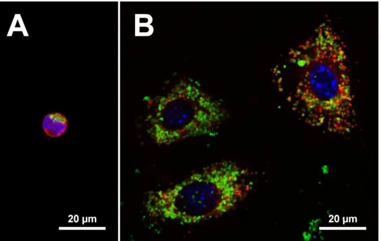
Fig. 1. Visualization of intracellular aluminium adjuvant. Confocal images of the centre sections of cells showing the nucleus as blue (DAPI) and aluminium adjuvant as green (lumogallion). A. The suspension cell line THP-1 co-cultured with aluminium adjuvant showing the cytoplasma as red after staining with CellTrace™ Far Red. B. The adherent cell line GL261 co-cultured with aluminium adjuvant showing lysosomes as red after staining with LysoTracker® Deep Red and as orange dots upon co-localization with aluminium adjuvant.
In a vaccine formulation the aluminium adjuvants are dispersed as small particles in water. These particles vary in size (from nanometres to micrometre) and the antigen, the molecule the immune
Atlas of Science
another view on science http://atlasofscience.org
system is supposed to respond to, is attached to their surface
The immune system comprises cells that help the body to battle pathogens, and can briefly be divided into sentinel cells and effector cells. Monocytes, neutrophils and dendritic cells are all sentinel cells, and they circulate the body and sample screen their surroundings for foreign materials. If they find a danger associated molecular pattern (DAMP) or another unknown
compounds, the sentinel cells become activated and trigger an immune response. This results in the maturation of effector cells, such as B and T cells. The sentinel cells are extremely good at removing cell debris and foreign micrometer sized material by a process called phagocytosis, in which the cells engulf the material. Hence, upon contact with aluminium adjuvants, these cells will engulf the adjuvant particles.
Fig. 2. Digital visualization of intracellular aluminium adjuvant. Digitally rendered image of a cell showing the nucleus as blue (DAPI), the actual cell as partially transparent red (CellTrace™ Far Red), and aluminium adjuvant as green (lumogallion). The image was generated from 25 individual cross section images taken with a microscope at different depths of the cell, out of which the centre image is depicted in Figure 1A. The digital image shows that the aluminium adjuvant is localized as particles inside the cell. For a better visualization, the cell can be seen rotating in a video (see below). The digitalization was made in Bitplane Imaris 8.1.2.
The shortage of knowledge regarding the immune stimulating properties of aluminium adjuvants has been hampered by the lack of easy and reliable methods of tracing engulfed aluminium
adjuvants. However, lumogallion a stain that earlier has been used to assay and detect aluminium ions, can also be used to stain aluminium adjuvant particles. Upon contact with lumogallion the adjuvant particles become fluorescent and emits light of the wavelengths 525 to 650 nm. The advanced microscope technologies and image analyses of today makes it possible to follow
fluorescent labelled structures inside a cell at a high resolution allowing easy intracellular detection
Atlas of Science
another view on science http://atlasofscience.org
of aluminium adjuvants. Figure 1 shows confocal microscopy images of the centre of cells showing the intracellular presence of aluminium adjuvants as green dots due to lumogallion staining.
Lumogallion staining enables intracellular tracking of aluminium adjuvants in viable cells. Engulfed material generally ends in an acid organelle called lysosome and figure 1B shows an example of intracellular tracing of aluminium adjuvant in which some of the engulfed aluminium adjuvant co-localize in a cellular compartment having low pH, demonstrating the presence of aluminium adjuvant in lysosomes (orange dots). Furthermore, images from confocal microscopy can be processed into three dimensional images enabling improved visualisation of the intracellular localization of engulfed aluminium adjuvant (Fig. 2).
http://atlasofscience.org/wp-content/uploads/2016/02/Video-Svensson-and-Eriksson.mp4
The staining of the cells shown in figures 1 and 2 was performed on living cells and no fixation and sectioning of the cells were needed. Hence, detection and tracing of aluminium adjuvants using lumogallion can be used to study living cells in real time as they take up the particles, revealing the dynamics of engulfment and intracellular pathways of aluminium adjuvants. It can also be used to detect and track cells that engulf aluminium adjuvants after injection of vaccine formulations in animals. Altogether, lumogallion will be a helpful tool unravelling the mystery of aluminium adjuvants and helping us in our efforts designing efficient and save vaccine formulations and protocols.
Andreas Svensson1 and Håkan Eriksson2
1
Lund Stem Cell Center, Lund University, Sweden
2
Biomedical Science, Malmö University, Sweden
Publication
Al adjuvants can be tracked in viable cells by lumogallion staining. Mile I, Svensson A, Darabi A, Mold M, Siesjö P, Eriksson H
J Immunol Methods. 2015 Jul
Powered by TCPDF (www.tcpdf.org)