ACTA UNIVERSITATIS
Digital Comprehensive Summaries of Uppsala Dissertations
from the Faculty of Medicine
890
Carotid Artery Wall Layer
Dimensions during and after
Pre-eclampsia
An investigation using non-invasive
high-frequency ultrasound
Dissertation presented at Uppsala University to be publicly examined in Sal IX,
Universitetshuset, S:t Olofsgatan 10B, Box 256, 751 05 Uppsala, Thursday, May 23, 2013 at 13:00 for the degree of Doctor of Philosophy (Faculty of Medicine). The examination will be conducted in Swedish.
Abstract
Akhter, T. 2013. Carotid Artery Wall Layer Dimensions during and after Pre-eclampsia: An
investigation using non-invasive high-frequency ultrasound. Acta Universitatis Upsaliensis. Digital Comprehensive Summaries of Uppsala Dissertations from the Faculty of Medicine
890. 66 pp. Uppsala. ISBN 978-91-554-8641-9.
Pre-eclampsia is associated with increased risk of cardiovascular disease (CVD) later in life. The ‘gold standard’ for estimating cardiovascular risk - ultrasound assessment of the common carotid artery intima-media thickness (CCA-IMT) - does not convincingly demonstrate this increased risk. The aim of this thesis was to examine whether high-frequency (22 MHz) ultrasound assessment of the individual CCA intima and media layers and calculation of the intima/ media (I/M) ratio - can indicate the increased cardiovascular risk after pre-eclampsia. After validation of the method in premenopausal women with systemic lupus erythematosus (SLE) who have a recognized increased risk of CVD, women during and after normal and preeclamptic pregnancies were investigated.
Assessment of the individual artery wall layers reliably demonstrated the increased cardiovascular risk in premenopausal women with SLE, while CCA-IMT did not. The artery wall layer dimensions in women with SLE were comparable to those of postmenopausal women without SLE and were 30 years older.
Among the women with normal pregnancies negative changes to the artery wall later on in the pregnancy were seen in those with lower serum estradiol, older age, higher body mass index or higher blood pressure early in the pregnancy. About one year postpartum, both the mean intima thickness and the I/M ratio had improved, compared to values during pregnancy. These findings support the theory that normal pregnancy is a stress on the vascular system.
Women who developed pre-eclampsia (mean age 31 years) had thicker intima layers, thinner media layers and higher I/M ratios, both at diagnosis and one year postpartum, than women with normal pregnancies, indicating increased cardiovascular risk.
Women with a history of severe pre-eclampsia (mean age 44 years; mean 11 years since the last delivery) had thicker intima layers and higher I/M ratios than women with a history of normal pregnancies, indicating long-standing negative vascular effects.
Assessment of individual CCA wall layers, but not of CCA-IMT, provided clear evidence of the well-known increased cardiovascular risk in women with SLE or pre-eclampsia. The method has the potential to become an important tool in reducing cardiovascular morbidity and mortality in these women through early diagnosis and intervention.
Keywords: Systemic lupus erythematosus, normal pregnancy, pre-eclampsia, high-frequency
ultrasound, common carotid artery, intima/media ratio, cardiovascular disease.
Tansim Akhter, Uppsala University, Department of Women's and Children's Health, Akademiska sjukhuset, SE-751 85 Uppsala, Sweden.
© Tansim Akhter 2013 ISSN 1651-6206 ISBN 978-91-554-8641-9
To my parents and
my daughter Tamanna
List of Papers
This thesis is based on the following papers, which are referred to in the text by their Roman numerals.
I Leonard D∗, Akhter T∗, Nordmark G, Rönnblom L, Naessen T. Increased carotid intima thickness and decreased media thickness in premenopausal women with systemic lupus erythematosus: an investigation by non-invasive high-frequency ultrasound. Scand J Rheumatol. 2011;40 (4):279-82.
II Akhter T, Larsson A, Larsson M, Wikström A-K, Naessen T. Ar-tery wall layer dimensions during normal pregnancy: a longitudinal study using noninvasive high-frequency ultrasound. Am J Physiol Heart Circ Physiol. 2013 Jan;304(2):H229-34.
III Akhter T, Wikström A-K, Larsson M, Naessen T. Individual Common Carotid Artery Wall Layer Dimensions, but not Carotid-IMT, Indicate Increased Cardiovascular Risk in Women with Preeclampsia:an investigation by non-invasive high-frequency ul-trasound. Submitted
IV Akhter T, Larsson M, Wikström A-K, Naessen T. Individual Ar-tery Wall Layer Dimensions Indicate Increased Cardiovascular Risk in Previous Severe Preeclampsia: an investigation using non-invasive high-frequency ultrasound. Submitted
∗ Both authors contributed equally to this work.
Contents
Introduction ... 11
Cardiovascular disease ... 11
Definition and classification of cardiovascular disease ... 11
Risk factors for cardiovascular disease ... 11
Cardiovascular disease in women ... 12
Estrogen and its vascular effects in women ... 12
The common carotid artery ... 13
Atherosclerosis ... 13
Cardiovascular disease, atherosclerosis and intima-media thickness ... 14
Separate estimates of the intima and media layers and calculation of intima/media ratio ... 15
Systemic lupus erythematosus ... 15
Normal pregnancy ... 16
Definition ... 16
Physiological changes during normal pregnancy ... 17
Endocrine changes during pregnancy ... 17
Normal pregnancy effects on common carotid artery intima-media thickness and subsequent risk of cardiovascular disease ... 17
Pre-eclampsia ... 18
Definition and classification of pre-eclampsia ... 18
Risk factors for pre-eclampsia ... 18
Pathophysiology of pre-eclampsia ... 19
Pre-eclampsia and risk of cardiovascular disease ... 20
Severity of pre-eclampsia and risk of cardiovascular disease ... 21
Pre-eclampsia and its effects on common carotid artery intima-media thickness ... 23
Aims of the study ... 24
Specific aims of the individual studies ... 24
Study populations ... 25
Study I ... 25
Study II ... 26
Study III ... 26
Methods ... 29
Assessments and data collection ... 29
Study I ... 29
Study II ... 30
Study III ... 30
Study IV ... 30
Ultrasound assessment of the common carotid artery ... 31
Ethical considerations ... 32
Statistical methods ... 32
Results ... 34
Study I ... 34
Study II ... 35
Study III ... 37
Study IV ... 40
Discussion ... 43
Cardiovascular disease in women, especially after pregnancy complications ... 43
Cardiovascular disease and carotid artery wall imaging ... 43
Carotid artery wall imaging in premenopausal women with systemic lupus erythematosus ... 45
Carotid artery wall imaging and normal pregnancy ... 45
Carotid artery wall imaging and pre-eclampsia ... 46
Methodological consideration ... 47
Conclusions ... 49
Future plans ... 50
Summary in Swedish- sammanfattning på svenska ... 51
Studie I ... 52
Studie II ... 52
Studie III ... 53
Studie IV ... 53
Acknowledgements ... 54
References ... 57
Abbreviations
ACR American College of Rheumatology
AHA American Heart Association
AUC Area under the ROC curve
BMI Body mass index
CAD Coronary artery disease
CCA Common carotid artery
CCA-IMT Common carotid artery-intima media thickness
CI Confidence interval
CHD Coronary heart disease
CV Coefficient of variation
CVD Cardiovascular disease
DBP Diastolic blood pressure
DHEA Dehydroepiandrosterone
E2 Estradiol
EOPE Early onset pre-eclampsia
ICD International classification of diseases
I/M ratio Ratio of intima thickness to media thickness
IMT Intima media thickness
LDL Low density lipoprotein
LGA Large for gestational age
LOPE Late onset pre-eclampsia
MAP Mean arterial pressure
MHz Mega Hertz
MI Myocardial infarction
PE Pre-eclampsia
ROC Receiver operating characteristic
RR Relative risk
SBP Systolic blood pressure
SD Standard deviation
SGA Small for gestational age
SLE Systemic lupus erythematosus
Introduction
Cardiovascular disease
Cardiovascular disease (CVD) is the number one cause of death globally; more people die annually from CVD than from any other cause. The World Health Organization (WHO) estimated that 17.3 million people died from CVD in 2008, representing 30% of all global deaths.1 Low- and
middle-income countries are disproportionately affected by CVD. In the year 2030, it is estimated that more than 23 million people will die from CVD, mainly
from coronary heart disease (CHD) and stroke.1 CVD affects people in their
mid-life years, undermining the socioeconomic development not only of the affected individuals but also of their families and nations.
Definition and classification of cardiovascular disease
CVD comprises a group of disorders of the heart and blood vessels which includes:1
Atherosclerotic cardiovascular disease
• CHD
• Cerebrovascular disease • Peripheral arterial disease
Non-atherosclerotic cardiovascular disease
• Rheumatic heart disease • Congenital heart disease
• Deep vein thrombosis and pulmonary embolism
Risk factors for cardiovascular disease
Clinical studies have identified several factors that increase the risk of CVD. These risk factors can be divided into those that are modifiable and those that are non-modifiable:2
Non-modifiable risk factors
• Older age • Male sex • Heredity
Modifiable risk factors
• Tobacco smoke • Dyslipidemia • High blood pressure • Diabetes mellitus • Obesity
• Physical inactivity
• Extensive alcohol consumption • Stress
Cardiovascular disease in women
CVD is the largest single cause of mortality among women. More than 8.6 million women die of CVD each year around the world; these account for
one third of all deaths in women.3 In Europe 54% of deaths in women and
43% of deaths in men are from CVD.4 CVD is the main cause of female
deaths in Sweden. In the year 2011, 39% of all female deaths in Sweden
were caused by CVD.5 Previous reports have focused primarily on male or
shared male and female risk factors where women were under-represented.6
Further, differences in management7 and mortality rate8 between men and women after acute cardiac diseases have been observed. Moreover, it has been reported that women are more likely than men to have unrecognised
myocardial infarctions (MIs).9 The American Heart Association (AHA) has
published separate guidelines for CVD prevention in women.10 Nonetheless,
there remains a need for further investigations into female-specific risk fac-tors for CVD and the atherosclerotic process in women.
The onset of CVD occurs approximately ten years later in women than in men, which could be the result of the protective effects of female sex hor-mones before menopause.11 Because of this older age at presentation of CVD, women are more likely to also suffer from co-morbidities such as diabetes and hypertension.
Estrogen and its vascular effects in women
The atheroprotective effects of estrogen are thought to be mediated mainly by direct action on the artery wall and alterations to the serum lipid
concen-trations.12 The direct effects of estrogen on the artery wall have two
compo-nents, one is rapid and non-genomic and the other is long term and ge-nomic.13 Estrogen receptors have been identified in both vascular smooth
muscle cells14 and endothelial cells;15 these cells are able to bind estrogen
with high affinity.
In the Women’s Ischemia Syndrome Evaluation study, Merz et al. showed that hypothalamus-mediated ovarian dysfunction and estrogen defi-ciency in premenopausal women were associated with premature coronary
artery atherosclerosis.16 In a similar study, Hanke et al. reported that
premenopausal women with angiographically confirmed coronary artery disease (CAD) had lower serum estradiol (E2) concentrations than control subjects.17
The common carotid artery
The common carotid artery (CCA) is an elastic artery. The right CCA is a branch of the brachiocephalic artery which arises from the thoracic aorta, whereas the left CCA arises directly from the thoracic aorta.18 The pulsation is stronger in the upper part of the artery, which lies more superficially than the lower part.
The CCA wall is composed of three layers: the intima, the media and the adventitia, in order from the inside out. The intima is composed of the endo-thelium and a thin layer of underlying fibrocollagenous tissue. The media is mainly composed of elastic fibres, in contrast to the media of a muscular artery, which is composed almost entirely of smooth muscle. The adventitia is mainly composed of collagen.
Atherosclerosis
Atherosclerosis is the main underlying pathology in CVD.19-21
Atherosclero-sis develops from oxidized low-density lipoprotein (LDL) molecules. LDL begins to accumulate in the arterial walls when the levels in the blood exceed
the capacity of the macrophages to remove it.22 These lipid depositions lead
to an inflammatory reaction in the arterial wall.22 In response to the
inflam-mation, macrophages are attracted to the arterial wall where they absorb the oxidized LDL to form specialized foam cells. When the foam cells die, they release a large number of products, causing the formation of a lipid core.22, 23
The foam cells also release growth factors particularly platelet-derived growth factors, which results in proliferation of smooth muscle cells and their migration from the media to the intima, towards the lipid core. Smooth muscle cells synthesize collagen, leading to the formation of a fibrous cap over the affected area, in an attempt to repair and stabilize the lesion. If the repair process is successful, the atherosclerotic plaque will be stable and will remain asymptomatic. If inflammation dominates over the repair mecha-nisms, the plaque may become active or unstable and this could lead to rup-ture of the fibrous cap and thrombosis.22
Atherosclerosis typically begins in early adolescence. According to an au-topsy study, among children aged 10-14 years who died of non-cardiovascular causes, more than half showed some evidence of
age of 26 years, the prevalence of any coronary atherosclerosis was 8.5%.25
Atherosclerotic lesions, which change the vascular anatomy, develop over decades.
Cardiovascular disease, atherosclerosis and
intima-media thickness
The atherosclerotic process in the artery wall is an important factor in the pathogenesis of CVD. Angiographic and Doppler evaluations have been used for more than 30 years to determine the incidence and development of atherosclerosis, but the evaluations have mainly been restricted to severe atherosclerosis (stenosis).26-28 In the late 1980s, advances in ultrasound reso-lution techniques provided the opportunity to quantify and monitor athero-sclerosis from its precursor lesion to occlusive disease using a non-invasive technique.
To assess the early progression of atherosclerosis it is necessary to visual-ize the artery wall itself. The high coincidence of carotid atherosclerosis with vessel pathology in other vasculature makes it an adequate window for in-vestigating systemic atherosclerosis.29-31 The use of peripheral arteries such as the carotid arteries as a vascular endpoint for assessing the extent of ath-erosclerosis is widely accepted. The present ‘gold standard’ for non-invasive assessment of the development of atherosclerosis is to measure the thickness of the combined intima and media layers of the common carotid artery (CCA-IMT) using 7-10 MHz ultrasound. The AHA recommend using this
method; an increase in CCA-IMT is seen as a surrogate marker for CVD.32
Several studies have demonstrated a correlation between an increase in CCA-IMT and the occurrence of cardiovascular risk factors33-35 and in-creased CCA-IMT has been shown to be an independent predictor of future CVD.36-40 However, when Wald et al. performed a meta-analysis of 18
stud-ies, they found that CCA-IMT measurement was not a sufficiently reliable screening process to discriminate between individuals with and without CHD.41 Further, in another meta-analysis, Costanzo et al. found that a
de-crease in CCA-IMT did not reflect a reduction in cardiovascular events.42 Similarly, Lorenz et al. and Den Ruijter et al. found no association between carotid intima-media thickness (IMT) progression and cardiovascular events.43, 44
Separate estimates of the intima and media layers and
calculation of intima/media ratio
CCA-IMT measurements have been used to assess atherosclerosis for many years. Despite the large-scale use of this measurement, interpretation of CCA-IMT ultrasound results has been questioned. For example, a validation study in cadavers by Gamble et al. showed that CCA-IMT results with 10 MHz ultrasound corresponded better to the total artery wall thickness
(ad-ventitia, media and intima) than to the intima + media thickness.45 Further,
Adams et al. found a poor correlation between CCA-IMT results and the degree of atherosclerosis assessed by coronary angiography.46
Intimal thickening is probably one of the earliest signs in the atheroscle-rotic process. However, it is not possible to obtain separate estimates of the intima and media layers using 7-10 MHz ultrasound. Mallery et al. and McPherson et al. measured the individual thicknesses of the intima and the media and the combined intima-media with intravascular high-frequency ultrasound (20 and 12 MHz, respectively), and showed good correlations with histological section results.47, 48 Further, with increasing age and devel-opment of atherosclerosis, the intima continues to increase in thickness while
the media decreases, as demonstrated by both histomorphometry49 and
intra-vascular high-frequency ultrasound.50 Thus, assessment of the individual
intima and media thicknesses could be more appropriate than assessment of the combined CCA-IMT for imaging the effects of vascular aging. This method has been validated in an animal model.51
Non-invasive high-frequency ultrasound has been used to demonstrate that 70-year-old subjects with prevalent CVD had significantly thicker inti-ma layers, thinner media layers and higher I/M ratios than subjects without CVD, whereas CCA-IMT measurements did not show any significant
differ-ences.52 The same method clearly demonstrated the expected effects of aging
and long-term postmenopausal oestrogen therapy on the CCA wall layers.53
The method has also been used to demonstrate the effects of conditions such as recurrent depression54 and stem cell transplantation,55 which are suspected to be associated with an increased risk of CVD. Thus, the use of intravascu-lar high-frequency ultrasound to measure the individual intima and media layers and calculation of the I/M ratio seem preferable to measuring the CCA-IMT, the present ‘gold-standard’ for non-invasive assessment of the artery wall status.
Systemic lupus erythematosus
Systemic lupus erythematosus (SLE) is a systemic inflammatory disease that mainly affects women. Incidence rates of SLE vary from 1 to 10/100,000 person-years.56 The exact pathoetiology of SLE remains elusive. An
ex-tremely complicated and multifactorial interaction among various genetic and environmental factors is probably involved.57 American College of Rheumatology (ACR) classification criteria are usually used in most SLE studies to define the disease.58 The criteria include nine clinical and two
im-munological criteria and SLE is present if a patient fulfils four or more of these eleven criteria.
CVD is reported to be a major cause of both morbidity and premature mortality in SLE patients.59 There is strong evidence of an increased
inci-dence of CVD events such as CAD in women with SLE.60, 61 Younger
wom-en with SLE, betwewom-en the ages of 35 and 44 years, have an up to 50-fold
increased risk of MI compared to women of similar age without SLE.60 The
pathogenesis of premature CVD in patients with SLE is thought to be medi-ated by accelermedi-ated atherosclerosis, probably as a result of a complex inter-play between traditional and non-traditional risk factors.62, 63 The use of ca-rotid ultrasound in several studies has demonstrated signs of accelerated atherosclerosis in SLE patients, with a higher prevalence of carotid plaques and coronary artery calcification than in controls.64, 65
Assessment of the CCA-IMT using 8-10 MHz frequency ultrasound has often been used to study the development of atherosclerosis in women with SLE. However, the results of these studies have been variable and difficult to interpret. A well executed review of premature CVD in patients with SLE has shown, in fact, that CCA-IMT values in SLE patients have been higher than, similar to, or even lower than those in healthy controls.65 A lower CCA-IMT value would imply a reduced risk of cardiovascular events, ac-cording to the classical interpretation of CCA-IMT results32 and this is
in-consistent with the documented increased risk of CVD in this group. Thus, conventional CCA-IMT measurement has been controversial with respect to non-invasive assessment of cardiovascular risk in patients with SLE. It is postulated that estimates of the individual artery wall layers could be prefera-ble, providing an alternative method of imaging the increased CVD risk in premenopausal women with SLE.
Normal pregnancy
Definition
Pregnancy without complication, resulting in delivery after gestational week 37 of an infant with a weight appropriate for its gestational age.
Physiological changes during normal pregnancy
A number of substantial cardiovascular changes occur during pregnancy to adapt and adjust the body to the changed requirements caused by the preg-nancy. The most important changes are increases in stroke volume and heart rate that cause a rise in cardiac output, which is increased by 1 L/min by as early as 8 weeks into gestation.66, 67 Further, the plasma volume increases and the peripheral vascular resistance decreases. The decrease in peripheral vascular resistance and the subsequent generalized vasodilatation result in a decrease in both the systolic and diastolic blood pressures (SBP and DBP), which reach their lowest levels in the middle of the second trimester and rise slightly again afterwards.66
However, normal pregnancy is also associated with a negative cardiovas-cular profile, including an increase in arterial stiffness,68 mild systemic
in-flammation,69, 70 altered lipid and lipoprotein profiles,71 and impaired glucose
tolerance.72
Endocrine changes during pregnancy
The placenta is a large endocrine organ, which produces huge, coordinated amounts of different hormones to establish and successfully maintain the pregnancy. Steroid hormones such as estrogen, progesterone, testosterone, and dehydroepiandrosterone (DHEA) are included in these. Mean circulating levels of E2 rise dramatically during pregnancy from pre-pregnancy levels of about 100 pg/ml (367 pmol/L) to about 15,000 pg/ml (55,050 pmol/L) at
term.73 Mean plasma progesterone levels also rise dramatically during
preg-nancy, to reach about 130,000 pg/ml at term (compared to about 10,000 pg/ml in the luteal phase).73 Mean testosterone and DHEA levels increase during pregnancy.73 The main effect of estrogen is on the cardiovascular system, while progesterone prevents maternal rejection of the fetus through its anti-inflammatory and immunosuppressive effects.73
Normal pregnancy effects on common carotid artery
intima-media thickness and subsequent risk of cardiovascular disease
Reports of changes in the CCA-IMT during pregnancy are rare. One small study by Mersich et al. showed fairly stable mean CCA-IMT values,
as-sessed once in each trimester.74 Normal pregnancy and effects on IMT later
in life have been examined with varying results. Wolff et al. showed that CCA-IMT was significantly thicker in multiparous (≥ 4 births) than in pri-miparous women,75 which was also suggested by Skilton et al..76 In contrast, Kharazmi et al. found no significant differences in CCA-IMT between
mul-tiparous and primiparous women.77 In a case-control study, Blaauw et al.
showed that at about 6 months postpartum the CCA-IMT was thicker in primiparous women with previous normal pregnancy than in nulliparous
women.78 Thus, results about the effects of normal pregnancy on CCA-IMT
have been controversial. The relationship between normal pregnancy and risk of later CVD has been poorly investigated. In a study of parity and risk of maternal CVD later in life, Parikh et al. showed that women who had had ≥ 5 births had an increased risk of CVD [hazard ratio 1.47; 95% confidence
interval (CI) 1.37-1.57] compared to women who had had 2 childbirths.79 To
our knowledge, there have been no previous reports on the longitudinal as-sessment of the individual artery wall layer dimensions during normal preg-nancy, using non-invasive high-frequency ultrasound.
Pre-eclampsia
Pre-eclampsia (PE) is a pregnancy-specific syndrome which affects 3-5% of all pregnancies.80 Worldwide, PE is a leading cause of maternal and perinatal morbidity and mortality.81
Definition and classification of pre-eclampsia
According to the International Society for the Study of Hypertension in
Pregnancy (ISSHP)82 PE is defined as new-onset hypertension and
pro-teinuria after gestational week 20, with normalization of blood pressure within 3 months postpartum. Hypertension is defined as an SBP ≥ 140 mmHg and/or DBP ≥ 90 mmHg. Proteinuria is defined as leakage of protein ≥ 300 mg/24 hours or a spot urine protein/creatinine ratio ≥ 30 mg/mmol.
Risk factors for pre-eclampsia
Examples of risk factors for PE include:83
Age (older age) Parity (primiparity) Previous history of PE Family history of PE
Pregnancy with more than one fetus
Time between pregnancies (more than 10 years) Diabetes mellitus
Chronic hypertension Renal disease
Autoimmune disease Antiphospholipid syndrome
Pathophysiology of pre-eclampsia
The pathophysiology of PE is still not completely understood but it is thought to be a multisystem condition. It has long been hypothesized that PE
is a two-stage syndrome.84 The first stage is mediated by reduced placental
perfusion, possibly as a result of abnormal placentation. During normal plac-entation, cytotrophoblasts invade the maternal decidua and penetrate and replace the endothelium of the walls of the adjacent spiral arteries. This re-sults in remodeling of the arterial wall, loss of the smooth muscle, and dilata-tion of the arteries.85 Preexisting maternal constitutional factors, such as
hypertension, diabetes and antiphospholipid syndrome, are associated with
abnormal placentation and these increase the risk of developing PE.86
Poor placentation leads to placental hypoxia, which results in production of increased amounts of factors, such as inflammatory markers (e.g. tumor necrosis factor-α and interleukin-6)70 and anti-angiogenic factors (e.g. solu-ble fms-like tyrosine kinase-1 and endoglin).87, 88 These factors are then
re-leased into the maternal circulation where they induce the second stage in the pathogenesis of PE: endothelial dysfunction, activation of the coagula-tion cascade, and vasoconstriccoagula-tion. These processes result in the clinical complex of PE.89
Maternal constitutional factors in the pathophysiology of pre-eclampsia
In many women with PE, perfusion is not sufficiently reduced to be identi-fied by Doppler measurements of the uterine arteries.90 Furthermore, many do not show typical signs of PE on pathological-anatomical examination of the placenta.90 Despite a four-fold greater risk that the infants of women with
PE will be born small for their gestational age (SGA) compared with those from normal pregnancies, the majority of infants from pre-eclamptic preg-nancies are not SGA.91-93 On the other hand, pregnancies without signs of
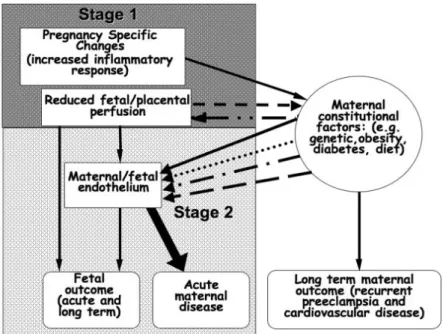
maternal PE, but with preterm births94 or intrauterine growth restriction95 often manifest abnormal features of placentation identical to those seen in PE. Thus, it is proposed that, in some instances, the abnormal placentation interacts with maternal constitutional factors to result in the PE syndrome (Figure 1).84
Figure 1. Maternal fetal interactions in the pathogenesis of pre-eclampsia. From Roberts, JM. et al. Placenta 2009; 30 (Supplement A): 32-37. Reprinted with per-mission from the Elsevier publishing group.
Pre-eclampsia and risk of cardiovascular disease
Several studies have shown that women examined 2-17 years after prior PE have higher blood pressure, increased insulin resistance, and higher levels of triglycerides.96-98 Endothelial dysfunction is the key factor in the pathogene-sis of atheroscleropathogene-sis and this dysfunction is common in women with
previ-ous PE.99-101 It is now a well known fact that the metabolic syndrome, which
is a risk factor for atherosclerosis and subsequent CVD,102, 103 is also a risk factor for developing PE.104 The metabolic syndrome is defined as high blood pressure, high fasting blood glucose and triglycerides, low levels of
high-density lipoprotein cholesterol, and abdominal obesity.102 Women with
this syndrome can respond to pregnancy in an abnormal way and can
devel-op features of PE.104 With increasing age, women with a history of PE have
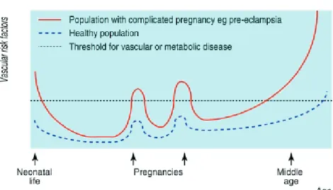
Figure 2. Risk factors for vascular disease are identifiable during excursions into the metabolic syndrome of pregnancy. From Sattar, N. et al. BMJ 2002; 325: 157-160. Reprinted with permission from the BMJ publishing group.
A meta-analysis by Bellamy et al. included 25 prospective or retrospective
cohort studies carried out between 1960 and 2006.106 In this analysis, women
with previous PE had relative risks (RR) of 2.16 (95% CI 1.86-2.52) for ischemic heart disease and 3.70 (95% CI 2.70-5.05) for hypertension, com-pared to women with previous normal pregnancies, with mean follow-up times of 11.7 and 14.1 years, respectively. In addition, women who oped PE had higher all-cause mortality rates than women who did not devel-op PE. In another meta-analysis, McDonald et al. found similar results, indi-cating an increased risk of developing CVD (RR 2.33) in women with a
his-tory of PE compared to women with normal pregnancies.107
Severity of pre-eclampsia and risk of cardiovascular disease
In a meta-analysis by McDonald et al., the RR of developing cardiac disease was 5.36 (95% CI 3.96-7.27) in women with previous severe PE compared to 2.00 (1.83-2.19) in women with mild PE (Figure 3).107
Figure 3. Risk of cardiac disease according to the severity of pre-eclampsia. From McDonald, SD. et al. Am Heart Journal 2008; 159: 918-30. Reprinted with permis-sion from the Right Link publishing group.
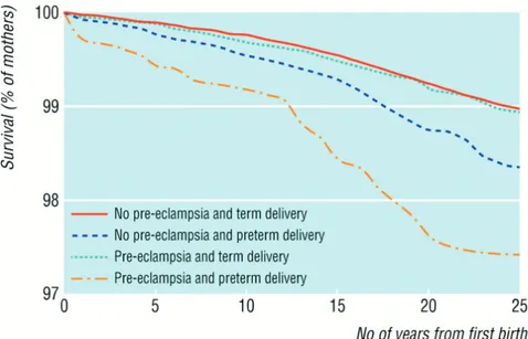
In a study by Arnadottir et al., women with hypertensive disorders of preg-nancy had a higher risk of CVD-related death than normotensive controls (RR 1.60; CI 1.29-1.98). The risk was more evident if they were suffering from PE, especially severe PE.108 Further, Irgens et al. showed that the long-term all-cause mortality rate was 1.2-fold (CI 1.02-1.37) higher in women with PE than in women with normotensive pregnancies. The risk was in-creased to 2.71-fold (CI 1.99-3.68) higher in women who had PE and
deliv-ered preterm, than in women without PE and term delivery (Figure 4).109 In
particular, the risk of death from cardiovascular causes in women with PE and preterm deliveries was 8.12-fold higher than in women with normoten-sive term deliveries.109
Figure 4. Long-term survival of mothers after pre-eclampsia. From Irgens, HU. BMJ 2001; 323: 1213-7. Reprinted with permission from the BMJ publishing group.
Pre-eclampsia and its effects on common carotid artery
intima-media thickness
It is now well established that PE is an independent risk factor for subse-quent CVD106, 107 as a result of endothelial dysfunction leading to develop-ment of atherosclerosis. Chambers et al. measured flow-mediated arterial dilatation in women with previous PE about 3 years postpartum and showed that these women had impaired endothelial function compared to women with previous uncomplicated pregnancies.100 Similar findings have been shown in several other studies.99, 101 As outlined previously, measurement of
the CCA-IMT is recommended and the test is often used as a marker of sub-clinical atherosclerosis.34, 36-38Whether PE is associated with increased
CCA-IMT has only rarely been investigated and the results are not convincing. For example, Blaauw et al. investigated a group of women with previous early-onset PE (EOPE), about 6 months postpartum. They found no significant differences in CCA-IMT between women with previous PE and women with
previous normal pregnancies.78 Similarly, Haukkamaa et al. found no
signif-icant differences in CCA-IMT between women with and without previous PE.110
Because of the documented increased risk of CVD in women with a histo-ry of PE and the inconsistent findings associated with CCA-IMT, it appears that there is a need for an alternative non-invasive method to assess cardio-vascular risk in these women.
Aims of the study
The overall aim of the thesis was to investigate whether measurement of the individual intima and media thicknesses and calculation of the I/M ratio during and after pre-eclamptic pregnancy could provide physical evidence of the well documented increased cardiovascular risk in these women.
Specific aims of the individual studies
I. To validate the method for non-invasive ultrasound assessment of
the individual CCA wall layer dimensions in young, premenopausal women with SLE with known increased risk of CVD.
II. To monitor changes in the CCA wall layer dimensions during and after normal pregnancy.
III. To compare the CCA wall layer dimensions at the time of PE
diag-nosis and about one year postpartum, with values in normal pregnant women of similar stage.
IV. To compare the CCA wall layer dimensions in premenopausal
Study populations
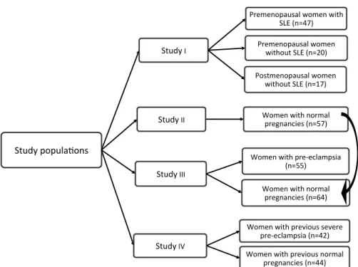
Figure 5. Flow-chart of the study populations.
The study populations were from both urban and rural areas of Sweden, with varying levels of education. Both nulli-/primiparous and parous women were included in the studies. Postmenopausal women and women receiving hor-mone replacement therapy were not eligible for inclusion in Studies I and IV. In Studies II-IV, women were not included if they had chronic hyperten-sion, renal disease, SLE, or pre-gestational or gestational diabetes, or if they were pregnant with more than one fetus at the index pregnancy.
Study I
Forty-seven premenopausal women, fulfilling four or more of the ACR clas-sification criteria for SLE were included. Consecutive patients were invited to participate at the time of outpatient visits at the Rheumatology
Depart-!"#$%&'('#)*+(,-& !"#$%&.& /0121,('*#-*)&3(21,&34"5& !67&8,9:;<& /0121,('*#-*)&3(21,& 34"5(#"&!67&8,9=><& /(-"21,('*#-*)&3(21,& 34"5(#"&!67&8,9?;<&
!"#$%&..& @(21,&34"5&,(02*)&'01A,*,B41-&8,9C;<&
!"#$%&...& @(21,&34"5&'01D1B)*2'-4*& 8,9CC<& @(21,&34"5&,(02*)& '01A,*,B41-&8,9E:<& !"#$%&.F& @(21,&34"5&'01G4(#-&-1G101& '01D1B)*2'-4*&8,9:=<& @(21,&34"5&'01G4(#-&,(02*)& '01A,*,B41-&8,9::<&
ment at University Hospital, Uppsala, Sweden. Two groups of women with-out SLE served as controls: 20 premenopausal women and 17
postmenopau-sal women were recruited from a previous study.53
Study II
The women were recruited during their first routine visit at two of the Upp-sala County’s antenatal clinics (Figure 6). Fifty-seven women with a normal pregnancy and expected normal pregnancy outcome remained in the third trimester evaluation. At the time of the postpartum examination, three wom-en were pregnant again and one woman had moved away from Swedwom-en. Thus, 53 women remained in the final evaluation.
Figure 6. Flow chart of the study population, Study II.
Study III
Cases: Fifty-five women with PE were recruited when they were admitted to
the antenatal clinic at the Department of Obstetrics and Gynecology at Uni-versity Hospital in Uppsala, Sweden. During the postpartum examination, 5
112 women were
asked to participate
Did not want to participate, 34 Miscarriages, 9 1st (baseline) examination (n=69) Miscarriages, 3 Dropouts, 3 2nd examination (n=63)
Moved away from Sweden, 1 Were pregnant again, 3
Postpartum examination
(n=53)
3rd examination
(n=57)
women were pregnant again and 2 did not want to participate. Thus, 48 women remained in the postpartum evaluation.
Controls: The control group comprised 64 women with healthy pregnancies.
Most of the women in the control group participated in Study II and were recruited during their routine visit at two of Uppsala County’s antenatal clin-ics.111 Eight women were recruited during a visit to the emergency outpatient department at University Hospital, Uppsala. In the postpartum examination, four women were pregnant again, one did not want to participate and one had moved out of Sweden. Thus, 58 women remained in the postpartum examination.
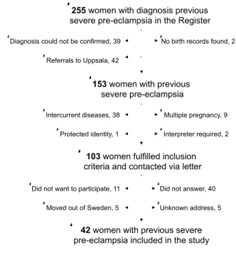
Study IV
Cases: Women between 40 and 50 years of age with one or more
pregnan-cies complicated by severe PE were included in this study. We used the Reg-ister of diagnosis at University Hospital, Uppsala, Sweden, to identify wom-en with a history of previous severe PE. The Swedish versions of the Inter-national Classification of Diseases (ICD) -9 and -10 were used to identify the women (ICD-9: 642F and ICD-10: 014.1A and 014.1B). Before inclu-sion, the birth records were reviewed for confirmation of the diagnosis. We found 255 women with a diagnosis of severe PE in the Register. Forty-two women remained in the study after exclusions of those whose delivery rec-ords could not be tracked, whose diagnosis could not be confirmed, who were referred from other regions, who had chronic diseases or twin/triplet pregnancies, who did not answer or declined participation in the study, or who had moved out of the country, etc.
(
Figure 7).Figure 7. Flow chart for the recruitment process of cases in Study IV.
Controls: Women between 40 and 50 years of age who had had solely one or
more normal pregnancies were recruited. Women living in Uppsala County, Sweden, many of them working at the Uppsala University Hospital, were recruited by personal invitation.
No birth records found, 21
255 women with diagnosis previous
severe pre-eclampsia in the Register Diagnosis could not be confirmed, 39
Referrals to Uppsala, 42
153 women with previous
severe pre-eclampsia
Intercurrent diseases, 38 Multiple pregnancy, 9
103 women fulfilled inclusion
criteria and contacted via letter Did not want to participate, 11 Did not answer, 40
Unknown address, 5 Moved out of Sweden, 5
Interpreter required, 2
42 women with previous severe
pre-eclampsia included in the study Protected identity, 1
Methods
Assessments and data collection
Gestational age was defined as completed weeks of gestation based on the second trimester routine ultrasound dating, at pregnancy weeks 16-18. Height and weight were measured and body mass index (BMI) was calculat-ed in kg/m2. Data were collected about age, reproductive history, smoking
habits, medical history and medications. Blood pressure was measured after about 15 minutes’ rest, in the supine position, on the right upper arm, with automated (Study I) or manual (Studies II-IV) blood pressure equipment (Umedico, with a calf-size appropriate for the arm circumference). Mean arterial pressure (MAP) was calculated as DBP + ⅓ (SBP - DBP) (Studies III & IV).
The delivery records were checked and data were collected about possible pregnancy-related complications, gestational week at delivery, and the birth weight of the baby. A healthy pregnancy was defined as a normotensive pregnancy with delivery of a baby with normal birth weight [within ± 2 standard deviations (SD) of the mean birth weight for gestational age],112 in
gestational week 37 or later. Pre-eclampsia was defined as new-onset hyper-tension (SBP ≥ 140 mmHg and/or DBP ≥ 90 mmHg, observed on at least two separate measurements ≥ 6 hours apart), combined with proteinuria (≥ 2 on a dipstick or a 24-hour urine sample showing leakage of ≥ 300 mg albu-min/24 hours), both after gestational week 20. PE was diagnosed as severe when blood pressure was pronounced (SBP ≥ 160 mmHg and/or DBP ≥ 110 mmHg) and/or the proteinuria was massive (≥ 5 g/24 hours). PE was catego-rized into EOPE, when it was diagnosed before gestational week 34 and late onset PE (LOPE), when it was diagnosed in gestational week 34 or later. Preterm birth was defined as birth before gestational week 37. SGA and large for gestational age (LGA) babies were defined as those with birth weights ≥ 2 SDs below or above the reference population’s mean birth weight for the gestational age.112
Study I
The investigation of SLE patients included an interview and a physical ex-amination by a rheumatologist. SLE disease activity was determined using
the modified SLE Disease Activity Index.113 Cumulative disease damage
was measured using the Systemic Lupus International Collaborating
Clin-ics/American College of Rheumatology (SLICC/ACR) damage index.114 A
venous blood sample was collected during the examination for routine chem-ical analyses and assessment of complement levels.
Study II
Participants were examined three times during pregnancy, in the first (be-tween gestational weeks 11 and 13), the second (be(be-tween gestational weeks 21 and 24) and the third (between gestational weeks 34 and 37) trimesters and once more, about one year postpartum, when the women had ended lac-tation and started to menstruate. A venous blood sample was collected from each subject, at each examination. After collection, the blood samples were kept at room temperature for about half an hour before being centrifuged for 10 minutes at 2000g. The serum samples were then separated and stored at – 70ºC until the level of E2 was analyzed, using a Cobas E instrument (Roche Diagnostics, Mannheim, Germany). The total coefficient of variation (CV) of the instrument for analysis of E2 was 2.6% at 13,000 pmol/L.
Study III
The participants were examined twice. The first examination was performed when the women were diagnosed with PE. The mean pregnancy duration at inclusion for controls was similar to that for the PE group. The second exam-ination was performed about one year after delivery, when the women had ended lactation and started to menstruate.
Study IV
The participants were examined once, at inclusion. At the examination the women were interviewed about the date of their last delivery, their own his-tory of CVD events and CVD-related risk factors, and any regular medica-tion. Angina pectoris, MI, stroke and other vascular events were categorized as cardiovascular events and CVD-related risk factors included hypertension, diabetes mellitus, and hyperlipidemia.
Ultrasound assessment of the common carotid artery
The CCA wall layers were imaged using high-resolution ultrasound equip-ment fitted with a broadband probe at 22 MHz center frequency (Osteoson, Minhorst Company, Meudt, Germany). The method has been extensively described in previous reports.52, 53 In brief, the artery wall layers were exam-ined with the women sitting upright and looking straight ahead after they had rested for about 15 min. The transducer was applied at the point of maximal pulsation of the left CCA, in front of the sternocleidomastoid muscle. The depth of penetration was up to 20 mm. The three-layer image of the pulsat-ing near wall showed two echo-dense zones (the adventitia and the intima) with an echo-lucent area (the media) in between followed by the echo-lucent artery lumen (Figure 8). About twenty point estimates of the artery wall, not adjusted to the cardiac cycle, were saved on a PC by one researcher. The individual artery wall layer dimensions were measured off-line for all partic-ipants by another researcher, who was blinded with regard to the study group and the time of assessment. The means of about 10 technically acceptable measurements were calculated and used in the analysis. In our laboratory, the calculated intra-reader CV was about 4.3% for intima thickness and about 4.1% for media thickness. The inter-reader variability was about 5.4% for intima thickness and about 3.2% for media thickness.
Figure 8. Ultrasonographic image of the common carotid artery near wall, obtained by non-invasive high-frequency (22 MHz) ultrasound. C, cutis; SC, subcutis; A, adventitia; M, media and I, intima. From Akhter, T. et al. AJP-Heart Circ Physiol 2013; 304: H229-H234. Reprinted with permission from the RightLink publishing group.
Ethical considerations
The local Ethics Committee of the Medical Faculty of Uppsala University approved all studies, and informed written consent was obtained from each subject included in the studies.
Statistical methods
The results are presented as mean (standard deviation) or number (%). Dif-ferences in proportions between groups were tested by Chi-square tests. For continuous variables, between-group differences were tested using the Mann-Whitney U-test and within-group differences using the Wilcoxon Signed Rank test. Between-group differences for the main outcomes were presented as mean differences (95% CI), in addition to the non-parametric test (Studies III & IV). Between-group differences in artery wall layer di-mensions were also adjusted for differences in BMI and MAP, using the
non-parametric Willett´s residual method.115 Correlations were analyzed
using the Spearman Rank Correlation test. Receiver operating characteristic (ROC) curve analysis was undertaken to illustrate and compare the discrimi-natory capacities of estimation of individual artery wall layer dimensions, combined CCA-IMT and MAP to correctly predict prevalent PE (Study III) or to correctly discriminate between women with regard to previous severe PE (Study IV).
The level of significance was set at a p value ≤ 0.05. All statistical anal-yses were performed using Statistica version 9, Statsoft Inc. (Study I) or the SPSS, version 20.0 (SPSS Inc. PASW statistics, Chicago, IL, USA) (Studies II-IV).
Results
Study I
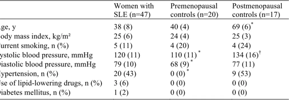
At the time of investigation, SLE disease was latent for all patients. Some of the traditional cardiovascular risk factors differed significantly between SLE
patients and controls (Table 1).Five women in the SLE group had had CVD
events (angina pectoris, MI or stroke) whereas none in the control group had.
Table 1. Traditional cardiovascular risk factors in women with SLE and controls.
Women with
SLE (n=47) Premenopausal controls (n=20) Postmenopausal controls (n=17)
Age, y 38 (8) 40 (4) 69 (6)*
Body mass index, kg/m² 25 (6) 24 (4) 25 (3) Current smoking, n (%) 5 (11) 4 (20) 4 (24) Systolic blood pressure, mmHg 120 (11) 110 (11) * 134 (16)†
Diastolic blood pressure, mmHg 79 (10) 68 (9) * 77 (11)
Hypertension, n (%) 20 (43) 0 (0) * 9 (53)
Use of lipid-lowering drugs, n (%) 3 (6) 0 (0) 0 (0) Diabetes mellitus, n (%) 1 (2) 0 (0) 0 (0) Mean (standard deviation) or number (%)
* p < 0.001 and † p < 0.01 compared to SLE patients (non-parametric test).
Artery wall layer dimensions
In the premenopausal women with SLE, the mean carotid intima was thicker (0.19 vs 0.12 mm), the media was thinner (0.45 vs 0.68 mm), and the I/M ratio was higher (0.45 vs 0.20) than in the age-matched healthy premenopau-sal control women (all p < 0.0001) (Figure 9). Further, in the women with SLE versus postmenopausal women who were 30 years older, the intima (0.19 vs 0.14 mm, p < 0.0001) and the media (0.45 vs 0.34 mm, p < 0.001) were thicker, while the I/M ratio was similar (0.45 vs 0.45 mm). The total CCA-IMT was lower in SLE patients than in age-matched controls (0.64 vs 0.80 mm, p < 0.01) but higher than in the postmenopausal women (0.64 vs 0.48 mm, p < 0.0001). SLE patients with a history of either CVD events or hypertension had significantly thinner media than patients without such a history, p < 0.05. Patients treated with azathioprine or mycophenolate mo-fetil had a thinner media than patients not treated with these drugs, p < 0.05.
Figure 9. Carotid artery near-wall dimensions assessed using 22 MHz ultrasound in premenopausal women with SLE compared to pre- and postmenopausal women without SLE. Mean ± SD. **** p < 0.0001 and *** p < 0.001 compared to SLE.
Correlation analysis between artery wall layer dimensions and SLE disease activity index
In the SLE patients, higher SLICC/ACR damage index, higher cumulative dose of prednisone, and higher C3d/C3 ratio were associated with a thinner carotid media, all p < 0.05. There were no significant correlations between age, disease duration or antimalarial medication and intima or media thick-ness or I/M ratio.
Study II
The mean age of participants in Study II was about 30 years, 50% were nul-liparous and very few were current smokers (4%). As expected, BMI in-creased during pregnancy and dein-creased again postpartum. SBP was stable during pregnancy and decreased postpartum (p < 0.05), whereas DBP showed a decrease in the second trimester assessment (p < 0.05), but was otherwise stable.
Artery wall layer dimensions
The mean values for intima and media layers and I/M ratio remained fairly stable during pregnancy. However, by about one year postpartum, the mean intima thickness and the I/M ratio had decreased (improved) compared to all trimesters, all p < 0.001 (Table 2). The combined CCA-IMT was also fairly stable during pregnancy and had decreased by one year after delivery. There were no significant differences in artery wall layer dimensions between
smokers and non-smokers or between nulliparous and parous women, at any of the assessment points.
Table 2. Artery wall layer dimensions in each trimester during pregnancy (n=57) and about one year postpartum (n=53).
Characteristics Trimester Postpartum
First Second Third
Gestational length (wk) or 13 (1) 23 (1) 37 (1) 12 (1.3) time postpartum (mo)
Intima thickness (mm) 0.11 (0.02) 0.11 (0.02) 0.11 (0.02) 0.08 (0.01) *
Media thickness (mm) 0.57 (0.14) 0.54 (0.15) 0.57 (0.16) 0.54 (0.11) Intima/media ratio 0.20 (0.05) 0.21 (0.06) 0.20 (0.06) 0.16 (0.04) *
Intima media thickness (mm) 0.68 (0.15) 0.65 (0.16) 0.67 (0.16) 0.62 (0.11)†‡
Mean (standard deviation)
* p < 0.001 compared to 1st, 2nd and 3rd trimester; † p < 0.01 compared to 1st trimester; and ‡ p < 0.05 compared to 3rd trimester (non-parametric test).
Correlations between first trimester characteristics and artery wall layer dimensions in different trimesters
Lower levels of serum E2, older age, and higher BMI and BP in the first trimester were often associated with a numerically thicker carotid intima layer, a thinner media layer and a higher I/M ratio, especially in the second trimester. Thus, in the second trimester, the I/M ratio was significantly lower in women with higher serum E2 levels, and higher in women who were old-er, or had higher BMI, SBP or DBP (all p < 0.05). In the third trimestold-er, the associations were similar but less apparent than those in the second tri-mester.
Correlations between base line characteristics and changes in the artery wall layer dimensions
With regard to changes in the artery wall dimensions from the first to the second trimesters, first trimester higher serum E2 levels were associated with a reduced I/M ratio, whereas a higher BMI, SBP and DBP were all associat-ed with an increasassociat-ed I/M ratio (Table 3). Further, older age was associatassociat-ed with an increased intima thickness (p < 0.05). Higher serum E2 levels were associated with an increased media thickness (p < 0.05), whereas higher BMI, SBP and DBP were associated with a reduced media thickness (all p < 0.05) (Table 3). Similar, but less apparent associations were found with re-gard to changes from the first to the third trimesters (data not shown).
Table 3. Association between maternal characteristics in the first trimester and changes in the carotid artery wall layer dimensions from the first to second tri-mesters.
First Trimester Characteristics Changes From the First to Second Trimester Intima thickness Media thickness Intima/media ratio
Serum estradiol -0.09 0.34* -0.36†
Age 0.31* -0.09 0.18
Body mass index 0.15 -0.29* 0.34*
Systolic blood pressure 0.12 -0.28* 0.27*
Diastolic blood pressure 0.23 -0.44‡ 0.43‡
Values are Spearman rank correlation coefficients. * p < 0.05; † p < 0.01; ‡ p < 0.001
Study III
Women with PE did not differ significantly from women with normal preg-nancies regarding mean maternal age, smoking habits or gestational duration at inclusion, but they were more often nulliparous and had a higher BMI, SBP, DBP and MAP in the first trimester, at inclusion and about one year postpartum (Table 4). Among women with PE, 42% had EOPE, 69% had severe PE and 85% were on anti-hypertensive medication. Gestational dura-tion at birth was on average 4 weeks shorter in the PE group than in controls (p < 0.0001). Infants born to pre-eclamptic mothers had a significantly lower mean birth weight than infants born to mothers with normal pregnancies, both before and after adjustment for gestational duration (p < 0.001).
Table 4. Clinical characteristics of the study population in Study III.
Characteristics
Pre-eclampsia Normal Pregnancies First trimester At inclusion Post- First trimester At inclusion Post- partum partum Gestational duration (wk) 35 (4) 35 (5)
Body mass index (kg/m2) 28 (6) * 33 (7) * 28 (7) ‡ 24 (3) 27 (4) 24 (4)
Systolic BP (mmHg) 124 (11) * 146 (12) * 120 (9) * 114 (9) 115 (9) 111 (7) Diastolic BP (mmHg) 78 (7) * 91 (11) * 78 (8) * 65 (7) 70 (7) 69 (7)
MAP (mmHg) 94 (8) * 109 (10) * 92 (8) * 81 (7) 84 (7) 83 (7) Means (standard deviation). BP, blood pressure; MAP, mean arterial pressure.
* p < 0.0001 and ‡ p < 0.001 compared to corresponding values in normal pregnancies
(non-parametric test).
Artery wall layer dimensions
At inclusion, women with PE had a thicker CCA intima (p < 0.0001), a thin-ner media (p < 0.001) and a higher I/M ratio (p < 0.0001) than controls. The differences in CCA intima thickness and I/M ratio remained significant be-tween study groups after adjustment for first trimester BMI and MAP (both p < 0.0001) (Table 5). At the postpartum examination, about one year after
delivery, the intima thickness and I/M ratio had decreased (improved) in both PE patients and controls (both p < 0.0001) (Table 5), but the differences between study groups remained highly significant (both p < 0.0001) (Table 5). In contrast, there were no significant differences in CCA-IMT between PE patients and controls, either during pregnancy or at the postpartum exam-ination.
Table 5. Common carotid artery wall layer dimensions in women with pre-eclampsia, at the time of diagnosis and about one year postpartum and in women with normal pregnancies.
CCA wall Pregnancy Postpartum
layer PE Normal Pregnancy % diff PE Normal Pregnancy % diff dimensions Intima (mm) 0.18 (0.03) *,† 0.11 (0.02) +64 0.12 (0.02) *,†,‡ 0.08 (0.01)‡ +50 Media (mm) 0.47 (0.12) § 0.55 (0.14) -15 0.49 (0.12)⎢⎢ 0.54 (0.11) -9 I/M ratio 0.41 (0.14) *,† 0.20 (0.05) +105 0.26 (0.08) *,†,‡ 0.16 (0.04)‡ +63 IMT (mm) 0.65 (0.13) 0.66 (0.15) -1.5 0.61 (0.12) 0.63 (0.11) -3 Mean (standard deviation) or percentage differences. CCA, common carotid artery; I/M, intima/media; IMT, intima-media thickness; PE, pre-eclampsia
⎢⎢p < 0.05 ; §p < 0.001 and * p < 0.0001 (non-parametric test) compared to normal pregnancy.
†p < 0.0001 compared to normal pregnancy, after non-parametric adjustment for body mass
index and mean arterial pressure in the first trimester and postpartum, respectively.
‡ p < 0.0001, compared to corresponding values during pregnancy.
In women with PE, none of the artery wall layer dimensions differed signifi-cantly with parity, EOPE, LOPE, severity of PE or antihypertensive therapy.
ROC curve analysis to discriminate between women with and without prevalent PE
At inclusion, the area under the ROC curve (AUC) value for the CCA intima thickness was 0.98 and that for the I/M ratio was 0.94, thus correctly differ-entiating women with regard to prevalent PE at diagnosis (Figure 10A). One year postpartum, the corresponding AUC values were 0.95 and 0.90, respec-tively (Figure 10B). In contrast, estimates of the combined CCA-IMT were not useful for differentiating women with regard to prevalent PE, with AUC values of 0.49 during pregnancy and 0.46 about one year postpartum. AUC values for MAP were 0.97 and 0.83, respectively (Figures 10A and B).
Figure 10. Receiver operating characteristic (ROC) curves illustrating area under the curve (AUC) values for individual artery wall layer dimensions, combined intima-media thickness of the common carotid artery and MAP with regard to pre-eclampsia, A) at diagnosis, and B) about one year postpartum. I/M, intima/media; IMT, intima-media thickness; MAP, mean arterial pressure.
Study IV
Women with a history of previous severe PE did not differ significantly re-garding age, smoking habits, time since last delivery, or BMI from the con-trols, but they were more often primiparous and had significantly higher SBP, DBP and MAP. Women with a history of previous PE were more often suffering from prevalent hypertension and using antihypertensive medication than controls. Further, there were two women in the previous PE group who had had CVD events (Table 6).
Table 6. Clinical characteristics of women with previous severe pre-eclampsia (n=42) and women with previous normal pregnancies (n=44).
Clinical Characteristics Pre-eclampsia Normal Pregnancies
Maternal age (y) 44 (3) 44 (3)
Primiparous, n (%) 11 (26) * 4 (9)
Current smoking, n (%) 3 (7) 3 3 (7) Time since last delivery (y) 11 (5) 11 (5) Body mass index (kg/m2) 27 (5) 25 (4)
Systolic blood pressure (mmHg) 128 (15) ‡ 119 (12) Diastolic blood pressure (mmHg) 82 (8) ‡ 76 (8)
Mean arterial pressure (mmHg) 97 (10) ‡ 91 (8) Antihypertensive therapy, n (%) 8 (19) * 2 (5)
Cardiovascular disease events, n (%) 2 (5) 0 (0) Mean (standard deviation) or number (%)
* p < 0.05 and ‡ p < 0.01 compared to normal pregnancies (non-parametric test).
Among the women with previous severe PE at the index pregnancy, 64% had an EOPE, 86% were delivered preterm and 14% had infants born SGA.
Artery wall layer dimensions
Women with previous severe PE had significantly thicker CCA intima layers and higher I/M ratios (both p < 0.0001) and non-significantly thinner media layers (p = 0.08) than women with previous normal pregnancies. After ad-justment for BMI and MAP, the differences in intima thickness and I/M ratio were still highly significant (both p < 0.0001). However CCA-IMT did not differ significantly between women with and without previous severe PE (Table 7).
Table 7. Common carotid artery wall layer dimensions in women with previous severe pre-eclamsia (n=42) and in women with previous normal pregnancies (n=44).
CCA wall layers Pre-eclampsia Normal Pregnancies Mean diff (95% CI) % diff Intima (mm) 0.13 (0.02) *,† 0.08 (0.01) 0.05 (0.04, 0.06) +63 Media (mm) 0.50 (0.11) 0.54 (0.11) -0.04 (-0.09, 0.01) -7 I/M ratio 0.27 (0.07) *,† 0.15 (0.03) 0.12 (0.10, 0.14) +80 IMT (mm) 0.63 (0.12) 0.61 (0.12) 0.01 (-0.04, 0.06) +3 Mean (standard deviation). CCA, common carotid artery; I/M, intima/media; IMT, intima media thickness; CI, confidence interval.
* p < 0.0001 (non-parametric test)
† p < 0.0001, after non-parametric adjustment for body mass index and mean arterial pressure.
Within the group of women with previous severe PE, those with a history of either CVD events or prevalent hypertension had thicker CCA intima layers and higher I/M ratios than women without such a history (all p < 0.05). Pri-miparous women had higher I/M ratios than women who were parous (p < 0.05). No significant differences in artery wall layer dimensions were found with regard to smoking habits or types of PE (EOPE or LOPE; repeated or not repeated PE; preterm or term PE; PE with or without SGA birth).
ROC curve analysis to discriminate between women with and without previous severe PE
In the ROC curve analysis, both the carotid intima layer and the calculated I/M ratio strongly discriminated between women with and without previous severe PE; the AUC value was 0.98 for intima thickness and 0.93 for the I/M ratio. In contrast, estimates of the combined CCA-IMT were not useful for discriminating between women with and without previous PE, with an AUC value of 0.52. The AUC value for MAP was 0.69 (Figure 11).
Figure 11. Receiver operating characteristic (ROC) curves illustrating area under the curve (AUC) values for different artery wall layers, combined intima media thick-ness and MAP, to discriminate between women with and without previous severe pre-eclampsia. I/M, intima/media; IMT, intima-media thickness; MAP, mean arterial pressure.
Discussion
Cardiovascular disease in women, especially after
pregnancy complications
Cardiovascular disease is the main cause of mortality in women, all over the world. Globally, more women die from CVD than from cancer, malaria, tuberculosis and HIV combined.3 According to the AHA, mortality from CVD has declined in the last 25 years, but significantly less for women than for men. Further, the global population is increasing in number and age, resulting in a marked increase in the number of women at risk for CVD.
PE is associated with an increased risk of CVD later in life.106, 107 Preg-nancies resulting in preterm delivery and birth of SGA infants are also asso-ciated with increased risk of CVD morbidity and mortality in later life.116, 117 It has been demonstrated that pregnancies resulting in preterm birth94 and
SGA infants95 but without maternal signs of PE show features of abnormal
placentation that are identical to those seen in PE. Preterm birth can be caused by inflammation and infection,118 and the process of atherosclerosis is also largely inflammatory in its origin.22 A possible mechanism for
intrauter-ine growth retardation is maternal hemodynamic maladaptation, to which maternal constitutional factors such as obesity or diabetes could contribute.119 Thus, several pregnancy complications could have potential
negative effects on cardiovascular risk. Cardiovascular studies in women are therefore warranted, especially in women who are at higher risk after preg-nancy complications, such as after PE. Early diagnosis of women at risk of CVD could help to determine early intervention to reduce the cardiovascular morbidity and mortality.
Cardiovascular disease and carotid artery wall imaging
Measurement of CCA-IMT by 7-10 MHz ultrasound has been used for a long time for non-invasive assessment of vascular health in patients with CVD, or as an indicator of progression or regression of CVD. Although the CCA-IMT is considered the 'gold standard' for assessment of vascular sta-tus,32 the interpretation of CCA-IMT measurements has been questioned. There are no significant and consistent differences in CCA-IMT measure-ments between individuals with or without CVD, according to a