Acta Psychiatr Scand. 2021;143:363–374. wileyonlinelibrary.com/journal/acps
|
363 O R I G I N A L A R T I C L EBrain structure and clinical profile point to neurodevelopmental
factors involved in pedophilic disorder
Christoph Abé
1|
Roberth Adebahr
2,3|
Benny Liberg
1|
Christian Mannfolk
4|
Alexander Lebedev
1|
Jonna Eriksson
5|
Niklas Långström
6,7|
Christoffer Rahm
4This is an open access article under the terms of the Creative Commons Attribution License, which permits use, distribution and reproduction in any medium, provided the original work is properly cited.
© 2020 The Authors. Acta Psychiatrica Scandinavica published by John Wiley & Sons Ltd. 1Department of Clinical Neuroscience,
Karolinska Institutet, Stockholm, Sweden
2ANOVA, Karolinska University
Hospital, Stockholm, Sweden
3Department of Clinical Sciences
(Psychiatry), Umeå University, Umeå, Sweden
4Centre for Psychiatry Research,
Department of Clinical Neuroscience, Karolinska Institutet, and Stockholm Health Care Services, Stockholm, Sweden
5Psychiatry South, Stockholm, Sweden 6Department of Medical Epidemiology
and Biostatistics, Karolinska Institutet, Stockholm, Sweden
7National Board of Health & Welfare,
Stockholm, Sweden Correspondence
Christoph Abé, Department of Clinical Neuroscience, Karolinska Institutet, Nobels väg 9, 17177 Stockholm, Sweden. Email: christoph.abe@ki.se
Funding information
ALF agreement between the Swedish government and the county councils, Grant/Award Number: SLL20150518 and SLL20160555; Söderström König Foundation; the Fredrik and Ingrid Thuring Foundation, Grant/Award Number: FITS-2015-00157; Swedish Society for Medical Research, Grant/ Award Number: P14-0136; Swedish Society for Medicine, Grant/Award Number: SLS-501421 and SLS-886481; The Center for Psychiatry Research, Department of Clinical Neuroscience, Karolinska Institutet, Grant/Award Number: CPF-99/2016
Abstract
Objective: Pedophilic disorder (PD) is characterized bypersistent, intense sexual
at-traction to prepubertal children that the individual has acted on, or causes marked distress or interpersonal difficulty. Although prior research suggests that PD has neu-rodevelopmental underpinnings, the evidence remains sparse. To aid the understand-ing of etiology and treatment development, we quantified neurobiological and clinical correlates of PD.
Method: We compared 55 self-referred, help-seeking, non-forensic male patients
with DSM-5 PD with 57 age-matched, healthy male controls (HC) on clinical, neu-ropsychological, and structural brain imaging measures (cortical thickness and sur-face area, subcortical and white matter volumes). Structural brain measures were related to markers for aberrant neurodevelopment including IQ, and the 2nd to 4th digit ratio (2D:4D).
Results: PD was associated with psychiatric disorder comorbidity and ADHD and
autism spectrum disorder symptoms. PD patients had lower total IQ than HC. PD individuals exhibited cortical surface area abnormalities in regions belonging to the brain's default mode network and showed abnormal volume of white matter underly-ing those regions. PD subjects had smaller hippocampi and nuclei accumbens than HC. Findings were not related to history of child-related sexual offending. IQ cor-related negatively with global expression of PD-cor-related brain features and 2D:4D correlated with surface area in PD.
Conclusions: In the largest single-center study to date, we delineate psychiatric
co-morbidity, neurobiological and cognitive correlates of PD. Our morphometric find-ings, their associations with markers of aberrant neurodevelopment, and psychiatric comorbidities suggest that neurodevelopmental mechanisms are involved in PD. The findings may need consideration in future development of clinical management of PD patients.
K E Y W O R D S
cerebral cortex, neurodevelopmental disorder, neuroimaging, pedophilic disorder, psychiatric comorbidity
1
|
INTRODUCTION
Pedophilic disorder (PD) is a paraphilic disorder defined by the Diagnostic and Statistical Manual of Mental Disorders, 5th edition (DSM-5)1 as a persistent sexual attraction to pre-pubescent children, where the individual has acted on these sexual urges, or they cause marked distress to the afflicted individual or interpersonal difficulty. Beyond furthering di-agnostics, risk assessment, and treatment for individuals with PD, improved knowledge about its neurobiological origins could aid the prevention of child sexual abuse.
Previous studies have linked PD to conditions where neu-rodevelopmental factors play substantial roles suggesting that neurodevelopmental factors contribute to the development of PD.2–4 These factors include genetic and environmental influences on the brain, such as prenatal androgens3 affect-ing brain development, cognitive functionaffect-ing, and disorder development. Thus, presumable brain alterations in PD are proposed to be present since early childhood or emerging ad-olescence. In line with aberrant neurodevelopment, individu-als with PD presented with lower intelligence compared with individuals attracted to adults.5 Other PD-related neuropsy-chological deficits include lower processing speed together with impaired attention and prefrontal executive functions, such as response inhibition, task switching, and cognitive reasoning.4,6,7 Research investigating anthropometric signs linked to in utero conditions suggest associations between PD and minor physical anomalies,8 shorter height9 and left-hand-edness.4 Notably, a higher 2D:4D ratio (the ratio between 2nd and 4th digit length), a putative marker for prenatal androgen exposure,10 has been associated with paraphilic interest11 but not been investigated specifically in PD. Other indications for neurodevelopmental influences originate from studies addressing psychiatric comorbidity.4 Gerwinn et al12 found an association between pedophilia and symptoms of the neurodevelopmental disorder attention deficit hyperactivity disorder (ADHD). However, links between PD and autism spectrum disorder (ASD) remain unknown.
A handful of previous, small-scale neuroimaging studies have addressed putative neurobiological underpinnings of PD. These studies indicate cortical abnormalities in various brain regions and volumetric white matter and amygdala al-terations pointing toward altered emotional and arousal net-works in PD. Despite heuristic value, prior studies report inconsistent results4,6,13 and suffer from substantial short-comings; small sample sizes, inadequate control groups, and limiting region of interest approaches. Moreover, the most re-cent and largest study combined data acquired with multiple brain scanners.13 This previous report suggested that brain abnormalities may rather be attributed to sexual offending behavior than to PD (or pedophilic interest) per se.13 A recent review argued that neuroimaging in PD is still in its infancy and suggested replication in larger samples while controlling
for potential confounding factors; sexual offending, sexual orientation, psychiatric comorbidity, and medication use.6 Hence, the contribution of neurodevelopmental mechanisms to PD remains unclear.
1.1
|
Aims of the study
The aim of this single-center study was to comprehensively assess measures of brain structure, psychiatric morbidity, and psychological function in a consecutive sample of help-seek-ing men with DSM-5 PD and to compare them with healthy controls. Another goal was to investigate the relations be-tween PD-related structural brain abnormalities and cogni-tive function (IQ) and 2D:4D ratios, as a marker for prenatal androgen exposure.
2
|
MATERIAL AND METHODS
2.1
|
Participants
We performed this study within the framework of Pedophilia at Risk – Investigations of Treatment and Biomarkers (PRIOTAB), a clinical trial and research pro-ject.14 With a case-control design (PRIOTAB-CC), we aimed to identify clinically useful biomarkers and risk factors by investigating PD-related neurobiology using magnetic resonance imaging (MRI), psychiatric assess-ment, and cognitive testing. The study was conducted at ANOVA, Karolinska University Hospital, Stockholm,
Significant outcomes
• Patients with pedophilic disorder showed symp-toms of neurodevelopmental disorders.
• Pedophilic disorder was associated with structural brain abnormalities in the default mode network. • Brain abnormalities were related to markers of
prenatal androgen exposure.
Limitations
• The cross-sectional study design does not allow for inference regarding causality.
• Structural neuroimaging cannot assess the brain regions’ functional role in the development of pedophilic disorder.
• We studied self-referred individuals with pedo-philic disorder. Conclusions about individuals not seeking clinical help should be drawn with caution.
Sweden, a multidisciplinary clinic and research center for sexual medicine, andrology, and transgender medicine. Self-referred, help-seeking subjects with PD were re-cruited through PrevenTell, a Swedish national helpline. PD was confirmed using a structured interview based on DSM-5 criteria. Age-matched, male healthy controls (HC), screen-negative for PD, were recruited in the Stockholm catchment area through Karolinska Trial Alliance and ad-verts on Karolinska Institutet's homepage. More details on screening and recruitment procedures can be found in the supplemental material or elsewhere.14 Fifty-five adult male individuals with PD and 57 HC completed baseline clini-cal assessments with four homosexual and eight bisexual participants in each group, respectively. A total of 54 PD patients and 50 HC provided MRI data of sufficient qual-ity for the current analyses. All participants provided writ-ten informed consent and the study was approved by the Swedish Central Ethical Review Board.
2.2
|
Psychiatric comorbidity and
clinical symptoms
The Swedish version of the Mini International Neuropsychiatric Interview (M.I.N.I.) 6.0.0 tapped co-existing psychiatric disorders.15 Using the 1999 WHO Consultation on Child Abuse Prevention guidelines,16 par-ticipants were also asked whether they had been sexually abused or exploited before age 15 years.
Symptoms indicative of autism spectrum disorder (ASD) were assessed with the abridged version of the Ritvo Autism and Asperger Diagnostic Scale (RAADS-14), covering mentalizing deficits (7 items), social anxiety (4 items), and sensory reactivity (3 items). We investi-gated ASD-related symptoms using two established cut-off scores. The first threshold (>13) is based on the suggested screening cut-off, where results ≤13 imply a non-ASD diagnosis with a negative predictive value of 99%. The stricter cut-off (>23) is linked to a positive predictive value of 37% of receiving a full ASD diagnosis in unselected general population samples.17
ADHD symptoms were assessed with the 18-item Adult ADHD Self-Report Scale – Screen (ASRS-v1.1).18 Each item was rated on a 5-point Likert scale (0 = never; 4 = very often). Indicated ADHD was coded “1” for re-spondents endorsing 4+ part A items with “sometimes” or “often” (depending on item), and “0” otherwise. The Alcohol Use Disorders Identification Test (AUDIT)19 and the Drug Use Disorders Identification Test (DUDIT)20 self-reports were used to screen for substance use disor-ders. We used the Hypersexual Behavior Inventory (HBI-19)21 self-report to assess hypersexual behavior, indicated by an HBI score >52.
2.3
|
General cognitive ability
The Swedish version of the Wechsler Adult Intelligence Scale–IV (WAIS-IV) was used to assess general cogni-tive ability.22 The full-scale intelligence quotient (IQ) was estimated from subtests of intellectual functioning across four cognitive areas: Verbal Comprehension Index (VCI), Perceptual Reasoning Index (PRI), Working Memory Index (WMI), and the Processing Speed Index (PSI).
2.4
|
Anthropometric measurements
A research nurse measured participants’ height, weight and the lengths of index finger (2nd digit) and ring finger (4th digit) of the right hand for assessment of the 2D:4D digit ratio.10 Digits were measured from the basal crease to the tip using a Vernier caliper. Handedness was assessed with the Swedish version of the Edinburgh Handedness Inventory self-report.23
2.5
|
Structural MRI image
acquisition and processing
Brain magnetic resonance imaging (MRI) scans were per-formed at the Karolinska University Hospital, Huddinge, Sweden, using a 3T medical scanner (Siemens Prisma). We obtained measures of cortical volume, thickness, and surface area for each participant from structural T1-weighted images using the semi-automated cortical surface reconstruction and parcellation methods provided by FreeSurfer 6.0.24,25 FreeSurfer also provided segmentation of subcortical vol-umes, cortical parcellations defined by the Desikan atlas, and segmentation of white matter (WM) volumes of regions underlying these cortical parcellations. These measures were extracted for secondary analyses, including multivariate classification analyses (described below). Details on image acquisition and processing are described in the Supporting Information.
2.6
|
Statistical analyses
2.6.1
|
Demographic and clinical variables
Group differences in demographic, anthropometric, and clin-ical measures were assessed with IBM SPSS Statistics ver-sion 25. We used t tests for normally distributed data (tested by skewness), the Mann–Whitney U test for skewed data and χ2-tests for dichotomous variables. We provided Cohen's d values as effect size (ES) measures, calculated as standard-ized mean differences for parametric data, via biserial rank
correlations for non-parametric data, or from frequency dis-tributions for categorical data.
2.6.2
|
Case-control differences in structural
brain imaging measures
Group differences in cortical brain imaging measures were performed on vertex level. In the main analysis, we tested for the effect of group (PD vs HC; independent variable of inter-est) on cortical thickness and WM-surface area (dependent variables), while correcting for age (regressor of no interest). Cortical volume comparisons were performed for complete-ness. Secondary analyses comparing groups on subcortical volumes and regional WM segmentations were conducted in SPSS v25, using multiple univariate analyses of covari-ance (ANCOVAs). More details on analytical methods are described in the Supporting Information.
2.6.3
|
Sensitivity tests (testing for potential
confounds on brain imaging findings)
For sensitivity analyses testing for potential confounders, we extracted cortical thickness, surface area, and volume meas-ures averaged over significant clusters obtained in the main analysis. First, we compared these measures between groups to validate our main findings (Table S2). We then tested for potential confounding effects by demographic or clinical variables (e.g., comorbidities, medication use) on group dif-ferences by entering them as additional covariates (one at a time) in the statistical model. In additional tests, when fewer than ten participants had a specific comorbidity/medication, we repeated confounder analyses one at a time after having excluded those individuals. More methodological details are provided in the Supporting Information.
2.6.4
|
Correlations with 2D:4D
(interpretational purpose)
To investigate the potential role of prenatal androgens on corti-cal structure in PD, we tested for associations between 2D:4D and cortical measures on vertex level, as well as subcortical and WM volumes. Details are described in Supporting Information.
2.6.5
|
Multivariate pattern and
classification analysis
To test whether global morphometric brain patterns ex-pressed in PD are linked to clinical and behavioral character-istics, we used partial least squares (PLS) classification26 to
categorize between PD and HC. Using FreeSurfer parcellated regional cortical, WM, and subcortical volume measures, we computed a single score for each subject that characterized the magnitude of expression of the identified PD-related abnormality pattern (here referred to as global morphomet-ric PD abnormality expression score). We then tested for associations between global morphometric PD scores and 2D:4D, total IQ, and variables that affected our main out-come measures indicated by sensitivity analyses (see above). Classification analyses were performed in R (see Supporting Information for details).
3
|
RESULTS
3.1
|
Socio-demographic, sexuality, and
psychiatric characteristics
Table 1 presents group comparisons of socio-demographic and clinical variables.
Pedophilic disorder and HC individuals differed numeri-cally in BMI and education level. However, these differences were not statistically significant. PD patients had moderately and significantly smaller intracranial volume than HC sub-jects and were more often unemployed. There were mar-ginal but non-significant differences in adult-oriented sexual orientation and sexual abuse victimization before age 15. Compared with HC, substantially larger proportions of PD men self-reported sexual offending and had additional psy-chiatric disorders. Specifically, more individuals with PD had major depressive episodes, dysthymia, social anxiety disorder, obsessive-compulsive disorder (OCD), and anti-social personality disorder. More individuals with PD were currently taking an antidepressant or another psychoactive medication, respectively. Significantly more PD than HC participants reported clinically relevant levels of hypersexu-ality, ADHD, and ASD symptoms; effect sizes were moder-ate to large. Albeit PD patients were on average still within the population average range (93–107),22 PD subjects had significantly lower IQ than HC participants, effects were large and driven mainly by differences in verbal comprehen-sion. We found that 2D:4D ratios were slightly but non-sig-nificantly larger in PD than in HC men. We tested the effects of demographic and clinical variables on main neuroimaging outcomes in sensitivity tests.
3.2
|
Case-control differences in structural
brain imaging measures (vertex-wise main
analysis)
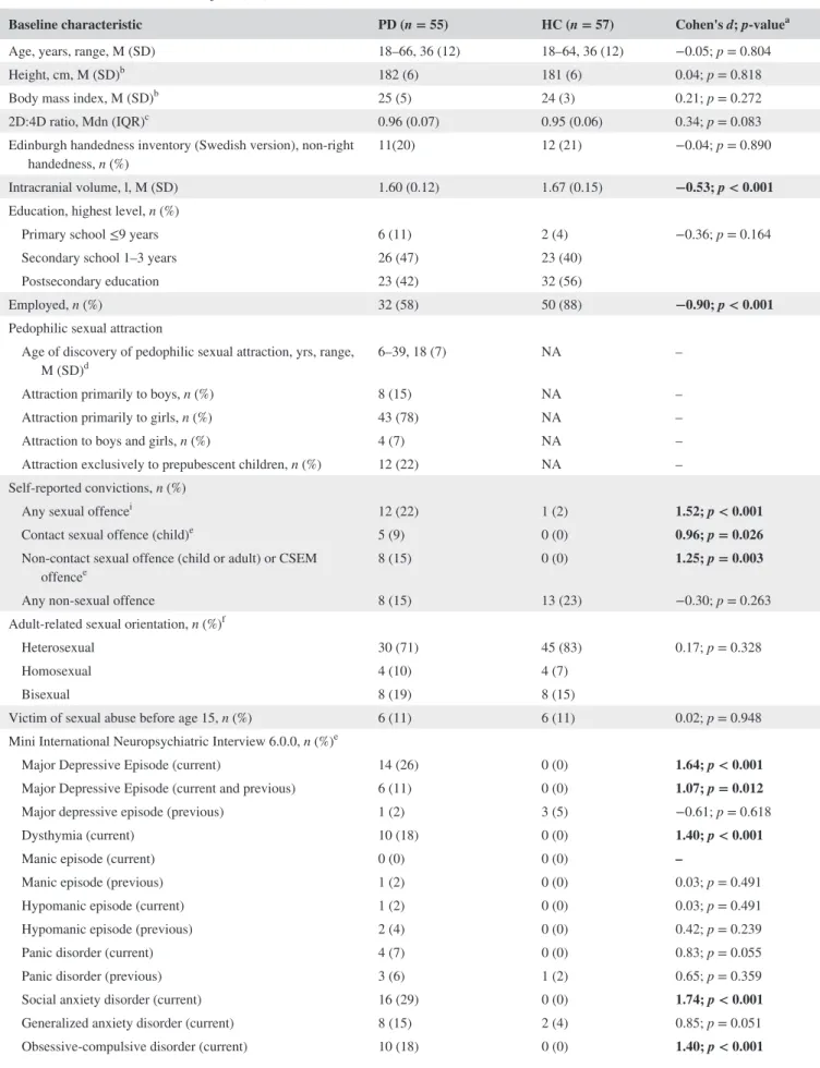
Figure 1 depicts results of vertex-wise cortical struc-ture analyses. PD showed smaller cortical surface area in
TABLE 1 Socio-demographic, sexuality, and psychiatric characteristics among male participants with DSM-5 Pedophilic Disorder (PD) and matched non-clinical male control subjects (HC)
Baseline characteristic PD (n = 55) HC (n = 57) Cohen's d; p-valuea
Age, years, range, M (SD) 18–66, 36 (12) 18–64, 36 (12) −0.05; p = 0.804
Height, cm, M (SD)b 182 (6) 181 (6) 0.04; p = 0.818
Body mass index, M (SD)b 25 (5) 24 (3) 0.21; p = 0.272
2D:4D ratio, Mdn (IQR)c 0.96 (0.07) 0.95 (0.06) 0.34; p = 0.083
Edinburgh handedness inventory (Swedish version), non-right
handedness, n (%) 11(20) 12 (21) −0.04; p = 0.890
Intracranial volume, l, M (SD) 1.60 (0.12) 1.67 (0.15) −0.53; p < 0.001 Education, highest level, n (%)
Primary school ≤9 years 6 (11) 2 (4) −0.36; p = 0.164
Secondary school 1–3 years 26 (47) 23 (40)
Postsecondary education 23 (42) 32 (56)
Employed, n (%) 32 (58) 50 (88) −0.90; p < 0.001
Pedophilic sexual attraction
Age of discovery of pedophilic sexual attraction, yrs, range,
M (SD)d 6–39, 18 (7) NA –
Attraction primarily to boys, n (%) 8 (15) NA –
Attraction primarily to girls, n (%) 43 (78) NA –
Attraction to boys and girls, n (%) 4 (7) NA –
Attraction exclusively to prepubescent children, n (%) 12 (22) NA – Self-reported convictions, n (%)
Any sexual offencei 12 (22) 1 (2) 1.52; p < 0.001
Contact sexual offence (child)e 5 (9) 0 (0) 0.96; p = 0.026
Non-contact sexual offence (child or adult) or CSEM
offencee 8 (15) 0 (0) 1.25; p = 0.003
Any non-sexual offence 8 (15) 13 (23) −0.30; p = 0.263
Adult-related sexual orientation, n (%)f
Heterosexual 30 (71) 45 (83) 0.17; p = 0.328
Homosexual 4 (10) 4 (7)
Bisexual 8 (19) 8 (15)
Victim of sexual abuse before age 15, n (%) 6 (11) 6 (11) 0.02; p = 0.948 Mini International Neuropsychiatric Interview 6.0.0, n (%)e
Major Depressive Episode (current) 14 (26) 0 (0) 1.64; p < 0.001
Major Depressive Episode (current and previous) 6 (11) 0 (0) 1.07; p = 0.012
Major depressive episode (previous) 1 (2) 3 (5) −0.61; p = 0.618
Dysthymia (current) 10 (18) 0 (0) 1.40; p < 0.001
Manic episode (current) 0 (0) 0 (0) –
Manic episode (previous) 1 (2) 0 (0) 0.03; p = 0.491
Hypomanic episode (current) 1 (2) 0 (0) 0.03; p = 0.491
Hypomanic episode (previous) 2 (4) 0 (0) 0.42; p = 0.239
Panic disorder (current) 4 (7) 0 (0) 0.83; p = 0.055
Panic disorder (previous) 3 (6) 1 (2) 0.65; p = 0.359
Social anxiety disorder (current) 16 (29) 0 (0) 1.74; p < 0.001
Generalized anxiety disorder (current) 8 (15) 2 (4) 0.85; p = 0.051
Obsessive-compulsive disorder (current) 10 (18) 0 (0) 1.40; p < 0.001
clusters comprising (i) bilateral ventromedial prefrontal cortex (vmPFC, also including medial superior frontal, me-dial orbitofrontal, and rostral anterior cingulate cortex), (ii) caudal and posterior cingulate cortex, precuneus and para-central cortex, supramarginal and superior/inferior parietal cortex, (iii) left fusiform/inferior temporal and lingual cortex (iv) middle and superior temporal cortex, (v) insula, lateral prefrontal cortex (pars opercularis/triangularis, caudal/rostral
middle frontal), extending into pre- and postcentral cor-tex. Yet not as pronounced, PD subjects also showed lower cortical volume in regions largely in agreement with those where surface area abnormalities were found. We observed no group differences in cortical thickness. A more detailed description, corresponding cluster statistics, and uncorrected results are available in the Supporting Information (Figure S1, Tables S1 and S2).
Baseline characteristic PD (n = 55) HC (n = 57) Cohen's d; p-valuea
Post-traumatic stress disorder (current) 1 (2) 0 (0) 0.03; p = 0.491
Psychotic syndrome (current) 1 (2) 0 (0) 0.03; p = 0.491
Any psychotic symptoms (current) 2 (4) 0 (0) 0.42; p = 0.239
Anorexia nervosa (current) 0 (0) 0 (0) –
Bulimia nervosa (current) 0 (0) 1 (2) 0.01; p = 1.00
Antisocial personality disorder (lifetime) 9 (16) 1 (2) 1.32; p = 0.008
Psychoactive medication, n (%)
Antidepressant 15 (27) 3 (5) 1.05; p = 0.002
Otherg 12 (22) 3(5) 0.90; p = 0.009
Ritvo Autism and Asperger Diagnostic Scale – screening toolh
Total sum (range 0–42), Mdn (IQR) 18 (20) 4 (5) 0.99; p < 0.001
Mentalizing subscale score (range 0–21), Mdn (IQR) 10 (12) 2 (3) 0.98; p < 0.001 Sensory reactivity subscale score (range 0–9), Mdn (IQR) 3 (4) 0 (3) 0.77; p < 0.001 Social anxiety subscale score (range 0–12), Mdn (IQR) 4 (8) 1 (3) 0.88; p < 0.001
Total score >13, n (%) 32 (60) 5 (9) 1.57; p < 0.001
Total score >22, n (%) 17 (32) 2 (4) 1.41; p < 0.001
Adult ADHD Self-Report Scale – Screen (range 1–6), total
score >3, n (%)h 20 (38) 9 (16) 0.65; p = 0.009
Alcohol Use Disorders Identification Test (range 0–40), total
score >7, n (%)h 6 (11) 9 (16) −0.21; p = 0.495
Drug Use Disorders Identification Test (range 0–44), total
score >5, n (%)e,h 4 (8) 2 (4) 0.45; p = 0.426
Hypersexual Behavior Inventory (range 19–95), total score
>52, n (%)h 33 (62) 1 (2) 2.50; p < 0.001
Wechsler Adult Intelligence Scale–IVb
Full-scale IQ, Mdn (IQR) 101 (23) 114 (15) −0.82; p < 0.001
Verbal comprehension index, Mdn (IQR) 104 (23) 118 (14) −1.02; p < 0.001
Perceptual reasoning index, Mdn (IQR) 110 (18) 113 (20) −0.29; p = 0.127
Note: For reasons of completeness, we exchanged a frequency of 0 to 1 in effect size calculations to obtain Cohen's d estimates.
Abbreviations: CSEM, child sexual exploitation material; NA, not applicable. aBolded figures are significant at p < 0.05 (two-tailed).
bMissing data from 1 HC. cMissing data from 1 PD participant.
dFour PD participants reporting they “had always known” about their pedophilic sexual attraction were excluded from this comparison. eExpected counts were <5 in one or more cells and p-values calculated with Fisher's Exact Test.
fPD participants exclusively attracted to children were excluded (n = 12), missing data from 1 PD and 3 HC participants. gOther psychoactive medications included stimulants, mood stabilizers, and sleep medication, e.g., antihistamines. hMissing data from 2 PD participants.
iOne PD patient reported both contact and non-contact sexual offences.
3.3
|
Case-control differences in
subcortical and WM volume (secondary
analyses)
Compared with HC, PD subjects demonstrated lower (bilat-eral) hippocampal and right accumbens volumes (Table S3). PD showed lower WM volumes in areas that largely align with brain regions in which cortical WM-surface area abnormalities were found (Table S4). No PD > HC patterns were present.
3.4
|
Sensitivity tests
Our results generally remained robust when correcting for de-mographic and clinical variables, with a few exceptions; for example, when correcting for intracranial volume (ICV) or IQ, several group comparisons of WM and subcortical volumes were no longer statistically significant. We want to empha-size that controlling for ICV and IQ may indirectly control for the PD phenotype and disguise effects of interest, which may explain the observed decrease in significance in some brain areas.27 Hence, results obtained when correcting for these fac-tors should be interpreted with caution. When correcting for sexual offending, group differences in WM volume underly-ing right caudal anterior cunderly-ingulate, right paracentral, and left posterior cingulate cortex were no longer statistically signifi-cant. Results of sensitivity tests are presented in more detail in the Supporting Information (Table S5 and S6).
3.5
|
Correlations with 2D:4D
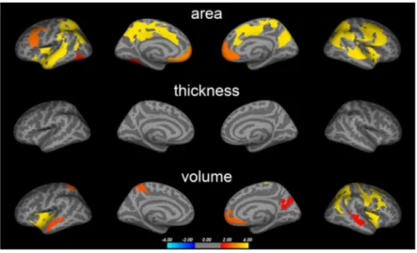
We found both significant positive and negative correla-tions between 2D:4D and cortical surface area, thickness, and volume in PD patients (Figure 2). These areas largely overlapped with regions in which PD-related abnormalities were found (Figure 3). The regions included the inferior fron-tal cortex (pars triangularis, pars opercularis; positive), pre-cuneus (negative), medial superior frontal and caudal ACC (positive), superior parietal (negative), inferior parietal (posi-tive), middle and inferior temporal (posi(posi-tive), and the insula cortex (positive). In line with this, we also found suggestive correlations between 2D:4D and WM volume underlying the right caudal ACC (positive) and bilateral superior parietal volumes (negative) (Table S8).
3.6
|
Multivariate classification and
pattern matching
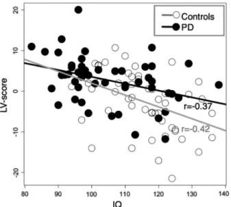
IQ exhibited a consistent negative association with the global morphometric PD abnormality expression score, which was characterized mainly by reductions in parietal, temporal, and prefrontal volumes (Figure S6). The relationship between the global morphometric PD abnormality expression score and IQ was present in the full sample and within PD and HC groups, respectively (Table S7 and Figure 4), indicating that associations are not driven by group differences.
FIGURE 1 Brain abnormalities in PD (main analysis). Colored clusters represent areas in which significant differences in cortical surface area, thickness, and volume were found between PD and HC subjects. Data underwent Monte Carlo cluster-wise corrections for multiple comparisons (see Figure S1 for uncorrected results). Statistical significance is displayed on a log(p)-scale where positive values (warm colors) represent the PD < HC contrast. No negative values (cold colors) reflecting PD > HC patterns were observed. Cluster-wise statistics are given in Table S1
4
|
DISCUSSION
In this single-center study of pedophilic disorder (PD), we present a PD profile from detailed characterization of clinical symptom-atology, cognitive functioning, and brain abnormalities. Brain measures included cortical thickness, surface area, and volume, as well as subcortical and regional white matter (WM) volumes. Additionally, we investigated associations between brain struc-ture and IQ as well as prenatal androgen exposure (2D:4D).
4.1
|
Indications for neurodevelopmental
contributions to PD
We found that PD was associated with smaller cortical sur-face area in several brain regions. Overall, these findings
remained when adjusting for IQ variability. In contrast, no group differences in cortical thickness were observed. Our analyses revealed that cortical surface abnormalities were more pronounced in the vicinity of WM (Figure S4). Consistent with this, we observed smaller regional WM vol-umes in patients with PD compared to HC participants in re-gions where surface abnormalities were found. This agrees with a previous study that reported prefrontal, microstruc-tural WM matter alterations in PD using diffusion tensor imaging.28 The exclusive alteration of surface area, but not cortical thickness, in PD could be related to the fact that these two imaging phenotypes are influenced by distinct develop-mental mechanisms.29,30 For example, genetic influences on cortical thickness and surface area appear to differ.29,30 Thus, our findings suggest that mechanisms affecting surface area may play a role in PD. Although the exact mechanisms behind
FIGURE 2 2D:4D correlations. Correlations between 2D:4D ratios and cortical surface area, thickness, and volume in PD. Colored areas represent brain regions in which significant correlations were observed after multiple comparison correction (see Figure S3 for uncorrected results). Statistical significance is displayed on a -log(p) scale. Cold colors represent negative and warm colors positive correlations, respectively
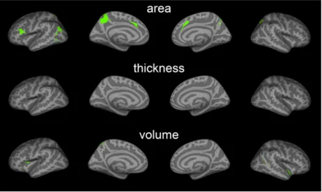
FIGURE 3 2D:4D correlations and PD-abnormality conjunction maps. Green areas indicate the regional overlap between areas where 2D:4D correlations (Figure 2) and PD-related abnormalities were found (Figure 1)
determination of cortical thickness and surface area remain largely unknown, they are believed to exert effects during the pre- and perinatal periods of neurodevelopment.29–31
Pedophilic disorder has been suggested to be associated with aberrant neurodevelopment.2–4 Consistent with that no-tion, we found that the 2D:4D ratio, a marker of prenatal an-drogen exposure, correlated with both cortical surface area and WM volume in those brain regions where PD differed from HC participants. Thus, our findings support prenatal androgen exposure as a possible determinant of the PD neu-ral signature. Androgen effects have been linked to myelin-ation,32 affecting WM volume and in turn cortical surface area, but not thickness.33 Although this is a plausible neuro-developmental explanation to our findings, the exact mech-anisms that contribute to the brain abnormalities found and the origin of the observed correlations are likely complex and cannot be derived from this study. Further research is needed to investigate this emerging hypothesis.
Moreover, we found lower IQ in PD compared with HC men, primarily driven by differences in verbal comprehen-sion, and the expressed PD-related brain abnormality pattern correlated negatively with total IQ. This is in agreement with a previous study reporting that IQ-mediated cortical differ-ences between controls and individuals with pedophilic inter-est.13 Since IQ is highly influenced by neurodevelopmental factors, this may suggest that the overall PD-related struc-tural abnormality pattern could arise from neurodevelopmen-tal mechanisms. This possibility, however, and the mediating mechanisms behind the link between IQ and PD-related brain abnormalities remain unsettled.
Moreover, PD patients expressed clinical symptoms of the neurodevelopmental disorders ADHD and ASD.1 Previous studies have reported higher prevalence of ADHD among in-dividuals with PD. Here, we demonstrate that this also applies to ASD symptoms. Interestingly, developmental trajectories of cortical surface area, but not thickness, have been shown to deviate from normal in autism.34 This aligns with the present findings of different cortical surface area but not thickness. Although autistic traits may be highly relevant for the clini-cal profile of PD, we controlled for it in sensitivity analyses, indicating that observed structural brain abnormalities were specifically related to PD and not to ASD symptoms per se.
4.2
|
Brain regions affected in PD
Although we did not replicate previously reported amygdala alterations in PD,6 our cortical surface area findings largely agreed with those of a recent multi-center study,13 not only with respect to outcome measure but also to anatomical loca-tion. However, the brain abnormalities observed in the previ-ous study were reportedly related to offender status rather than pedophilic interest. This discrepancy could be explained by differences in study design and analytical strategies, but also by limited statistical power and potential confounding both due to the merging of small samples investigated with numerous different MRI scanners in previous studies. We were able to use the same MRI scanner and controlled for several potential confounds, including sexual offending. Our sensitivity analyses indicated that the abnormalities reported here were indeed related to PD, and not to sexual offender status or other clinical and demographic variables.
Specifically, we found PD-related abnormalities in the ventromedial prefrontal cortex (vmPFC), posterior cingulate, precuneus, and inferior parietal lobule, including the angular gyrus and supramarginal cortex. These brain structures are important hub regions of the brain's default mode network (DMN).35–37 The DMN also contains the lateral middle tem-poral cortex, temtem-poral pole, temporoparietal junction (TPJ), and hippocampus35–37; regions also altered in PD participants. Hence, the structural abnormality pattern observed in PD occurs primarily in DMN regions. This network is involved in internal and self-related processes, but also in social cog-nition including thinking about others, their emotions and thoughts.35–37 The DMN is also recruited when imagining future consequences of one's own actions and moral rea-soning.35–37 Intriguingly, the DMN is impaired in several psychiatric conditions, including ADHD and ASD, and puta-tively involved in the clinical expression of these disorders.37 Notably, the DMN also interacts with other brain regions and networks with similar functions. For example, PD subjects showed abnormalities in the lateral prefrontal and anterior in-sula cortex, key regions for cognitive control, decision-making,
FIGURE 4 Correlation between global morphometric PD abnormality expression score (LV-score) and IQ. Relationships in this plot are quantified with Pearson correlation coefficients: r = −0.37 (p = 0.005) and r = −0.42 (p = 0.001) for PD and control groups, respectively
social cognition, and integration of self-related and emotional processes.38,39 Behavioral aspects reportedly associated with PD in forensic samples include impulsivity and risk taking as well as impaired social skills, empathy, moral reasoning, and self-reflection.4,40 Hence, we speculate that the brain ab-normalities observed here may relate to increased PD-related behaviors. However, this cannot be concluded from this study, and may only apply to a sub-group of patients. Notably, not all PD patients showed pronounced ASD symptoms. Further, it remains unknown how observed brain abnormalities relate to the development of the core symptom of PD; the sexual attraction to prepubescent children. Within the PD group, we observed brain differences related to pedophilic sexual orien-tation (sexual attraction toward girls and/or boys), which do not appear to align with regions commonly associated with adult-related sexual orientation41 (Figure S2). The present findings may constitute initial steps toward understanding the complex interplay of neurobiological factors underlying sex-ual attraction patterns in PD.
In conclusion, in the largest single-center study to date, we investigated self-referred, help-seeking patients with DSM-5 PD in a non-correctional, clinical setting. Our re-sults suggest that PD co-occurs with substantial psychiatric morbidity including depressive and anxiety disorders, clini-cal symptoms of ADHD and ASD, and somewhat lower IQ, albeit still in the normal range. We report that PD is related to cortical surface area alterations and underlying white mat-ter abnormalities, primarily in brain regions belonging to the default mode network. The results were not confounded by demographic or clinical variables, and importantly, unrelated to self-reported sexual offence history. The neuroanatomical findings, their association with IQ and 2D:4D, and the symp-tom profile suggest that PD may have neurodevelopmental underpinnings. The results could inform the development of preventive and supportive interventions for PD patients.
4.3
|
Strengths and limitations
This study is of substantial clinical relevance through the comprehensive characterization of a representative clini-cal sample of self-referred and help-seeking men with PD. Potential limitations are that structural neuroimaging can-not assess the functional involvement of brain regions in the expression of PD. Functional MRI studies are necessary to quantify the neural correlates of cognitive and behavioral as-pects of PD. Second, the cross-sectional study design does not allow for inference regarding causality. Third, it is unknown whether PD patients with psychiatric comorbidity, including symptoms of ASD, are more prone to seek help. However, the investigated sample is representative for self-referred individuals with PD in a clinical, non-correctional setting. Finally, although a history of sexual offending behavior did
not explain our findings, prospective studies following PD patients over longer periods of time might help identify clini-cal, cognitive, and neurobiological variables that relate to the risk of child sexual abuse.
ACKNOWLEDGEMENTS
This study was funded by the Swedish Society for Medicine (SLS-501421and SLS-886481), the Swedish Society for Medical Research (P14-0136), the Söderström König Foundation; the Fredrik and Ingrid Thuring Foundation (FITS-2015-00157), the Center for Psychiatry Research, Department of Clinical Neuroscience, Karolinska Institutet (CPF-99/2016), and by the ALF agreement between the Swedish government and the county councils (SLL20150518 and SLL20160555). We thank Maria Andersson, Kerstin Eriksson, Pia Jaensen, and Susanne Jarlvik Alm from Karolinska University Hospital for assistance with data col-lection, and Tomas Jonsson, Maria Kristoffersen-Wiberg, and Torkel Brismar for help with the trial organization. We thank the staff at ANOVA and PrevenTell for participant recruitment.
CONFLICT OF INTEREST
None.
AUTHORS’ CONTRIBUTIONS
CR and BL contributed with study design and conceptual-ization. CA conceived analyses, performed most brain im-aging analyses, and wrote the first draft of the manuscript. RA planned and performed analyses of clinical and behav-ioral measures together with NL. CM performed analyses on white matter and subcortical measures and AL conducted classification analyses. CA, RA, NL, CM, and AL created tables and figures. All authors contributed with interpretation of data, critically revised the manuscript for intellectual con-tent, approved the final version of the manuscript for publica-tion, and agree to be accountable for all aspects of the work.
PEER REVIEW
The peer review history for this article is available at https:// publo ns.com/publo n/10.1111/acps.13273.
DATA AVAILABILITY STATEMENT
Data access and codes can be obtained on reasonable request.
ORCID
Christoph Abé https://orcid.org/0000-0002-1680-8480 Alexander Lebedev https://orcid.org/0000-0002-0319-2357
REFERENCES
1. American Psychiatric Association. Diagnostic and Statistical Manual of Mental Disorders, 5th edn. Arlington, VA: American Psychiatric Association; 2013.
2. Fazio RL. Toward a neurodevelopmental understanding of pedo-philia. J Sex Med. 2018;15(9):1205-1207.
3. Becerra Garcia JA. Etiology of pedophilia from a neurodevelop-mental perspective: markers and brain alterations. Rev Psiquiatr Salud Ment. 2009;2(4):190-196.
4. Tenbergen G, Wittfoth M, Frieling H, et al. The neurobiology and psychology of pedophilia: recent advances and challenges. Front Hum Neurosci. 2015;9:344.
5. Blanchard R, Kolla NJ, Cantor JM, et al. IQ, handedness, and pe-dophilia in adult male patients stratified by referral source. Sex Abuse. 2007;19(3):285-309.
6. Mohnke S, Muller S, Amelung T, et al. Brain alterations in paedo-philia: a critical review. Prog Neurogibol. 2014;122:1-23. 7. Suchy Y, Eastvold AD, Strassberg DS, Franchow EI. Understanding
processing speed weaknesses among pedophilic child molest-ers: response style vs. neuropathology. J Abnorm Psychol. 2014;123(1):273-285.
8. Dyshniku F, Murray ME, Fazio RL, Lykins AD, Cantor JM. Minor physical anomalies as a window into the prenatal origins of pedo-philia. Arch Sex Behav. 2015;44(8):2151-2159.
9. McPhail IV, Pedophilia CJM. Pedophilia, height, and the magnitude of the association: a research note. Deviant Behavior. 2015;36(4):288-292. https://doi.org/10.1080/01639 625.2014.935644
10. de Sanctis V, Soliman AT, Elsedfy H, Soliman N, Elalaily R, Di Maio S. Is the second to fourth digit ratio (2D:4D) a bio-marker of sex-steroids activity? Pediatr Endocrinol Rev. 2017;14(4):378-386.
11. Rahman Q, Symeonides DJ. Neurodevelopmental cor-relates of paraphilic sexual interests in men. Arch Sex Behav. 2008;37(1):166-172.
12. Gerwinn H, Weiss S, Tenbergen G, et al. Clinical characteristics associated with paedophilia and child sex offending - differen-tiating sexual preference from offence status. Eur Psychiatry. 2018;51:74-85.
13. Lett TA, Mohnke S, Amelung T, et al. Multimodal neuroimaging measures and intelligence influence pedophile child sexual offense behavior. Eur Neuropsychopharmacol. 2018;28(7):818-827. 14. Landgren V, Malki K, Bottai M, Arver S, Rahm C. Effect of
go-nadotropin-releasing hormone antagonist on risk of committing child sexual abuse in men with pedophilic disorder: a randomized clinical trial. JAMA Psychiatry. 2020;77(9):897–905.
15. Sheehan DV, Lecrubier Y, Sheehan KH, et al. The Mini-International Neuropsychiatric Interview (M.I.N.I.): the development and vali-dation of a structured diagnostic psychiatric interview for DSM-IV and ICD-10. J Clin Psychiatry. 1998;59(Suppl 20):22-33. 16. World Health Organization. Report of the Consultation on Child
Abuse Prevention, 29–31 March 1999. WHO, Geneva: Violence and Injury Prevention Team & Global Forum for Health Research; 1999.
17. Eriksson JM, Andersen LM, Bejerot S. RAADS-14 Screen: va-lidity of a screening tool for autism spectrum disorder in an adult psychiatric population. Mol Autism. 2013;4(1):49.
18. Kessler RC, Adler L, Ames M, et al. The World Health Organization Adult ADHD Self-Report Scale (ASRS): a short screening scale for use in the general population. Psychol Med. 2005;35(2):245-256.
19. Saunders JB, Aasland OG, Babor TF, de la Fuente JR, Grant M. Development of the Alcohol Use Disorders Identification Test (AUDIT): WHO collaborative project on early detection
of persons with harmful alcohol consumption–II. Addiction. 1993;88(6):791-804.
20. Berman AH, Bergman H, Palmstierna T, Schlyter F. Evaluation of the Drug Use Disorders Identification Test (DUDIT) in crimi-nal justice and detoxification settings and in a Swedish population sample. Eur Addict Res. 2005;11(1):22-31.
21. Reid RC, Garos S, Carpenter BN. Reliability, validity, and psy-chometric development of the Hypersexual Behavior Inventory in an outpatient sample of men. Sex Addict Compulsivity. 2011;18(1):30-51.
22. Wechsler D. WAIS IV: Wechsler Adult Intelligence Scale – Fourth Edition. Stockholm: Swedish version. NCS Pearson Inc.; 2010. 23. Oldfield RC. The assessment and analysis of handedness: the
Edinburgh inventory. Neuropsychologia. 1971;9(1):97-113. 24. Fischl B, Dale AM. Measuring the thickness of the human cerebral
cortex from magnetic resonance images. Proc Natl Acad Sci USA. 2000;97(20):11050-11055.
25. Fischl B, van der Kouwe A, Destrieux C, et al. Automatically parcel-lating the human cerebral cortex. Cereb Cortex. 2004;14(1):11-22. 26. Wold S, Ruhe A, Wold H, Dunn WJ III. The collinearity problem
in linear regression. The partial least squares (PLS) approach to generalized inverses. SIAM J Sci Stat Comput. 1984;5(3):735-743. 27. Hyatt CS, Owens MM, Crowe ML, Carter NT, Lynam DR, Miller
JD. The quandary of covarying: a brief review and empirical ex-amination of covariate use in structural neuroimaging studies on psychological variables. NeuroImage. 2020;205:116225.
28. Cantor JM, Lafaille S, Soh DW, Moayedi M, Mikulis DJ, Girard TA. Diffusion tensor imaging of pedophilia. Arch Sex Behav. 2015;44(8):2161-2172.
29. Winkler AM, Kochunov P, Blangero J, et al. Cortical thickness or grey matter volume? The importance of selecting the phenotype for imaging genetics studies. NeuroImage. 2010;53(3):1135-1146. 30. Rakic P. Specification of cerebral cortical areas. Science.
1988;241(4862):170-176.
31. Bhardwaj RD, Curtis MA, Spalding KL, et al. Neocortical neuro-genesis in humans is restricted to development. Proc Natl Acad Sci USA. 2006;103(33):12564-12568.
32. Bielecki B, Mattern C, Ghoumari AM, et al. Unexpected central role of the androgen receptor in the spontaneous regeneration of myelin. Proc Natl Acad Sci USA. 2016;113(51):14829-14834. 33. Cafiero R, Brauer J, Anwander A, Friederici AD. The concurrence
of cortical surface area expansion and white matter myelination in human brain development. Cereb Cortex. 2019;29(2):827-837. 34. Mensen VT, Wierenga LM, van Dijk S, et al. Development of
cortical thickness and surface area in autism spectrum disorder. Neuroimage Clin. 2017;13:215-222.
35. Mak LE, Minuzzi L, MacQueen G, Hall G, Kennedy SH, Milev R. The default mode network in healthy individuals: a systematic review and meta-analysis. Brain Connect. 2017;7(1):25-33. 36. Raichle ME. The brain's default mode network. Annu Rev
Neurosci. 2015;38:433-447.
37. Andrews-Hanna JR, Smallwood J, Spreng RN. The default network and self-generated thought: component processes, dynamic con-trol, and clinical relevance. Ann N Y Acad Sci. 2014;1316:29-52. 38. Craig AD. How do you feel–now? The anterior insula and human
awareness. Nat Rev Neurosci. 2009;10(1):59-70.
39. Tanji J, Hoshi E. Role of the lateral prefrontal cortex in executive behavioral control. Physiol Rev. 2008;88(1):37-57.
40. Cohen LJ, Galynker II. Clinical features of pedophilia and implica-tions for treatment. J Psychiatr Pract. 2002;8(5):276-289.
41. Abé C, Rahman Q, Langstrom N, Ryden E, Ingvar M, Landen M. Cortical brain structure and sexual orientation in adult females with bipolar disorder or attention deficit hyperactivity disorder. Brain Behav. 2018;8(7):e00998.
SUPPORTING INFORMATION
Additional supporting information may be found online in the Supporting Information section.
How to cite this article: Abé C, Adebahr R, Liberg B,
et al. Brain structure and clinical profile point to neurodevelopmental factors involved in pedophilic disorder. Acta Psychiatr Scand. 2021;143:363–374. https://doi.org/10.1111/acps.13273