Research
Report number: 2019:26 ISSN: 2000-0456 Available at www.stralsakerhetsmyndigheten.se
Analysis of trends in brain tumour
incidence in Sweden
.
2019:26
Authors: Jonas NilssonMichael Bergqvist Georg Holgersson Jacob Järås Stefan Bergström Roger Henriksson
SSM 2019:26
SSM perspective
Background
In the past few years, a discussion has been ongoing in both the research community and the media as to whether exposure to radiowaves from the use of mobile phones has an impact on the incidence of brain tumours in the general population. Ever since the introduction of mobile phones in the 1980s, their use has increased dramatically and has now spread to almost all parts of the world. According to the International Telecommunication Union, there were nearly 7 billion mobile phone subscriptions globally in 2014. Because this distribution is so widespread, there is a great need to investigate possible impacts on health. In 2011, the International Agency for Research on Cancer (IARC) gathered experts in the field to form a working group for discussion of the outcomes of relevant studies. The decision was made to classify mobile phones as “possibly carcinogenic to humans” mainly based on indications of an elevated risk of gliomas, a kind of brain tumour.
Sweden is one of the countries where mobile phone use blossomed early on, for which reason incidence trends based on Swedish registries are of per-tinent interest in the puzzle of knowledge that needs to be put together to form a solid foundation for risk assessments. However, it is important to keep in mind that this kind of analysis can never prove the lack of, nor the exist-ence of, a correlation. Incidexist-ence trends can be influexist-enced by a wide range of factors, apart from direct registry factors (changed coding practice, and the like), in addition to environmental factors and changed lifestyle patterns; other possible aspects are not only protective factors, but also risk factors whose strength is variable over time. Another aspect that should be consid-ered is that some illnesses are characterised by a long latency period. Conse-quently, they may not manifest themselves until many years after an exposure situation. Nonetheless, it is reasonable to assume that a strong link between radiowave exposure and brain tumour development would have a clear impact on incidence trends within a period of 15 years following large-scale imple-mentation of mobile telephony.
Results
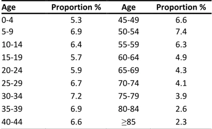
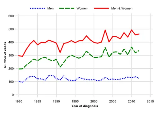
This project initially compiled data from the Swedish Cancer Registry for the purpose of identifying all brain tumour diagnoses made between the years 1980 and 2013, and subsequently estimating the incidences of the brain tumour types of low-grade gliomas, high-grade gliomas, and meningiomas during this period. Also, via Statistics Sweden (SCB), data was collected on these patients’ incomes in order to analyse a possible correlation between income and incidence. It transpired that there was a time lag in the statistics that risked causing substantial underestimation of the number of identi-fied cases in 2013. The findings from the entire period have been taken into account in this report, though the presentation of incidence trend param-eters has consequently been limited to the period 1980-2012. In total during the period 1980-2012, 30,255 primary brain tumours were identified on the part of 30,142 patients.
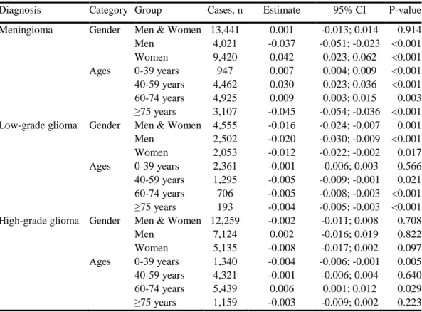
In the case of the meningioma tumours, there was an increased incidence in the age group 0-74. Patients over 75 years of age showed a decrease in the
SSM 2019:26
annual incidence. The overall conclusion, based on all the age groups, was that no significant incidence trend was observed for meningiomas.
The researchers observed a slight decrease in the annual incidence of high-grade brain tumours among patients in the age group 0-39 years, and an increase in the age group 60-74 years of age (p=0.029). On the whole, high-grade brain tumours showed no overall incidence trend.
The outcomes demonstrated a statistically significant decrease in the annual incidence of low-grade gliomas during the period 1980-2012. In terms of age group classification, this significant decrease in low-grade glioma incidence was observed in all age groups above the age of 39. However, these findings should be interpreted cautiously. It is clear that the outcomes largely depend on selecting where the survey begins and ends. Nonetheless, there does not seem to be any long term, upward trend.
No clear difference could be discerned between males and females in terms of incidence trends for all the types of tumours, with the exception of low-grade gliomas (p=0.001). The outcomes from the incidence’s dependency on income are irregular, or rapidly variable, on the part of higher incomes. This indicates an all too low statistical volume and provides no information about possible correlations.
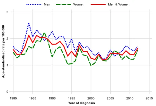
Overall, there was no clear increase in the annual incidence of the stud-ied tumour types from the years 1980-2012, a period of time during which mobile phone use increased sharply. Nevertheless, it should be noted that this study did not investigate a specific correlation between mobile phone use and brain tumour incidence. Many factors have an influence on the development of brain tumours.
All the same, the observation has been made that the presented findings do not provide evidence that any environmental factor encompassing a large proportion of the population (for instance, mobile phone use) has had a material impact on the risk of development of brain tumours between 1980 and 2012.
Relevance
The present report provides support for the previous assessment made by the Swedish Radiation Safety Authority: Exposure to radiowaves while using mobile phones does not pose any significant risks leading to brain tumours. The recommendation to observe precautions by using hands-free equipment during mobile phone calls nevertheless remains in effect, mainly due to long-term uncertainties and indications of biological effects in animal studies. Need for further research
As this study has only had access to a somewhat complete body of data up to and including 2012, it is strongly recommended to carry out an update. It would be appropriate to launch this kind of study in 2021, with an analysis of statistics up to and including 2017.
Project information
Contact person SSM: Torsten Augustsson Referens: SSM 2016-498 / 7030054-00
2019:26
Authors:
Date: December 2019
Report number: 2019:26 ISSN: 2000-0456 Available at www.stralsakerhetsmyndigheten.se Jonas Nilsson Michael Bergqvist Georg Holgersson Jacob Järås Stefan Bergström Roger Henriksson
Analysis of trends in brain tumour
SSM 2019:26
This report concerns a study which has been conducted for the Swedish Radiation Safety Authority, SSM. The conclusions and view-points presented in the report are those of the author/authors and do not necessarily coincide with those of the SSM.
1
Swedish Radiation Safety
Authority
Research protocol SSM2015-3265
No support for increased brain tumour incidence
between the years 1980 and 2012, based on data
from the Swedish National Register.
Authors:
Jonas Nilsson1+3+5, Michael Bergqvist1+2+3, Georg Holgersson1+2, Jacob Järås4, Stefan Bergström1+2 and Roger Henriksson3+4
1 Center for Research & Development, Uppsala University/ County
Council of Gävleborg, Gävle Hospital, SE-801 87 Gävle, Sweden
2 Department of Oncology, Gävle Hospital, SE-801 87 Gävle, Sweden 3 Department of Radiation Sciences & Oncology, Umeå University
Hospital, SE-901 87 Umeå, Sweden
4 Regional Cancer Center Stockholm-Gotland
5 Department of Radiology, Gävle Hospital, SE-801 87 Gävle,
2
Table of contents
Introduction
1. Radiation - ionising versus non-ionising ... 6
2. Brain tumours ... 6
The 2007 WHO Classification ... 6
Astrocytic tumours ... 7
Oligodendroglial tumours ... 9
Oligoastrocytic tumours ... 9
Ependymal tumours ... 10
Choroid plexus tumours ... 10
Other neuroepithelial tumours ... 11
Neuronal and mixed neuronal-glial tumours ... 11
Pineal tumours ... 12
Embryonal tumours ... 12
Tumours of the cranial and paraspinal nerves ... 13
Meningeal tumours ... 14
Tumours of the sellar region ... 15
3. In vitro studies ... 17
Apoptosis and cell damage ... 17
Heat shock proteins ... 18
P53 ... 19
Genotoxicity ... 20
4. Epidemiologic studies investigating the role of mobile phones and brain tumour development ... 21
1998-2002 ... 21
2003-2007 ... 24
3
2013-2015 ... 41
Materials and Methods
5.1 Electronic database searches ... 435.2 Statistical methods ... 43
Results
6.1 Overall results ... 466.2 Results in graphic presentation ... 48
Discussion ... 60
4
Sammanfattning
Under de senaste åren har det debatterats huruvida miljöfaktorer kan påverka incidensen av hjärntumörer i den generella populationen. En av de huvudsakliga faktorer som har omnämnts i dessa sammanhang är användandet av mobiltelefoner. Sedan introduktionen av
mobiltelefoner under 80-talet har användandet ökat kraftigt och når nu ut till nästan alla världens hörn. Enligt International
Telecommunication Union fanns det år 2014 nära 7 miljarder mobiltelefonabonnemang världen över. Eftersom utbredningen är så omfattande så är det av högsta vikt att ta reda på dess potentiellt skadliga effekter hos människa. Under 2011 samlade the International Agency for Research on Cancer (IARC) flertalet experter inom området för att i en arbetsgrupp diskutera resultaten av aktuella studier. Man beslutade att riskklassificera mobiltelefoni som ”möjligt carcinogen för människan” baserat på tecken till en ökad risk för uppkomst av hjärntumörtypen gliom.
Primära hjärntumörer är de tumörer som har sitt ursprung i hjärnan, hjärnhinnorna, kranialnerverna, hypofysen eller tallkottkörteln. Tumörerna delas i sin tur upp i subgrupperingar beroende på dess egenskaper och aggressivitetsgrad enligt World Health Organization (WHO) fyrgradiga klassifikationssystem. Låggradiga hjärntumörer (WHO grad 1-2) beskriver tumörer med låg tillväxthastighet och sparsam infiltrationsförmåga till omgivande vävnader. Höggradiga gliom (WHO grad 3-4) beskriver tumörer med hög tillväxthastighet, stor infiltrationsförmåga och ett aggressivt beteende.
I denna rapport har vi samlat in data från det svenska cancerregistret för att identifiera samtliga hjärntumördiagnoser mellan åren 1980-2012 och för att sedermera beräkna incidensen av hjärntumörtyperna låggradiga gliom, höggradiga gliom samt meningiom under denna period. Vidare samlade vi via Statistiska Centralbyrån (SCB) in data på inkomst hos dessa patienter för att analysera eventuella samband
5
mellan inkomst och incidens. Totalt identifierades 30 255 primära hjärntumörer hos 30 142 patienter.
Avseende meningiom såg vi en ökning av incidensen i åldrarna 0-39, 40-59 och 60-74, medan en sjunkande incidenstrend sågs i
åldersgruppen ≥ 75 år. Män och kvinnor visade på en minskning samt ökning av incidensen, respektive. Sammantaget alla åldrar sågs ingen signifikant incidenstrend av meningiom.
Vi såg en diskret minskning av årliga incidensen av höggradiga gliom hos patienter i åldersgruppen 0-39 år och en diskret ökning i
åldersgruppen 60-74 år. Ingen skillnad mellan kön iakttogs.
Sammantaget förelåg ingen övergripande incidenstrend av höggradiga gliom.
Vi kunde visa en statistiskt signifikant minskning av den årliga incidensen av låggradiga gliom under tidsperioden 1980-2012. Avseende åldersgrupperingar såg vi denna signifikanta minskning av låggradiga gliom i samtliga åldersgrupper över 40 år. Även för män och kvinnor kunde vi se denna signifikanta minskning av
incidenstrend för låggradiga gliom.
Vid låga inkomster (0-100 tkr/år) sågs lätt sjunkande incidens för meningiom och låggradiga gliom. Vid inkomstgrupperna >100-200 tkr/år och >200-360 tkr/år sågs lätt ökande incidenser för samtliga tumörentiteter. Data gällande inkomst bör dock tolkas med stor försiktighet.
Sammantaget har det inte skett någon ökning av den årliga incidensen för undersökta tumörentiteter från 1980-2012, en tidsperiod då
mobiltelefonanvändande har ökat. Data ska tolkas med försiktighet eftersom att vi inte har undersökt det faktiska sambandet mellan mobiltelefoner och hjärntumörer och det kan finnas ytterligare hittills okända faktorer som kan påverka utvecklingen av hjärntumörer.
6
Introduction
1. Radiation: ionising versus non-ionising
Energy emitted from a source is referred to as radiation. Typical electromagnetic radiation behaves in a “wave-like” manner. Examples of electromagnetic radiation include radio waves, infrared light, visible light, ultraviolet light, X-rays and gamma rays. These only differ in wavelength and frequency.1
Ionizing radiation, for example X-rays and gamma rays, has enough energy to interact with an atom in terms of removing an electron from the orbit of the atom. As a result, the atom becomes charged or
ionised.
Non-ionizing radiation, however, does not have sufficient energy to remove the electron causing ionisation. This type of radiation includes electric and magnetic fields, radio waves, infrared, visible and
ultraviolet radiation.1 Even though ultraviolet radiation is normally classified as non-ionizing radiation, shorter wavelengths can cause molecules to ionise and it has been proven that UV-radiation has a carcinogenic effect.
2. Brain tumours
2.1 The 2007 WHO classification
Based on histopathological assessment, tumours in the central nervous system are divided as per the 2007 WHO classification, using a four-point scale. Per definition a brain tumour is an abnormal tissue within the cranium including the brain, cranial nerves, meninges, skull, pituitary gland and pineal gland.2 In the clinical setting, the grading
system is an important tool to decide choice of therapy, particularly adjuvant radiation and chemotherapy.
There are more than 100 types of brain tumours, mostly emerging from the cells that support the brain cells. These cells are called glial
7
cells and tumours consisting of glial cells are called gliomas.
The grading definition is made by a pathologist based on histology of the tumour. A tumour with a histology close to normal is generally defined as grade I, whilst a tumour with an abnormal histology is classified as a high grade tumour, WHO grade III or IV. High grade tumours (WHO III-IV) are regarded as malignant and these tumours are generally fast growing, often relapse and may spread to other parts of the brain or the spinal cord. Low grade tumours (WHO I-II), on the other hand, generally grow slowly, are less likely to relapse, normally do not spread and may be treated by surgery alone.3
2.2 Astrocytic tumours
Astrocytic brain tumours arise from astrocytes in the brain, which are supporting cells surrounding neurons, giving structural and metabolic support. The astrocytes also regulate ion concentration in the
extracellular space, modulate the synaptic transmission and transmitter reuptake, and are an important part of the blood-brain barrier. There are few known etiologic factors to the development of astrocytic brain tumours. A minority of patients have a hereditary predisposition and in these patients a familial aggregation of brain tumours is often present. Another known etiologic factor is ionizing radiation.4
The astrocytic tumours include all WHO grades within its subgroups. According to the WHO classification, the following subgroups of astrocytic tumours are listed:
Pilocytic astrocytoma (grade I), Subependymal giant cell astrocytoma (grade I), Pleomorphic xanthoastrocytoma (grade II), Pilomyxoid astrocytoma (grade II), Diffuse astrocytoma (grade II), Anaplastic astrocytoma (grade III), Glioblastoma (grade IV), Giant cell glioblastoma (grade IV) and Gliosarcoma (grade IV).5
Pilocytic astrocytoma
The yearly incidence of pilocytic astrocytoma in the United States in the late 1990s was about 0.23 per 100,000 with about 700 new cases
8
per year. Pilocystic astrocytoma generally forms sacks of fluid (cysts) and is classified as one of the most benign astrocytomas of all types. It is normally seen in children or young adults, and is often removed with surgery alone.4,6
Subependymal giant cell astrocytoma
Subependymal giant cell astrocytoma is a ventricular tumour that is
quite common in patients with tuberous sclerosis with a prevalence of 6% to 15% in these patients.4,6 It is a slow-growing tumour that usually forms within the walls of fluid-filled spaces in the brain.7
Pleomorphic xanthoastrocytoma
Pleomorphic xanthoastrocytoma is a rare histologic type of brain
tumour and accounts only for less than 1% of all astrocytomas. Most commonly it affects children and young adults. In most cases this type of tumour affects the temporal lobes of the brain, and thereby they frequently present with seizures.8
Diffuse astrocytoma
Diffuse astrocytomas often contain microcysts and mucous-like fluid.6
The incidence of these tumours is about 0.1 per 100 000 persons per year, and it accounts for about 35% of all astrocytic brain tumours. In North America, 1500 to 1800 new cases are diagnosed each year.4 Diffuse astrocytoma is either of the fibrillary, germistocystic or
protoplasmic type. It has a tendency to invade surrounding brain tissue but usually grows at a quite slow pace. Treatment of diffuse
astrocytomas is usually surgery when the tumour is anatomically accessible and can be completely removed. When treating children or young adults radiation as additional therapy can be discussed. When the tumour is inaccessible or cannot be completely removed, radiation as additional therapy is also often used.6
Anaplastic astrocytoma
The incidence of anaplastic astrocytoma is about 0.49 per 100.000 people per year, and the mean age at diagnosis is 40 years.4 This
high-9
grade tumour tends to invade surrounding tissue, making radical surgery difficult. Treatment includes surgery followed by radiation therapy and additional chemotherapy can also be used.6
Glioblastoma
In Europe, the incidence of glioblastoma is estimated to be about 3.55 new cases per 100.000 people per year and glioblastomas accounts for about 69% of all incident cases of astrocytic and oligodendroglial tumours - and is thereby the most frequent histologic type.9 The tumour might contain cystic material, calcium deposits, blood vessels and a mixed grade of cells. Treatment includes surgery followed by radiation therapy. Chemotherapy is also often given at the same time as the radiation therapy (concomitant).6
2.3 Oligodendroglial tumours
Oligodendroglial tumours arise from glial cells in the cerebral hemispheres. They normally occur in the cerebral white matter and have a cellular appearance. The tumours are divided into WHO grade II and an anaplastic WHO grade III, which is more aggressive and has a poorer prognosis. The incidence of oligodendrogliomas is about 5-19% of all intracranial tumours. The median survival for the
oligodendroglial grade II tumours range from 4-10 years, and the prognosis for the anaplastic type (grade III) is only 3-4 years.10
Treatment includes surgery in both grades of tumours. If the tumour is incompletely removed and if the tumour is anaplastic, additional radiotherapy is a standard therapy choice. Chemotherapy is often used after radiation therapy in the anaplastic grade III tumours.10
2.4 Oligoastrocytic tumours
Oligoastrocytic tumours contain a mixture of oligodendroglial and astrocytic differentiation, and are estimated to account for about 5% to 10% of all infiltrative gliomas. These tumours are divided into
oligoastrocytomas (WHO grade II) and anaplastic oligoastrocytomas (WHO grade III).11 The tumour can arise within the cerebral
10
hemispheres of the brain but is usually present in the frontal or temporal lobes. Oligoastrocytomas usually develop in young adults and middle-aged people and are rarely seen in children.12
Standard therapy is surgery if the tumour is accessible. Radiation therapy and chemotherapy can also be used depending on tumour grade and complete removal during surgery or not.12
2.5 Ependymal tumours
Ependymal tumours include subependymoma (WHO grade I), myxopapillary ependymoma (WHO grade I), ependymoma (WHO grade II) and anaplastic ependymoma (WHO grade III) based on histologic appearance. Subependymoma is a rare type of tumour with a benign character. Myxopapillary ependymoma is almost exclusively located to the cauda equina (lower part of the spinal cord).
Ependymomas and anaplastic ependymomas arise from transformation
of normal ependymal cells in the brain. They represent 6% to 9% of primary brain tumours and account for about 30% of all brain tumours in children younger than 3 years. The mean age of diagnosis is 4 years. In children the tumour is usually located in the fourth ventricle (in the central parts of the brain), whilst in adults and adolescents it normally presents within the spinal canal. In older children and adults, radiotherapy followed by surgical resection is standard therapy. In children younger than 3 years chemotherapy is recommended instead of radiotherapy to avoid adverse radiation effects.13
2.6 Choroid plexus tumours
This group of tumours includes choroid plexus papilloma (WHO grade I), atypical choroid plexus papilloma (WHO grade II) and the more malignant choroid plexus carcinoma (WHO grade III).
Choroid plexus papilloma
Choroid plexus papilloma is a benign type of intraventricular tumour that accounts for about 1% of all brain tumours, 2-6% of all paediatric brain tumours and 0.5% of all adult brain tumours. About 85% of all
11
choroid plexus papillomas develop in children under 5 years of age. Treatment should include radical surgical excision and is often curative. The majority of these tumours are benign, but a small percentage can be malignant.14,15
Choroid plexus carcinoma
Choroid plexus carcinoma is a more malignant type of brain tumour
with an annual incidence of less than 0.1 per 100 000 people in
Europe.16 The initial therapy choice is surgical resection, and adjuvant radiotherapy and chemotherapy have been demonstrated to increase survival and may be indicated for aggressive disease.17
2.7 Other neuroepithelial tumours
Angiocentric glioma
Angiocentric glioma (WHO grade I) is a very rare type of brain
tumour first identified in 2005. The tumours are typically slow-growing with supratentorial location and treatment with total surgical resection seems to have excellent results. If complete resection cannot be achieved, additional radiotherapy is indicated.18,19
Choroid glioma of the third ventricle
Choroid glioma of the third ventricle (WHO grade II) is a rare type of
tumour that arises from ependymal cells in the roof of the third ventricle. It mainly affects adult women with a mean age of
approximately 45. Complete surgical resection is standard therapy.20,21
2.8 Neuronal and mixed neuronal-glial tumours
This group of tumours includes gangliocytoma (WHO grade I), gangliogliomas (WHO grade I), anaplastic gangliogliomas (WHO grade III), desmoplastic infantile astrocytoma and ganglioglioma (WHO grade I), dysembryoplastic neuroepithelial tumour (WHO grade I), central neurocytoma (WHO grade II), extraventricular neurocytoma (WHO grade II), cerebellar liponeurocytoma (WHO grade II), paraganglioma of the spinal cord (WHO grade I), papillary
12
glioneuronal tumour (WHO grade I) and rosette-forming glioneuronal tumour of the fourth ventricle (WHO grade I).5 These rare and mostly benign tumours occur most often in children and young adults. They derive from ganglion-type cells, which are a group of nerve cells. Treatment is often surgical resection.22
2.9 Pineal tumours
The pineal gland is located in the middle of the brain and synchronises hormonal release with phases of the light-dark cycle. The exact
function of this gland, however, remains unclear. Tumours developed from the gland account for about 0.4-1.0% of all brain tumours in adults and 3.0-8.0% of all brain tumours in children. Based on
histology the tumours differentiate into Pineocytoma (WHO grade I), Pineal parenchymal tumour of intermediate differentiation (WHO grade II-III), Pineoblastoma (WHO grade IV) and Papillary tumour of the pineal region (WHO grade II-III). Treatment includes surgery and, depending on histologic diagnosis, additional radiotherapy and
chemotherapy can be used.23
2.10 Embryonal tumours
These highly malignant brain tumours often develop in children, and are highly cellular and invasive. All three kinds: medulloblastoma, CNS primitive neuroectodermal tumour (PNET), and atypical teratoid/rhabdoid tumour, are WHO grade IV.24
Medulloblastoma
Medulloblastoma is the most common type of brain tumour in
children and young adults, and accounts for about 20% of all primary brain tumours in people younger than 19. Peak incidence is between five and nine years of age, and the tumour is rarely seen in people older than 40 years.25
Primitive neuroectodermal tumour (PNET)
Primitive neuroectodermal tumour is a highly malignant medulloblastoma-like brain tumour that occur primarily in the13
cerebellum but can spread to other parts of the brain. This tumour also mainly affects children.26,27
Atypical teratoid/rhabdoid tumours
Atypical teratoid/rhabdoid tumours are a rare, highly malignant brain tumour that account only for about 1-2% of all primary brain tumours diagnosed. However, in children younger than 3 years of age, the tumours account for approximately 10-20% of all primary brain tumours. Given the rarity of this type of tumour, there are no standard treatment recommendations.28
2.11 Tumours of the cranial and paraspinal nerves
Schwannoma
Schwannoma is a benign (WHO grade I) type of tumour that arises
from the Schwann cell. The Schwann cell plays a vital role in
maintaining the axons of the neurons in the peripheral nervous system (PNS).29 Vestibular Schwannoma/Acoustic neuroma is a type of
Schwannoma that arises from the vestibular portion of the eighth cranial nerve (responsible for hearing). The incidence is
approximately 1 per 100 000 people / year. The mean age at diagnosis is about 50 years and the tumours are unilateral (occur on only one side) in 90% of cases.30 They account for about 6% of all intracranial tumours. Most cases are sporadic without known aetiology, but there is a familial autosomal dominant type called neurofibromatosis type 2 where the patients have bilateral (on both sides) vestibular tumours as well as other intracranial tumours. Treatment can be radiation therapy, surgery or observation.31
Neurofibroma
Neurofibroma is also a benign (WHO grade I) type of tumour located
along a nerve or nervous tissue. The tumour is mainly associated with the genetic diseases neurofibromatosis type 1 and 2 (NF-1 / NF-2). In NF-1, neurofibromas can occur throughout the body, including tumour lesions under the skin. NF-2 on the other hand is associated with
14
bilateral acoustic neuromas, meningiomas, other types of
schwannomas and ependymomas.32 The incidence of NF 1 is about 1 in 2 600-3 000 individuals, whereas the incidence of NF-2 is about 1 in 36 000 – 40 000 individuals. Treatment most commonly includes surgical resection of the tumour32,33
Perineuriomas
Perineuriomas (WHO grade I, II & III) are a rare type of nerve sheath
tumour that can be spread throughout the body.34
High grade malignant peripheral nerve sheath tumours
High grade malignant peripheral nerve sheath tumours (MPNST)
(WHO grade II, III & IV) are a rare type of sarcoma originating from Schwann cells, fibroblastic cells or perineural cells in peripheral nerves. The tumour is mostly seen in NF-1. Treatment can include surgical resection, radiation therapy and chemotherapy.35
2.12 Meningeal tumours
Meningeal tumours arise from the meninges which are 3 connective tissue layers that cover the brain and spinal cord.
Meningiomas
Meningiomas are common primary tumours in the central nervous
system (CNS), accounting for about 36% of all primary brain tumours and 53.5% of all non-malignant tumours. The benignant (WHO grade I) tumour is slow-growing and develops from meningothelial cells of the arachnoid layer (one of the three connective tissue layers).36
Atypical meningiomas
Atypical meningiomas (WHO grade II) represent 20-35% of all
meningiomas. These tumours tend to grow faster, and sometimes relapse after seemingly complete surgical resection.37 Anaplastic
meningioma (WHO grade III) is a rare malignant type of meningioma
with a high rate of recurrence and death. Treatment often includes surgical resection followed by radiotherapy.38
15
Meningeal haemangiopericytoma
Meningeal haemangiopericytoma (WHO grade II) is a rare
mesenchymal tumour that accounts for about 2.5% of all meningeal tumours and 1% of all intracranial tumours. After treatment with macroscopic surgical resection they often recur with a local recurrence rate up to 91%.39
Atypical meningeal haemangiopericytoma
Atypical meningeal haemangiopericytoma (WHO grade III) is an even
rarer type of haemangiopericytoma with only a few reported cases. Because of the rarity of this tumour, no definite treatment guidelines have been established.40
Haemangioblastoma
Haemangioblastoma (WHO grade I) is a rare type of benign vascular
neoplasm. It accounts for about 1-2.5% of all intracranial tumours. In about one quarter of all cases, the tumour is associated with a
hereditary syndrome called von Hippel-Lindau disease. Standard treatment is surgical resection unless the risk of operation outweighs the benefits.41
2.13 Tumours of the sellar region
Sella turcica (Turkish chair) is a saddle-like compression in the base of the skull where the pituitary gland is located. Tumours arising in this area are gathered in this anatomically based classification group according to WHO classification system.5
Craniopharyngioma
Craniopharyngioma (WHO grade I) is a slow-growing tumour that
usually occupies the sellar region. The tumour is more common in children with an incidence worldwide of about 1.4 cases per million children per year. In children craniopharyngiomas account for about 5-10% of all intracranial tumours and 56% of all tumours in the sellar region. Overall the tumour accounts for 1-3% of all intracranial tumours and 13% of all tumours in the sellar region.42 Treatment
16
includes either gross total resection or limited resection followed by radiotherapy.43
Granular cell tumours of the neurohypophysis
Granular cell tumours in general can arise anywhere in the body but in rare cases occur in the sellar region. In these cases they are called
Granular cell tumours of the neurohypophysis, they mostly have a
benign behaviour and are classified as WHO grade I. Because of the rarity, there have not been any large systematic studies and therefore there are no standard treatment guidelines. Total or partial surgical resection is, however, mostly mentioned in the literature as a good option.44
Pituicytoma
Pituicytoma (WHO grade I) is a very rare low-grade spindle cell
astrocytic tumour originating in the neurohypophysis, just above the sella turcica. Until 2011 only 31 cases were reported in the literature. The best treatment seems to be total surgical resection.45
Spindle cell oncocytoma of the adenohypophysis
Spindle cell oncocytoma of the adenohypophysis is a very rare type of
neoplasm in the sellar region that was established as a
clinicopathological diagnosis by WHO in 2007. It normally shows a benign behaviour and is therefore characterised as WHO grade I.46
17
3. In vitro studies
In vitro studies are made to investigate specific individual cells.
Because of the complicity of interactions between cells and their environmental surroundings in situ (animal and/or human) they cannot completely mimic the real condition and can therefore only be used to assess cell toxicity and risk assessment. Without in situ studies, results cannot be used to predict toxicity and risk in humans.47 In the last decade, there have been several in vitro studies regarding potential cell-damage due to mobile phone radiofrequency electromagnetic field (RF EMF) exposure. In theory damage to the cell DNA could increase the risk of brain tumour development. In general however, RF EMFs do not have enough energy to induce DNA-damage.48 It has, however, been suggested that indirect effects of cell stress due to exposure to RF EMF could cause cell cycle disturbances.49
3.1 Apoptosis and cell damage
In 2012 a study group in China investigated the potential altering effect by mobile phone RF EMF on already established glial cancer cells from rats as well as on normal astrocytes. In the study, they exposed astrocytes and glial cells to 1950-MHz for 12, 24 or 48 hours and found an increased apoptosis (programmed cell death) due to damage in the mitochondria. And increased expression of caspase-3, a hallmark of apoptosis, was also found. The study did not, however, show any significant tumour development due to the exposure.50 Due to the findings, another study was made in 2015 to investigate the relationship between mobile phone use and human glioblastoma development. In the study, human glioblastoma cell lines were exposed to 1950-MHz RF EMF for 12, 24 or 48 hours. No effect on apoptosis nor proliferation was seen in the human glioblastoma cells. Neither was there any structural change in the exposed human glioblastoma cells compared to the unexposed cells. The results also showed that exposure to RF EMF did not alter the migration, invasion
18
capacity nor the development or progression of human glioblastoma cells.51
In 2010 a pilot study was made showing tumour-promoting effects by life-long RF EMF exposure to mice treated by carcinogenic agent in utero.52 A replication study was made in 2015 with higher numbers of animals per group. They found significantly higher numbers of
tumours in lungs and livers as well as lymphomas compared to controls. However, the results did not show any increase in brain tumours due to EMF exposure.53 To our knowledge, no follow-up study has been made to confirm the results regarding lung-expression and liver cancers or lymphomas.
In line with these results, Hook et al. 2004 studied whether RF
radiation could induce DNA-damage or apoptosis. They used cultured cell lines and exposed them to five types of frequency/modulation with SARs at 2.4, 3.2 or 24 W/kg and 2.6 or 26 mW/kg respectively for 24 hours. Results showed that there was no statistically significant difference in DNA damage or apoptosis between RF exposed cells and controls.54
3.2 Heat shock proteins
Heat shock proteins (HSPs) are proteins in our cells involved in diverse functions including assembly of multiprotein complexes, transportation of polypeptides and regulation of protein folding. Phosphorylation of HSPs is normally a response to cellular stress.55 In 2001 French et al. proposed that repetitive RF EMF exposure to human cells leads to upregulation of HSPs, which alters normal cell function and leads to cancer.56 Leszczynski et al. 2002 studied the results of 1 hour non-thermal 900-MHz mobile phone RF EMF exposure on cultured human endothelial cells. They showed an increased phosphorylation of HSP27 and stated the hypothesis that mobile phone RF EMF exposure could facilitate the development of brain cancer by inhibiting a specific apoptosis pathway, and also by increasing blood-brain barrier permeability through stabilisation of
19
endothelial cell stress fibers.57
Following these results, however, Hirose et al. 2007 similarly cultured cell lines from human glioblastoma and fibroblasts from foetal lung. These cell lines were exposed to 2.1425 GHz signals and did not induce HSP27 phosphorylation, suggesting that exposure to RF signals up to 800 mW/kg could not induce heat shock response in these human cells.55 Similar results were found by Chauhan et al. 2006, who exposed cultured cell lines to 1.9 GHz pulse-modulated RF fields for 6 hours. They found no evidence that RF EMF exposure to human cells increase HSPs, thus no evidence of cell stress due to non-thermal radiation.58
3.3 p53
In our cells we have an important cell regulation protein called p53. As a result of cell stress, this protein can accumulate and be activated through phosphorylation. If this happens it induces apoptosis
(programmed cell death), mediates cell cycle arrest (allows cell
damage repair) and stimulates DNA-repair. It is therefore an important tumour suppressor involved in preventing cancer.59 Bourthoumieu et
al. 2013 analysed whether p53 levels were affected by GSM-900 MHz RF EMF exposure to human amniotic cells for 24 hours. The study did not, however, show any accumulation or activation of p53
compared to controls following RF exposure, suggesting that exposure to RF EMF up to 4 W/kg does not induce p53 pathway and cellular stress in human cells.59
These results were in accordance with the study by Li et al. 1999. In this study they exposed human fibroblast cells to 837 MHz
continuous-wave microwave irradiation for 2 hours. In both exposed cells and controls, the p53 levels remained unchanged.60
Hirose et al. 2006 also analysed the potential effects on p53 expression due to EMF radiation. They used cultured human glioblastoma cells and human fibroblasts from foetal lungs and
20
80 mW/kg or to continuous wave (CW) RF fields at 2.1425 GHz. They did not, however, find any differences in p53 levels in exposed cells compared to controls, suggesting that exposure to low-level RF signals does not induce p53-dependent apoptosis, DNA-damage or other stress response in human cells.
3.4 Genotoxicity
To study the structure and properties of chromosomes and
chromosomal behaviour after RF exposure, Bourthoumieu et al. 2010 used cultured human amniotic cells and exposed them to GSM-900 MHz for 24 hours at a specific absorption rate of 0.25 W/kg. The genotoxic effects were assessed using R-banded karyotyping, allowing visualisation of all chromosomal rearrangements. Results showed no cytogenetic effects after exposure.61
In 2008 Hirose H et al. evaluated morphology of cultured cells continuously exposed to 2.1425 GHz RF fields at specific absorption rates of 80 and 800 mW/kg from mobile base stations for 6 weeks. Results showed that there is no evidence of malignant transformation of cells due to RF field radiation.62
In 2006 Qutob et al. exposed cultured glioblastoma cells to non-thermal 1.9 GHz pulse-modulated RF field for 4 hours at specific absorption rates of 0.1, 1.0 and 10.0 W/kg to assess the potential effect on gene expression of the glioblastoma cells. The results however did not show any change in gene expression, suggesting that RF EMF in these specific absorption rates do not affect tumour progression in human glioblastoma cells.63
21
4. Epidemiologic studies investigating the role
of mobile phones and brain tumour
development
4.1 1998-2002
In the late 90s, concerns were expressed about the potential risk of brain tumour development due to exposure of RF EMF, especially those from mobile phones. On January 16, 1998, the International Agency for Research on Cancer decided to launch a study on health consequences of exposure to weak RF EMF exposure (300 Hz – 300 GHz). The main reason was to find out whether there was an
association with brain tumours and other types of head cancers.64 By this time, several other studies were also made to investigate the subject.
One of the first case control studies was made by Hardell et al. in 2000. They included a total of 233 living men and women, aged 20 to 80 years, who had a histopathologically verified brain tumour and lived in the Uppsala-Örebro region (1994-1996) or in the Stockholm region (1995-1996). A non-significantly increased risk of brain tumour in the temporal or occipital lobe was shown on the same side as the mobile phone had been used. No significant dose-response relationship between mobile phone use and brain tumour development could be seen.65
In 2000 Muscat et al. presented another case control study. This included a total of 469 men and women aged 20 to 80 years with primary brain cancer and 422 matched controls without brain cancer. The study was conducted in five US academic medical centres
between 1994 and 1998. Results showed no association between brain cancer and mobile phone use. A non-significant increase in
22
seen, however.66
A third large case control study was published by PD Inskip et al. 2001. This was the largest study conducted until that point in time. It included 782 patients in four different hospitals in the United States. Of all the patients 489 had histopathologically confirmed glioma, 197 had meningioma and 96 had acoustic neuroma. Another 799 patients with non-malignant conditions were included as controls. The results did not, however, show any associated risk between mobile phone usage and brain tumour development, but there was a non-significant increase in acoustic neuroma.67 None of the above mentioned case control studies could find any significant association between the side of brain tumour development and the side on which the mobile phone was used.
In 2001 Johansen et al. published a Danish cohort study including 420 095 subjects. The study design was to identify mobile telephone subscribers in Denmark from 1982 until 1995, linked to the Central Population Register to collect personal information and also to match this against the Danish Cancer Registry to study whether there was an association between mobile telephone subscription and development of malignancies. They did not see any increased incidence of tumours of the brain and nervous system among the subscribers compared to the general population. Neither was there any dose-response
relationship on malignancies based on number of years as a
subscriber. As in the case control studies mentioned above, this study could not find any anatomic clustering that could be related to the side of mobile phone usage.68
At this time the increased media focus on the possible relationship between mobile phone usage and brain tumour development caused the Center for Devices and Radiological Health (CDRH) of the US Food and Drug Administration respond in October 1999, stating “the available science does not allow us to conclude that mobile phones are absolutely safe, or that they are unsafe. However, the available
23
scientific evidence does not demonstrate any adverse health effects associated with the use of mobile phones”.69
In 2002 Hardell et al. again investigated the subject when they included a total of 1 617 patients aged 20 to 80 years of both sexes in a case control study. All cases had histopathologically verified brain tumour and one matched control was selected from the Swedish Population Register. Patients were collected from the Uppsala-Örebro, Stockholm, Linköping and Gothenburg medical regions of Sweden. A questionnaire was used to assess exposure and a total of 1 429 patients and 1 470 controls answered. Results showed a significantly increased risk with analogue telephone (450 or 900 MHz) usage with an odds ratio of 1.3 (95% confidence interval (CI) 1.02-1.6). With a tumour induction period of more than 10 years, the risk increased even further with an odds ratio of 1.8 (95% confidence interval 1.1-2.9). Increased risk was also found in patients with a tumour in the temporal area exposed to microwaves from analogue telephones in the ipsilateral side of the brain, though non-significant. No increased risk was found in digital telephones (defined as 070 as prefix of telephone number). When analysing pathologic subtypes of temporal tumours, they found an association between analogue telephone usage and acoustic
neuromas with an odds ratio of 3.5, confidence interval 1.8-6.8. The risk of meningiomas was also increased with an odds ratio of 4.5, confidence interval 1.0-20.8.70
A register-based case control study was also published in 2002 by Auvinen et al. In this study a total of 398 brain tumours and 34 salivary gland cancers were included (every diagnosed patient between 20 and 69 years of age in Finland in 1996). There were five controls per patient, matched from the population register. Only 13% of brain tumour cases, 12% of salivary gland cancer cases and 11% of controls had ever had a personal mobile phone subscription. Both analogue and digital mobile phones were included, but the average duration of subscription on digital mobile phones was less than one
24
year whereas it was 2-3 years for analogue phones. Results showed that overall mobile phone usage did not associate with brain tumours or salivary gland cancers. There was however a weak association between analogue mobile phone usage and gliomas.71
4.2 2003-2007
In 2004 an epidemiological study was published by Lönn et al. that included national data from the national cancer registers in Denmark, Sweden, Norway and Finland. They obtained data and calculated annual age standardised incidence rate of all intracerebral tumours from 1969-98. There was an annual incidence increase in the late 1970s and early 1980s, coinciding with improved diagnostic methods being introduced. However, since the introduction of mobile phones in 1983 with only few users prior to 1994, no increase in annual
incidence could be found, suggesting that mobile phone usage does not associate with brain malignancies.72
To investigate the anatomical distribution of brain tumours, A Kahn et al. 2003 included 73 Irish neurosurgical patients with unilateral
histopathologically confirmed glioma and studied whether there was an association with the hand in which the mobile phone was used. They did not find any statistically significant association in this matter.73 A potential bias in this study is that they hypothesised that
the patients held their phone in the dominant hand side, which of course may not be true.
In January 2004 the Independent Advisory Group on Non-ionising Radiation, led by Professor Swerdlow at the Institute of Cancer Research, London, reviewed the experimental and epidemiological studies published so far to assess the evidence of adverse health effects from radiofrequency emissions, especially those associated with mobile phones and base stations. The review stated that the research did not give cause for concern, but it cautions about the variable quality of studies meaning that adverse health effects could therefore not be ruled out. It said that “the possibility therefore
25
remains that there could be health effects from exposure to radiofrequency fields below guideline levels; hence continued research is needed”.74
Based on the findings on acoustic neuroma from the four studies mentioned above (Johansen et al, Inskip et al, Muscat et al. and Hardell et al.), Christensen et al. 2004 aimed to investigate the potential association between mobile phone use and risk of acoustic neuroma. They included 106 incident cases between 2000 and 2002 in Denmark. They did not find any increase in risk of acoustic neuroma (relative risk 0.90 with a confidence interval of 0.51 – 1.57), nor did they found any increased relative risk of using mobile phones for more than 10 years over that of short-term users. Also, the anatomical position of the tumours did not associate with the side of the head where the telephone was mostly used.75
To further study the risk of acoustic neuroma, Lönn et al. also published a case control study in 2004 and included a total of 148 cases aged 20 – 69 years diagnosed with acoustic neuroma between 1999 and 2002 in Sweden and matched them to 604 controls. Mobile phone RF exposure data was provided by face-to-face interviews for the majority of cases and controls. Results showed that the overall relative risk for regular use of mobile phones was 1.0 (95%
confidence interval 0.6-1.5). The long-term effects, that is over at least 10 years of mobile phone usage, did however show an increased relative risk for acoustic neuroma with an odds ratio of 1.9 (0.9-4.1). When analysing ipsilateral mobile phone use of at least 10 years, the relative risk was estimated to be 3.9 (1.6-9.5), indicating an increased risk of acoustic neuroma in long-term (more than 10 years) usage of mobile phones.76
Following these results, another study was published by Lönn et al. in 2005 to investigate the long-term effects of mobile phone use and brain tumour risk. They included all new cases of gliomas and mengiomas in patients aged 20 to 69 years, diagnosed in the Umeå,
26
Stockholm, Gothenburg and Lund regions between 2000 and 2002. A total of 371 glioma cases and 273 meningioma cases and 674 controls were included. Exposure collection was made by face-to-face
interviews for the majority of both cases and controls. Results showed that for regular mobile phone use, regardless of duration, the odds ratio was 0.8 (95% CI 0.6-1.0) for glioma and 0.7 (95% CI 0.5-0.9) for meningioma. When adjusting for duration of mobile phone use, no further odds ratio was increased for either gliomas or meningiomas. No increase was seen either when analysing the subgroups of digital and analogue mobile phones respectively. No anatomical association to the side where the mobile phone was used was seen.77
A similar study was made in 2005 by Christensen et al. who published a nationwide population-based case control study to investigate
whether there was an association between mobile phone usage and brain tumours - that is gliomas and meningiomas respectively. All incident cases of glioma and meningioma in Denmark between 2000 and 2002 in ages 20 to 69 years were included. A total of 252 people with glioma and 175 people with meningioma as well as 822
population-based controls matched for age and sex were included. Mobile phone usage was associated with low relative risk of high-grade glioma with an odds ratio of 0.58 (95% CI 0.37-0.90). No increased risk was seen in either low-grade gliomas or meningiomas due to use of mobile phones.78
To investigate the potential geographical differences (in urban and rural areas) of association between mobile phone usage and brain tumours, Hardell et al. 2005 analysed 1 429 cases of brain tumours diagnosed in the central part of Sweden between 1997 and 2000, aged 20 to 80, as well as 1 470 matched controls. One reason for this geographical study was that the output power from mobile phones differs in urban and rural areas due to increased distance between base stations, with a higher output in rural areas than in urban areas.
27
living in urban areas compared to those living in rural areas using digital mobile phones; OR 1.4 (95% CI 0.98-2.0) and 0.9 (95% CI 0.8-1.2) respectively. After more than five years latency time results showed an odds ratio of 3.2 (95% CI 1.2-8.4) and 0.9 (95% CI 0.6-1.4) respectively. These results should be carefully interpreted though, because of low numbers in the subgroup analysis.79
In 2005 Hardell et al. published a case control study on the use of mobile phones and cordless phones and the potential association with acoustic neuroma and meningioma. Cases were included from 2000 until 2003, both men and women aged 20 to 80 years at time of diagnosis. Recruiting areas included the Linköping and
Uppsala/Örebro medical regions. All had a histopathological diagnosis of brain tumour. Controls were included from the national population registry, living in the same areas as their matched subjects. A total of 305 cases with meningioma and 84 cases with acoustic neuroma were included. Another 24 cases of benign brain tumours were not
calculated due to low numbers. When using analogue mobile phones, the odds ratio for meningioma was 1.7 (95% CI 0.97-3.0). With a latency period of more than 10 years, the odds ratio increased to 2.1 (95% CI 1.1-4.3). For digital and cordless mobile phones, there was a tendency towards increased risk for meningioma, however this was not statistically significant.
For acoustic neuroma, use of analogue mobile phones yielded an odds ratio of 4.2 (95% CI 1.8-10), increasing to 8.4 (95% CI 1.6-45) with a latency period of more than 15 years. This result is, however, based on low numbers (4 cases, 12 controls). Use of digital mobile phones gave an odds ratio of 2.0 (95% CI 1.05-3.8), increasing to 2.7 (95% CI 1.3-5.7) with a latency period of 5-10 years. The more than 15 year latency time could not be conclusive since only 1 case was analysed. For cordless phones, no significant increase was seen. The conclusion was that use of analogue mobile phones is a significant risk factor for acoustic neuroma.80 In 2013 Hardell et al. published results from a pooled analysis based on the previously mentioned case control study
28
and from a further study including subjects diagnosed with acoustic neuromas between 2007-2009. In total 316 participant cases and 3 530 controls were included. Use of analogue mobile phone yielded an OR of 2.9 (95% CI 2.0–4.3), increasing with > 20 years latency period to OR 7.7 (95% CI 2.8–21). Use of digital 2G mobile phones showed an OR of 1.5 (95% CI 1.1–2.1), increasing to 1.8 (95% CI 0.8–4.2) with > 15 years latency time. Cordless phones showed an OR of 1.5 (95% CI 1.1–2.1), increasing to 6.5 (95% CI 1.7-26) with latency time > 15 years. Digital wireless phones (2G and 3G mobile phones and cordless phones) yielded OR of 1.5 (95% CI 1.1-2.0), increasing to 8.1 (95% CI 2.0-32) with a latency period > 20 years.81 Hardell et al. also published a case control study in 2006 to investigate the association between mobile and cordless phones and malignant brain tumours. As in their previous study, they included cases in Linköping and
Uppsala/Örebro region between 2000 and 2003, with both men and women aged 20 to 80 years. A total of 359 cases of malignant brain tumours were included, of whom 248 had astrocytoma (204 cases of high grade and 44 cases of low grade astrocytoma). 69 cases had “other malignant” brain tumours. Of all malignant brain tumours, use of analogue mobile phones showed an odds ratio of 2.6 (95% CI 1.5-4.3), increasing to 3.5 (95% CI 2.0-6.4) with a latency period of more than 10 years. The use of a digital mobile phone gave an odds ratio of 1.9 (95% CI 1.3-2.7), increasing to 3.6 (95% CI 1.7-7.5) if having used the digital mobile phone for more than 10 years. A significantly increased risk was also seen in cordless phone use, with an odds ratio of 2.1 (95% CI 1.4-3.0), and with more than 10 years latency period the odds ratio was increased to 2.9 (95% CI 1.6-5.2). In total, using a multivariate analysis analogue, digital, and cordless phones showed an increased risk of having a malignant brain tumour.82
Hardell et al. also used the data collected from the previous study to investigate whether there is an association with brain tumours in different age groups. The results showed an increased risk in using an analogue mobile phone with an odds ratio of 1.31 (95% CI 1.04-1.64),
29
and with ipsilateral use the odds ratio increased to 1.65 (95% CI 1.19-2.30). The highest risk was found in the age group 20-29 years (odds ratio 5.91 (95% CI 0.63-55)) for ipsilateral use, but these results should be interpreted carefully because of low numbers (6 cases, 1 control). Furthermore analysing > 5 year latency time, the highest odds ratio was also found in the 20-29 age group using analogue mobile phones (odds ratio 8.17 (95% CI 0.94-71)) and using cordless phones yielded an odds ratio of 4.30 (95% CI 1.22-15). These results were also based on low numbers - 7 cases, 1 control and 13 cases, 5 controls respectively, and should therefore be interpreted with caution.83
In October 2005, the INTERPHONE collaboration published results based on a multinational case control study on association between mobile phones and acoustic neuromas. The United Kingdom (UK) as well as Denmark, Finland, southern and central parts of Norway and the Stockholm, Gothenburg and Lund regions of Sweden conducted those studies. A total of 678 cases of acoustic neuroma and 3 553 controls were included, and all studies followed the core protocol of the INTERPHONE Study, coordinated by the International Agency for Research on Cancer, though including a wider age range. The cases were diagnosed between 1999 and 2004 and ages included were 20-69 in the Nordic countries, 18-59 in the southeast of England and 18-69 in northern UK. A majority of the exposure collection was carried out by face-to-face interviews. Results showed no increased risk of acoustic neuroma in either analogue or digital mobile phone use (odds ratio 0.9 (95% CI 0.7-1.2) and 0.9 (95% CI 0.7-1.1) respectively). No association could be seen in latency periods up to more than 10 years, nor were they able to find any association to lifetime hours of use of either mobile phone type. A trend was, however, seen in the risk of acoustic neuroma in ipsilateral use of mobile phone if first use was more than 10 years previously (odds ratio 1.3 (95% CI 0.8-2.0)) and with cumulative years of use more than 10 years yielded an odds ratio of 1.8 (95% CI 1.0-3.3). The trend
30
was not statistically significant (Fisher’s exact test: p = 0.4 and p=0.11 respectively). Also, self-reported use of exposure could be a recall bias, especially after the brain tumour diagnosis, since the cases might believe that mobile phone exposure caused their tumour. In
conclusion, no associated risk between mobile phone use and acoustic neuroma was seen.84
To further investigate the possible association between mobile phone use and gliomas, in 2006 Hepworth et al. published a case control study in the UK, including cases diagnosed between 2000 and 2003. There were five centres in the UK involved in the study and the cases included patients aged 18-69 or 18-59. The majority of exposure information was gathered by face-to-face interviews. A total of 966 cases and 1 716 controls were included. Results showed an odds ratio of 0.94 (95% CI 0.78-1.13) for regular users. Neither was any
increased risk of gliomas associated with lifetime years of use, cumulative hours of use or cumulative numbers of calls. Neither was there any association with rural or urban areas as seen in Hardell et al. 2005.79 There was, however, a significant odds ratio of 1.24 (95% CI 1.02-1.52) for tumours on the ipsilateral side of the phone use, as was there a reduced odds ratio of 0.75 (95% CI 0.61-0.93) for contralateral (the other side than the use of mobile phones). The authors suggest that these results might be due to recall bias of which side the phone has been used.85 Many comments have been made by other authors since the publication, saying that the study has many flaws: i.e. “controls were more affluent than cases”, “non-participating controls were more likely than participating controls not to use cellphones”, “The reference group was never/non-regular cellphone users and because this reference did not exclude cordless phones, the reference group cannot be described as unexposed”, etc.86 The last remark
clearly points to a relevant flaw in the study.
Another study was published in 2006 by the German INTERPHONE Study Group to assess the risk of glioma and meningioma by using
31
mobile phones and cordless phones. The case control study was made in three different regions in Germany, including a total of 366 cases of glioma and 381 cases of meningioma aged 30-69. Another 1 494 control subjects were collected using a population registry. Overall, results showed no increased risk of having neither gliomas nor meningioma using a mobile phone. Respective odds ratios were 0.98 (95% CI 0.74-1.29) and 0.84 (95% CI 0.62-1.13). Using a mobile phone for more than 10 years, an increased risk was seen with an odds ratio of 2.20 (95% CI 0.94-5.11). This result is, however, based on low numbers (12 cases, 11 controls), and should be interpreted
carefully. Furthermore, this study did not analyse analogue and digital phones separately as in other studies. No increased risk was seen in using cordless phones: an odds ratio of 0.90 (95% CI 0.66-1.23) was seen in the group using cordless phones for more than 5 years.87
In 2006 Muscat et al. published a study where they analysed time trends in age-adjusted incident rate of neuronal cancers. Results showed, however, that the age-adjusted incidence rate between 1973-1985 was 0.01/100.000 (95% CI 0.00-0.02) and between 1986-2002 the age-adjusted incidence rate was 0.01/100.000 (95% CI -0.01-0.01), indicating that introduction of mobile phones does not affect the risk of neuronal cancers.88
Similarly, a Swiss study group analysed the age adjusted mortality rate of neuroepithelial tumours in Switzerland to evaluate whether there had been an increase since the widespread use of mobile phones. The study period was divided as before and after 1987 - the time at which analogue mobile phones were introduced. Time trend analysis showed generally increasing mortality rates between 1969 and 1987. In 1987-2002, however, results showed a fairly stable mortality rate in all age groups with the exception of increases that were seen in
mortality rates in elderly people (60-74 and 75+). In younger people, however, who are more frequent users of mobile phones, no increase in mortality rate was seen.89
32
Based on the INTEPHONE study in five North European countries, we reported findings on the publication in October 2005 by
Schoemaker et al.84 in which associated risk between mobile phone use and acoustic neuroma was assessed. Further analysis of a possible association with glioma was made by Lahkola et al. in 2007 and the study design is described above. The population-based case control study included 1 521 cases of glioma and 3 301 controls. Results showed that there was no increased risk of glioma in mobile phone users (odds ratio 0.78 (95% CI 0.68-0.91). Nor was there any increased risk when analogue mobile phones and digital mobile phones were analysed separately. No association could be seen with duration of use, years since first use, cumulative number of calls or cumulative hours of use. An increased risk of glioma was, however, found when mobile phones were used for more than 10 years on the ipsilateral side where the tumour was located (odds ratio 1.39 (95% CI 1.01-1.92)). The number of cases was quite low in this analysis (77 cases, 117 controls), and p value for trend was 0.04 - hence just about significant, and the results should therefore be interpreted with caution.90
In 2007 another population-based case control study was published by Klaeboe et al. including patients aged 19-69 from southern parts of Norway with either glioma, meningioma or acoustic neuroma diagnosed between 2001 and 2002. The reason for the study was to test whether mobile phone use could increase the incidence of these intracranial brain tumours. A total of 289 glioma cases (response rate 77%), 207 meningioma cases (71%) and 45 acoustic neuroma cases (68%) were included as well as 358 controls (69%). Results showed that use of a mobile phone is not associated with increased risk of glioma (odds ratio 0.6 (95% CI 0.4-0.9), meningioma (odds ratio 0.8 (95% CI 0.5-1.1) or acoustic neuroma (odds ratio 0.5 (95% CI 0.2-1.0)). No increasing trend was seen for glioma or acoustic neuroma over time since first regular use, cumulative use of mobile phones or increasing duration of regular use. For meningioma, the trend
33
mentioned showed a tendency to increase, but this was not significant. The conclusion is that no association could be seen between mobile phones and intracranial brain tumours. This was, however, a study with fairly low numbers and with a low response rate, hence there is a potential selection bias of both cases and controls.91
In 2007 Hardell et a. summarised the results of 16 case control studies and two cohort studies to investigate if long-term use of mobile phones is associated with acoustic neuroma and glioma in a meta-analysis. Long-term use was defined as 10 or more years, and only 11 of the identified studies gave results for this latency period. Of seven studies on acoustic neuroma, four had a latency period eligible for the study, and three of these studies showed a significantly increased risk of acoustic neuroma if exposed to microwaves, especially if used on the ipsilateral side. In the meta-analysis, ipsilateral mobile phone use for acoustic neuroma with a latency period of 10 or more years gave an odds ratio of 2.4 (95% CI 1.1-5.3).
Nine studies analysed the risk of gliomas, and of these a total of six studies were eligible for the inclusion criteria of latency period of 10 years or more. All studies showed increased odds ratios. The meta-analysis for ipsilateral mobile phone use for glioma gave an odds ratio of 2.0 (95% CI 1.2-3.4) using a latency period eligible to the study.92
4.3 2008-2012
In 2008, however, P Kan et al. also published a meta-analysis on mobile phone use and brain tumour development. A total of nine studies were included, and in the pooled analysis of all tumour types regardless of latency period the risk for mobile phone use and brain tumour development yielded an odds ratio of 0.90 (95% CI 0.81-0.99. For all brain tumour types with a latency period of 10 years or more (five studies), a slightly increased odds ratio was seen: OR 1.25 (95% CI 1.01-1.54). No separate analysis was made on acoustic neuroma and long-term use of mobile phones. The overall results on acoustic neuroma, however, regardless of latency time, yielded an odds ratio of
34
0.96 (95% CI 0.83-1.10). Interestingly the pooled analyses for analogue mobile phone use compared to unexposed patients gave an odds ratio of 1.13 (95% CI 0.83-1.54), whereas digital phone use yielded an odds ratio of 0.86 (95% CI 0.68-1.09) compared to the unexposed group. When analogue mobile phone use was compared to digital mobile phone use, the pooled odds ratio was 1.22 (95% CI 1.06-1.41). The authors suggest that the slightly increased long-term risk (OR 1.25) could be explained by the confounding relationship between duration of use and mobile phone type, since long-term users were also analogue mobile phone users.93
Another case control study was published by Takebayashi et al. in 2008. The authors estimated the SAR (specific absorption rate) inside the tumours based on the anatomical location of the tumours and on the intracranial radiofrequency distribution. A total of 88 glioma cases, 132 meningioma cases, 102 pituitary adenoma cases and 683 matched controls were included. Exposure assessment was made using the INTERPHONE protocol with face-to-face interviews. Results showed that the maximal SAR was below 0.1 W/kg, which is not enough to cause thermal effects in the cell. The odds ratio in mobile phone users was 1.22 (95% CI 0.63-2.37) for glioma, 0.70 (95% CI 0.42-1.16) for meningioma and 0.90 (95% CI 0.50-1.61) for pituitary adenoma. In the laterality analysis the odds ratio for
ipsilateral and contralateral use were 1.24 (95% CI 0.67-2.29) and 1.08 (95% CI 0.57-2.03) for glioma, and 1.14 (95% CI 0.65-2.01) and 0.65 (95% CI 0.37-1.13) for meningioma, respectively. No increasing trend in risk could be seen in relation to cumulative latency time nor cumulative call time. Neither was any increased risk observed
regarding cumulative exposure levels inside the tumour, as defined by estimated SAR.94 The study design has, however, later criticised for having selected controls with a higher socioeconomic status, hence probably more frequent users of mobile phones. Also the possible recall bias of exposure may have contributed to false results.95
35
To investigate the possible association between mobile phone usage and pituitary tumours, Schoemaker et al. published a population-based case control study in May 2009. The authors included patients aged 18 -59 from Southeast England between December 2000 and February 2005. A total of 291 cases and 630 controls were included. Results showed that there was no increased risk associated with mobile phone usage overall: odds ratio 0.9 (95% CI 0.7-1.3), nor to a latency time of more than 10 years: odds ratio 1.0 (95% CI 0.5-1.9). Neither was there any association to 10 or more years of cumulative use: odds ratio 1.1 (95% CI 0.5-2.4).96
In 2009 Khurana et al. published another meta-analysis to review the potential long-term effects of mobile phone use and brain tumour development. To be included, the studies had to be published in a peer-reviewed journal. Incorporation of both latency time of more than 10 years as well as laterality were also inclusion criteria. A total of eleven epidemiological studies were included. The pooled data analysis showed an overall odds ratio of 1.9 (95% CI 1.4-2.4) in glioma if a mobile phone was used on the ipsilateral side for more than 10 years. Overall odds ratios in acoustic neuroma and
meningioma were 1.6 (95% CI 1.1-2.4) and 1.3 (95% CI 0.9-1.8) respectively. Statistical significance was thus seen in glioma and acoustic neuroma, but not in meningioma. Based on the analysis, the authors suggest that there is in fact a link between long-term mobile phone usage and the development of an ipsilateral brain tumour.97 Myung et al. published yet another meta-analysis in 2009 on the issue. Epidemiologic studies that were of case-control design, investigating associations between mobile phones or cordless phones and malignant or benign tumours were selected. The authors included a total of 23 case control studies, including 12 344 cases and 25 572 controls. Pooled data analysis showed no increased risk in overall use of mobile phones when analysing all 23 studies included: odds ratio 0.98 (95% CI 0.89-1.07) for malignant and benign tumours. In subgroup analysis