ACTA UNIVERSITATIS
UPSALIENSIS UPPSALA
2013
Digital Comprehensive Summaries of Uppsala Dissertations
from the Faculty of Pharmacy
178
Effects of Dehydration and
Blockade of the
Renin-Angiotensin System in the
One-humped Camel (Camelus
dromedarius
)
MAHMOUD AL HAJ
ISSN 1651-6192 ISBN 978-91-554-8757-7 urn:nbn:se:uu:diva-207574
Dissertation presented at Uppsala University to be publicly examined in C8:321, Biomedical center, Husargatan 3, Uppsala, Wednesday, October 16, 2013 at 13:15 for the degree of Doctor of Philosophy (Faculty of Pharmacy). The examination will be conducted in English. Abstract
Al Haj, M. 2013. Effects of Dehydration and Blockade of the Renin-Angiotensin System in the One-humped Camel (Camelus dromedarius). Acta Universitatis Upsaliensis. Digital Comprehensive Summaries of Uppsala Dissertations from the Faculty of Pharmacy 178. 52 pp. Uppsala. ISBN 978-91-554-8757-7.
The one-humped or the dromedarian camel is a pseudo-ruminant mammal, well adapted to the hot and dry climates of the desert. Its ability to withstand torrid heat and extreme desiccation is of paramount importance to its survival. The studies presented in this thesis were designed to investigate and document the effect of dehydration in the presence or absence of angiotensin II (Ang II) AT1 receptor blocker (losartan) on blood constituents, electrolytes, hormones, neurotransmitters as well as liver and kidney enzymes in a subset of dehydrated camels and to compare them with hydrated camels. Additionally, we studied the response of atrial natriuretic peptide (ANP) and B-type natriuretic peptide (BNP) and revealed for the first time the cardiac storage form of BNP in the camel heart. Dehydration induced significant increments in packed cell volume (PCV), white blood cells (WBC), gamma glutamyl-transferase (GGT), serum sodium, creatinine and urea levels, and a doubling in plasma cortisol and arginine vasopressin (AVP) levels. At the same time dehydration caused significant decrease in body weights, plasma insulin like growth factor-1 (IGF-1) and its binding protein-3 (IGFBP-3), and a 50% decrement in ANP and BNP levels. Moreover, dehydration with and without losartan resulted in significant changes in stress hormones and anti-oxidants in plasma, liver and kidney homogenates. Losartan on one hand enhanced the effect of dehydration resulting in significant increases in sodium, creatinine and urea levels. In addition losartan raised the binding affinity of Ang II AT2 receptors in the small intestine with 8-fold and with 16-fold for liver AT1 receptors, indicating that Ang II AT1 and AT2 receptor binding sites were present in camel's small intestine while only AT1 receptor binding sites were found in the camel liver. One the other hand losartan resulted in significant decrease in body weights impaired the rise in anti-diuretic hormone and reduced aldosterone level. Finally, we showed that the proBNP is the storage form of BNP in the camel heart.
Keywords: Blood, biochemical analysis, catecholamine, colorimetric assay, cortisol, dehydration, dromedary camel, glutathione, hematological analysis, HPLC, insulin- like growth factor-1, losartan, plasma, receptors, radioimmunoassay, serum
Mahmoud Al Haj, Uppsala University, Department of Pharmaceutical Biosciences, Box 591, SE-751 24 Uppsala, Sweden.
© Mahmoud Al Haj 2013 ISSN 1651-6192 ISBN 978-91-554-8757-7
3
My deep sense of gratitude To great many people who helped and
Supported me without whom this project would have been A distant reality
4
List of Papers included in the thesis
This thesis is based on the following Papers, which are referred to in the text by their Roman numerals:
I. Al Haj M, Kazzam E, Nagelkerke NJ, Nyberg F, Nicholls MG and Adem A. Effect of Dehydration in the Presence and Absence of the Angiotensin Receptor Blocker Losartan on Blood Constituents in the Camel. Journal of Medical Sciences(2011); 4(2): 73-78.
II. Al Haj M, Adem A, Chandranath IS, Benedict S, Pathan JY, Nagelkerke N, Nyberg F, Lewis LK, Yandle TG, Nicholls GM, Frampton CM, Kazzam E. Responses to Dehydration in the One-Humped Camel and Effects of Blocking the Renin-Angiotensin System. PLoS One. 2012; 7(5):e37299. Epub 2012 May 18.
III. Al Haj M, Kazzam E, Amir N, Nyberg F, Adem A. Changes in insulin-like growth factor-1 and IGF-binding protein-3 in camel plasma during dehydration in the presence and absence of losartan. Comp Clin Pathol(2012) 21:1745–1749 DOI
10.1007/s00580-012-1562-y.
IV. Adem A, Al Haj M, Benedict S, Yasin J, Nagelkerke N, Nyberg F, Yandle TG, Frampton CM, Lewis LK, Nicholls MG, Kazzam E. ANP and BNP Responses to Dehydration in the One-Humped Camel and Effects of Blocking the Renin- Angiotensin System. PLoS ONE 2013; 8(3): e57806.
doi:10.1371/journal.pone.0057806.
V. Al Haj M, Kazzam E, Amir N, Nyberg F, Nicholls MG, Adem A. Effects of Dehydration and Blockade of Angiotensin II AT1 Receptor on Stress Hormones and Anti-Oxidants in the one-humped camel (manuscript submitted)
5
List of additional papers
I. Rizk DE, Mensah-Brown EP, Chandranath SI, Ahmed I, Shafiullah M, Patel M, Al-Haj M, Adem A. Effects of ovariectomy and hormone replacement on collagen and blood vessels of the urethral submucosa of rats. Urol Res. 2003 Jul; 31(3):147-51. Epub 2003 Apr 1.
II M. Alhaj Ali, E. Mensah-Brown, S.I. Chandranath, E. Adeghate and A. Adem. Distribution of Insulin like Growth Factor-1 (IGF-1) and its Receptor in the Intestines of the One-humped Camel (Camelus dromedarius). Growth Factors, September– December 2003 Vol. 21 (3–4), pp. 131–137.
III Al Ali MA, Nyberg F, Chandranath SA, Ponery AS, Adem A, Adeghate E. (2006). Effect of high- calorie diet on the prevalence of diabetes mellitus in the one-humped camel (Camelus dromedarius). Annals of New York Academy of Sciences 1084:402-410.
IV Ali MA, Nyberg F, Chandranath SI, Dhanasekaran S, Tariq S, Petroianu G, Hasan MY, Adeghate EA, Adem A.Distribution of neuroendocrine cells in the small and large intestines of the one-humped camel (Camelus dromedarius). Neuropeptides. 2007 Oct; 41(5):293-9. Epub 2007 Aug 3.
V T.A. Abbas, M. Alhaj Ali and H. Abu Damir. Thiamine (vitamin B1) status in the blood of young, pregnant, lactating and racing dromedary camels (Camelus Dromedarius) in UAE. Revue Méd. Vét. 2008, 159, 11, 545-550.
VI Abu Damir H, Abbas TA, Alhaj Ali M. Copper status in breeding and racing camels (Camelus dromedarius) and response to cupric oxide needle capsules. Trop Anim Health Prod. 2008 Dec; 40(8):643-8. doi: 10.1007/s11250-008-9143-4. Epub 2008 Mar 13.
VII Hassen Abu Damir, M. Alhaj Ali, T. A. Abbas, E. A. Omer, A. M. Fihail. Narasin poisoning in the dromedary camel. Comparative Clinical Pathology (10 January 2012), pp. 1-7, doi: 10.1007/s00580-011-1403-4 Key: citeulike: 10221098
6
Contents
1. Introduction ……….9 Definition ………...…...9 Classification ………...10 Domestication………..10 Distribution ………..10 General anatomy ………..11Physiological and behavioral adaptations ……….………..….12
Blood constituents ………....12
Renin-Angiotensin system ………....13
Insulin-like growth factor-1 ………..14
Catecholamines ………...14
Oxidative stress ……….15
Natriuretic peptides ………...15
2. Aims ………...16
3. Materials and methods ………17
Experimental Animal procedures ………...17
Body weights and Blood sampling ………17
Tissue collection ………...………….18
Techniques used ……….. ……...18
Complete Blood Count (CBC)……….18
Serum biochemical examination ……….18
Radioimmunoassay technique ……….19
HPLC technique ………..19
Receptor binding assay………..20
Colorimetric assay...………..……21
7
4. Results and discussion ………22
Effect of Dehydration in the Presence and Absence of Angiotensin Receptor Blocker Losartan on Blood Constituents in the Camel (Paper I)………....22
Responses to Dehydration in the One-Humped Camel and Effects of Blocking the Renin-Angiotensin System (Paper II)………...…25
Changes in insulin-like growth factor-1 and IGF-binding protein-3 in camel plasma during dehydration in the presence and absence of losartan (Paper III)……….. 30
ANP and BNP Responses to Dehydration in the One-Humped Camel and Effects of Blocking the Renin- Angiotensin System (Paper IV)...33 Effects of Dehydration and Blockade of Angiotensin II AT1 Receptor on Stress
Hormones and Anti-Oxidants in the one-humped camel (Paper V)…………..37
5. Conclusions ………43
6. Acknowledgements ……….………44
8
Abbreviations
ACTH Adrenocorticotropic hormone ALB Albumin
ALP Alkaline phosphatase ALT Alanine aminotransferase AST Aspartate aminotransferase ANP Atrial natriuretic peptide BNP B-type natriuretic peptide CBC Complete blood count Cr Creatinine
FAO Food and agricultural organization GH Growth hormone
GGT Gamma glutamyl transferase GSH Glutathione
HB Hemoglobin
HPLC High-performance liquid chromatography
IGF-1 Insulin-like growth factor-1
IGFBP-3 Insulin-like growth factor Binding Protein-3 LDH Lactate dehydrogenase
MCH Mean corpuscular hemoglobin
MCHC Mean corpuscular hemoglobin concentration MCV Mean corpuscular volume
PCV Packed cell volume
RBC Red blood cells count RAS Renin-angiotensin system
RAAS Renin-angiotensin-aldosterone system RIA Radioimmunoassay
TP Total protein WBC White blood cells count
9
Introduction
Definition and Description
A camel is a large hornless, long necked pseudo-ruminant mammal having even-toed hooves and fatty deposits known as hump on its back with long cushion footed spindly legs. The term camel is derived from the Arabic word “Gamal” meaning “beauty”. Camels are considered as pseudo-ruminants, because they differ from true ruminants in a few anatomical features: Adult camels have two incisor teeth in their upper jaw, while, true ruminants do not have. Camels lack an omasum (the third division of the stomach in true ruminants) which is considered the water reabsorbing portion of the stomach. Camels do not have gallbladder and their hooves have been reduced to claw-like toes, projecting beyond the pads [1].
10
Classification:
Camels of the world comprise two broad groups, the old-world group and the new-world group. Both groups are members of the order: Artiodactyla (even-toed ungulates); Suborder: Tylopoda (pad-footed); Family: Camelidae and Genuses: Camelus. The genus Camelus comprises both old and new-world Camelids. The old world group comprises two species: the Dromedarius (one-humped) and the Bactrianus (two-(one-humped) and the New-World group comprise four species (Alpaca, Guanaco, Llama, and Vicuna) all new world Camelids are hump-less. All members of the Camelidae family have the same number of chromosomes (74), but the evolutionary changes that occurred due to gene mutation or minor chromosome rearrangements resulted in the existing variation in Camelids [2].
Domestication
The one-humped camel was domesticated around 3000 B.C. in southern Arabia [3], mainly for its meat and milk [4]. Scientists believe that ancestors of the modern camel lived in North America 40 million years ago, wandering across the Alaskan' land bridge' to Asia and eventually Africa. Remains of camels have been found in old Palestine, dating to 1800 B.C. [5]. The two-humped camel was domesticated on the borders of Iran and Turkmenistan and spread to southern Siberia, Mongolia and China. The Bactrian camel is stockier than the dromedary and its body is covered by thick wool and has two-humps. This thesis deals mainly with the one-humped camel or the dromedary that lives in the hot deserts and semi-desert areas of the world. Camels are still valued for their ability as transportation of package, riding and working animals; in addition, they are kept as providers of milk, meat, wool and hides in many parts of Africa and Asia.
Distribution
The One-Humped Camel is widely distributed in the hot arid areas of Africa, Middle East, and Asia, whereas the Two-Humped Camel is found in the cold parts of China, India, USSR and Australia. According to the United Nations animal distribution statistics in 2009, Food and Agricultural Organization (FAO) showed that the total number of camel population in the World is around 19 million [6]. 89% of the camel population (17 million) is the one-humped camel while, 11% (2 million) is the two-humped camel. Around 12.1 million heads of the one-humped camel are found in Africa and 4.9 million heads in Asia. 84% of African camels and 60.1% of the world's one-humped camel population is found in five East African countries (Somalia, Sudan, Kenya, Eritrea and Djibouti). Camel population is declining in many parts of the world; particularly in the non-tropical areas [7].
11
Figure. 2 Camel world distributions (Redrawn from Köhler-Rollefson, 1991)
General anatomy
Dromedaries are noted for their thick eyelashes and small, hairy ears. Their eyes are large and are protected by a double row of long curly eyelashes that keep out dust and thick eyebrows shield the eyes from the desert sun. Their flat and leathery pads have two toes. While walking the pads spread and prevent their feet from sinking in the desert sand. They walk by moving both legs on one side, simultaneously hence the rolling motion gait. Adult males grow to a height of 1.8–2.0 meters, and females to 1.7–1.9 m. Body weight is usually in the range of 400– 600 kg for males, with females being 10% lighter. They come in colours varying from brown to almost black. Male dromedaries have a soft palate, which they inflate to produce a deep pink sack, called “laha” in Arabic, hanging out of their mouth to attract females during the mating season. The gestation period is 13 months and the newborn walks immediately after birth. Camels chew cud like ruminants, but they differ from true ruminants in a few anatomical
12
features, such as adult camel has two incisor teeth in their lower jaws; they lack an omasum, the third stomach division of ruminants, which is considered the water reabsorbing portion of the stomach; they have no gallbladder; and the hooves have been reduced to claw-like toes, projecting beyond the pads [1].
Physiological and behavioral adaptations
The camel's physiological adaptation to its desert environment is due to its extremely low rate of water turnover, which is accomplished by minimal use of evaporative cooling, low urinary output, and its ability to extract water from undigested feed residues [8]. Camels can lose up to 30 percent of their body weights without ill effects, while other mammals may die from circulatory failure when water loss involves 12% of their body weights [9, 10]. Moreover, this loss can be replenished in a matter of minutes when it gets access to water [11]. The camel has the lowest water-turnover of all animals [12] and is able to regulate water and salt uptake from the colon and their excretion from the kidneys [13]. Camels do not need to sweat to lower body temperature; they can increase their body temperature from 34°C in the early morning to 41°C in the late afternoon, at which time the environment cools greatly, thus conserving water [10]. The water-deprived camel reduces its body metabolism [14] thus conserving water. Camels can also reduce heat effect by presenting the smallest possible body surface area to the sun-rays by being less active during the hot hours of the day [15]. In addition, camels can change their body covering from wool in the winter to a smooth shiny reflecting hair in the summer. The hump does not serve as water reservoir, nor as an energy reserve, but it is an accumulation of body fat that leaves the subcutaneous tissues without fat, thus allowing an efficient cooling [1]. The
mechanism involved in water conservation by the one-humped camel is not well understood, but probably it involves several body systems including central and peripheral nervous system, urinary and digestive systems.
Blood constituents
Haematological and biochemical analysis of blood can often provide valuable information regarding health and sickness of an animal. Since the camel is an adaptable species, standard hematological and serum biochemical values need to be determined in a number of animals in variable environmental and physical conditions. Measurements of hematological indices such as packed cell volume (PCV), hemoglobin (HB), red blood cells (RBC), white blood cells (WBC), mean corpuscular volume (MCV), mean corpuscular hemoglobin (MCH) and mean corpuscular
13
hemoglobin concentration (MCHC) were measured using fully automatic hematology analyzer. Determination of biochemical indices such as blood urea, creatinine, TP, GGT, ALT and TBili was performed using an ACE chemistry analyzer. A camel’s RBC is nucleated and has an oval shape, unlike other mammals, which have circular and anucleated RBC. The advantage of having oval blood cell is to facilitate their flow in the dehydrated camel. They can also withstand high osmotic variation, making them much less likely to burst when a camel drinks a large quantity of water.
Renin-Angiotensin system (RAS)
RAS or RAAS is a hormone system that regulates blood pressure and fluid or water balance. The circulating and central RAS contribute to water and sodium balance through a constellation of actions and interactions. Ang II acts through two receptor subtypes, AT1 and AT2 receptors that are cloned and characterized pharmacologically [16, 17]. Most of the classic physiological effects of Ang II, such as sodium and water retention, vasoconstriction, aldosterone and vasopressin release are mediated by the AT1 receptor [18]. In addition RAS is an important contributor to arterial pressure, activity of the sympathetic nervous system, and secretion of ACTH,
aldosterone, cortisol, ANP and BNP. In some species, RAS is an important regulator of thirst and AVP secretion and controller of water excretion by the kidney [19]. Drinking is controlled by a number of factors dominant amongst which are tissue osmolality and vascular (fluid) volume [20]. Dehydration causes low blood volume, and this leads to an increase of renin secretion from the kidneys. Renin cleaves plasma angiotensinogen into angiotensin I, which is then hydrolyzed to the highly active octapeptide angiotensin II, which in turn elicits drinking. Dehydration in camel is known to stimulate the activity of RAS [21]. It is unclear, however, whether this activation of the RAS contributes importantly to maintenance of water and electrolyte
homeostasis and to the neurohormonal and renal responses to water deprivation in the camel. The unique ability of the camel to survive extreme water deprivation suggests that extrapolation of data from other species might not be applicable to the camel. With the advent of drugs such as losartan, which specifically block the AT1 receptor, it is possible to dissect the physiological role of RAS under various circumstances including dehydration. Accordingly, we have studied blood constituents, serum electrolytes, and the hormonal and renal responses to dehydration in camels with and without blockade of the AT1 receptor. Our hypothesis was that the RAS is an important regulator of neurohormonal and electrolyte balance and renal function during water deprivation in camels. Therefore, we predicted that camels with blockade of the AT1 receptors would retain less water during dehydration (as a result, they lose more body weight and exhibit a greater rise
14
in plasma sodium, urea and creatinine) and would display lower plasma levels of ADH, cortisol, aldosterone, ANP and BNP than camels, which did not have blockade of the RAS.
Insulin-like growth factor-1
Insulin like growth factor-1 (IGF-1), a mediator of growth hormone (GH), is a low molecular mass polypeptide, found in highest concentration in serum [22]. It is synthesized and secreted primarily by the liver via a GH dependent process and circulates in the blood complexed to a specific carrier protein [23], the so-called IGF binding protein (IGFBP) family. IGF-1 is homologous to insulin regarding its amino acid sequence and biological activity [24]. It exerts insulin like metabolic activities, such as glucose and amino acid transport and promotes DNA synthesis [25] and thus regulates reproduction and differentiation in a variety of cell types and tissues [26]. Camel diet comprises dry and coarse feed from the desert environment that is well known for its scarcity of water and green roughages. It would be justified therefore to expect the camel gut to require a very high rate of proliferation and differentiation and these require the presence of high level of IGF-1 and its receptors [27].
Catecholamines
The catecholamines represent a group of signaling compounds derived from the amino acid tyrosine and contain a catechol group. They are important as neurotransmitters and hormones in the sympatho-adrenal nervous system. They are present in the central nervous system but are also produced and released by the adrenal glands into the blood circulatory system during physical or emotional stress. The adrenal gland is divided into cortex and medulla. The cortex is divided into three distinct layers that produce steroid hormones and accessory sex hormones, while the adrenal medulla has two cell types that produce the two important catecholamines epinephrine (EP) and norepinephrine (NE) [28, 29]. EP results in an increase in heart rate, contraction of blood vessels, and dilatation of the respiratory air passages, in addition to its role in fight-or-flight response of the sympathetic nervous system [30]. Norepinephrine is secreted by certain nerve endings of the sympathetic nervous system, and by the medulla of the adrenal glands. Its primary function is to help in maintaining a constant blood pressure by stimulating certain blood vessels to constrict when the blood pressure falls below normal. A third member of the
catecholamine family is dopamine (DA), a simple organic neurotransmitter, which plays a number of important physiological roles in the body. DA is synthesized in the body mainly by neurons and cells in the medulla of the adrenal glands. It plays a major role in the brain system that is responsible for reward-driven learning. DA is a neurotransmitter as well as a biochemical
15
precursor of NE, in many non-neural tissues it cannot be converted to NE and is synthesized independently of nerve activity [31]. DA regulates fluid and electrolyte balance by direct and indirect actions in the kidney, blood vessels, adrenal glands gastrointestinal tract, sympathetic nervous system and brain centers [31]. The free form of plasma DA is nearly equivalent to that of EP and 20% that of NE and this free form makes up less than 2% of total plasma DA [32]. Dehydration causes an increase in catabolic stress hormones that are correlated with an increase in vascular resistance [32].
Oxidative stress
Oxidative stress is any disturbances in the normal redox state of cells that will cause toxic effect due to production of peroxides and free radicals leading to damage of all components of the cell, including proteins, lipids, and DNA. Thus, oxidative stress can cause disruptions in normal mechanisms of cellular ability to detoxify the reactive intermediates or to repair the resulting damage. Glutathione (GSH) is considered as the mother antioxidants with different functions, it is the major endogenous antioxidant produced by cells to neutralize the free radicals and reactive oxygen compounds, it is also essential for the immune system to exert its full potential and participates in proliferation of lymphocytes [32]. Glutathione exists in reduced (GSH) and oxidized (GSSG) states, in healthy cells and tissue, more than 90% of the total glutathione pool is in GSH form and less than 10% exists in GSSG form. The ratio of the GSH/GSSG is used to evaluate oxidative stress status in biological systems [33]. Dehydration is mentioned to cause oxidative damage to body cells by either random formation of intermolecular disulphide bridges, or by uncontrolled oxidation of thiols to sulphonic acids [34]. GSH as cellular anti-oxidant can protect thiol-containg enzymes and can directly scavenge free radicals [35].
Natriuretic peptides
Natriuretic peptides are also called cardiac hormones. They belong to a family of endogenous polypeptide mediators mainly of cardiac origin with natriuretic and vasodilator effects. Atrial natriuretic peptide (ANP) is a 28-amino acid polypeptide secreted by atrial myocytes in response to distension. While, brain natriuretic peptide (BNP) is a 32-amino acid mainly secreted by ventricles in response to stretch. They Increase glomerular filtration resulting in aqueous diuresis and natriuresis (sodium excretion in urine). They cause vasodilation and decrease of arterial pressure. They are also involved in inhibition of aldosterone and renin secretion and perhaps of antidiuretic hormone. ANP and BNP could decrease the feeling of thirst and appetite for salt. It has overall opposite effects to Ag II.
16
Aims
The general aim of this thesis was to investigate the effect of dehydration in the presence and absence of the Angiotensin II AT1 receptor blocker losartan on vascular and neuro-hormonal systems in the one-humped camel as well as to characterize and elucidate the amino acid structure of the camel B-type natriuretic peptide (BNP) and its storage form in the camel heart.
The specific aims of this thesis were to:
- Study the Effect of Dehydration in the Presence and Absence of the Angiotensin Receptor Blocker Losartan on Blood Constituents in the Camel.
- Study the Responses to Dehydration in the One-Humped Camel and Effects of Blocking the Renin-Angiotensin System.
- Measure changes in insulin-like growth factor-1 and IGF-binding protein-3 in camel plasma during dehydration in the presence and absence of losartan.
- Study ANP and BNP Responses to Dehydration in the One-Humped Camel and Effects of Blocking the Renin- Angiotensin System.
- Study Stress Hormones and Anti-oxidant Responses to dehydration and Blockade of
Angiotensin II AT1 Receptor in the One-humped Camel (Camelus dromederius) (manuscript).
17
Materials and Methods
Experimental animal procedures:Thirty male camels, aged 4-5 years and weighing 290 – 348 kg were used for these studies. Male camels were selected because males are normally used as meat providers and they are not expensive to buy, while females are kept for breeding, milk production and in racing activities, so they are very expensive. The camels were divided into three groups: the first group was the control (n=6), they were allowed free access to feed and water. The second group (n=6) and the third group (n=18) were given food ad-libitum and were denied free access to water for 20 days. The second group was daily injected with Ag II AT1 receptor blocker losartan at a dose of 5mg/Kg I/V. (Merck, USA) for 20 days (Treated group). Six camels from the third group (Dehydrated group) were allowed free access to water on day 21 (rehydrated) for 24 hours (Rehydrated group). All camels were maintained on dry hay for the first week of the experiment and green-hay for the remainder of the study period. All camels were kept under shade in a corral during the hot summer months of June and July in the Arab gulf region when the ambient temperatures varied between 400C and 480C and the humidity reaches up to 30%. Losartan was
used in the second group of camels in order to dissect the role of Ag II in the dehydrated camels. The study protocol for this thesis was approved by the animal ethics committee of the United Arab Emirates University. The approval ID was AE/03/38. The camels were daily visited by an experienced veterinarian to ensure their wellbeing.
Body weights and Blood samples:
Measuring Body weights for the live camels in the field was very difficult, if not practically impossible. Therefore, we calculated the body weights of our camels, using the formula live weight (kg) = Shoulder height x chest girth x hump girth x 50 [36] on basal day and then every third day during the 20 days of the experiment. Blood samples were obtained without apparent discomfort to the camels from the external jugular vein, daily between 0800 and 1000 AM, two days before the start of the experiment (baseline values) and then on the 3rd, 5th, 10th, 15th, and 20th day of water deprivation. A total of 50 ml whole blood was collected from each camel on
each occasion in different vacutainers, one as whole blood with anticoagulant (K3-EDTA), the
second vacutainer without anticoagulant for serum biochemical analysis. Another 10 ml is stored as a whole blood in –80oC. The remaining 20 ml were centrifuged at -10 0C and separated in 2
different tubes as Plasma (CP + code) and Serum (CS + code), and stored in -80oC. Six samples were drawn from each camel (50 x 6 = 300 ml WB) at six time points. Blood for the
18
measurement of hormones were collected in a special vacutainer with k3-EDTA placed on ice,
centrifuged at + 4ºC within one hour after collection and plasma aliquots were stored at -80ºC until analyzed. Blood from the rehydrated group was collected at four time points that is at 1st,
4th, 12th, and 24 hour after rehydration.
Tissue collection:
All camels except the rehydrated group were sacrificed on day 21 from the starting day of the experiment. Tissue samples collected from each camel were whole brain, cortex, thalamus, hypothalamus, pituitary gland, spinal cord, dorsal-ganglions, kidneys, pancreas, liver and from different parts of the digestive tract. Some of the samples were kept in Zamboni fixative and some were kept in dry ice and stored at -80°C until further use for Western-blot,
immunohistochemistry, receptor-binding, colorimetric assay and receptor autoradiographic studies. The rehydrated camels were sacrificed after 24 hours of rapid rehydration and similar tissues were collected.
Techniques used
Complete Blood Count (CBC)
In the study presented in paper I complete blood count (CBC) analysis was done using fully automatic hematology and chemistry analyzers. Body weights were calculated at baseline and thereafter every third day. Whole blood with anticoagulant (K3-EDTA) was used for
hematological examination including; packed cell volume (PCV), hemoglobin (HB), red blood cells (RBC), white blood cells (WBC), mean corpuscular volume (MCV), mean corpuscular hemoglobin (MCH) and mean corpuscular hemoglobin concentration (MCHC) using fully automatic hematology analyzer (CELL-DYN 3700 system-ABBOTT-USA).
Serum biochemistry analysis
In the studies presented in papers I and II. Whole blood without anticoagulant was used for serum biochemical analysis using ACE chemistry analyzer (Alfa Wassermann-USA). The blood was centrifuged and serum was collected within one hour after blood withdrawal. The
biochemical analysis include urea, creatinine (Cr), sodium (Na), potassium (K), total protein (TP), alkaline phosphatase (ALP), GGT, calcium (Ca), and inorganic phosphorus (P), iron (Fe), Copper (Cu), using ACE chemistry analyzer (Alfa Wassermann-USA). Hematological
19
Radioimmunoassay technique
In the studies presented in papers III, IV & V assays for plasma hormone levels were performed using commercial radioimmunoassay (RIA) kits for aldosterone, antidiuretic hormone (ADH), cortisol, IGF-1and IGFBP-3 in addition to ANP and BNP levels in plasma, all reagents were purchased from Diagnostics Systems Laboratories, USA and plasma renin activity (PRA) from Diasorin, USA. For the examination of cardiac immunoreactive BNP, hearts from 2 healthy camels with free access to feed and water were obtained immediately upon slaughter, placed on dry ice and stored at -800C. Plasma and camel heart tissue were couriered on dry ice to the
Endolab in Christchurch New Zealand for measurements of ANP and BNP. Briefly, plasma samples were extracted on solid-phase C18 cartridges (Sep-Pak, Waters Corp., Milford, MA), prior to RIA the assay for BNP used an antiserum to porcine BNP-26 (Bacxhem cat number T-4195), porcine BNP-26 for standards and radiolabelled porcine BNP-26 as tracer. ANP was measured in the same extracts as BNP employing a locally raised antiserum (R31) [37, 38].To characterize the form of BNP in camel heart, approximately 1g of left or right atrial or ventricular tissue was diced, boiled for 5 minutes in 10 volumes of distilled water containing 0.1% Triton X-100, acidified to 1M with acetic acid and homogenized for 2 minutes at 24,000 rpm (Ultra-Turrax T25, IKA). Following centrifugation, the extract was separated on a size exclusion HPLC column (Toyo Soda, Tokyo, Japan), equilibrated with 0.1% TFA in 20% acetonitrile at 0.5 ml/minute and collected in 0.5ml fractions into tubes containing 10µl of 1% triton X100. The fractions were dried, re-suspended in assay buffer and measured by RIA. The column was calibrated with the following standards (molecular weight): recombinant human proBNP containing an additional methionine residue at its N-terminus (12,037), human amino terminal proBNP (8,457), human BNP (3,464) and human angiotensin II (1,046). Parallelism of extract dilutions with the RIA standard curve were performed on serial 2-fold dilutions of left and right atrial and ventricular tissue extracts. Plasma samples were extracted on Sep-Pak cartridges and reconstituted as concentrated solutions, then serially diluted prior to RIA. All hormone
measurements from all camels were measured in the same assay to avoid inter-assay variability. The intra-assay coefficient of variation varied between 4.5% for aldosterone to and 7% for cortisol.
HPLC technique
In the study presented in paper V plasma catecholamine levels were determined using a high performance liquid chromatography (HPLC) as described by Eldrup et al., [39]. Briefly, 2ml
20
plasma was added to 15mg acid washed alumina and 1.0 ml Tris buffer (1.5M, 8.5 pH), vortex mixed for 5 seconds, and mixed for a further 5 min in a rotary mixture. Alumina was allowed to settle and the supernatant aspirated to waste. The alumina was washed twice with double distilled water. After centrifugation the washing was discarded and the catecholamines eluted with 100ul of 0.1M acetic acid, and 50ul of this solution injected into the system.
Receptor binding assay
In the studies presented in papers II, III, IV & V, competition binding assays were performed. In order to determine whether the dose of losartan administered was sufficient to affect AT1 receptors, liver and intestine sections were obtained at slaughter from 3 camels of each group. Measurement of Ang II receptor binding to camel intestine and liver tissues was determined using receptor binding assay as described by Rosenstrom et al., [40]. Aliquots of a prepared tissue fraction (0.2 mg of protein/sample) were incubated with varying concentrations of unlabeled Ang II with 36 pM 125I-Ang II in a final volume of 0.5 ml of binding buffer containing 50mM
Tris-HCl (pH 7.4), 100mM NaCl, 10mM MgCl2, 1mM EDTA, 0.2% BSA, 0.025% Bacitracin, 10µM
leupeptin, 10µM bestin, 10µM pepstatin A and 1µM PMSF in the presence of 1µM losartan (AT1 selective antagonist) to block AT1 receptors or 1µM PD-123319 (AT2 selective
antagonist) to block AT2 receptors. After incubation in a shaking water bath for 120 min (AT1) and 90 min (AT2) at 25°C, the samples were immediately filtered under vacuum through Whatman GF/B glass fiber filters that had been presoaked in 0.3% PEI using a Brandel automatic 36-sample cell harvester and washed 3 times with 3 ml of ice cold Tris-HCl buffer (pH 7.4) containing 1% BSA. Both filtration and washing were completed within 20 seconds. The filters were then dried, transferred to tubes and the amount radioactivity trapped on the filters was measured by a gamma counter (1470 WIZARD). Specific binding was determined as total binding minus nonspecific binding. The total binding measured in the absence of unlabeled Ang II was less than 10% of the total radioactivity in the sample. Nonspecific binding of 125I-Ang II to
membrane preparations determined in the presence of 1µM unlabeled Ang II was less than 15% of the total binding. The characteristics of Ang II binding was determined by using six different concentrations (0.001-100n M). IC50 values were obtained using the non-linear curve-fitting
program GraFit (Erithacus Software, UK). Ki values were calculated from IC50 values using
Cheng and Prusoff equation: Ki = IC50/1 + [L]/Kd. All results were presented as mean ± SEM of three experiments each done in triplicate.
21
Colorimetric assay
In the study presented in Paper V glutathione (GSH) concentrations in plasma, and liver homogenate were determined spectrophotometrically as described by Tietze, F. [41]. Briefly Liver specimens were homogenized using a polyteron homogenizer (ultra turrax,T-25,IKA laboratories) in 5% 5-sulfosalicylic acid (SSA) on ice with 3 volumes, using Sigma (St. Louis, USA) colorimetric GSH assay kit according to manufacturer’s protocol. Another seven volumes of 5% SSA Solution were added till even suspension was achieved. Incubated for 10 min at 4ºC and centrifuged at 10,000 x g for 10 min at 4ºC, the supernatant were immediately stored at -70 ºC until the GSH assay. The amount of GSH was measured spectrophotometrically by its reaction with 5,5¢-dithiobis (2-nitrobenzoic acid) to yield a yellow chromophore. The absorbance was read at 412 nm and total GSH concentration was expressed as µM/ml.
Statistical analysis
All data were presented as means ± SEM. In papers I, II, III and IV differences between the three groups of camels for body weight, serum biochemistry and plasma hormone levels were
determined using 2-way analysis of variance (ANOVA) with time as a repeated measures factor. Significant differences were identified by ANOVA; the level of significance at individual time points was established by Fisher’s protected least-significant difference tests. P-values of less than 0.05 were considered to be statistically significant. In paper V calculations were performed with the SPSS, Version 18.0 statistical software package.
22
Results and discussion
Effect of Dehydration in the Presence and Absence of Angiotensin Receptor Blocker Losartan on Blood Constituents in the Camel (Paper I)
Camels are adapted to endure water deprivation for sustained periods of time without ill effects [8]. A number of studies have been carried out on blood chemistry and hematology of the one-humped camel with and without dehydration [42, 43] the results are often conflicting presumably because of disparate methods of analysis, differences in seasons chosen for the studies as well as gender and age of the camels. In addition, we are not aware of studies assessing directly the potential role of the renin-angiotensin system in responses to sustained dehydration in the camel. Accordingly, we have examined hematological and biochemical variables before and during 20 days of dehydration and determined whether these responses are affected by blockade of the AT1 receptors. Differences in the measured indices at baseline between the three (Control, Treated and Dehydrated) groups were small and not statistically significant (Table-1). The mean body weight for the control animals increased progressively during the study period from 302±16 to 318±14 Kg. By contrast, the mean body weight for both dehydrated groups decreased
progressively across dehydration. The decrease on day 20 reached 39.1% for the treated (348±21 to 212±5 Kg) and 34.5% for the dehydrated (290±8 to 190±6 Kg). Whole blood analysis showed that PCV did not change significantly during the 20 days of the experiment in control and dehydrated animals; this result is in line with the observations of Yagil and Zine Filali [44, 45]. However, a significant increase (p<0.05) was observed in the treated camels at the end of the dehydration period. The most obvious explanation is that treated camels lost more water and hence developed a lower plasma volume than the dehydrated animals. This possibility is supported by the greater loss of body weight in this group of camels. In addition, serum
creatinine was significantly increased in both treated and dehydrated camels compared to control. Furthermore, plasma protein concentration, a useful indicator of plasma water volume [45] increased slightly (7.14+0.4 g/dl) compared to control camels, though not significantly in the treated versus dehydrated or control groups. The change in plasma protein concentration with dehydration in the present study was greater by 5% than in other reported studies [46] most likely because the period of dehydration was longer for our camels. WBC revealed significant increase (p<0.01) in both treated and dehydrated camels compared to time matched control (Table 1). The increase in WBC observed in the dehydrated camels could possibly be due to the stimulation of the RAS. However, the significant increase of WBC observed in the treated group could possibly
23
be due to the effect of RAS mediated through a different receptor than that blocked by losartan. Since, it was reported that angiotensin receptors (AT1 and AT2) antagonists alone or in combination failed to block completely the effects of Ang II suggesting that another Ang II receptor may be functional in WBC [47]. Alternatively, it is possible that the size and/or shape of RBC might also be affected during dehydration in the presence of losartan indicating that the renin-angiotensin system may play a role in the morphology of red blood cells during dehydration in the camel.
Table 1. Changes in hematological parameters of control, losartan treated and dehydrated camels. All Values are mean ± SE. Significance from control is denoted by * P<0.05 &** P<0.01.
The biochemical analysis in the three groups revealed statistically significant increase in serum urea and creatinine levels (p<0.01 and p<0.05 respectively) in both treated and dehydrated groups compared to control. No significant differences between the treated and dehydrated groups were seen. Our results also showed that GGT level was significantly lower (p<0.01) on day 20 in the treated group compared to control and the dehydrated groups. All other parameters including iron, copper, calcium, alanine, total bilirubin and aminotransferase were not
significantly changed between the three groups by the end of the study period (Table-2). Control Losartan Dehydrated Basal day Day 20 Basal day Day 20 Basal day Day 20 PCV (%) 25.9±0.9 25.7±0.5 26.1±1.1 30.5±1.9
*
26.7±0.7 28.9±1.9 Hb g/liter 12.1±0.4 12.1±0.3 12.3±0.6 14.3±1.2 13.2±0.4 13.5±0.6 MCH Pg 13.5±0.3 13.6±0.2 13.5±0.3 13.5±0.2 13.9±0.2 14.2±0.2 MCHC g/dl 46.4±0.6 46.9±0.3 47.2±0.6 46.6±0.9 46.9±0.4 46.6±0.3 MCV Fl 29.2±0.7 28.9±0.3 28.7±0.7 29.0±0.5 29.8±0.4 30.5±0.5 RBC M/ul 8.9±0.4 8.9±0.1 9.1±0.4 10.6±0.8 9.5±0.3 9.7±0.4 WBC K/ul 10.7±1.2 11.6±0.7 10.5±1.6 15.3±0.7**
10.2±1.2 13.4±0.9**
24
Finally, 20 days of dehydration with or without losartan in camels caused significant loss in body weights for both dehydrated groups compared with time matched control. Weight loss was more pronounced in the treated group than in the dehydrated, albeit no significant differences were seen between them. Losartan enhanced the effect of dehydration and resulted in significant increase in PCV, Urea and creatinine levels. These data suggest that blockade of AT1 receptors compromises water retention during dehydration in the camel and that dehydration with or without losartan has major effects on biochemical, hematological, kidney and liver enzymes and that the renin–angiotensin system plays a role during dehydration.
Table 2. Biochemical changes in serum of control, losartan treated and dehydrated camels. All
Values are mean ± SE. Significance from control is denoted by * P<0.05, and from Basal day is denoted by # P<0.05.
Control Losartan Dehydrated Basal day Day 20 Basal day Day 20 Basal day Day 20 GGT u/l 20.8± 2.2 33.6±2.4 18.2±1.6 15.2±5.4*# 19.3±2.6 33.3±3.5 ALT u/l 19.3±1.7 16.0±2.4 21.5± 3.4 21.0 ±4.3 20.1± 0.9 21.1± 1.9 T-bill Mg/dl 0.2± 0.03 0.2±0.02 0.17± 0.01 0.2±0.09 0.16± 0.01 0.2±0.04 T.P g/dl 6.3± 0.2 5.6± 0.1 6.19± 0.2 7.14±0.4 6.6± 0.2 6.76±0.1 Urea Mg/dl 15.7± 0.4 11.2±1.3 13.3±1.3 39.1± 6.8** 16.5+ 0.9 34.9 ± 3** Creatinine Mg/dl 1.4± 0.1 1.5±.0.5 1.3± 0.1 3.7±1.3* 1.3± 0.06 2.4±0.3* Albumin g/dl 3.1± 0.07 2.8±0.01 3.1± 0.1 3.4±0.10 3.1± 0.1 3.3±0.1 Fe (µg/dl) 59.3±4.3 62.8±5.3 52.5±6.4 60.3±5.3 60.8±3.3 59.1±3.4 Cu (µg/dl) 68.7± 1.7 79.2±2.5 61.8±1.2 75.8±6.5 65.4±1.4 75.2±2.2 Ca Mg/dl 8.9± 0.7 9.2±0.05 10.1±0.2 10.9±0.2 10.2± 0.2 10.9±0.1
25
Responses to Dehydration in the One-Humped Camel and Effects of Blocking
the Renin-Angiotensin System (Paper II)
In this study we investigated the role of RAS in plasma electrolytes, renal function and hormonal responses after long term (20 days) dehydration with or without AT1 receptor blocker (losartan) in a subset of dehydrated camels versus hydrated camels (control). We hypothesized that the RAS might play a central role in maintaining water and electrolytes homeostasis during dehydration. It is well known that Ang II acts through two receptor subtypes, AT1 and AT2 that have been cloned and characterized pharmacologically [48, 49]. The most common physiological effects of Ang II, such as sodium and water retention, vasoconstriction, aldosterone and
vasopressin release are mediated by AT1 receptor [21]. In order to determine whether the dose of losartan administered was sufficient to affect the binding of Ang II to AT1 receptors we used Ang II receptor binding assay [40]. The results of the displacement study showed that Ang II receptors in the camel liver were exclusively of the AT1subtype. Our earlier results (paper I) showed that body weights of the camels decreased by 34.5% across 20 days of water deprivation in the dehydrated group and by 39.1% in the losartan treated group. In addition changes in serum sodium level revealed significant increase (p<0.001) in both losartan treated and dehydrated groups of camels (Table 3). The overall increase was greater in the losartan group versus dehydrated, albeit, no significant differences were seen between the two dehydrated groups. These results indicate that there was an effect of losartan on these receptors, as anticipated. Renal function was also affected by dehydration and losartan, because serum urea and creatinine levels were significantly increased (p<0.001) across dehydration in both losartan treated and dehydrated groups compared to control (Table 3). Likewise, it had been reported in the camel, that
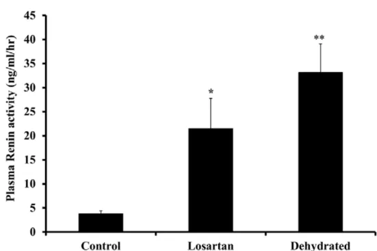
dehydration resulted in an increase in serum urea and creatinine reflecting, a major decline in glomerular filtration rate [51]. Plasma renin activity (PRA) increased significantly on day 20 of dehydration for the losartan treated group (P<0.05), while for the dehydrated group was (P< 0.01) without any significant difference between the treated and dehydrated groups (Fig. 3). The hormone systems including PRA, arginine vasopressin (AVP) and aldosterone together with the RAS, under most circumstances serve to retain water and/or sodium and to maintain circulatory and renal status.
26
Table 3. Plasma levels of Na, Urea and Creatinine in control, losartan treated and dehydrated
camels. Data are shown as Mean ±SEM. Significance from control is denoted by *P< 0.05, **P< 0.01 and ***P< 0.01.
Group of camels Base-line level Day 20 level Na (mM/L) control 141.3 ± 1.2 145.2 ± 1.1 losartan 144.2 ± 1.2 180.7 ± 4.3*** dehydrated 144.6 ± 0.7 175.3 ± 2.2*** UREA (mg/dL) Control 15.7 ± 1.3 11.2 ± 1.3 Losartan 16.4 ± 0.8 39.2 ± 6.8** Dehydrated 13.3 ± 1.4 34.9 ± 3.1** CREATININE (mg/dL) Control 1.43 ± 0.1 1.5 ± 0.1 Losartan 1.33 ± 0.07 2.5 ± 0.4* Dehydrated 1.28 ± 0.2 2.2 ± 0.1*
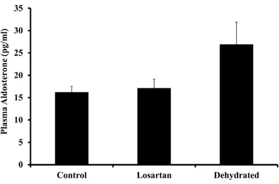
Although earlier studies in camels have documented responses in AVP, RAS and aldosterone to dehydration [51, 21, 13], the duration of these studies has been relatively brief (8–14 days), time-matched controls (non-dehydrated animals) were not used. In this study, we have documented changes in all of these hormones after a longer period (20 days) of water deprivation. Plasma aldosterone concentrations remained steady over 20 days in control animals; however, they increased variably but not statistically significant in both dehydrated groups (Fig. 4). The modest rise in aldosterone levels in the dehydrated camels contrasted with a decline in losartan- treated camels compared with control camels. In congruence with earlier observations [52, 53], we noted little change in the circulating levels of aldosterone in the dehydrated camels despite vigorous activation of the RAS.
27
Figure 3. Plasma renin activity in control, losartan treated and dehydrated animals on day 20 of
dehydration. Data are shown as mean ± SEM. Significant difference from control is denoted by *P<0.05, **P<0.01.
One possible explanation for this minimal response in circulating aldosterone levels in the dehydrated animals is the substantial rise in serum sodium and presumed, though not measured, increase in plasma osmolality. Studies from the 1960s and 1970s in experimental animals and man demonstrated that an increase in extracellular sodium concentration can itself suppress aldosterone production and also inhibit the stimulatory actions of its known secretagogues. It had been observed that an increase in the plasma sodium concentration of 6–15 mmol/L in blood supplying the auto-transplanted sheep adrenal gland inhibited the aldosterone secretory response to sodium depletion [54].
28
Figure 4. Plasma aldosterone levels in control, losartan-treated and dehydrated camels on day 20
of dehydration. Data are shown as mean ± SEM. Changes in aldosterone level in dehydrated camels is not statistically significant compared to control camels.
Furthermore, the aldosterone secretory response to infused Ang II in this sheep model was inversely related to the plasma sodium concentration [55]. Increases in plasma sodium
concentration of lesser magnitude than in the current study were shown to inhibit the aldosterone secretory response to Ang II or potassium by the isolated perfused canine adrenal gland [56]. Furthermore, studies in healthy humans [57] and in a patient with diabetes insipidus and adipsia [58] indicate that an increase in serum sodium concentration reduces aldosterone levels and inhibits it responsiveness to known stimuli. Finally, support for a modulating effect of circulating sodium concentration on aldosterone secretion comes indirectly from studies of volume depletion in camels induced by the intravenous injection of furosemide where, in contrast to the present situation of water depletion, plasma sodium levels declined [59] and PRA and plasma aldosterone levels increased vigorously (more than 4-fold) and in parallel.
29
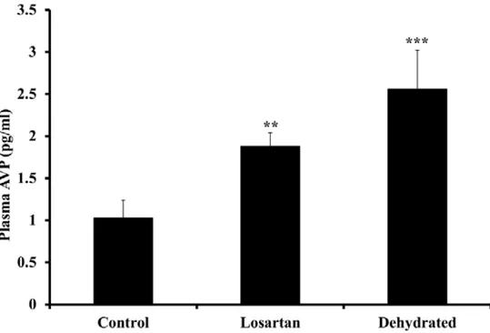
Figure 5. Plasma arginine-vasopressin levels in control, losartan treated and dehydrated camels
on day 20 of dehydration. Data are shown as mean ± SEM. Significant difference from control is denoted by **P<0.05, ***P<0.01.
In this study, dehydration stimulated a significant increase in plasma AVP (Fig. 5). The most obvious explanation for this is the rise in serum sodium (and osmolality) together with the inevitable, though unmeasured fall in circulating volume and arterial pressure. It is also possible that the substantial increase in RAS activity contributed a high level of Ag II concentration, that resulted in stimulating AVP secretion [60], an effect enhanced during water deprivation and inhibited by water-loading [60, 61]. Some support for this contention comes from our
observation that plasma AVP levels were lower after 20 days of dehydration in camels receiving losartan compared with those without losartan, although no significant differences were seen between the two dehydrated groups. These results suggest that RAS plays an important role in the maintenance of water and electrolytes balance during dehydration and shows that our hypothesis is correct, since we found greater disturbances in water balance, electrolytes, hormones and renal function in the dehydrated group when the RAS was blocked using losartan.
30
Changes in insulin-like growth factor-1 and IGF-binding protein-3 in camel
plasma during dehydration in the presence and absence of losartan (Paper III)
The aim of the study presented in paper III was to determine the effect of dehydration in the presence and absence of the angiotensin II AT1 receptor antagoniost losartan on plasma IGF-1 and IGFBP-3 in the dromedary camel and to compare them with a group of hydrated controls. All vertebrates maintain plasma osmolality and extra cellular volume primarily by regulating the ingestion and excretion of water and electrolytes; an elevation in plasma osmolality, and consequent cellular dehydration, is the most potent stimulus of thirst [62]. Insulin-like growth factor-I (IGF-I) is produced primarily by the liver as an endocrine hormone and in target tissues as a paracrine or autocrine. IGF-1production is stimulated by growth hormone (GH) and can be retarded by under nutrition. IGF-1 mediates chondrogenic effects of GH in promoting long bone growth, and regulates the proliferation and differentiation of various cell types, including bone marrow cells. Regarding its amino acid sequence IGF-1 shows partial homology with the pro-insulin peptide, and also exhibits pro-insulin-like bioactivities [63, 64]. IGF-1 is expressed at low concentrations in the stromal elements of normal pancreas tissue [65, 66]. IGFBP is a family of proteins that possess unique biological properties, and are well-described modulators of IGF activity at the cellular level [67]. A number of studies have been carried out on IGF-1 and its receptors in the one-humped camel as well as hormonal levels with and without dehydration, we are not aware of studies assessing directly IGF-1 and IGFBP-3 responses in presence of Ang II AT1 receptor antagonist to sustained dehydration in the camel. In previous study [68] we showed that the one-humped camel unlike other mammalians has a high concentration of IGF-1 receptors in the duodenal mucosa compared to other parts of the digestive tract. It also possesses a higher concentration of IGF-1 receptors in its mucosa compared to the muscle layer. This could be a significant feature necessary for the regenerative ability of the duodenal mucosa in the one-humped camel; as the duodenum is the first part of the gut that comes in contact with the roughages. In the present study we observed no significant changes in the level of plasma IGF-1 concentration in the control animals compared with their base-line level after 20 days of the experiment. In contrast, the effect of dehydration with or without losartan on the circulating levels of IGF-1 in the losartan treated and dehydrated groups were profound from the early days of dehydration. On day 5 of dehydration both groups showed a decrease of 45% for the
dehydrated and 60% for the treated compared to their baseline and control. The level of IGF-1 decreased progressively as the days of dehydration continued. On day 20 of dehydration the IGF-1 level for the dehydrated group reached 80% from the base line level as well as from the
time-31
matched control; while, the losartan treated group showed higher reduction (89%) in IGF-1 level (Table.4) . The exact reason for this major drop in IGF-1 is not well known, but one possible explanation could be due to the reduced basal metabolic rate as a result of reduced food and water intake. This may affect the liver function resulting in decreased IGF-1 synthesis. Another possible explanation could be that these dehydrated camels reduce most of their body synthetic processes as an adaptation mechanism to preserve water and reduce energy required. It is known that the endocrine system mediates many of the physiological responses to the homeostatic and climatic demands of salt and water transport [69]. Previous studies have suggested a role for IGF-1 in the regulation of intestinal water uptake reduction and osmoregulation in fishes [70]. It was also reported that hyperosmotic stress may increase the GH sensitivity in the gills and liver of flounders, and consequently improve IGF-I production [71].
Table-4. Plasma IGF-1 (ng/ml) levels in control, losartan treated and dehydrated camels. Data
are shown as Mean ± SEM. Significance of Basal versus losartan and dehydrated is denoted by *P< 0.05 & **P<0.01. While, significance of Control versus losartan and dehydrated is denoted by $$P<0.01 & $$$P<0.001).
Camels Group Baseline level (n=3) Day 5 level (n=5) Day 10 level (n=6) Day 20 level (n=6) Control 331.0±92 319.8±79 326.3±54 334.7±33 Losartan 319.7±25 126.5±19* 82.8±15**$$$ 32.4±11***$$$ Dehydrated 315.7±25 173.0±73 118.3±28**$$$ 62.0±22***$$$ The effect of 20 days of dehydration in presence of losartan on the circulating IGFBP-3 was 26% decrease compared to their basal values and their controls. The effect of both dehydration and losartan on the treated camels was significant on day 10 of dehydration and remained steady (26%) to the last day of the experiment. The dehydrated group showed 22% decrease on day 10 and 29% on day 20 of dehydration compared to their baseline value and to time-matched control. The decrease in IGFBP-3 level for the dehydrated group was greater than the treated group; albeit, no significant differences were seen between them (Table 5). Previous studies have suggested a role for IGF-1 in the regulation of intestinal water uptake reduction and osmoregulation in fishes [70].
32
Table-5. Plasma IGFBP-3 (ng/ml) levels in control treated and dehydrated camels at baseline,
day 5, 10 and 20 of dehydration. Data are presented as Mean ± SEM. Significance of Basal versus losartan and dehydrated is denoted by (*P< 0.05, **P<0.01 and *** P<0.001) and Control versus losartan and dehydrated by ($$P<0.01, $$$P<0.001). Camels Group Baseline level
(n=3) Day 5 level (n=5) Day 10 level (n=6) Day 20 level (n=6) Control 410.0±19 396.8±32 406.2±21 405.0±13 Losartan 406.7±71 387.8±15 300.4±15*$$ 302.6±21***$$$ Dehydrated 396.0±31 397.0±42 308.6±13*$$ 281.6±19***$$$
The ratio of GH/IGF-1 axis has been suggested to be essential in water homeostasis. Little is known about the role of IGF-1 in dehydration. From the present study it is evident that dehydration resulted in a reduction in both levels of IGF-1 and IGFBP-3, which may reflect a disturbance in growth hormone level in this dehydrated camels. The effects caused by losartan on the IGF-1 and IGFBP-3 levels suggest a possible role of RAS in the present context. It was indicated that endurance horses with dehydration and electrolyte disturbances exhibited activation of the renin-angiotensin-aldosterone-vasopressin axis [72]. Moreover, studies in several species have demonstrated that intracerebroventricular administration of AT1 receptor antagonist inhibited homeostatic responses to thermal dehydration [73]. The effects of the AT1 blocker on IGF-1 is not clear but earlier studies have shown that IGF-1 may up-regulate the AT1-receptor [74] and this may account for an important interaction that may have implications for the observed effect of losartan.
33
ANP and BNP Responses to Dehydration in the One-Humped Camel and
Effects of Blocking the Renin- Angiotensin System (Paper IV)
The objective of the study presented in paper IV was to investigate and compare the response of ANP and BNP in the circulation of hydrated, losartan treated and dehydrated camels; and to document the cardiac storage form of BNP in the camel heart. For measurements of plasma BNP, we first determined which antibody and standard should be used in our radioimmunoassay. This is vital since, by contrast with ANP, the amino acid structure of BNP is known to vary
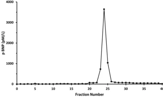
considerably across species and even amongst mammals. Using polyclonal antibody raised to porcine BNP, camel plasma from control, losartan and dehydrated camels together with extracts from left and right ventricle and left and right atria diluted in parallel with standard porcine BNP1-32 (Figure 6). In contrast, there was no cross reactivity to standard human BNP1-32 and hence no parallelism, for camel heart extracts in a radioimmunoassay which used an antibody to human BNP1-32 (data not shown). Size exclusion HPLC of extracts from camel atrial tissue demonstrated a single immunoreactive peak in fractions 24-26 (Figure 7) which eluted one fraction later than recombinant human proBNP standard but well ahead of synthetic human NTproBNP and BNP1-32 standards. This indicates that immunoreactive BNP in the camel heart is in the form of proBNP with an approximate molecular weight of 11,600 Da, and close to the theoretical molecular weight of camel proBNP (11,734 Da) calculated from the dromedary sequence provided by Osman et al and in Gene bank: BAD21300.1 [75]. Further, it is evident that little or no immunoreactive BNP is stored as smaller molecular weight BNP or NT-proBNP. Our observations in this regard are congruent with an earlier study of camel natriuretic peptide cDNA which concluded that camel BNP has 94% identity with porcine BNP [75]. Our additional observation, using size-exclusion HPLC is that, as in many species including sheep [76], the dominant form of immunoreactive BNP in camel heart is a large molecular weight form consistent with the proBNP hormone.
With regard to previous studies of the cardiac natriuretic peptides, 14 days of dehydration in camels was reported to have no effect on circulating levels of ANP [53]. In contrast, we observed that plasma ANP levels were substantially and statistically significant lower after 20 days of dehydration compared with time-matched controls. This response is unsurprising given that stretch of the cardiac chambers, generally considered the major stimulus to ANP release [77], would certainly have decreased across the dehydration period and, presumably, overcame any stimulatory action from increased activity of the RAS [77].
34
Figure 6. Dilutions of extracts of plasma (top panel) and heart (lower panel) from two camels in
35
Figure 7. A single peak of immunoreactive BNP on size exclusion HPLC using camel atrial
tissue extract by radioimmuno assay with standard porcine BNP and antibody directed against porcine BNP. This peak corresponded exactly with that of large molecular weight proBNP, well separated from BNP1-32 and NT-proBNP, using the same technique for extracts of human heart (data not shown).
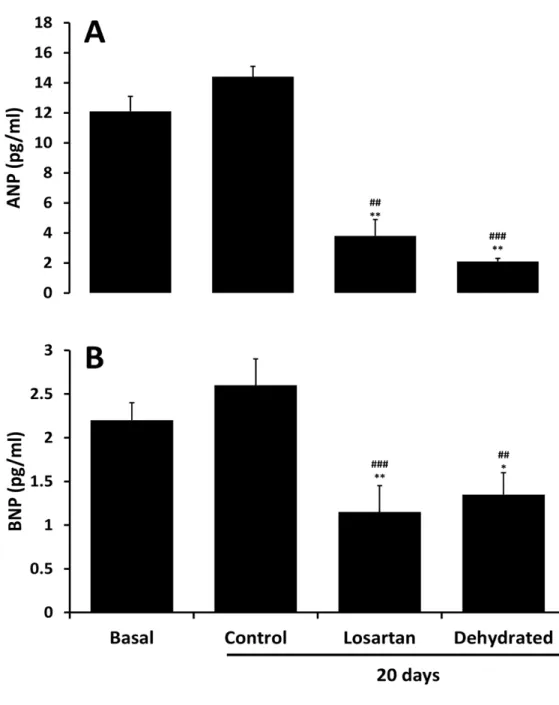
Plasma levels of ANP were approximately 6-fold higher than concomitant BNP levels at baseline (Figure 8). Whereas both ANP and BNP levels tended to rise over 20 days in control camels but not significantly, they decreased substantially and similarly across 20 days of dehydration with and without losartan administration (Figure 8). As for ANP, dehydration resulted in a clear-cut decline in circulating levels of BNP. Again, as for ANP, we presume that any stimulatory action on BNP synthesis and release from activation of the RAS was overcome by reduced stretch of cardiac chambers during prolonged dehydration. Teleologically, these responses in plasma ANP and BNP levels would be seen as logical in being protective against excessive sodium and water loss. If, as would be expected, arterial pressure declined across dehydration in our camels, the natriuretic and diuretic actions of these peptides would be diminished [78, 79] thereby tending to further limit urinary loss of sodium and water. Nevertheless, caution is needed in this discussion since, to our awareness; there are no in-depth studies in camels regarding the regulation neither of cardiac natriuretic peptide release nor on their renal effects under conditions of water-depletion versus repletion.
36
Figure 8. Plasma levels of ANP (top panel) and BNP (lower panel) in pmol/L at baseline and 20
days in control, losartan treated and dehydrated camels. Data are shown as mean±SEM. *P< 0.05, **P<0.01 denotes basal versus treated and dehydrated. While ##P<0.01, ###P<0.001 denotes control versus treated and dehydrated.
37
In summary, 20 days of dehydration induced substantial suppression of plasma ANP and BNP concentrations. Blockade of the AT1 receptor had little or no effect on the plasma ANP and BNP responses to dehydration. Finally, the storage form of B-type natriuretic peptide in the one-humped camel is large molecular weight proBNP.
l
Effects of Dehydration and Blockade of Angiotensin II AT1 Receptor on
Stress Hormones and Anti-Oxidants in the one-humped camel
(Paper V)
In the study presented in paper V we documented and compared plasma levels of catecholamines cortisol, glutathione and malondialdehyde in plasma, liver and kidney homogenates after long term dehydration in the presence and absence of Ag II AT1 receptor blocker (losartan) versus levels in non-dehydrated camels, using HPLC, radioimmunoassay and colorimetric assays. Catecholamines as neurotransmitters and adrenomedullary hormones influence almost all tissues and many body functions. Together with other neuronal and hormonal systems they play a significant role in regulating a number of physiological processes. Our results demonstrated that dehydration activated some components of the sympathetic nervous system as reflected in the circulating levels of norepinephrine and dopamine and deactivated the circulating plasma level of epinephrine (Figure 9). Blockade of AngII AT1 receptors tended to overstress the effects of dehydration, although differences from dehydration alone failed to reach levels of statistical significance. The main cause for the increased level of plasma norepinephrine in these
dehydrated camels is not immediately apparent. The most apparent explanation could be due to the physical stress caused by dehydration alone or in combination with the effect of losartan. This increase in norepinephrine and dopamine levels may have played a positive role in the regulation of their cardiovascular and metabolic systems. Recently it was reported that hormones released during stressful conditions are used to supply the body with the energy required for breaking down the body metabolites [22-80]. On the other hand, plasma dopamine level was significantly increased after 20 days of dehydration in both treated and dehydrated groups compared to time matched control. Dopamine was reported to regulate fluid and electrolyte balance by direct and indirect actions in the kidney, blood vessels, gastrointestinal tract, adrenal glands, sympathetic nervous system, hypothalamus and other brain centers [23-30].
38
Figure 9. Catecholamines concentration (pg/ml) in plasma of Control, Treated and Dehydrated
Camels. Data shown are mean ± SEM. Asterisks denote * P<0.05, ** P<0.01 and *** P<0.001. Significant difference from control values
Plasma cortisol levels increased during dehydration to more than 2-fold for the dehydrated and 3- fold for the treated group when compared with time-matched control (Figure 10). The increase in cortisol level was higher in the treated versus dehydrated camels, albeit, no significant difference between them was observed. The main cause for the vigorous rise in plasma cortisol in these dehydrated camels is not immediately apparent. The most obvious possibility is that the hypothalamic-pituitary axis was stimulated during dehydration resulting in a rise in plasma adrenocorticotropic hormone (ACTH) and thence cortisol. This could be the result of “stress” or possibly due to activation of the renin-angiotensin system since angiotensin II, at least under some circumstances, can stimulate or enhance the release of corticotropin-releasing factor [81] and ACTH [82, 83] and, in addition, is capable of stimulating cortisol secretion directly from the
* ** *** * ** ***
0
50
100
150
200
250
300
350
400
450
500
Epinephrine
Norepinephrine
Dopamine
Catecholamines (pg/ml)
Control Losartan Dehydrated
39
adrenal fasciculata [84, 85] or augmenting the effect of ACTH on cortisol production [86]. It had been reported that cortisol had the capacity to interfere with a principal mechanism of resistance to dehydration, by inhibiting the effects of arginine vasopressin in dogs [87]. Other studies showed that effect of external heat load could be associated with significant increments of plasma cortisol [88].
Fig. 10. Plasma Cortisol concentration (µg/dl) in Control, Treated and Dehydrated camels. Data
are shown as percentage concentrations compared to control.
Dehydration enhanced by losartan activated body antioxidants resulting in a significant increase in plasma GSH levels after 20 days of dehydration compared to control (Figure 11). Liver homogenate GSH level was also significantly increased in both treated and dehydrated groups compared to control. However, liver homogenate GSH level was more affected by losartan than dehydration alone. The liver is known to have the highest content of antioxidant and antioxidant enzymes indicating that it plays an important role in pro-oxidants detoxification [89]. GSH level