Faculty of Veterinary Medicine and Animal Science
Seroprevalence and risk factors for Rift
Valley fever and Capripoxvirus in small
ruminants in the border region of
Tanzania - Zambia
Emelie Olovsson
Uppsala 2019
Seroprevalence and risk factors for Rift Valley
fever and Capripoxvirus in small ruminants in
the border region of Tanzania - Zambia
Emelie Olovsson
Supervisor: Jonas Johansson Wensman, Department of Clinical Siences (KV), Swedish
University of Agricultural Sciences (SLU)
Assistant Supervisor: Sara Lysholm, Department of Clinical Science (KV), Swedish
University of Agricultural Sciences (SLU)
Assistant Supervisor: Gerald Misinzo, Department of Veterinary Microbiology and
Parasitology, Sokoine University of Agriculture in Morogoro (Tanzania)
Examiner: Johanna Lindahl, Department of Clinical Siences (KV), Swedish University of
Agricultural Sciences (SLU)
Degree Project in Veterinary Medicine
Credits: 30
Level: Second cycle, A2E Course code: EX0869 Place of publication: Uppsala Year of publication: 2019
Online publication: https://stud.epsilon.slu.se
Cover illustration: Sheep herd in Momba district. Photo by Emelie Olovsson
Key words: Infectious diseases, RVFV, CaPV, SGPV, sheep and goat pox, seroprevalence, risk
factors,Tunduma, Momba, goats, sheep, livestock
Nyckelord: Infektionssjukdomar, RVFV, CaPV, SGPV, får- och getkoppor, seroprevalens, riskfaktorer, Tunduma,
Momba, getter, får, boskap
Sveriges lantbruksuniversitet
Swedish University of Agricultural Sciences Faculty of Veterinary Medicine and Animal Science Department of Clinical Science
SUMMARY
Tanzania is a country where poverty is still high and many households are dependent on agriculture to support their families. Small ruminants, such as sheep and goats, make up an important part of agriculture; they are cheap to buy and can easily be sold or exchanged for the farmers. The animals therefore function as a living bank and should unforeseen expenses arise, the money can be made available by selling an animal. This means that the health of these animals is important socio-economically for the farmers.
Rift Valley fever (RVF) and sheep and goat pox (SGP) are two diseases that OIE have listed as notifiable. Rift Valley fever virus (RVFV) is an arbovirus transmitted by arthropod vectors such as mosquitoes. It is mainly infecting ruminants such as sheep, goats, cattle, buffaloes and camels, but is also a zoonotic disease and can infect humans. When domestic ruminants are infected, massive abortions can be seen in all stages in pregnant animals and a high fatality rate in young animals. Sheep and goat pox virus (SGPV) is a Capripoxvirus (CaPV) that belongs to
Capripoxvirus genus. The virus is mainly transmitted through direct contact with infected
animals, but indirect transmission through environment, or mechanical, through biting vectors, is also possible. Animals infected with SGPV show clinical signs of fever, loss of appetite, increased salivation and ocular and nasal discharge. After a few days, papules appear in the skin and on mucous membranes, even inside the body, which can cause serious and fatal complications. Young animals suffer more from the disease and the case fatality rate can be high. For farmers in rural communities, both diseases can have significant negative socio-economic impact, due to the loss of production and animals. The gender-equality between men and women may also be affected since women often are the main caretaker of the livestock. This master thesis was performed as a Minor Field Study (MFS) that investigated the seroprevalence of RVF and SGP in Tanzania, in the two districts Momba and Tunduma close to the border of Zambia. The aim was to evaluate the seroprevalence in sheep and goats to understand the epidemiology of these diseases in the southwestern part of Tanzania and also investigate associated risk factors. Of the samples collected, 484 were from goats and 7 from sheep. Totally 16 of 491 analyzed samples were seropositive for RVFV, giving a seroprevalence of 3.3% on an individual level. All seropositive animals were goats, 93.8% females and 6.2% males. In total 31.7% (13/41) of the villages had seropositive animals, with a seroprevalence within the villages ranging up to 25%. The majority of the farmers reported that they utilized communal grazing system for their animals, where the majority of sheep and goats were reported to have daily contact with other domestic livestock. Only few sheep and goats had contact with wild ruminants. In this study, farmers buying their animals or had farmers in the same village buying their animals from markets, had significantly more seropositive animals
In this study only a single animal was seropositive for CaPV, a female goat belonging to the Momba district.
CONTENT
Introduction ... 1
Aim ... 1
Litterature review ... 1
Tanzania ... 1
Animal and wildlife ... 2
Agriculture ... 2
Momba and Tunduma ... 2
Rift Valley fever ... 3
Etiology ... 3 Epidemiology ... 3 Transmission ... 5 Clinical manifestation... 7 Diagnostics ... 7 Vaccination ... 8
Sheep and goat pox... 8
Etiology ... 8 Epidemiology ... 9 Transmission ... 10 Clinical manifestation... 11 Diagnostics ... 11 Vaccine for SGP ... 12
Material and methods ... 12
Study area and study design ... 12
Animals and sampling ... 13
Antibody detection ... 14
RVFV competitive ELISA ... 14
Double antigen ELISA for Capripox ... 15
Questionnaire ... 16
Statistical analyses ... 16
Results ... 16
Study area and animal and sampling ... 16
Antibody detection ... 17 Questionnaires ... 21 Management routines ... 21 Disease managment ... 21 Trade... 22 Animal health ... 23 Public health ... 24 Farmer details ... 25 Discussion ... 27 Conclusions ... 29
Acknowledgment ... 30
Populärvetenskaplig sammanfattning ... 31
References ... 34
1
INTRODUCTION Aim
The purpose of this study was to investigate the epidemiology of Rift Valley fever and Capripox in the southern part of Tanzania, close to the border of Zambia. To achieve the aim of the study, blood was collected from goats and sheep in two separate districts, Tunduma and Momba. Also, all farmers were asked questions (appendix 1) from a questionnaire regarding associated risk factors. Serum was analyzed by ELISA to investigate the seroprevalence of the selected infectious diseases. This master project was managed through close collaboration with scientists in Tanzania (Sokoine University of Agriculture) and is a smaller part of a PhD project with the aim to investigate the following infectious diseases; Rift Valley fever, capripox, peste des petits ruminants and foot and mouth disease and analyzing the risk factors for spread of these diseases in both Tanzania and Zambia.
LITTERATURE REVIEW Tanzania
The country Tanzania is located in East Africa just below the equator with a total area of 947 303 km² (Central Intelligence Agency, 2018; Nationalencylopedin, 2018; Utrikesdepar-tementets landguide, 2018). The United Republic of Tanzania is made up by the mainland of Tanzania and the island of Zanzibar and is bordering eight countries; Zambia, Mozambique, Rwanda, Burundi, Congo, Uganda, Malawi and Kenya. Zanzibar has its own parliament and serves as a self-governing region. Tanzania consists of 31 regions governed by a president. The capital is Dodoma, located centrally in the country, but previously it was Dar es Salaam. Dar es Salaam is still the most important trading city in Tanzania, located by the Indian Ocean. Ethnic diversity in the country is great and consists of around 125 different ethnical groups, and more than 100 languages are spoken within Tanzania (Central Intelligence Agency, 2018; Nationalencylopedin, 2018; Utrikesdepartementets landguide, 2018). Swahili is a Bantu language, which is one of four language families in Africa, and among the most common used in lower courts and primary school. English is introduced in secondary school and used in higher education and in higher courts.
Over the year, the country has two different rain seasons, one longer that occurs from March to May and one shorter rain season between October and January (Utrikesdepartementets landguide, 2018). During both rain seasons, the rainfall ranges between 750-1400 mm and spatial distribution varies over the country, but a greater amount of rain is seen closer to large water such as lakes or oceans (Hamisi, 2013). The dry season is mainly during the months from July to September.
Tanzania is administratively divided into 31 regions, and the regions are subdivided into 169 districts (Central Intelligence Agency, 2018; Nationalencyklopedin, 2018; Utrikesdepar-tementets landguide, 2018). The district is further divided into so called wards, and the ward
2
consists of different numbers of villages. The districts are the smallest administrative units with the responsibility for management of human and livestock diseases.
Animal and wildlife
Tanzania is famous for its beautiful nature and wildlife and almost a third of the landscape is national parks, game reserves, conservation areas and marine parks (Central Intelligence Agency, 2018; Nationalencylopedin, 2018; Utrikesdepartementets landguide, 2018). Tanzania’s national parks contain around 20% of Africa’s large mammal species, and the national parks attract tourists from all over the world, in particularly Serengeti National park in the north which is one of the main attractions. It is the second largest park and famous for its massive migration of wildebeests and zebra. Another park, the Ngorongoro Crater, is the largest caldera intact in the world. This caldera contains a rich wildlife and was in 1959 established as a conservation area by the United Nations Educational, Scientific and Cultural Organization (UNESCO). It is since appointed as a World Heritage site and through that legally protected by international treaties.
Agriculture
Human population in Tanzania is estimated to be around 56 million people and within the country poverty is still high (Central Intelligence Agency, 2018; Nationalencylopedin; Utrikesdepartementets landguide). Many people make a living through running their own farms, and small ruminants such as sheep and goats play an important role in the agriculture. They are often cheap to buy and easy to sell or trade for money, and these animals therefore have a big economic impact for the farmers. The animals can function as a living bank and when expenses arise the farmers can sell off some animals. The animals also contribute with products such as milk, wool, meat and skin, and are therefore important as food resources and for livelihood resilience for rural householders. The health management of small ruminants involves both genders and contributes to a more gender-equal society (Galie et al., 2017).
Momba and Tunduma
The two districts, Tunduma and Momba, belong to the Mbeya region (Wikipedia). In January 2016, Mbeya was divided, and the western half was renamed Songwe region. These districts are close to the border of Zambia. Tunduma town is on the border between Tanzania and Zambia and has border posts for the Tanzam Highway and Tazara railway, which link the two countries together. The 1,860 km long Tazara railway starts in Dar es Salaam in the east of Tanzania and reaches to Kapiri Mposhi in the central province of Zambia.
The border between Tanzania and Zambia is regularly crossed by civilians and vehicles (Africa business communities and Lusaka times). In recent time, a new border post has been established that has simplified the cross-border movement. It is possible that it will affect the animal trade and movements of animals between the countries.
3
Rift Valley fever
Etiology
Rift Valley fever (RVF) is caused by Rift Valley fever virus (RVFV) which belongs to the family Bunyaviridae and genus Phlebovirus (Epiwebb; Pepin et al., 2010). It has a spherical shape and is enveloped, just like other bunyaviruses and is about 90-110 mm in diameter (Pepin
et al., 2010; Sherman et al., 2009). It consists of a single-stranded RNA with three segments of
negative polarity: S (small), M (medium) and L (large) (Ikegami 2012). The virus has glycoprotein-peplomers, and these glycoproteins are important in the viral cycle and responsible for viral uptake of the virus in to cells (Pepin et al., 2010).
Rift Valley fever virus is sensitive to heat, desiccation, acid pH and disinfectiants (Quinn et al., 2011). Several of the viruses in the Bunyaviridae family are arboviruses, and thus transmitted through arthropods vectors, and this is also the case for RVFV.
The Rift Valley fever virus is primarily affecting and causing severe disease in ruminants such as sheep, goats, cattle and buffalos, but also camels have been found with the disease (WHO, 2018). Rift Valley fever virus can also infect humans which makes this a zoonotic disease. Amongst humans the virus causes acute clinical signs such as fever, headache, joint pain and abdominal pain. This therefore makes Rift Valley fever an important disease in a one health perspective.
Epidemiology
The first described outbreak of the disease was announced in July 1930 in Kenya by Daubney & Hudson (1931). The clinical manifestation was a high case fatality rate in new-born Merino lambs in a farm in the Rift Valley. The outbreak occurred when the farmer changed the lambing season from October and November to July and August. After the first outbreak, additional outbreaks were reported seventeen years later in 1947 (Sindato et al., 2014).
Rift Valley fever is endemic in sub-Saharan Africa (Rich & Wanyoike, 2010; Dar et al., 2013; Chevalier et al., 2005). A large outbreak was reported in Egypt 1977-1979, when 100 000 animals became ill and also a large number of people, around 200 000, were affected and approximately 600 of them died from the disease (Bowen, 2011). This was the first time the disease was discovered north of Sahara. In the year 1997 and 1998, the disease was found in Somalia, Kenya and Tanzania. Large amounts of animals like sheep, cattle, goats and camels became ill and many of them died during the outbreak (Bowen, 2011).
Rift Valley fever virus has a complex epidemiology, involving a wide range of factors where the key factors are considered to be rainfall and flooding, contact between animals, breeding sites of mosquitoes, movement and availability of livestock (Himeidan et al., 2014). These mentioned factors along with the complexity of different mosquito species, which can carry and spread the virus, make transmission possibly over big area of land. The presence of competent vectors in RVF-free countries with the ongoing climate change strongly suggests that the disease should be on the list of the most significant emerging viral threats to the public and
4
veterinary health (Pepin et al., 2010). The World Organization for Animal Health (OIE) has listed RVF as a notifiable disease (OIE, 2014).
Rift Valley fever in Tanzania
The livestock populations in Tanzania are the third largest on the African continent, with an estimated 25 million cattle, 16.7 million goats, 8 million sheep, 2.4 million pigs and 36 million chickens (United Republic of Tanzania ministry of livestock and fisheries development, 2015). The major part is located in the northern and central regions of the country (Chengula et al., 2013).
In the country, a total of 10 RVF outbreaks have been reported in the past 80 years, the first in 1930 and then in 1947, 1957, 1960, 1963, 1968, 1977-1979, 1989, 1997-1998 and 2006-2007 (Sindato et al., 2014). Sindato and others conducted a retrospective study based on disease reported data on district and village level to investigate the spatial and temporal pattern of the RVFV outbreaks. Generally, the outbreaks occur between the months of December to June, the virus has been seen spreading southward, from Ngorongoro district in the north to Ulanga district in the south. In total, 39.2% of all districts in Tanzania have suffered from outbreaks varying in size. In Tanzania, the epidemiological features seem to be the same as in other countries. What makes Tanzania unique is that it is the only country with two branches of the Great Rift Valley ecosystem, the eastern and western, and these ecosystems are associated with RVFV (Sindato et al., 2014). These branches involve the whole country, starting in the north where it traverses from Kenya and carry out through the country to the southern parts.
Spatial investigations in Tanzania show that the northern part of the country appears to be the starting point for all outbreaks (Sindato et al., 2014). It is assumed to be an initial amplification site for the virus, and leading to spreading and progressive infiltration of RVFV to southern parts of Tanzania. The mechanism of spatial spreading is still unknown, but the authors propose the possibility of passive and active movements of mosquitoes and uncontrolled movement of livestock within the country. The authors conclude that the outbreaks of RVF in Tanzania seem to be heterogeneously distributed and the transmission of the virus seems to vary between areas and during seasons. That suggestion is consistent with the findings in other studies where seropositive animals were found during an inter-epidemic period in Morogoro and Arusha regions seven years after a major outbreak (Wensman et al., 2015). The authors concluded that it is an indication for the virus still circulating in low numbers in Tanzania. Furthermore, the authors take into consideration that the farmers in rural Tanzania is poorly prepared when the next outbreak arise due to no vaccinations against the disease are implemented.
In the beginning of the early outbreaks during the 1900s, Tanzania had a poor awareness of the disease, and inefficient recording systems and lack of capacity for diagnostics likely contributed to a low number of reports (Sindato et al., 2014). After 1978, RVF was listed as a notifiable disease by OIE and after that, monitoring and diagnostics have been improved. This has suggested caused the number of reported cases to increase.
5
About 12-13 years ago, in 2006-2007, the latest major outbreak was reported in Tanzania. This outbreak started in late December of 2006, and in January 2007 local veterinarians in the Arusha area started to report cases of massive abortions and deaths that could be caused by RVF (Sindato et al., 2014; Chengula et al., 2013). After the first report, additional reports came from Manyara, Kilimanjaro, Dodoma, Tanga, Iringa and Morogoro regions. The outbreak ended in July 2007 and had at that point affected 10 of 21 regions in the country and on a district level 45 out of 120 (Chengula et al., 2013; Sindato et al., 2010).
During the inter-epidemic periods, since the major outbreak in 2006-2007, studies detecting antibodies in both adult and young animals has been conducted in different parts of Tanzania (Wensman et al., 2015; Sumaye et al., 2013). Detections of these antibodies has also been in regions with no previous history of RVF outbreaks. This important finding shows that RVFV is endemic and circulating in low levels, within the country.
Rift Valley fever in Zambia
The first outbreak of RVF in Zambia was reported in 1974 and was located in the central and southern parts of the country, but there were also reports from the northern part belonging to the Copperbelt province (Hussein et al., 1987). Rift Valley fever is considered to be endemic in Zambia but for the last two decades no cases has been reported (OIE, 2014). During 1982-1986, a study was carried out in a sentinel herd with indigenous breeds in Mumbwa, and a low level of seasonal activity (3-8%) were found (Davies et al., 1992). Although low level of activity of the virus has been documented, no RVF-associated abortions or deaths have been observed (Rostal et al., 2010; Davies et al., 1992). In 2012, a review demonstrated that Zambia’s environment is beneficial for the virus, and thereby possessing a threat for the livestock producing farmers (Dautu et al., 2012). Little research has been carried out to study the presence of RVFV amongst humans, livestock and wildlife in Zambia, but OIE data between 2005-2010 show that most countries bordering to Zambia have reported cases of RVF. The studies conducted have been during outbreaks and in high risk areas and therefore information gaps exist (Dautu et al., 2012). Knowledge about different types of strains of RVFV in Zambia is also missing.
Transmission
Rift Valley fever was first suspected to be spread indirectly, because no natural spreading between sheep in the laboratory was observed (Daubney and Hudson, 1931). They later identified the vector Taeniorhynchus brevipalpis to carry RVFV. Linthicum et al., 1985 identified the virus in a variety of the Diptera species, and in Aedes mcintoshi the virus was present in both adults and offspring. They suspected a transovarian transmission, i.e. when the female mosquito directly passes virus on to her offspring, which later has been confirmed (WHO, 2018; Favier et al., 2006). Other genera of mosquitoes such as Anopheles, Culex and
Eretmapodites can transmit RVFV, but this is mainly during epizootic periods when the level
of virus circulation is high (House et al., 1992). Rift Valley fever virus has been isolated in about 40 different species of mosquitoes that belong to seven different families: Aedes,
6
Aedes mosquitoes function as a primary vector. The mosquitoes bite an infected animal tissue,
feed on blood, and carry and spread the virus to susceptible animals, in particular ruminants (Bowen, 2011). Many animal hosts can be infected, for example sheep, cattle, goats, camels, buffaloes and others (WHO, 2018). Some studies have shown that sheep and cattle can be more susceptible to RVFV than goats (Bird et al., 2008). After susceptible animals become infected it takes between 3 to 5 days for them to become highly viremic and then spreading through also secondary vectors is possible (Bowen, 2011). Additional mosquito species have been experimentally infected with RVFV, and may act as amplifying secondary vectors (EFSA, 2005).
Heavy rainfall has been proven associated with outbreaks of RVF in Africa (Pepin et al., 2010). Most of the Aedes-vectors belong to the family of flood mosquitoes which favor laying eggs in flooded grassland areas and elevated water tables (Davies et al., 1985). For several years, the virus can be dormant in the mosquito eggs. The eggs can also survive for many years in the soil in dried flood beddings, and when rain season starts the eggs hatch (Fontenille et al., 1998; Linthicum et al., 1985). Several other risk factors have also been associated with the occurrence of the virus, and these includes climatic conditions such as higher temperature, geographical features, vegetation cover, human activities and livestock trade in the country (WHO, 2018). Rift Valley fever virus is not only transmitted through arthropod vectors but also through direct contact between susceptible hosts and infected animal tissues, body fluids and aborted materials (Pepin et al., 2010). Aborted materials, such as fetus and placenta, have been identified to contain large amounts of the virus.
In Zambia, no studies have been conducted regarding potential mosquito species that can spread the disease (Dautu et al., 2012). Different species of vectors have been observed to differ depending on region and season, within the country (WHO, 2018).
Potential European vectors
In Europe, there are potential vectors that should be able to carry and spread RVFV (Rolin et
al., 2013). In order for the virus to be introduced into a new region, two factors are needed,
optimal environmental conditions and introduction of virus are required for a period of time. The authors conclude that the conditions are still not optimal for the virus to be spread in to Europe, but the risk of the pathogen being sporadically introduced is likely to be relatively high (Rolin et al., 2013).
The European Food Safety Authority (EFSA) has highlighted the need of increased knowledge about which of the mosquito species in Europe that have the ability to carry and transmit the virus (Verteirt et al., 2013). In this study, periods of peaks of Aedes and Culex mosquito species in some areas could pose a risk, a great quantity of mosquitoes was mainly observed in the coastal areas in countries of the Mediterranean Basin.
7
Clinical manifestation
The virus primarily affects ruminants such as cattle, sheep, goats and buffaloes (FAO, 2003) and cause a diffuse clinical picture with nasal and ocular discharges, fever, colic, vomiting and hemorrhagic diarrhea (Ikegami, 2012; SVA, 2018). The incubation time is about 3 days (Bowen, 2011). The main clinical manifestation is epidemic abortions (so called abortion storms) in pregnant animals infected in all stages (Epiwebb, 2018). Rate of abortion can be as high as 90-100% in affected pregnant animals in all stages (OIE, 2018).
Rift Valley fever virus has been detected in several tissues and cells in the body (Pepin et al., 2010). After infection, a local replication occurs in the regional lymph node that leads to viremia with systemic spreading (Bowen, 2011). The systemic spreading is affecting internal organs, in particularly the liver where necrosis can occur, but also the spleen can be affected. Viral replication has been seen in other tissues like adrenal glands, lung and kidney tissues. In some rare cases the virus can affect the brain and cause encephalitis. Animals often die because of necrosis in the liver, renal insufficiency and shock, frequently with severe hemorrhagic (Bowen 2011).
Out of all ruminants, it seems like sheep are affected most seriously by RVFV (Pepin et al., 2010). But out of all ruminants that can be affected it is the young animals that are more susceptible to the virus (Epiwebb; SVA; Pepin et al., 2010). In young animals, sudden death can occur without any clinical signs, while in some cases a high fever develops and the animal dies after a day. This causes a high case fatality rate (Chengula et al., 2014), and in young animals younger than one week, the case fatality rate can be as high as 90% (Quinn et al., 2011). Diseases like listeriosis, Q-fever and toxoplasmosis have similar clinical manifestation, but the abortion storms that occur when RVFV infects ruminants make other differential diagnosis more unlikely (Pepin et al., 2010).
Rift Valley fever virus can infect humans and therefore RVF is also a zoonotic disease (Reed
et al., 2012). The incubation time is from 3 to up to 12 days before symptoms first starts to
appear. In humans, RVFV causes symptoms of acute fever, headache and pain in muscles and joints (Ikegami, 2013). The symptoms often decline after 4-7 days, when the viremia declines and antibodies start to develop (OIE, 2018). In some humans, complications can occur such as bleedings, liver failure and encephalitis (WHO, 2018). The case fatality rate is 1%. Transmission of RVFV can occur through arthropods vectors but also from organs and body fluids of infected animals to humans (OIE, 2018). Occupational groups, such as farmers, slaughterhouse workers and veterinarians, are therefore at higher risk of infection (WHO, 2018). In previous studies in the Mbeya regions, that include Momba and Tunduma district, a seroprevalence of 5.2% has been observed in humans (Heinrich et al., 2012).
Diagnostics
International organizations such as OIE have expressed demands for the development of high quality and safe diagnostic tests for RVFV. Working with RVFV pose some problems, it
8
requires high biosecurity and biocontainment safe facilities when handling (Pepin et al., 2010). Rift Valley fever virus is considered to be a potential bioweapon.
Several kinds of diagnostic methods are available to diagnose RVF and different techniques are used, such as antigen detection, virus isolation, nucleic acid amplification techniques and detection of specific antibodies (Pepin et al., 2010; OIE, 2009; Epiwebb, 2018; SVA, 2018). To detect RVFV, amplification of RNA fragment can be done by a newer more efficient molecular diagnostic assay with reverse transcriptase polymerase chain reaction (RT-PCR) and reverse transcription loop-mediated isothermal amplification (RT LAMP) (OIE, 2018; Pepin et
al., 2010). These processes can take a long time before providing results, but it has a high
sensitivity for detecting the virus RNA.
Enzyme-linked immunosorbent assay (ELISA) is one of several serological methods, and the technique is used to identify RVFV specific antibodies (Pepin et al., 2010). Depending on how the ELISA is designed, it can detect IgG, IgM or total antibodies. The recommended ELISA is the one based on RVFV recombinant antigens (Pepin et al., 2010; Jansen van Vuren et al., 2007).
Vaccination
Outbreaks of RVF can be prevented through vaccination programs, where both modified live attenuated and inactivated virus vaccines can be used (WHO, 2018). The live attenuated vaccine only requires one dose for long-term immunity, but spontaneous abortion has been seen in pregnant animals (WHO, 2018). The inactivated virus vaccine requires multiple doses for immunity but no side effect with spontaneous abortion has been seen. Efficient and safe vaccines for both medical and veterinary use are, however, still lacking (Pepin et al., 2010). Recently, highly immunogenic vaccines have been developed, and they will probably replace live attenuated vaccines, but further evaluations are required to confirm safety and efficacy of this vaccine (Ikegami, 2017). Old vaccines, such as MP-12 vaccine, have been tested on pregnant ewes, they were subcutaneously vaccinated and all ewes delivered healthy lambs (Morill et al., 1987). Ikegami, 2017 suggests that further testing of vaccines, such as MP-12, would be an option when the market for RVF vaccines is small. To evaluate the MP-12 vaccine, in both humans and animals, would not require additional investment.
Sheep and goat pox
Etiology
Sheep and goat pox viruses (SGPV) are Capripoxviruses (CaPV), belonging to genus Capripox, sub-family Chorodipoxvirinae and family Poxviridae. Capripoxviruses are large, (170-260 nm in diameter), enveloped, and double-stranded DNA viruses (Carn 1993; Tulman et al., 2002; Buller et al., 2005). Sheep pox virus (SPPV), goat pox virus (GTPV) and lumpy skin disease virus (LSDV) are a series of CaPVs affecting domestic ruminants and causes various pox diseases. Genome sequences of SPPV, GTPV and LSDV show that they are highly similar with more than 96% of the nucleotides identical (Tulman et al., 2002). Sheep pox virus and GTPV appear to be specific for each host species, but recent isolates show the ability to infect both
9
hosts, and are therefore concluded as SGPV (Babiuk et al., 2009). The varieties of strains for SGPV are phylogenetically distinct from each other and are named based on the host species that it has been identified from (Kitching et al., 1989; Kitching 1986; Tulman et al., 2002). They cause different clinical diseases in either sheep or goats, and some strains are similarly pathogenic in both species (Babiuk et al., 2008). Sheep and goat pox virus is vulnerable to direct sunlight, but in wool and hair it can remain viable for up to three months (Epiwebb, 2018; OIE, 2018). In shaded dirty pens, SGPV can survive for up to six months.
The CaPVs are an economically important group of viruses because of their severe impact on deprived rural communities and small-scale farmers in endemic regions (FAO, 2013). World Organization for Animal Health classified SGP as a notifiable disease in 2014. Sheep and goat pox are among the most common diseases in sheep and goats entailing a huge economic loss for affected countries. For farmers, the production losses can be significant and affect their socio-economic standing (Yeruham et al., 2007). Sheep and goat pox is also limiting the international trade of animals and animal products (OIE, 2018).
Epidemiology
Sheep and goat pox viruses are spread over a variety of continents over the world. In the Middle East, SGPV is endemic in countries like Turkey, Iran, Syria and Iraq (OIE, 2014; Mangana et
al., 2008). Sheep and goat pox viruses are mostly restricted to Asia and north of the equator in
Africa.
In Europe, SGP is an exotic disease but several neighboring countries are endemically infected with SGP (Kitching, 2004). After the veterinary service failed in Syria, uncontrolled movements of livestock pose a major risk of spreading the diseases to Europe (FAO, 2013). Sporadic outbreaks have been reported in European countries, mostly in Greece (Mangana et
al., 2007). In Greece, it is mainly sheep pox that has been reported, and during some outbreaks,
like the one in 2007 when the case fatality rate was high, it was concluded that possibly the sheep pox virus was of a highly virulent strain or the hosts had a low immunity to the virus. The affected herds were mixed with both sheep and goats and serologically it was proven that antibodies to CaPV were present in goats, but no clinical signs were reported. The geographic position of Greece makes the country an important area for control of introduction of SGP to Europe, due to the fact that neighboring countries are enzootic (Mangana et al., 2007). Greece has applied stamping-out/non-vaccination policy since 1992 whenever there is an outbreak in the country.
EFSA evaluated the possible ways of SGP introduction into Europe in 2014, and the most important risk factors identified were movements of people and vehicles across the borders (EFSA, 2014). The movement can be immigrants passing through endemic areas carrying the virus over long distances, but also tourists, farmers, veterinarians and animal care workers can transmit the virus over the border when they are visiting animal facilities. EFSA also concluded that movement of infected animals over the border is assumed to be the most efficient way to introduce SGP to new areas but it is considered to be less important than movement of people and vehicles (EFSA, 2014).
10
Sheep and goat pox is endemic throughout the northern and central part of Africa (Carn, 1993), and many reports of SGP have also been from East African countries, namely Sudan and Kenya (Enan et al., 2013; Ahmed, 2012; Elshafie & Ali, 2008; Davies et al., 1985). Sheep and goat pox has recently been serologically proven to be present in Ethiopia and considered to be the most important disease in the country (Fentie et al., 2017).
Sheep and goat pox in Tanzania
In Tanzania, no reports of SGPV have been done until 2018, CaPV were detected during a massive outbreak of respiratory disease in 2016 in sheep and goats (Kgotlele et al., 2018). This outbreak was located to the Ngorongoro district in the north of Tanzania and confirmed occurrence of co-infection with pathogens that are associated with respiratory distress such as PPR.
Sheep and goat pox in Zambia
In Zambia, no reports have been made about presence of SGPV (OIE, 2018). Outbreaks of LSDV on the other hand have been reported within the country. Clinical signs of lumpy skin disease (LSD) were first described in Zambia in 1929, and were first believed to be a result of either poisoning or hypersensivity to insect bites (FAO, 2013). Thereafter, cases occurred between 1943 and 1945 in neighboring countries such as Botswana, Zimbabwe, Mozambique and in South Africa (Green, 1959; Von Backstrom, 1945).
Transmission
In natural infection, SGPV enters the respiratory tract through aerosols associated with close contact with infected animals (Radostits et al., 2006). Inhalation of aerosols to the respiratory tract is the primary transmission route of the virus (Kitching & Taylor 1985). Experimental studies of SGPV transmission have used intradermal inoculation or administration by mouth or nose (Bowed et al., 2008).
Sheep and goat pox viruses are extremely tolerant in the environment and can survive for a long time in shaded dirty pens (Epiwebb, 2018; OIE, 2018). Therefore, spread through indirect transmission is possible. Virus has also been found in urine and feces leading to contamination of the environment (Bowed et al., 2008). Wool of previously infected animals has also been identified as a risk factor for spreading the disease, because it can contain virus for a long time after the animals have recovered (EFSA, 2014). The virus is however sensitive to direct sunlight.
Different kinds of vectors have been shown to spread SGPV, such as biting flies (Stomoxys
calcitrans) (Kitching et al., 1989; Mellor et al., 1987; Kitching & Mellor; 1986). In other
studies, no virus transmission was observed with lice or fleas in sheep, even though virus was isolated from infected sheep (Kitching & Mellor, 1986). Despite this, some researchers suggest that mechanical transmission through biting insects might be more common than previously suspected (Bowed et al., 2008), because the skin of infected animals contains high titers of virus (Babiuk et al., 2009; Bowed et al., 2008).
11
In summary, transmission of SGPV occurs through different pathways and both by direct and indirect contact with oronasal secretions, aerosols, and respiratory droplets produced by acutely infected animals (Verma et al., 2011; Kitching & Taylor, 1985).
Clinical manifestation
Sheep and goat pox can affect animals in all age groups and causes severe pox diseases in small ruminants (Kitching 2004; Radostits et al., 2006; Epiwebb, 2018). Signs can be acute to mild and in some cases subclinical. The morbidity rate can in susceptible herds be up to 75-100% and the case fatality rate in flocks with young and old animals could be as high as 100%. In some flocks, the case fatality rate can also be 90%, due to the fact that animals simultaneously suffer from another viral condition, such as peste des petits ruminants (PPR) (Radostits et al., 2006). In Europe, the naive population of sheep and goats are more susceptible to SGP than those in African and Asia (EFSA, 2014) were SGP are endemic.
The incubation period is from 5 to 6 days up to 2 weeks (Bowed et al., 2008; Epiwebb, 2018; OIE, 2018). Sheep and goat pox has an acute progression with clinical signs such as swelling of the nostrils, nasal discharge with high viscosity, serous discharge from the eyes and marked depression (Radostits et al., 2006; Bowed et al., 2008; Epiwebb, 2018; OIE, 2018). These signs start together with pyrexia (about 40-42ºC), difficulties in breathing and loss of appetite. After a few days, lesions start to occur on un-wooled skin in the face, around the lips and on the eyelids, as well as in the mucosa of buccal, respiratory, digestive and uro-genital tracts. At first, the lesions arise as macules, and then develop into papules. The papules become ulcerated and necrotic and in the next 5-10 days scabs will forms. Lesions can cover the entire body but are visible in hairless parts of the skin, oral cavity and mammary glands. After the animals have been infected, it takes from 6 to up to 10 days before infected animals start secreting virus from the nose, conjunctiva and oral cavity. Virus occurrence was found to be correlated with the appearance of ulcerated lesions on mucosal surfaces (Bowed et al., 2008). These ulcerative lesions could be seen in mucous membranes in the mouth, nasal cavities and throughout the digestive and respiratory system. Those lesions can cause serious clinical signs because of complications due to secondary bacterial infections. Also, in infected animals involvement has been observed of the lymphoid tissues, liver and spleen with detection of virus. In adult animals, it is not common with the systemic complications seen in young animals, but abortion and secondary pneumonia can be observed and the lesions are primarily observed on the skin on the underside of the tail. Adult animals often recover after 3 to 4 weeks with permanent depressed scars. Shedding of virus has experimentally been observed to occur up to 41 days after inoculation for goats and 64 days for sheep from nasal discharge.
Diagnostics
World Organization for Animal Health (OIE) has categorized SPPV and GTPV as notifiable diseases, because of their potential for rapid spread and considerable economic impact. Therefore, diagnostic monitoring plays an important role to identify the virus spread to susceptible livestock.
12
Sheep and goat pox is a clinical diagnosis due to the characteristic clinical manifestations with lesions, species affected and post mortem findings (SVA,2018 & OIE, 2018). The clinical signs are similar to diseases such as foot and mouth disease (FMD), dermatophilosis/streptothricosis, photosensitization and PPR, and therefore it is necessary to use laboratory methods to confirm the diagnosis. The generic CaPV real-time polymerase chain reaction (PCR) is the gold standard and detects CaPV DNA, but does not differentiate between different virus species (EFSA, 2014). In a recent study, it has been suggested that PCR assay based on the RPO30 gene can be used to identify all CaPV infections (Mahmoud and Khafagi, 2016).
The test most used, after PCR, is serological test such as ELISA. The ELISA will not distinguish between different strains of SPPV, GTPV and LSDV, it will only detect the group of antibodies against CaPV. Together with PCR, ELISA is considered to be the most sensitive and specific diagnostic tests (EFSA, 2014).
To eradicate SGP the same strategy can be adopted that was followed in case of rinderpest (OIE, 2018). That includes serological surveillance and vaccination, with initial mass vaccination. For a country to be declared SGP-free it requires a period of ten years free from the disease.
Vaccine for SGP
For SGP there are a variety of live attenuated and inactivated CaPV vaccines. To protect against SGP it is possible to use a single strain of CaPV for both species, and this vaccine gives protection for all field strains of virus regardless of their geographical origin (Kitching et al., 1986; Kitching & Taylor, 1985). The inactivated vaccines only have a short-term immunity (OIE, 2018).
A new generation of vaccine is under development using CaPV genome as a vector for the genes of other pathogens such as peste des petits virus (PPRV) (Tuppurainen et al., 2014). The possibility with this vaccine is that it will provide protection against SPPV, SGPV, LSDV and PPRV. This recombinant vaccine is not commercially available yet.
MATERIAL AND METHODS Study area and study design
This study was a cross-sectional serological survey that investigated the seroprevalence of RVF and SGP. The aim was to investigate the epidemiology of the diseases by detecting antibodies in serum collected from sheep and goats in villages. The study was conducted in the southern part of Tanzania in two districts, Tunduma and Momba (Fig. 1), close to the border of Zambia. These districts were selected from the Mbeya region due to the proximity to the Tazara railway and Tanzam highway, close to the Zambia border. In the Tunduma district, 8 villages were visited, and in the Momba district, 33 villages were visited. Villages included in the study were randomly selected from a list provided by a District Veterinary Officer (DVO) for Momba district, and a list provided by a District Livestock Officer (DLO) for Tunduma. For each
13
village, GPS-coordinates were recorded and before initiating blood sampling of the small ruminants, a written consent were signed by the farmer.
Fig 1. The approximate locations of the two districts, Tunduma and Momba, in the southwestern
part of Tanzania. Purple = Momba. Blue = Tunduma.
From every village, four households were selected using snowball sampling method. The first step was to approach a farmer in the village, sampling his/her goats and then ask for the next farmer with sheep and/or goats. The criteria for applying the method following questions were asked to the farmer;
1. Do you know a livestock keeper with less than 5 sheep and/or goats? 2. Do you know a livestock keeper with 5-15 sheep and/or goats?
3. Do you know a livestock keeper with more than 15 sheep and/or goats?
However, these criteria were not always met due to some villages only having farms with small number of livestock. In these cases farmers were asked giving direction to just any farm in the village with sheep and/ or goats.
Animals and sampling
This study was approved by an ethical committee (ILRI-IREC 2018-04). The sample size was calculated according to a method described by Humphrey, Cameron & Gunn (2004) to estimate a true prevalence on animal-level and herd-level. For estimation, a prevalence of 50% was used for a maximum sample size, and the calculation was based on the ELISA that gave the highest sample size which were RVF competitive ELISA. For RVF competitive ELISA the sensitivity
14
is 0.91-1 and the specificity 1, the confidence interval was 0.95 used and precision 0.05. The sample size required was 461 blood samples, and therefore, it was decided to test 491 animals. Blood samples were collected during a single field trip during September 2018.
Sheep and goats were randomly selected for blood sample collection. At every farm, collections of blood samples were conducted from at least three sheep and/or goats. However, in some farms the households had less than three animals, in those cases all animals were selected and sampled.
Fig. 2. Blood sampling during the fieldtrip in Momba district. Photo: Elsa Wilén.
Collection of blood was obtained from the jugular vein on restrained animals using a syringe, vacutainer, and blood collection tubes (BD Vacutainer, Plymouth, UK) (Fig. 2). From each individual, one serum tube was collected, and individual data was collected about species, breed, gender, age, clinical signs at time of blood collection and any clinical signs observed in the last 12 months.
After sampling, the serum tubes were put in a waist bag that was carried throughout the village. The tubes where then placed in a cooling bag in the car and kept there until the end of the day. The serum was separated and placed in cryotubes the same day or the day after sampling. During the field trips the samples were stored in different freezers with a temperature around – 9 ºC until transportation back to Morogoro.
Antibody detection
RVFV competitive ELISA
The test used in this study for detection of antibodies to RVFV was a competitive ELISA (ID Screen Rift Valley Fever Competition Multi Species, ID-vet, Grabels, France). This test detects antibodies that are directed to RVFV nucleoprotein in both serum and plasma. Detection of antibodies indicates exposure to RVFV by natural infection or vaccination. The competitive ELISA does not differentiate IgM from IgG antibodies. The sensitivity is 91-100% and the specificity is 100%, according to the manufacturer.
15
The ELISA was used according to the manufacturer’s instructions and are briefly described below. First, 50 µL of dilution buffer was added to each well. After this, 50 µL of each sample were added to corresponding well except for the wells where 50 µL of the positive and negative control were added. The plate was covered and placed in a shake incubator (Micro Shake ELISA Plate Shaker) for 1 hour ± 4 min at a temperature of 37 ºC (± 2 ºC), to allow homogenization of the samples through vibrations throughout the incubation period. After incubation, the plate was washed 3 times with 300 µL of wash solution. Then 100 µL of conjugate was added in to each well. The plate was then covered and incubated for 30 ± 3 min at a temperature of 21 ºC (± 5ºC). After another wash step. 100 µL of substrate solution was added to each well. The plate was then covered and incubated 15 ± 2 min at a temperature of 21 ºC (± 5ºC) in the dark. After that 100 µL of stop solution was added in each well in the same order as substrate solution was added to stop the enzymatic reaction. Followed by plate reading at 450 nm with a Erba Lisa Scan Ⅱ (Erba Mannheim). Calculations were made to check the validity of the positive and negative control. For a valid test the mean value of the negative test needed to be higher than 0.7 and quota of the positive control divided with the negative control should be lesser than 0.3. For each sample, the competition percentage value was calculated through dividing the sample optical density (OD) value with the OD value of the negative control, multiplied by 100. Samples with a competition percentage ≤ 40% were considered positive and a competition percentage ≥ 50% were considered to be negative, in between samples were considered to be doubtful.
Double antigen ELISA for Capripox
The test used in this study for detection of antibodies to CaPV was a double antigen ELISA (ID Screen Capripox Double Antigen Multi-species, ID-vet, Grabels, France). This test is designed to detect antibodies for CaPVs causing goat pox, sheep pox and lumpy skin disease. The diagnostic test can be used with serum or plasma from individuals of cattle, sheep, goat and other susceptible species. According to the manufacturer, it has been shown to have a specificity of >99.7% in CaPV free regions (ID-vet).
The ELISA was used according to the manufacturer’s instructions and are briefly described below. First, 50 µL of dilution buffer was added to each microwell. After this, 50 µL of each sample was added to corresponding microwell, except for the wells where 50 µL of the positive and negative control were added. The plate was covered and incubated for 90 ± 9 min at a temperature of 21 ºC (± 5ºC). After incubation, the plate was washed 5 times with 300 µL of wash solution. Then 100 µL of conjugate was added in each microwell. The plate was then covered and incubated for 30 ± 3 min at a temperature of 21 ºC (± 5 ºC). After another wash step. 100 µL of substrate solution was added to each well and the plate covered and incubated for 15 ± 2 min at a temperature of 21 ºC (± 5 ºC) in the dark. After that 100 µL of stop solution was added in each microwell in the same order as substrate solution was added to stop the enzymatic reaction, followed by plate reading at 450 nm with a Erba Lisa Scan Ⅱ (Erba Mannheim). Calculations were made to check the validity of the positive and negative control. For a valid test the mean value of the positive test needed to be higher than 0.350 and the ratio of the mean value of the positive and negative control to be higher than 3.
16
For each sample, calculations were made with the following formula:
Sample value − mean value of negative control
Mean value of positiv control − mean value of negative control x 100
Samples that were < 30% was considered negative and ≥ 30% were considered to be positive.
Questionnaire
In addition to blood sampling, a questionnaire in English was used to interview each farmer about the perceived socio-economic impact of infectious diseases. A PhD student from the Sokoine University of Agriculture conducted the questionnaire in Swahili since many farmers only spoke Swahili. The questionnaire contained multiple questions about management procedures, health status, contact with other herds and wildlife etc (Appendix 1).
Statistical analyses
The statistical analyses from the results of the serology were processed in Excel and the Chi-square calculations by Social Science Statistic (Stangroom, 2018). It was used to compare seropositivity between goats and risk factors. A confidence interval of 95% was used in this study.
RESULTS
Study area and animal and sampling
In total, blood samples were collected from 164 herds and each farmer or livestock keeper were interviewed. In the Momba district, 132 herds were sampled and in Tunduma district, 32 herds were sampled. Within both districts the flock size varied between 1-200 sheep and/or goats. In total 491 serum samples were collected, out of these 484 samples (98.6%) were from goats and 7 from sheep (1.4%) (Table 1). The total number of females was 405 and males 86. Table 1. Total numbers of animals presented in species and gender
Animals Total in % (number)
Goats Females Males 98.6 (484) 81.5 (400) 17.1 (84) Sheep Females Males 1.4 (7) 1.0 (5) 0.4 (2) Total Females Males 100 (491) 82.5 (405) 17.5 (86)
17
Of the total proportion of females, female goats accounted for 98.8% (400/405). The male goats accounted for 97.7% (84/86) of the total proportion of males. The female sheep accounted for 71.4% (5/7) of the total proportion of sheep, and the males for 28.6% (2/7).
All farmers reported that they owned goats and a minority reported that they owned sheep. Most farms had an average herd size between 5-15 goats (54.9% or 90/164) (Table 2). It was 4.3% (7/164) farmers that kept sheep and their average herd size were more than 15 sheep 71.4% (5/7).
Table 2. Herd size for each species
Species Herd size Number (%)
Goats 0-5 6-15 >15 34 (20.7) 90 (54.9) 40 (24.4) Sheep 0-5 6-15 >15 158 (96.4) 1 (0.6) 5 (3) Antibody detection
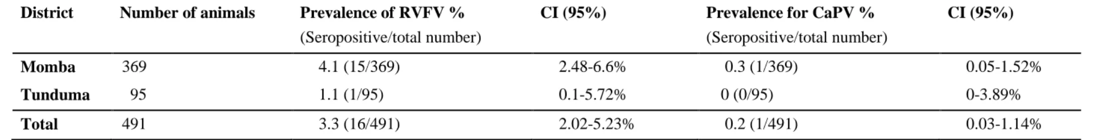
For RVFV, 3.3% (16/491) (confidence interval 95% gives 2.02-5.23%) out of the total number of animals were seropositive in the RVFV competitive ELISA on an individual level. None of the sheep was seropositive for RVFV. Out of 484 goat samples, 3.3% (16/484) was seropositive for RVFV on an individual level. The seroprevalence for female goats was 3.5% (14/400), and for male goats 2.4% (2/84), no statistic significant difference between genders (p-value 0.60). Females accounted for the larger proportion of all seropositive animals 87.5% (14/16). Totally 31.7% (13/41) of the villages had seropositive animals, the seroprevalence within the villages ranged from 0-25%. In 0.2% (1/491) of the samples the result was doubtful and in this study interpreted as negative.
Of 491 animals 75% (368/491) was older than 1 year and 25.1% (123/491) younger than 1 year (Table 3). The Momba district was 60% (296/491) older than 1 year and 20.1% (100/491) younger than 1 year. In Tunduma district it was 14.7% (72/491) animals older than 1 year and 4.7% (23/491) younger than 1 year.
18
Table 3. Total number of animals and the seroprevalence for RVFV and CaPV divided by specie,
gender and age
Species Total number (%) RVFV seropositive (%) CaPV seropositive (%) Goats Females < 1 year > 1 year Males < 1 year > 1 year 484 (98.6) 400 (81.5) 86 (17.5) 314 (64.0) 84 (17.1) 37 (7.5) 47 (9.6) 16 (3.3) 14 (3.5) 1 (1.2) 13 (4.1) 2 (2.4) 0 (0) 2 (4.3) 1 (0.2) 1 (0.3) 0 (0) 1 (0.3) 0 (0) 0 (0) 0 (0) Sheep Females < 1 year > 1 year Males < 1 year > 1 year 7 (1.4) 5 (1.0) 0 (0) 5 (1.0) 2 (0.4) 0 (0) 2 (0.4) 0 (0) 0 (0) 0 (0) 0 (0) 0 (0) 0 (0) 0 (0) 0 (0) 0 (0) 0 (0) 0 (0) 0 (0) 0 (0) 0 (0) Total Females < 1 year > 1 year Males < 1 year > 1 year 491 (100) 405 (82.5) 86 (17.5) 319 (65.0) 86 (17.5) 37 (7.5) 49 (10.0) 16 (3.3) 14 (3.5) 1 (1.2) 13 (4.1) 2 (2.3) 0 (0) 2 (4.1) 1 (0.2) 1 (0.2) 0 (0) 1 (0.3) 0 (0) 0 (0) 0 (0)
Out of all seropositive animals, 93.8% (15/16) were more than one year old. Distribution between the genders is seen in Table 3.
For CaPV 0.2% (1/491) out of the total number of animals were seropositive on the double antigen ELISA. The seropositive animal was a female goat more than 1 year old in the Momba district. The goat had had signs of coughing, difficult breathing and diarrhea during the past 12 months. Positive predictive value (PPV) for a seroprevalence of 0.2% of 50% with the CaPV tests specificity of 99.7%. The negative predictive value (NPV) becomes 99.8%.
20
Table 4. Individual prevalences for RVFV, CaPV and their confidence intervals (CI in the sampled districts) District Number of animals Prevalence of RVFV %
(Seropositive/total number)
CI (95%) Prevalence for CaPV % (Seropositive/total number)
CI (95%)
Momba 369 4.1 (15/369) 2.48-6.6% 0.3 (1/369) 0.05-1.52%
Tunduma 95 1.1 (1/95) 0.1-5.72% 0 (0/95) 0-3.89%
Total 491 3.3 (16/491) 2.02-5.23% 0.2 (1/491) 0.03-1.14%
21
Fig 3. A photo of an ELISA plate with 6 positive wells for RVFV ID Screen Rift Valley Fever Competition Multi Species.
Questionnaires
Management routines
All farmers (164/164; 100%) reported that they were using communal grazing as the main grazing system for their goats and sheep. A small number of farmers also utilized herding (6/164; 3.7%) and tethering (2/164; 1.2%) in addition to communal grazing. All farmers of the sampled herds reported that their sheep and goats had daily contact with other goats and sheep and the majority (160/164; 97.6%) also reported daily contact with cattle. In contrast, almost all farmers (159/164; 97%) reported that their goats and sheep had no contact with wild ruminants.
Of the two farmers that reported that their animals were in daily contact with wild ruminants (2/164; 1.2%), one had a RVF seropositive animal. This animal was a female goat older than 1 year.
Disease managment
A majority of the farmers (157/164; 95.7%) reported that they did not vaccinate their sheep and goats. Out of those that vaccinated their animals (7/164; 4.3%) all of them vaccinated their animals against contagious caprine pleuropneumonia (CCPP).
22
All farmers reported that they did not keep their sick animals separated from the rest of the herd.
Trade
All farmers answered that they never had bought animals from other countries. After acquiring new sheep and goats 96.3% (158/164) immediately let them mix with the rest of the herd and only six farmers (3.7%) reported that they did not let them mix immediately. All farmers with RVFV seropositive animals let their animals mix immediately after acquiring new animals. Table 5. Where the farmers bought their sheep and goats from
Location % (number)
From other farmers in the same village 86.0 (141) From farmers in other villages in the same district 42.7 (70)
From farmers in other district 3.0 (5)
At markets 18.8 (30)
From traders 1.8 (3)
Other ways 3.7 (6)
On the question of where farmers bought their sheep and/or goats from, they could answer several different options (Table 5). A majority of the farmers reported that they bought their sheep and goats from other farmers in their village. Of all the farmers, 18.3% (30/164) reported that they bought their animals from markets, and on a village level, 51.2% (21/41) of the villages had bought animals from markets. Farmers who bought their animals from markets or had neighbors who did, were more likely to have at least one RVFV seropositive animals (60.0% vs. 40%, p-value<0.000012). In the villages that bought their animals from markets, 38.1% (8/21) had at least one RVFV seropositive animals.
The farmer with the sole seropositive animal for CaPV had only bought new animals from other farmer in the same village, but the other farmers in that village reported that they had bought animals from markets.
23
Fig 5. The distribution of clinical signs that the farmers think was acceptable for
the animals to have and still be sold.
Fig 6. The distribution of clinical signs that the farmers think it was acceptable for
the animals to have and they would still buy it.
Most farmers (458/491; 93.3%; Fig 5) reported that they think it is acceptable to sell animals with signs of ocular and nasal discharge and cough. About 15% of the farmers (Fig 6) reported that signs of ocular and nasal discharge and coughing in sheep/goats were okay for the animals to have and the farmer would still buy it.
Animal health
The most common clinical signs that farmers reported that they had observed in the last 12 months were diarrhea, coughing and ocular and nasal discharge (Table 6). The observed abortion rate was 36% (59/164) in the herds and out of the farmers with abortion during the last 12 months 13.6% (8/59) had RVFV seropositive animals. Of the farmers with seropositive
0,0% 20,0% 40,0% 60,0% 80,0% 100,0% Runny eyes and nose
Coughing Diarrhea Abortion Blister and
sores
Which sheep/goat diseases is it okay
for a goat/sheep to have and it can
still be sold?
0,0% 20,0% 40,0% 60,0% 80,0% 100,0% Runny eyes and noseCoughing Diarrhea Abortion Blister and
sores
What diseases would you say that it
is okay for the goats to have and you
24
animals, 53.3% (8/15) had abortions in the last 12 months and out of those 72.7% (8/11) farmers had have abortion during the last 12 months. On a village level 42.3% (11/26) had seropositive animals during the last 12 months. There was no statistic significant difference between villages that had observed abortion and those who did not have observed any abortions.
Table 6. The most common clinical signs that the farmer had observed in the last 12 months
Signs of diseases Total number (%)
Diarrhea 133 (81.1)
Coughing 120 (73.2)
Abortion 59 (36.0)
Dying kids/lamb 57 (34.8)
Sudden death 66 (40.2)
Blisters and sores 25 (15.2)
Runny eyes and nose 120 (73.2)
Public health
Fig 7. The results of the question on which diseases those are ok for
animals to have and still be consumed by the farmers.
A large proportion of the farmers reported that they think it is okay to consume animals with a variety of clinical signs. Out of the signs (Fig 7) ocular and nasal discharge (153/164; 93.3%) was the most common that the farmers reported was ok for the animals to have and still be consumed. Abortion was one of the signs less frequently reported (135/164; 82.3%) as okay for the animals to have and still be consumed. Of the farmers with seropositive animals, 80% (12/15) of them reported that they did consume animals that had aborted. There was no statistic significant difference between the two groups.
0,0% 20,0% 40,0% 60,0% 80,0% 100,0% Runny eyes and nose
Coughing Diarrhea Abortion Blister and
sores
Which diseases are okay for the
goats/sheep to have and still be
25
Farmer details
Table 7. Gender and age of farmer and/or caregiver of the animal and the seroprevalence
Age % (number) Seropositive % (number) Years in school % (number) Seropositive % (number) Women <30 31-50 > 51 100 (41) 22.0 (9) 63.4 (26) 14.6 (6) 4.9 (2) 0 (0) 7.7 (2) 0 (0) Women 0 years 1-7 years > 7 years 100 (41) 29.3 (12) 63.4 (26) 7.3 (3) 4.9 (2) 0 (0) 3.8 (1) 33.3 (1) Men <30 31-50 > 51 100 (122) 14.8 (18) 63.1 (77) 22.1 (27) 10.7 (13) 16.7 (3) 10.4 (8) 7.4 (2) Men 0 years 1-7 years > 7 years 100 (122) 21.3 (26) 67.2 (82) 11.5 (14) 10.7 (13) 19.2 (5) 8.5 (7) 7.1 (1)
Among all farmers, 23.2% (38/164) reported that they had not gone to school. Out of the farmers, 9.1% (15/164) of them had RVFV seropositive animals and of all farmers with seropositive animals, 33.3% (5/15) was uneducated.
26
Table 8. Univariable analysis for risk factors associated with seropositivity for RVF at an individual animal level
Questions Answers RVF Seropositive RVF Seronegative P value < 0.05
Have you observed any symptoms in your sheep/goat in the last 12 months?
Yes (149) No (15) 15 1 431 44 0.681174 Not significant Where do you buy sheep and/or goats from? Within the district (136)
Outside the district (28)
14 2 393 82 0.618746 Not significant When was the last time you bought sheep and/or goats? Within the last 6 months (29)
More than 6 months ago (135)
2 14 85 390 0.578305 Not significant After acquiring new sheep and goats, do you let them
mix with your original heard immediately?
Yes (158) No (6) 16 0 457 18 0.739461 Not significant
27
DISCUSSION
In this master thesis, the aim was to investigate the seroprevalence of RVFV and CaPV among sheep and goats in the Momba and Tunduma districts, close to the border of Zambia. The individual seroprevalence for RVFV in sheep and goats in the two districts were 3.3% (16/491), no statistic difference was observed between the two districts. Only 1.4% of the animals included in this study were sheep and none was seropositive, but the population included was too small and no conclusion could be drawn. In Momba district the prevalence on a village level was 33.3% and for Tunduma 25%. This seroprevalence indicates that sheep and goats sometime during their lifetime have encountered RVFV due to either natural infection or through vaccination, the latter one is in this study considered unlikely. In this study, only goats were seropositive for RVFV, but a majority of the animals (98.6%) included in this study was also goats. Previous studies have identified sheep to have a higher seroprevalence of RVFV than goats (Blomström et al., 2016; Jeanmaire et al., 2011; Rostal et al., 2010). In Tanzania, a slightly higher number of goats than sheep have been seropositive in some district (Wensman
et al., 2015). In other studies, no significant differences between the two species been were
observed (Sumaye et al., 2013).
In this study, 3.8% of the females were seropositive compared to 1.2% of the males, but no significant difference between genders was observed. Out of all seropositive animals, all but one were more than one year of age, 14 being females and one male. The only seropositive animal under 1 year was a female. This could indicate presence of the virus with ongoing circulation of RVFV in the districts. Previously conducted studies detected young animals to be seropositive showing continuous circulation of RVFV in the northern and central part of Tanzania during an inter-epidemic period (Wensman et al., 2015). Sindato et al. (2014) concluded in their review that RVF outbreaks were mainly reported during periods of prolonged heavy rainfall. Sindato et al. (2010) suggest that when RVFV has been introduced to a new geographical area, it becomes endemic and favorable conditions in the environment allow a reactivation in a large scale. It is possible that RVFV is circulating in the Momba district where the animal under 1 year was found seropositive.
The age of the animals was estimated mainly based on the farmers’ information. Some owners knew exactly how old every individual was, while some farmers were not sure about the age and gave an approximated age. The animals included in this study would have to be over 4 months old, in some cases when the farmer was unsure about the age and the size of the animal was tiny, it was rejected too avoid sampling animals that were too young. Some animals were estimated by the farmer to be around one year. This could influence the interpretation of the seroprevalence between the age group, when results are presented under or above one year old. A supposed risk factor for transmission of RVF is contact with wildlife (Wensman et al., 2015). In this study, most farmers reported that there was no direct contact between sheep and goats and wild ruminants. Almost all farmers reported that their sheep and goats were in daily contact with cattle, and these animals graze further away from the livestock keepers and can therefore