swedish dent al journ al, supplement 220, 20 1 1 . d oct or al dissert a tion in odont ol og y m aria p igg malmö universit y 20 1 1 malmö university 205 06 malmö, sweden www.mah.se
maria pigg
chronic intraoral pain
– Assessment of diagnostic methods and prognosis
isbn/issn 978-91-7104-387-0/0348-6672 c hr onic intr a or al p ain
C H R O N I C I N T R A O R A L P A I N
Swedish Dental Journal, Supplement 220, 2011
© Copyright Maria Pigg, 2011 ISBN 978-91-7104-387-0 ISSN 0348-6672 Holmbergs, Malmö 2011
Department of Endodontics and
Department of Stomatognathic Physiology
Faculty of Odontology, Malmö University
Malmö, Sweden
2011
MARIA PIGG
CHRONIC INTRAORAL PAIN
The summary of this publication is also available at www.mah.se/muep
5
CONTENTS
ABSTRACT ... 7 POPULÄRVETENSKAPLIG SAMMANFATTNING (SUMMARY IN SWEDISH) ... 9 PREFACE ... 11 THESIS AT A GLANCE ... 12ACRONYMS AND DEFINITIONS ... 13
QST measures ...14
Taxonomy ...14
INTRODUCTION ... 16
Chronic and acute pain ...16
Orofacial pain ...17
Neuropathic pain ...18
Intraoral pain of odontogenic origin ...23
Intraoral pain of nonodontogenic origin ...24
The science of diagnostics ...28
Diagnostic tests in tooth pain investigation ...29
Somatosensory examination and QST ...36
Radiographic examination ...36
The importance of an accurate diagnosis ...38
Prognosis ...39
OBJECTIVES ... 40
HYPOTHESES ... 41
MATERIALS AND METHODS ... 42
Healthy subjects (I and II) ...42
Patients (III and IV) ...42
Data collection ...43
6
Somatosensory examination and diagnostic testing (I, II, IV) ... 44
Radiographic examination (III) ... 48
Self-report measures (III and IV) ... 48
Statistical analyses (I–IV) ... 51
Informed consent and ethical approval (I–IV) ... 53
RESULTS ... 54
Characteristics of intraoral thermal thresholds (I) ... 54
Reliability of intraoral QST (II) ... 55
CBCT and conventional image assessments in patients with AO and SAP (III) ... 58
Prognosis and prediction of outcome in AO (IV) ... 59
DISCUSSION ... 64
Somatosensory threshold assessment ... 65
Reliability of somatosensory testing ... 67
Radiographic examination in atypical odontalgia ... 68
Prognosis assessment ... 71
CONCLUSIONS, CLINICAL IMPLICATIONS, AND FUTURE WORK PLAN ... 77
AUTHORS' CONTRIBUTION ... 80
ACKNOWLEDGEMENTS ... 81
REFERENCES ... 84
7
ABSTRACT
The overall goal of this thesis was to broaden our knowledge of chronic intraoral pain. The research questions were:
What methods can be used to differentiate inflammatory, odontogenic tooth pain from pain that presents as toothache but is non-odontogenic in origin?
What is the prognosis of chronic tooth pain of non-odontogenic origin, and which factors affect the prognosis? Atypical odontalgia (AO) is a relatively rare but severe and chronic pain condition affecting the dentoalveolar region. Recent research indicates that the origin is peripheral nerve damage: neuropathic pain. The condition presents as tooth pain and is challenging to dentists because it is difficult to distinguish from ordinary toothache due to inflammation or infection. AO is of interest to the pain community because it shares many characteristics with other chronic pain conditions, and pain perpetuation mechanisms are likely to be similar.
An AO diagnosis is made after a comprehensive examination and assessment of patients’ self-reported characteristics: the pain history. Traditional dental diagnostic methods do not appear to suffice, since many patients report repeated care-seeking and numerous treatment efforts with little or no pain relief. Developing methods that are useful in the clinical setting is a prerequisite for a correct diagnosis and adequate treatment decisions.
Quantitative sensory testing (QST) is used to assess sensory function on skin when nerve damage or disease is suspected. A variety of stimuli has been used to examine the perception of, for
8
example, touch, temperature (painful and non-painful), vibration, pinprick pain, and pressure pain. To detect sensory abnormalities and nerve damage in the oral cavity, the same methods may be possible to use.
Study I examined properties of thermal thresholds in and around the mouth in 30 pain-free subjects: the influence of measurement location and stimulation area size on threshold levels, and time variability of thresholds. Thresholds for cold, warmth and painful heat were measured in four intraoral and two extraoral sites. Measurements were repeated 3 times over 6 weeks, using four sizes of stimulation area (0.125–0.81 cm2). The threshold levels were highly dependent on location but less dependent on measuring probe size and time variability was small, and this knowledge is important for the interpretation of QST results.
Study II applied a recently developed standardized QST examination protocol (intended for use on skin) inside the oral cavity. Two trained examiners evaluated 21 pain-free subjects on three occasions over 1–3 weeks, at four sites—three intraoral and one extraoral. Most tests had acceptable reliability and the original test instruments and techniques could be applied intraorally with only minor adjustments.
Study III examined the value of cone-beam computed tomography (CBCT) in pain investigations. Twenty patients with AO and 5 with symptomatic apical periodontitis (inflammatory tooth pain) participated. The results indicate that when AO is suspected, addition of CBCT can improve the diagnostic certainty compared to sole use of periapical and panoramic radiographs, especially because of the superior ability of CBCT to exclude inflammation as the pain cause.
Study IV assessed the long-term prognosis of AO, and analyzed potential outcome predictors. A comprehensive questionnaire including validated and reliable instruments was used to gather data on patient and pain characteristics and pain consequences from 37 patients in 2002 and 2009. Thirty-five percent of the patients reported substantial overall improvement at follow-up, but almost all still had pain of some degree after many years. The initial high level of emotional distress was unchanged. Low baseline pain intensity predicted improvement over time.
9
POPULÄRVETENSKAPLIG
SAMMANFATTNING
(SUMMARY IN SWEDISH)
Det övergripande målet med avhandlingen var att öka vår förståelse av kronisk smärta i munnen. Tre delarbeten syftar till att undersöka och utveckla diagnosmetoder för att särskilja tandsmärta av olika ursprung, och det fjärde delarbetet undersöker långtidsprognosen för atypisk tandsmärta samt prognospåverkande faktorer.
Atypisk odontalgia (AO), eller atypisk tandsmärta är ett allvarligt och kroniskt men relativt sällsynt smärttillstånd som uppträder i munhålans betandade område. Mycket tyder på att orsaken är skada på sensoriska nervceller, och bör betecknas neuropatisk smärta eller nervsmärta. Tillståndet yttrar sig som smärta i en tand eller i området där en tand har suttit, och är svårt att särskilja ifrån ”vanlig tandvärk” utlöst av inflammation eller infektion. AO liknar andra kroniska smärttillstånd på många sätt och det är troligt att samma eller liknande mekanismer underhåller smärtan.
Diagnosen ställs efter noggrann värdering av undersökningsfynd och patientens beskrivning av smärtsymptom och smärthistorik. Tandläkarens sedvanliga undersökningsmetoder förefaller inte tillräckliga; många patienter med AO beskriver upprepat vård-sökande och ett flertal behandlingsförsök med ringa eller helt utebliven smärtlindring. Det är angeläget att utveckla diagnos-metoder för att säkrare ställa rätt diagnos och därmed fatta riktiga behandlingsbeslut.
10
Kvantitativ känselundersökning (QST) används på hud för att mäta känselfunktion när man misstänker nervskada eller sjukdom i nervsystemet, som ofta medför förändrad känseluppfattning. En rad stimuli används för att undersöka känslighet för t ex beröring, temperatur, vibration, sticksmärta och trycksmärta. För att påvisa känselförändring och nervskada i munhålan borde det vara möjligt att använda samma undersökningsmetodik.
I delarbete I studerades temperaturtrösklar i och omkring munnen. Trösklar för kyla, värme och smärtsam värme mättes på smärtfria försökspersoner 3 gånger under 6 veckor. Mätningar gjordes även med varierande storlek på mätinstrumentets stimuleringsyta. Temperaturkänsligheten påverkades mycket av mätlokalisation men betydligt mindre av stimuleringsytans storlek, och tids-variationen var liten. Kunskapen har betydelse för tolkning av mät-resultat vid känselundersökning.
I delarbete II användes ett standardiserat testprotokoll för kvantitativ känselundersökning, nyligen utvecklat för att användas på hud. Två undersökare mätte känsel- och smärttrösklar i mun och ansikte på smärtfria försökspersoner vid tre tillfällen, och de flesta ingående testen uppvisade acceptabel tillförlitlighet. Metoden kan därmed användas i framtida forskning och på sikt möjligen även vid diagnostik.
Delarbete III undersökte värdet av volymtomografiundersökning (CBCT) i smärtutredning. Resultaten tyder på att volymtomografi kan öka den diagnostiska säkerheten vid misstänkt atypisk tandsmärta, eftersom metoden är överlägsen övriga röntgen-metoder för att påvisa inflammatoriska förändringar i käkbenet.
Delarbete IV undersökte långtidsprognosen för atypisk tandsmärta. Genom ett omfattande frågeformulär med validerade och tillförlitliga instrument och mätskalor insamlades patient- och smärtkarakteristika från 37 patienter med AO år 2002 och 2009. 35 % av patienterna angav väsentlig förbättring över tid, men nästan alla hade fortfarande smärtor i någon grad, och depressiva symptom förekom i oförändrat hög utsträckning. Låg ursprunglig smärtintensitet innebar större sannolikhet för en förbättrad situation på lång sikt.
11
PREFACE
This thesis is based on the following articles, which are referred to in the text by their roman numerals:
I Pigg M, Svensson P, List T. Orofacial thermal thresholds: time-dependent variability and influence of spatial summation and test site. J Orofac Pain 2011; 25(1):39-48.
II Pigg M, Baad-Hansen L, Svensson P, Drangsholt M, List T. Reliability of intraoral quantitative sensory testing (QST). Pain 2010; 148: 220-6.
III Pigg M, List T, Petersson K, Lindh C, Petersson A. Diagnostic yield of conventional radiographic and cone-beam computed tomographic images in patients with atypical odontalgia. Int Endod J 2011; 44(12):1092-101.
IV Pigg M, Svensson P, Drangsholt M, List T. Long-term prognosis of atypical odontalgia. A 7-year prospective study. Submitted for publication, October 2011.
The articles are reprinted with kind permission from the copyright holders: Blackwell Quintessence Publishing Co. Inc. (I), the International Association for the Study of Pain (II), and John Wiley & Sons, Inc. (III).
12 Thesis at a glance St udy O bjectiv es M et hods Illu stra tio ns M ai n f indi ngs/C oncl usi ons (I) Oro fa cia l th erm al th resh old s: tim e-dependent var iabi lit y and i nf luence of s pat ial su m m ati on an d test si te T o exam ine the ti m e vari abi lit y of int raoral th erm al th re sh ol ds and t he rel ati on sh ip b etw een th re sh old s a nd te st site locat ion and st im ul at ion area si ze. T herm al threshol ds w ere m eas ur ed i n 30 he al thy su bj ects o n tw o o ccasi on s, w it h 5 di ff erent st im ul at ion area si zes and i n 6 orof aci al si tes. O ro fa cia l lo ca tio n af fect s t he ther m al t hr es hol d l evel . In tra ora l th re sh old s re m ain reaso nab ly stab le o ve r ti m e,
and are onl
y m argi nal ly af fect ed by t he si ze of t he sti m ul ati on area. (II) Rel iabi lit y of int raoral quant it at ive s ens or y t es ti ng (Q ST ) T o determ ine the i ntra - and in te r-exam iner rel iabi lit y f or int raoral and f aci al Q ST in heal thy s ubj ect s, and t o eval uat e t he i nt raoral appl icabi lit y of Q ST . 21 heal thy s ubj ect s under w ent s om at os ens or y exam inat ion on t hree occas ions accor di ng t o a stan dard ized an d com prehensi ve Q ST pr ot oc ol . M ost Q ST m easures have accept abl e rel iabi lit y w hen appl ied i nt raoral ly. (III) Diagnost ic yi el d of convent ional radi ographi c and cone -beam com put ed tom ogr aphi c ( CBCT) im ages in pat ient s w it h at ypi cal odont al gi a T o determ ine w hether a CB CT exam inat ion i m proves th e id en tific atio n o f p atie nts w it h at ypi cal odont al gi a ( A O ) com pared t o radi ographi c exam inat ion w it h convent ional int raoral per iapi cal and panor am ic im ages. 20 pat ient s w it h A O and 5 pa tie nts w ith sym pto m ati c api cal peri odont it is ( SA P) under w ent r adi ogr aphi c exam inat ion w it h convent ional radi ographi c m et hods and C BC T. C BC T im proves i dent if icat ion of p atie nts w ith ou t inf lam m at ory changes of jaw bone t is sue . O vera ll, m ore fin din gs w ere m ad e w ith C BC T as an adj unct and fo r a m ajo rity of t he A O group (60% ) no pat hol ogy w as found w it h any m et hod. (IV ) Long -te rm p ro gn osis o f at ypi cal odont al gi a. A 7-year prospect ive st udy T o exam ine the l ong -te rm pr ognos is of A O and i dent if y fa cto rs p re dic tin g p ersiste nt pai n. Sel f-rep ort fo llo w -up dat a f or 37 pat ient s w ith A O w ere col lect ed i n 2009 and com pared t o basel ine dat a fr om 2002. A m aj ori ty of pat ient s w ith AO rep ort ed im provem ent ov er tim e, w hic h is a lso refl ected b y l ow er p ai n int ensi ty and f requency.
13
ACRONYMS AND DEFINITIONS
AFP Atypical facial pain AO Atypical odontalgia BMS Burning mouth syndrome
CBCT Cone-beam computed tomography
CPI Characteristic pain intensity
DFNS German Research Network on Neuropathic Pain GCPS Graded chronic pain severity
IASP International Association for the Study of Pain ICC Intraclass correlation coefficient
IMMPACT Initiative on Methods, Measurement, and Pain Assessment in Clinical Trials
JFLS Jaw functional limitation scale NRS Numerical rating scale (0–10)
PDAP Persistent dento-alveolar pain disorder PGIC Patients’ global impression of change scale
QST Quantitative sensory testing
RDC/TMD Research diagnostic criteria for TMD
RCT Randomized controlled trial
SAP Symptomatic apical periodontitis SCL-90R Symptom checklist 90 revised SF-36 Short-form health survey, 36 items
STARD Standards for Reporting of Diagnostic Accuracy
14
QST measures
CDT Cold detection threshold WDT Warmth detection threshold CPT Cold pain threshold
HPT Heat pain threshold
PHS Paradoxical heat sensation (on cold stimulus) TSL Thermal sensory limen
MDT Mechanical detection threshold MPT Mechanical pain threshold MPS Mechanical pain sensitivity
DMA Dynamic mechanical allodynia
WUR Wind-up ratio
PPT Pressure pain threshold VDT Vibration detection threshold
Taxonomy
Nociceptive pain
Pain that arises from actual or threatened damage to non-neural tissue and is due to the activation of nociceptors (IASP pain taxonomy 2011).
Neuropathic pain
Pain caused by a lesion or disease of the somatosensory nervous system (IASP pain taxonomy 2011).
Allodynia
Pain due to a stimulus that does not normally provoke pain (IASP pain taxonomy 2011).
Hyperalgesia
Increased pain from a stimulus that normally provokes pain (IASP pain taxonomy 2011).
15
Dysesthesia
An unpleasant abnormal sensation, whether spontaneous or evoked (IASP pain taxonomy 2011).
Paresthesia
An abnormal sensation, whether spontaneous or evoked (IASP pain taxonomy 2011).
Spatial summation
The ability of the nervous system to integrate nociceptive input from large areas. The sensory threshold and perceived intensity are affected by increasing the stimulation area, manifested as decreased pain threshold and increased pain intensity (Nie et al.
2009).
Temporal summation
The ability of the nervous system to integrate repetitive nociceptive input, i.e. increasing pain perception to repetitive stimulation at the same intensity (Nie et al. 2009).
Peripheral sensitization
Increased responsiveness and reduced threshold of nociceptive neurons in the periphery to the stimulation of their receptive fields (IASP pain taxonomy 2011).
Central sensitization
Increased responsiveness of nociceptive neurons in the central nervous system to their normal or subthreshold afferent input (IASP pain taxonomy 2011).
16
INTRODUCTION
Chronic and acute pain
The International Association for the Study of Pain currently defines pain as ‘an unpleasant sensory and emotional experience associated with actual or potential tissue damage, or described in terms of such damage’ (IASP pain taxonomy 2011). Nearly every human being has some personal experience of pain; it is one of the basic bodily sensations. While acute pain could be described as a survival mechanism, alerting us to impending or ongoing tissue damage and invoking a withdrawal reflex, chronic pain is more complex and has no clear-cut physiological value.
The concept of chronic pain embraces several different meanings depending on the situation: it is a complex, multi-dimensional condition. One popular definition is ‘pain that extends beyond the expected period of healing’, which usually refers to pain persisting for more than 3–6 months. Chronic pain may be associated with a chronic disease, such as rheumatoid arthritis or multiple sclerosis, and also with less well-defined disorders, such as irritable bowel syndrome, back pain, fibromyalgia, chronic headache, and TMD. Another type of chronic pain is pain that persists long after initial tissue damage has healed and occurs when plastic changes in the nervous system have become permanent.
Chronic pain is a very different condition from pain associated with acute tissue damage. Why some patients develop chronic pain conditions and others do not is not fully known, but genetic factors may be involved (Diatchenko et al. 2005). Psychological factors
17
seem to be intimately involved in the individual’s perception of pain, the transition of acute pain into chronic pain, and the maintenance of chronic pain (Turk 1999). Social and cultural contexts shape patients’ perceptions and responses to pain, and psychosocially dysfunctional individuals appear to be less able to develop effective coping strategies (Dworkin and Massoth 1994). The biopsychosocial model describes chronic pain as a multidimensional phenomenon, focusing on perceived illness rather than disease. Three main aspects of pain can thus be distinguished: the sensory (physiological), the affective (emotional and motivational), and the evaluative (cognitive) dimensions (Linton and Skevington 1999).
World-wide, chronic pain is one of the most common health problems today and affects approximately 10–30% of the adult population—a recent systematic review found a prevalence of 19% for moderate to severe general (non-cancer) chronic pain in Europe (Reid et al. 2011). The consequences of chronic pain are enormous costs in healthcare expenses, lost income, and lost productivity. Many studies have shown chronic pain to have a detrimental effect on patient-perceived health status, quality of life, activities of daily life and mental health (Breivik et al. 2006; Collett 2011). A substantial proportion of non-cancer chronic pain patients report not receiving pain treatment, and many others consider their pain treatment inadequate (Bekkering et al. 2011).
The most commonly reported non-cancer chronic pain disorders are musculoskeletal pain, neuropathic pain, fibromyalgia, osteoarthritis, and rheumatoid arthritis (Reid et al. 2011).
Orofacial pain
One of the most common locations for pain is the orofacial area. Orofacial pain is a term referring to oral pain, dental pain, and pain in the face above the neck, anterior to the ears and below the orbitomeatal line (Zakrzewska and Hamlyn 1999). In an epidemiological study including 45,711 households in the US, 21% of the population had experienced orofacial pain of some kind during the last 6 months. The pain was described as toothache,
18
oral sores, jaw joint pain, face or cheek pain, or burning mouth, and around one-fifth of the patients had experienced more than one pain symptom, or a combination of symptoms. Toothache when biting or chewing (or pain perceived as coming from a tooth) was by far the most frequently reported symptom, reported by 12% (Lipton et al. 1993).
In the dental office, a common reason for seeking emergency care is pain. Most orofacial pain conditions are of an acute, nociceptive character—such as pain from dental hypersensitivity, reversible and irreversible pulpitis, apical periodontitis and dental trauma— but others, while still mainly nociceptive in character, are more chronic—such as myofascial pain in the masticatory system and arthralgia of the temporomandibular joint. Still other pain conditions are neuropathic, such as trigeminal and pretrigeminal neuralgia, glossopharyngeal neuralgia, and posttraumatic neuralgia after injury or surgical trauma. Idiopathic tooth pain or atypical odontalgia (AO); burning mouth syndrome; headache presenting as dental pain; referred pain from the masticatory system, from neck and shoulder structures, or from ongoing angina pectoris; systemic disease; maxillary sinusitis; and psychosocial or behavioral factors are other possible causes of pain, and for various reasons, a correct diagnosis may be difficult (Svensson and Sessle 2004).
Neuropathic pain
Neuropathic pain was recently redefined by the IASP as a clinical description of ‘pain caused by a lesion or disease of the somatosensory nervous system’ (IASP pain taxonomy 2011). The term ‘lesion’ is commonly used when diagnostic investigations (e.g., imaging, neurophysiology, biopsies, and laboratory tests) reveal an abnormality or when there was obvious trauma. The term ‘disease’ is commonly used when the underlying cause of the lesion is known (e.g., stroke, vasculitis, diabetes mellitus, or genetic abnormality). Lesions or diseases of the neural tissues can occur either in the CNS or peripherally, and do not always cause pain. The prevalence of peripheral neuropathy is reported to be 2.4% in the general population, rising to 8% in ages above 55 years (Cruccu and Truini 2010).
19
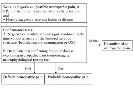
In the diagnostic descriptions and criteria for various disease entities, some are based mainly on clinical presentation (e.g., trigeminal neuralgia) and others on history (e.g., postherpetic neuralgia). Clinical decision-making thus requires knowledge of neuroanatomy and -physiology, a thorough review of patient-reported history and symptoms, and a comprehensive clinical examination with relevant and targeted clinical diagnostic testing, supplemented by laboratory tests if needed (Haanpää et al. 2011). Figure 1 depicts the recommended diagnostic process for patients with suspected neuropathic pain (Treede et al. 2008).
Figure 1. Grading system for neuropathic pain diagnosis (modified from Treede et al. 2008)
The diagnosis relies heavily on evidence of a damaged somatosensory system, represented by altered somatosensory function. The patient’s report of abnormal sensations, paresthesia or dysesthesia is one leg of this. The other is the clinician’s ‘bedside’ examination of sensory function: a qualitative (or semi-quantitative) assessment of nerve fiber function using simple and commonly available tools to examine touch (Aβ-fibers), cold
(Aδ-20
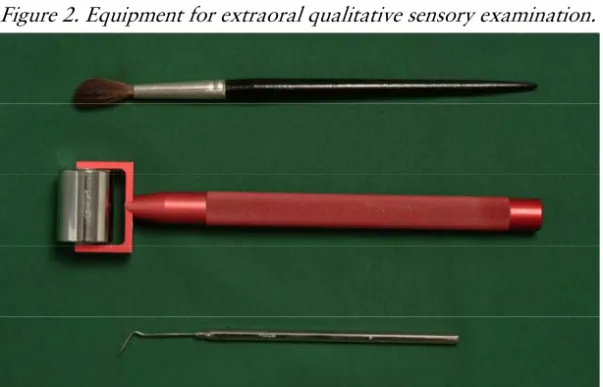
fibers) and pain (Aδ- and C-fibers) perception. Figure 2 depicts equipment for cutaneous qualitative sensory testing.
Figure 2. Equipment for extraoral qualitative sensory examination.
New guidelines on how to assess neuropathic pain in general were presented in 2004, and these have been updated (Cruccu et al.
2004; Cruccu et al. 2010; Haanpää et al. 2011). No criterion standard (or gold standard) for diagnosing neuropathic pain exists (no clinical or laboratory test can distinguish between a neuropathy without pain and a painful neuropathy), so the aim should be to confirm the diagnosis of an underlying neuropathy
that could rationally explain the pain problem reported by the patient. Using the same standards, recommendations for methods to assess neuropathic pain in the trigeminal area are under development (Svensson et al. 2004; Svensson et al. 2011).
Laboratory tests for diagnosing peripheral neuropathy and small fiber neuropathy in general include electroneuromyography (large fiber), microneurography, laser-evoked potentials, functional brain imaging (PET and fMRI), and skin biopsy (epidermal innervation). Quantitative sensory testing (QST) is a more comprehensive clinical method of assessing sensory nerve function than qualitative bedside testing. Until lately, a major drawback of QST has been a
21
lack of standardization in the choice of test modalities and in test performance recommendations, resulting in a lack of reference data for normal nerve function and lack of knowledge on the somatosensory characteristics of various conditions. In the last decade, however, substantial progress has been made in this field, and the validity and reliability of QST on cutaneous sites is now approaching the standard where it may be considered a clinically useful test method and not just a research tool. A few studies examining the orofacial area have also been presented (Juhl et al.
2008; List et al. 2008; Pfau et al. 2009). Recently, an international task force established by the IASP Special Interest Group on Oro-facial Pain (SIG-OFP) presented guidelines and recommendations for assessment of somatosensory function in patients with orofacial pain (Svensson et al. 2011).
In particular, a comprehensive QST protocol developed by the German Research Network on Neuropathic Pain (DFNS) has gained substantial interest in the neuropathic pain community (Rolke et al. 2006b). The DFNS protocol includes functional assessment of the sensory fibers: A, A, and C. A function is evaluated by measuring detection thresholds for mechanical touch and vibration, A and C function by measuring thermal detection and pain thresholds (cold, warmth, cold pain and heat pain) and mechanical pain thresholds for pinprick and pressure stimuli. In addition, signs of central sensitization are assessed by examining the occurrence of (i) paradoxal heat sensations on cold stimulation,
(ii) temporal summation to repeated pinprick pain stimuli, and (iii)
allodynia to a light stroking mechanical stimulus. One objection to somatosensory testing has been that it can be time consuming, which would limit its clinical usefulness. The DFNS protocol makes a comprehensive evaluation of somatosensory function possible in about 30 minutes per examined site, which is considered clinically feasible.
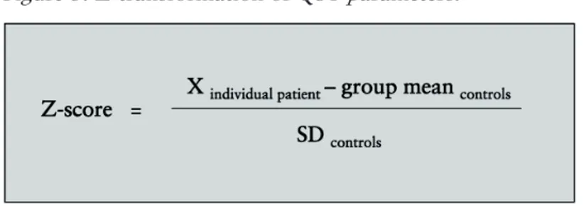
QST results can be transformed into Z-scores to describe a patient’s sensory profile. Z-transformation (Figure 3) of each sensory modality into a standard normal distribution allows the patient to be compared to a control group with normal sensory
22
function in a standardized way, independent of the stimulus modality’s unit of measurement.
Figure 3. Z-transformation of QST parameters.
According to this concept, a positive Z-score denotes increased sensitivity (gain of function) and a negative Z-score, decreased sensitivity (loss of function) in units of standard deviation (SD) of the control group (Rolke et al. 2006b). Figure 4 is an example of sensory profiles based on Z-scores.
Only the presence of signs of a disturbed somatosensory function, revealed through diagnostic testing, do not justify use of ‘neuro-pathic’ in the diagnosis, since diagnostic tests commonly yield inconclusive or inconsistent data. Somatosensory function may be altered in other pain conditions as well, such as acute nociceptive pain and functional pain, due to the physiological plasticity of the nervous system (Latremoliere and Woolf 2009).
23 Figure 4. Sensory profiles of three patients with AO, based on
Z-transformation of QST modalities (Z-scores)
Intraoral pain of odontogenic origin
Pain perceived as coming from one or more teeth in a region can be either odontogenic or non-odontogenic in origin. The most common by far is odontogenic tooth pain. Figure 5 lists possible origins of pain presenting as toothache.
Typical dental or dentoalveolar pain can present with a variety of symptoms ranging from intermittent and slight discomfort provoked by chewing, biting or tooth brushing to a continuous, spontaneous, severe, and highly disabling pain that affects many aspects of daily life. Pulpal and periapical inflammations are the most common causes of tooth pain. About one-third of all dental emergencies are reported to be endodontic, and about 90% of emergency appointments with pain as a symptom are prompted by pain from the dental pulp or the periapical region (Rossman et al.
2006). One review article reported the prevalence of dental pain to be 7–66% depending on the defining criteria used and the population studied (Pau et al. 2003).
24
Figure 5. Origins of tooth pain.
In inflammatory conditions, pain is induced through activation of normally functioning sensory nerve fibers; the pain is nociceptive in character. The affected peripheral nerve fibers include primary afferent neurons of the dental pulp, the gingiva and the periodontal ligament.
Intraoral pain of nonodontogenic origin
Besides tooth pain of odontogenic causes, the dentist is sometimes confronted with tooth pain caused by pathological processes outside the tooth (Fig. 5). The diagnostic process involves (i) a comprehensive evaluation of the patient’s description of symptoms and of events preceding the pain problem, (ii) clinical observations,
and (iii) the results of clinical and radiographic tests and
examinations. Most times, findings are straightforward and the dentist is able to arrive at a diagnosis quickly and with reasonable certainty. Other times, the differential diagnosis can be quite challenging: the clinical presentation may be complex and test
25
results inconclusive. In such cases, a conservative approach is often recommended and invasive treatment should be avoided until the diagnosis is more certain. For effective pain management, an accurate diagnosis should precede any treatment.
Atypical odontalgia (AO), also known as neuropathic tooth pain, phantom tooth pain (PTP), and persistent idiopathic facial pain (PIFP), is perhaps the most difficult to distinguish from toothache of dental origin. An international consensus collaboration, organized by the International RDC-TMD Consortium, recently introduced a new classification system for orofacial pain taxonomy based on ontological principles and adhering to the Ontology of General Medical Science (OGMS) guidelines. The collaboration examined AO as an example condition, and reviewed and revised the diagnostic criteria. Persistent dento-alveolar pain disorder (PDAP) was suggested as the new term, but it has not yet gained widespread acceptance (Nixdorf et al. 2011). AO has been described as tooth-related pain or pain located at a site where a tooth was extracted, in absence of clinical or radiographic evidence of tooth pathology or other relevant hard or soft tissue pathology explaining the pain (Melis et al. 2003). Pain has been ongoing for at least 6 months, is not paroxysmal in character, and is continuous or present during most of the day. Sleep is usually unaffected (Woda and Pionchon 1999). All adult ages and both sexes can be affected, but with a preponderance of women 45–60 years (List et al. 2007; Melis et al. 2003).
A recent systematic review found the prevalence of persistent pain after endodontic treatment to be 5.3%, with higher report quality studies suggesting > 7% (Nixdorf et al. 2010a). Studies examining nonsurgical and surgical endodontic treatment and retreatment were included. A meta-analysis of nine prospective studies reporting details on pain cause estimated the prevalence of non-odontogenic tooth pain as 3.4%, likely including patients with AO (Nixdorf et al. 2010b).
Patients with AO have often received multiple unsuccessful irreversible treatments aimed at pain relief (List et al. 2007;
26
Marbach and Raphael 2000). The etiology and mechanisms of pain development and pain perpetuation are still being debated. Deafferentation of primary afferent trigeminal nerve fibers has been suggested as the cause.
The theory that AO is neuropathic in origin and that neural mechanisms are involved in pain maintenance has been explored in many studies over the last decade. The evidence includes:
Pain onset is frequently reported in relation to an invasive dental or surgical procedure (such as endodontic treatment, endodontic surgery, tooth extraction, orthognathic surgery, facial fractures, dental implant placement, and dental injec-tions), in other words, a known or plausible trauma to trigeminal sensory fibers (List et al. 2007; Turp 2001).
Animal studies show that loss of tooth pulp in an inflamed environment produces a neuronal response with derangement of the periodontal plexus and axonal sprouting or neuroma formation (Holland 1995).
The human blink reflex (a trigeminofacial brainstem reflex) is delayed and reduced in patients with AO compared to pain-free control subjects, which may indicate impaired nerve function. Also, the blink reflex was altered on the painful and
the non-painful side, suggesting involvement of central mechanisms in these patients (Baad-Hansen et al. 2006b). Increased sensitivity to topical application of capsaicin occurs
in patients with AO compared to pain-free controls. The change in sensitivity is present on both painful and non-painful sides, indicating central sensitization (Baad-Hansen et al.
2006a; Baad-Hansen et al. 2007).
Studies on QST report sensory abnormalities in patients with AO, in response to both mechanical and thermal stimuli. Signs of hypersensitivity as well as hyposensitivity in the pain area occur, but results are partly contradictory between studies
27
(Lang et al. 2005; List et al. 2008), perhaps due to differences in assessment technique, test site (intraoral vs. extraoral), or test group inclusion criteria or to true heterogeneity in somato-sensory characteristics in the patient group. A standardized assessment method with confirmed reliability is likely to give better information on somatosensory function in these patients and allow comparison between pain conditions.
Application of a painful cold stimulus in the pain area produced a prolonged pain sensation (aftersensation) in patients with AO compared to controls. Moreover, the effect also occurred on the non-painful side, indicating involvement of central mechanisms (Zagury et al. 2011).
What makes differential diagnosis between odontogenic, inflammatory tooth pain and nonodontogenic, neuropathic tooth pain particularly challenging may be a combination of several factors:
The conditions share many clinical characteristics and can be similar in clinical presentation.
Toothache of odontogenic origin is much more common than neuropathic tooth pain, and is indeed the most likely explana-tion for pain localized to a tooth. It is familiar to the dentist, while pain conditions with lower prevalence are generally less familiar and therefore less easily suspected.
The diagnostic work-up commonly used in pain investigations is primarily designed to detect signs of inflammation; in prac-tice, test results indicating pathology are usually interpreted as inflammation, although alternative interpretations exist. A patient presenting with persistent pain is often emotionally
affected by the pain and presents a stressful situation to the dentist. To help the patient and remove the pain without delay, the dentist may base the treatment decision on less conservative diagnostic criteria and, for example, initiate
28
endodontic treatment despite absence of convincing signs of irreversible pulpal disease, thus accepting the risk of overdiagnosis (Reit and Petersson 2010)
There is a lack of diagnostic tests able to reliably distinguish nociceptive tooth pain from neuropathic tooth pain.
Dental emergency care in general is very efficient. Quick and efficient pain relief of toothache is usually possible, and most patients have come to expect this. But if the dentist cannot effect immediate pain relief due to, for example, diagnostic complexity, the patient is disappointed and feels the dentist has done something wrong. The patient may express a strong and urgent conviction of what is needed—endodontic treatment or extraction of the painful tooth—and seek care elsewhere until finding a dentist willing to provide this treatment; a behavior often referred to as “doctor shopping”.
The science of diagnostics
Diagnostics is the collected information about a condition (the patient’s story and clinical observations) that together with other information (e.g., knowledge of disease mechanisms and treatment efficacy) serves as the basis for a treatment decision. Diagnostic methods need to distinguish between health and disease, to evaluate degrees of disease, and to differentiate conditions from each other, making it possible to move forward in the process of clinical decision making (SBU, Swedish Council on Health Technology Assessment 2010). Making a diagnosis is often a complex task, and professionals will unavoidably vary in their decisions, depending on interpretation of clinical observations, level of knowledge, and personal experience, among other things. The diagnostic process has sometimes been regarded more as an act of art than of science. But clinical reasoning can—and should— be described in scientific terms, and diagnostic procedures structured and compared according to scientific criteria, which is necessary when deciding the usefulness of a method in a particular
29
situation. Some important properties of diagnostic tests are briefly outlined below.
Validity. The extent to which the test agrees with the “true” condition for which it is used as the diagnostic aid to identify.
Reference standard (criterion or gold standard). A reference test with optimal validity, used to evaluate other diagnostic tests.
Diagnostic accuracy. The extent to which a test correctly identifies what it is meant to identify; the overall agreement between the test and the reference standard.
Sensitivity and specificity. Two ways of expressing diagnostic accuracy. If the prevalence of the condition in the population is known, positive and negative predictive values
can be calculated.
Reliability. The reproducibility or repeatability of a test; to what extent a test can be repeated with the same results by another examiner (inter-examiner reliability) or by the same examiner on another occasion (test-retest or intra-examiner reliability).
For many diagnostic tests in wide use clinically, these properties are not fully known. The Standards for Reporting of Diagnostic Accuracy (STARD) initiative is an effort to improve the reporting quality of studies on diagnostic accuracy (Bossuyt et al. 2003). Their checklist for study design and manuscript preparation allows
(i) identification of possible sources of bias and (ii) evaluation of a test’s usefulness in various circumstances.
Diagnostic tests in tooth pain investigation
While the pathological mechanisms of pulpal and periapical inflammation have been vigorously studied, the literature describes the scientific properties of clinical diagnostic testing methods less well. Simple psychophysical tests are often useful in the clinical situation, despite not being very standardized. Dichotomous outcomes (response or no response) are generally used as guidelines in assessing endodontic disease.
30
Provocation of teeth and periapical tissues and pulpal sensibility assessment are common tests when endodontic disease is suspected. Accuracy—sensitivity and specificity—has been determined for some tests of pulp necrosis or presumed irreversible pulpitis (Table 1). The accuracy and variability of many tests are unknown, however, and test validity—to differentiate between pain conditions with similar presentation but of different origin—is often insufficiently examined. Tests applied in the diagnosis of a patient with toothache include:
Mechanical provocation of teeth Pain on percussion Pain on apical palpation Cusp loading Pulp testing Cold stimulus Heat stimulus Electrical stimulus Diagnostic anesthesia
Transillumination of natural teeth Provocation of referred pain
Palpation of the masticatory system Neck examination
24
Figure 5. Origins of tooth pain.
In inflammatory conditions, pain is induced through activation of normally functioning sensory nerve fibers; the pain is nociceptive in character. The affected peripheral nerve fibers include primary afferent neurons of the dental pulp, the gingiva and the periodontal ligament.
Intraoral pain of nonodontogenic origin
Besides tooth pain of odontogenic causes, the dentist is sometimes confronted with tooth pain caused by pathological processes outside the tooth (Fig. 5). The diagnostic process involves (i) a comprehensive evaluation of the patient’s description of symptoms and of events preceding the pain problem, (ii) clinical observations, and (iii) the results of clinical and radiographic tests and examinations. Most times, findings are straightforward and the dentist is able to arrive at a diagnosis quickly and with reasonable certainty. Other times, the differential diagnosis can be quite challenging: the clinical presentation may be complex and test
31
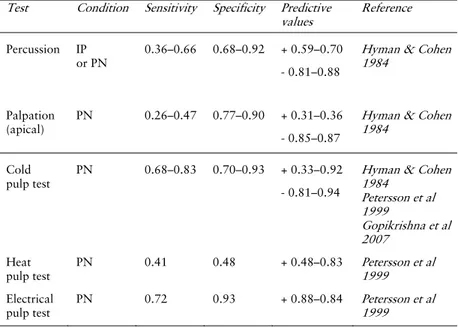
Table 1. Accuracy of intraoral diagnostic tests. IP: Irreversible pulpitis; PN: Pulp necrosis
Test Condition Sensitivity Specificity Predictive
values Reference Percussion IP or PN 0.36–0.66 0.68–0.92 + 0.59–0.70 - 0.81–0.88
Hyman & Cohen 1984
Palpation
(apical)
PN 0.26–0.47 0.77–0.90 + 0.31–0.36
- 0.85–0.87
Hyman & Cohen 1984
Cold pulp test
PN 0.68–0.83 0.70–0.93 + 0.33–0.92
- 0.81–0.94
Hyman & Cohen 1984 Petersson et al 1999 Gopikrishna et al 2007 Heat pulp test PN 0.41 0.48 + 0.48–0.83 Petersson et al 1999 Electrical pulp test PN 0.72 0.93 + 0.88–0.84 Petersson et al 1999
In inflammatory tooth pain, increased sensibility is the result of a complex series of interactions. Mediators are synthesized and/or released, and they activate nociceptive fibers. The affected neurons in turn release neuropeptides, which increase the inflammation further (neurogenic inflammation) and sensitize the nociceptors, thereby reducing the threshold for pain on stimulation such as percussion.
In nerve damage, the mechanisms are less clear but likely involve both peripheral and central sensitization. Changes in the reactivity of the sensory nervous system may provide alternative interpretations of diagnostic test results, and failure to appreciate this may lead to an incorrect diagnosis. Alternative test interpretations and implications for differential diagnostic accuracy are described below.
Test Condition Sensitivity Specificity Predictive
values Reference Percussion IP or PN 0.36–0.66 0.68–0.92 + 0.59–0.70 - 0.81–0.88
Hyman & Cohen 1984
Palpation (apical)
PN 0.26–0.47 0.77–0.90 + 0.31–0.36 - 0.85–0.87
Hyman & Cohen 1984
Cold pulp test
PN 0.68–0.83 0.70–0.93 + 0.33–0.92 - 0.81–0.94
Hyman & Cohen 1984 Petersson et al 1999 Gopikrishna et al 2007 Heat pulp test PN 0.41 0.48 + 0.48–0.83 Petersson et al 1999 Electrical pulp test PN 0.72 0.93 + 0.88–0.84 Petersson et al 1999
32
Mechanical provocation of teeth
Pain on percussion of a tooth is generally interpreted as presence of inflammation in the periapical tissues and considered a sign of either inflammation spread throughout the pulp (engaging the periodontium immediately apical to the tooth) or apical periodontitis. The inflammatory process also affects sensory nerves, and the pain threshold is lowered so that normally non-painful stimuli elicit pain. Allodynia and hyperalgesia are clinical signs of peripheral sensitization of nociceptors and the changes subsequently induced in higher order neurons (Hargreaves et al.
1994).
In AO, pain is often reported to increase on percussion of one or more teeth in the painful area (Merskey and Bogduk 1994). The usefulness of percussion testing in distinguishing between tooth-related pain conditions such as symptomatic pulpitis, symptomatic apical periodontitis, and neuropathic tooth pain is largely unknown.
Pain or tenderness on palpation of the region overlying the apex or apices of a tooth—a case of allodynia—is usually interpreted as presence of periapical inflammation subsequent to pulp necrosis. Patients with AO often report tenderness and increased pain on palpation of the painful region, including the periapical area (Graff-Radford and Solberg 1992; Merskey and Bogduk 1994). The validity of apical palpation to distinguish symptomatic apical periodontitis from neuropathic tooth pain has not been examined. Cusp loading with a FracFinder, Tooth Slooth, or similar instrument aims to detect incomplete fractures and cracks in teeth by selectively inducing force in various areas of the tooth. Loading a cusp that has a crack usually produces a sharp sensation of pulpal pain in the vital tooth. Responses vary, depending on location and extension of the crack, and are more doubtful in root-filled teeth. Teeth with symptomatic apical periodontitis are usually painful on biting (Tronstad 2009). Also in atypical odontalgia, AO pain often intensifies on biting and chewing (Melis
33 et al. 2003), but the validity of the test to differentiate pain conditions is unreported.
Pulp testing
In the trigeminal area, pulp testing may be useful in distinguishing pulpal pain and pain from other conditions, such as referred pain (Gopikrishna et al. 2009). A normal response to pulp testing may eliminate pulpal pathology. Pulp testing is usually done by thermal or electrical provocation of the pulp through the hard tissues of the crown.
Cold stimulation of teeth (e.g., applying ice or ethyl chloride) is often done to test dental pulp vitality. Vital pulp usually responds to stimulation with mild to moderate pain sensation. The underlying mechanism may involve the hydrodynamic flow of dentinal fluid causing movement in odontoblast processes and activation of adjacent free nerve endings (Olgart 1986).
Allodynia to cold, such as increasing pain in cold or windy weather, is frequently reported in AO, and cold stimulation of gingiva has elicited lingering pain in patients with AO (Zagury et al. 2011). Whether also responses to pulp cold testing can be altered in teeth in the painful region has not yet been examined. Heat stimulation of teeth (heated gutta-percha) is sometimes used for vitality testing but mostly to provoke bouts of toothache in patients presenting with intermittent pain of suspected pulpal origin, to determine which tooth is responsible. The hyperalgesia and allodynia here are clinical signs of nociceptive fibers’ increased and prolonged response to heat stimulation after sensitization by inflammatory mediators (Hargreaves et al. 1994). Whether heat stimulation of teeth can provoke or increase atypical tooth pain is unreported. Discomfort and increased pain on intake of hot food or drink is frequent in patients with pulpitis but has also been reported for AO (Merskey and Bogduk 1994).
Electrical stimulation of teeth (using an electronic pulp testing device) is frequently used to examine pulp vitality by testing the
34
responsiveness of pulpal nerves to electrical stimulation. It is unknown whether response to electrical pulp testing of teeth in the painful region is altered when nerve function is impaired.
Diagnostic anesthesia
If the pain is continuous or at least ongoing at the time of examination, efforts to extinguish the symptoms may provide diagnostic guidance (Reit and Petersson 2010). Individual tooth anesthesia can help localize the painful tooth by determining whether local anesthesia eliminates the pain. This method is useful primarily in the maxilla; mandibular blocks are less selective. But there are drawbacks: successful analgesia usually points up the offending tooth, but pain relief may also be due to the placebo effect, and failure to produce analgesia may have several causes: insufficient dosage, erroneous injection technique, or presence of referred pain or non-odontogenic tooth pain. In AO, diagnostic anesthesia was reported to produce pain relief in about half of the patients (List et al. 2006). Anesthetic agents are also used as pain treatment. In a study on trigeminal neuralgia, peripheral nerve blocks of 10% lidocaine produced pain relief in 34% of patients for 3–172 weeks. Responders to the lidocaine block had significantly lower pretreatment pain intensity and shorter pain duration than non-responders, possibly indicating mainly peripheral mechanisms for pain maintenance in patients where peripheral injections were effective (Han et al. 2008).
Transillumination of natural teeth
Optical testing can be helpful in detecting cracks and incomplete fractures in natural teeth. A strong, well-localized light (preferably fiberoptic) applied to the crown may help localize cracks and fractures since they block penetration of light through the tooth (Tronstad 2009).
Drawbacks to the test as a differential diagnostic aid include
(i) depending on the location and extension, the crack may be difficult to detect; (ii) in restored teeth, cracks can often be seen under magnification (e.g., microscopic inspection) and may not necessarily be related to any symptoms; (iii) pain present in a
35
cracked root-filled tooth is not easy to interpret; and (iv) the method is usually not helpful in crowned teeth.
Provocation of referred pain
Palpation of the masticatory muscles can also provoke pain in teeth. Pain is referred from the ‘true’ pain site (muscle fibers) to another peripheral site because first-order neurons from both regions converge onto the same central second-order neurons in the CNS. The pain is experienced as coming from a tooth, and palpation of the muscle site provokes increased local pain in the muscle and referred tooth pain (Okeson and Bell 2004). If masticatory muscle palpation provokes tooth pain, dental treatment will most likely not relieve the tooth pain since the pain probably originates in the masticatory muscles.
A neck examination yields information on mobility and range of movement of the neck and on musculoskeletal pain that may or may not be related to the tooth pain. Just as for the masticatory muscles, pain from the neck muscles may be referred to tooth-bearing areas through neural convergence (Simons et al. 1999). When increased tooth pain is provoked by neck manipulation, it is not unlikely that the origin of pain is to be found in neck structures, and dental treatment is thus not indicated.
Thermal stimulation of teeth with pulpitis frequently produces onset of pain. Due to neural convergence, both spontaneous and provoked pain can be perceived as coming from another tooth, on the ipsilateral side but often in the opposite jaw. Provocation test of suspected teeth then helps localize the offending tooth (Bender 2000).
Almost no single test is truly a differential diagnostic, because the result is rarely specific to any one condition. Lack of information on validity, suboptimal sensitivity and specificity as well as lack of standardization in testing procedures and equipment may limit the usefulness of a particular test. In intraoral pain investigations, multiple diagnostic tests are highly recommended, as well as considering alternative diagnoses.
36
The differential diagnosis of persistent pain localized to a tooth or a tooth region may require development of more specific methods. Long-term, the goal is to apply diagnostic methods that will help clinicians distinguish neuropathic, nonodontogenic tooth pain from inflammatory, odontogenic tooth pain with greater certainty.
Somatosensory examination and QST
AO is suggested to be a neuropathic pain condition. To fulfill the diagnostic criteria for neuropathic pain, altered function in the sensory nervous system must be present and possible to demonstrate. Patients may describe abnormal sensations (paresthesia or dysesthesia) such as discomfort, numbness, tingling, tenderness, itching, feelings of hollowness, enlargement, or swelling of the tissues; pain elicited on chewing, tooth brushing or touching the painful area; and pain elicited on eating or drinking cold or hot food or beverages.
No clinical diagnostic method is currently able to objectively evaluate somatosensory function in the oral cavity. Qualitative and quantitative sensory testing procedures that are used on other body sites for suspected neuropathic pain may conceivably be useful intraorally. Prior to clinical use in pain investigations, testing methods in the orofacial area should be evaluated for reliability, validity and clinical feasibility. Studies I and II have taken the first steps toward this goal.
Radiographic examination
Radiographic visualization of inflammation signs is a common diagnostic method. Periapical bone destruction (or defect) is assessed as a surrogate measure for periapical disease. One study found that, for identifying periapical inflammation, intraoral periapical images had a sensitivity of 0.55 and a specificity of 0.98; panoramic images had a sensitivity of 0.28 and a specificity of 1.00. CBCT was the reference standard and the prevalence of periapical inflammation was estimated as 64% (Estrela et al.
37
The low sensitivity of conventional radiographic images indicates a risk of false negative tests—failure to identify the disease. Sensitivity of CBCT is higher (in laboratory studies), but periapical bone destruction observed with CBCT has not been clearly correlated to histologically detected inflammation of human jaw bone tissue. Thus to what degree bone destruction observed with this method correlates with disease is unknown.
In patients with AO invasive treatment has usually been done, often repeatedly. This complicates the interpretation of any periapical findings. Periapical bone defects around apices of endodontically treated teeth may be present for considerable time without active disease, for example, during healing after endodontic treatment, after endodontic surgical interventions, and as residual defects in cortical bone (Christiansen et al. 2009). In such cases, a single radiographic examination is insufficient to diagnose ongoing disease; comparison over time is necessary, which is inconvenient when earlier radiographs are unavailable and the patient is in severe pain.
In addition, the conventional radiograph is a summation image— all structures present between the source of radiation and the sensor or film are reproduced at the same spot—thus the diagnostic information on whether a bone defect is truly localized around the apices of the suspected tooth is not clear. Another consequence is that structural noise may disturb reliable assessment of the periapical area; for example the zygomatic arch is often projected over the apices of the posterior maxillary teeth. Besides better visualization of the periapical area, CBCT may also yield information not found in the periapical image, such as additional roots, untreated root canals in endodontically treated teeth, and root perforations and fractures (Lofthag-Hansen et al. 2007). The diagnostic output of a radiographic examination could be improved in several ways: (i) increasing the ability to detect disease, (ii) decreasing structural noise, and (iii) improving the ability to judge whether findings in the periapical region are related to periapical disease, through improved 3D discrimination of the
38
image. CBCT is reported to be superior to intraoral periapical and panoramic radiography in all these respects, and study III examines the usefulness of CBCT for differential diagnosis of tooth pain.
The importance of an accurate diagnosis
The main reason why an accurate diagnosis is important is that the best treatment available for the condition can be provided as soon as possible, and ineffective or harmful treatment to the patient can be avoided.
Patients with chronic tooth pain are typically treated by general dentists and then referred to endodontic specialists, TMD specialists, or oral and maxillofacial surgeons when the pain appears to be treatment resistant. Repeated attempts at various dental treatments are made but prove ineffective, while more adequate treatment is postponed. A patient with AO consults on average 4–7 different professionals for the pain (List et al. 2007; Pfaffenrath et al. 1993); clear evidence of just how difficult it often is to find the accurate diagnosis and treatment. Case reports describe single patients having received multiple ortograde endodontic treatments and retreatments, endodontic surgical interventions and extractions for intractable pain (Marbach et al.
1982). Because pain due to endodontic disease and neuropathic tooth pain are perpetuated by different mechanisms, effective treatment will also differ.
Endodontic treatment aims mainly to remove the cause of pulpal or periapical inflammation, which is often microbial infection, and if necessary, also remove irreversibly inflamed pulp to promote preservation or restoration of healthy periapical conditions. Causal treatment normally resolves symptoms, and endodontic treatment (nonsurgical or surgical) or tooth extraction usually yields effective pain relief (Seltzer and Hargreaves 2002).
In neuropathic pain, symptoms are caused by abnormal function of the sensory nervous system. It may be impossible to restore normal function, so treatment must instead aim to reduce symptoms. In this case, further invasive dental treatment does not usually
39
improve the patient’s situation; on the contrary, many patients report increased or unchanged pain.
Prognosis
The prognosis for neuropathic tooth pain or AO has not been investigated in large, high-quality studies. The main reason for this may be that a large material is difficult to collect. The etiology of AO has long been debated, and diagnostic criteria have changed over time and are likely to change again. Reports of long-term outcome in patient cohorts with less specified pain conditions show that a majority of patients with chronic orofacial pain are still in pain many years after the pain investigation (Allerbring and Hägerstam 2004; Wolf et al. 2002).
From the patient’s perspective, it would be helpful to know the long-term outcome—is this pain likely to go away or not? For professionals, better knowledge is needed of what factors are important to consider in assessing the prognosis. Study IV of this thesis investigates the long-term prognosis for AO in a prospective study of 46 patients, for whom various aspects of clinical and psychosocial character had been thoroughly examined previously, and thus establishes the accuracy of the diagnosis with reasonable certainty (List et al. 2006; List et al. 2007; List et al. 2008).
40
OBJECTIVES
The general aims of this thesis were to (i) develop diagnostic methods for the investigation of persistent intraoral pain and (ii)
assess the long-term prognosis and risk factors for persistent pain in AO.
The specific aims of the studies on which this thesis is based were to:
Investigate time-dependent variability and influence of test site and stimulation area size on orofacial thermal thresholds for warmth, cold, and painful heat (I).
Investigate intra-examiner (test-retest) and inter-examiner reliability for intraoral and facial QST in healthy control subjects, and to evaluate the intraoral applicability of QST (II). Investigate whether the additional diagnostic yield of a CBCT
examination over conventional intraoral periapical and panoramic radiographs in patients suspected of having AO improves the identification of AO (III).
Examine the long-term prognosis of AO and identify factors predicting persistent pain in AO over 7 years (IV).
41
HYPOTHESES
Thresholds for perceived warmth, cold, and painful heat vary between different orofacial sites (I).
Intraoral thermal thresholds and size of stimulation area are correlated (I).
Intraoral thermal thresholds remain stable over a 6-week period (I).
Intraoral and facial sensory threshold reliabilities (between and within examiners) are acceptable and of a similar magnitude (II).
The additional information on anatomical structures in the pain area provided by CBCT examination—compared with conventional intraoral periapical and panoramic radiographs—improves the possibilities to identify AO (III). Self-report measures and clinical characteristics have predictive
value for the long-term outcome of AO (IV).
Patients with AO who show clinical signs of abnormal nervous function have a less favorable long-term prognosis (IV).
42
MATERIALS AND METHODS
Healthy subjects (I and II)
Study I
Thirty young healthy subjects, 15 men and 15 women, participated. The subjects were recruited from among dental students at Malmö University. The inclusion criterion was good health with no orofacial pain complaints. The exclusion criterion was dental treatment scheduled for during the study.
Study II
Twenty-one healthy subjects, 13 women and 8 men, were included. The subjects were recruited from the staff at Malmö University’s Dental School, or respondents to flyers and advertisement. The inclusion criterion was good health with no orofacial pain complaints; exclusion criteria were dental treatment scheduled for the time of the study and intake of medication during the time of the study (antidepressants, analgesics, or hypnotics).
Patients (III and IV)
Study III
Twenty patients with AO (18 women and 2 men) and 5 patients with symptomatic apical periodontitis (SAP, 3 women and 2 men) participated. All patients were recruited from the Departments of Stomatognathic Physiology or Endodontics or the Dental School Emergency Unit at Malmö University. The SAP group served as positive controls for the radiographic assessments, to ensure that
43
conventional radiography and CBCT images would depict periapical bone destruction as expected.
The inclusion criterion for the AO group was continuous or recurrent pain located in a region where a tooth had been endodontically or surgically treated or extracted, with no pathological cause detectable in clinical or intraoral radiographic examinations and persisting for more than 6 months.
For the SAP group, the inclusion criterion was continuous or recurrent pain from a tooth that was diagnosed with apical periodontitis after clinical and intraoral radiographic examinations.
Exclusion criteria for both groups were trigeminal neuralgia, herpes zoster, maxillary sinusitis, cluster headache, and paroxysmal hemicrania.
Study IV
The study examined 46 patients diagnosed with AO, previously described in a study by List et al. 2007. In 2002, these patients were recruited from four orofacial pain clinics in Sweden (the Specialist Public Dental Service Clinics for Stomatognathic Physiology in Linköping, Kalmar and Jönköping, and the Department of Stomatognathic Physiology at Malmö University).
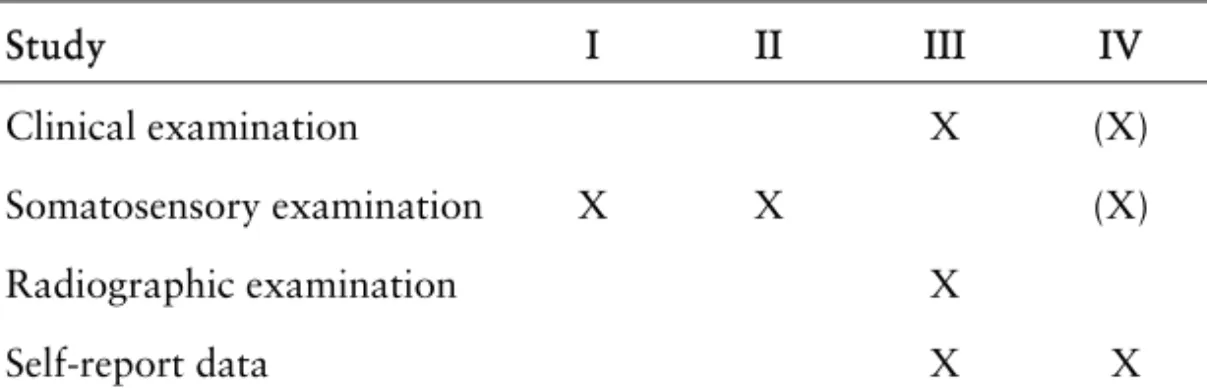
Data collection
Table 2 gives an overview of the type of data collected in the four studies.
44
Table 2. Data collection study by study.
Clinical examination (III and IV)
Study III
The clinical examination comprised a dental examination (assessment of pain location and pain on percussion or apical palpation, assessment of periodontal pocket depth, selective loading with a FracFinder, transillumination of natural teeth with an optical fiber [when possible], and sensibility testing of non-root-filled teeth), an examination of the masticatory system (according to the RDC/TMD), and a head and neck examination.
Study IV
No clinical data were collected at follow-up, but baseline clinical data from 2002 were available and in part analyzed as possible predictors of pain resolution or persistence (marked with [X] in Table 2) . A thorough dental examination, an examination of the masticatory system according to the RDC/TMD, and a neurological examination had all been done as part of the pain investigation of these patients.
Somatosensory examination and diagnostic testing (I, II, IV)
Assessment of thermal thresholds (I)
Cold detection thresholds (CDTs), warmth detection thresholds (WDTs), and heat pain thresholds (HPTs) were assessed at six sites: the mucosal side of the lower lip, the buccal gingiva adjacent to the first upper left premolar and the first lower left premolar, the tip of the tongue, the skin area just below the left eye, and an extra-trigeminal point on the hand. Threshold assessments were
Study I II III IV
Clinical examination X (X)
Somatosensory examination X X (X)
Radiographic examination X
45
made using an MSA Thermotest (Modular Sensory Analyzer, SOMEDIC, Hörby, Sweden) with a 0.81-cm2 contact surface intraoral probe. Baseline temperature was 37ºC for intraoral and 32ºC for extraoral sites. Cut-off temperatures were 10ºC (CDT) and 51ºC (WDT and HPT). Temperature ramp (rate of temperature change) was 1ºC/s. Subjects were instructed to press a stop button when the threshold was reached (i.e., when the sought-after stimulus was perceived), thereby terminating stimulation. At one intraoral site—the tip of the tongue—threshold measurements were also (i) performed with varying stimulation area size and (ii) repeated after 2 weeks and after 6 weeks.
(i) Plastic cover tips (Figure 6), successively reducing the size of contact area (between probe surface and oral mucosa) were manufactured for the intraoral thermode from 1-mm thick ethylene-propylene copolymer (thermal conductivity coefficient 0.12 W/m • K; Essix C+, Ortopro AB,
Gothen-burg, Sweden). Contact areas were 0.81 cm2
(uncovered = no plastic tip), 0.50 cm2, 0.28 cm2, 0.125 cm2, and 0.00 cm2 (fully covered = control). The purpose of this was to examine the influence of stimulation area size on threshold levels, and the occurrence of intraoral spatial summation (increasing stimulation area decreases the threshold level for when a stimulus is perceived).
(ii) Measurements with the 0.81 cm2 stimulation area were repeated after 2 weeks and after 6 weeks to examine the time variability of intraoral thermal thresholds.
Because the starting temperature was not the same for extra- and intraoral sites, delta values (deviation from starting temperature) were calculated and used in all comparisons between sites.
46
Figure 6. Intraoral probe and plastic cover tips used to vary stimulation area size.
Table 3. QST parameters (in order of testing) and equipment
Thermal testing
CDT Cold detection threshold MSA Thermotest WDT Warmth detection threshold MSA
TSL Thermal sensory limen MSA
PHS Paradoxical heat sensations on cold stimulation during TSL procedure
MSA
CPT Cold pain threshold MSA
HPT Heat pain threshold MSA
Mechanical testing
MDT Mechanical detection threshold Von Frey filaments MPT Mechanical pain threshold The PinPrick MPS Mechanical pain sensitivity The PinPrick
DMA Dynamic mechanical allodynia Cotton wisp, Q-tip, brush WUR Wind-up ratio for repetitive pinprick
stimulation
The PinPrick
VDT Vibration detection threshold Rydel-Seiffer tuning fork PPT Pressure pain threshold Pressure algometer