1
Green tea inhibits proteolytic enzymes in
GCF from patients with chronic
periodontitis
J. Boräng
A. Boucher
Handledare: H. Jansson, C. Wickström
Masteruppsats (30hp) Malmö högskola
Tandläkarprogrammet Odontologiska Fakulteten
2
ABSTRACT
Chronic periodontitis involves tissue destruction by matrix metalloproteinase, derived from the host cells, as part of the immunological response to bacterial virulence factors. Green tea has been studied for its health promoting properties, which includes anti-inflammatory effects. The effect is in part due to enzyme inhibition by tea polyphenols. The aim of this study was to further investigate the inhibitory effect of green tea, focusing on enzymatic activity in gingival crevicular fluid from patients with periodontal disease. Patients with chronic periodontitis were selected for participation in the study. Gingival crevicular fluid was extracted with micropipettes from the gingival sulci of the patients. Samples were treated with green tea and compared with untreated samples from the same subject. Fluorescence protease assay with casein as substrate was made using fourteen samples for detecting differences in caseinolytic activity. Zymogram assay using gelatin as substrate was done using four samples to test gelatinolytic activity and analyse molecular weights of the different enzymes. The fluorometric assay showed a significantly lower enzyme activity in samples mixed with green tea than untreated samples (p<0.001). The zymogram assay showed a difference in band strength which was most pronounced in the bands of molecular weight around 255 kDA, analogous to complexes of matrix metalloproteinase-9. In conclusion, green tea has been shown in this study to have a strong inhibitory effect on caseinolytic activity and a lesser, more specific, inhibitory effect on gelatinase activity.
Keywords: green tea, polyphenols, EGCg, GCF, periodontitis, MMP, inhibition, zymography,
3
SAMMANFATTNING
Kronisk parodontit orsakar vävnadsdestruktion till följd av matrixmetalloproteinasaktivitet. Dessa enzym härrör från värdcellerna och är en del av det immunologiska svaret på bakteriella virulensfaktorer. Grönt te har studerats för sina hälsofrämjande egenskaper, som omfattar bland annat anti-inflammatoriska effekter. Effekten beror delvis på enzyminhibering av tepolyfenoler. Syftet med denna studie var att ytterligare undersöka den inhiberande effekten av grönt te, med fokus på enzymatisk aktivitet i gingivalvätska från patienter med parodontal sjukdom. Patienter med kronisk parodontit valdes ut för att delta i studien. Gingivalvätska extraherades med mikropipetter från patienternas gingivala sulci. Proverna behandlades med grönt te och jämfördes med obehandlade prover från samma försöksperson. Fluorescens proteasanalys med kasein som substrat utfördes på fjorton prover för att detektera skillnader i kaseinolytisk aktivitet. Zymogramanalys med användning av gelatin som substrat utfördes på fyra prover, för att undersöka skillnader i gelatinolytisk aktivitet och analysera molekylvikter för de olika enzymerna. Den fluorometriska analysen visade en signifikant lägre enzymaktivitet i prover med tillsatt grönt te jämfört med obehandlade prover (p<0.001). Zymogramanalysen visade en skillnad i enzymaktivitet som var mest uttalad i banden för molekylär vikt runt 255 kDa, analogt med komplex av matrixmetalloproteinas-9. Sammanfattningsvis har det i denna studie påvisats att grönt te har en hämmande effekt på kaseinolytisk aktivitet och en mindre, mer specifik, hämmande effekt på gelatinasaktivitet.
4
INTRODUCTION
Gingivitis and periodontitis are inflammatory diseases, induced by microbial endo- and exotoxins. Tissue remodulation is a physiological constant occurrence in most of the human tissues. During a state of chronic inflammation the remodelling of tissue through enzymatic catalysis by matrix metalloproteases (MMPs) becomes greater than the synthesis of tissue, which therefore leads to a pathological shift in balance with tissue destruction as a result. Even if periodontitis is induced by bacterial virulence factors the largest part of collagenolytic activity comes from endogenous MMPs (1). The different types of MMPs vary in substrate specificity but can collectively degrade all tissue components that make up the periodontium (2). The leukocytes present at the inflamed site consist of approximately 90% neutrophil granulocytes (PMNs). They form a defence against foreign entities, such as microbes, through means of phagocytosis and extra- and intracellular degradation. The neutrophil granulocytes produce large amounts of MMPs for tissue degradation to support further leukocyte migration to the site. The two main types of PMN-secreted MMPs associated with periodontal disease are MMP -8 and -9 (3, 4). MMP -2 is mainly secreted by endothelial cells, fibroblasts and osteoblasts and is also found in increased levels in GCF during periodontal disease (5). The exocytosis of these enzymes occurs via vesicles in a pro- form and they are activated through protease cleaving of the pro domain (6-8). Several other cells of the periodontium: fibroblasts, epithelial cells and endothelial cells are capable of inducing active forms of MMPs (2). Thus inflammation in the periodontal tissue results in a greater quantity of MMPs in the gingival crevicular fluid (GCF) produced by both host cells and microbes (1, 9). Endogenous protease inhibitors exist in the tissue as a way to control the rate of tissue destruction during physiological remodulation. Tissue inhibitor of matrix metalloproteinase (TIMP), a specific endogenous inhibitor, binds to the MMPs and forms a complex, which inhibits further catalytic activity. Inhibition is achieved through binding of TIMP to the active Zn2+ site of the MMP which is required by the enzyme to attach to the substrate (2). During
5 inflammation, however the endogenous protease inhibitors are insufficient to maintain the balance due to increased MMP- production and activation (3).
Tea is derived from the plant leaves of Camellia sinensis. Green tea is a popular beverage in many parts of the world and studies have shown that green tea possess health promoting properties. These properties are associated with the polyphenols that tea contains. Polyphenols exists in varying types and quantities in all sorts of tea (black, green and semi-fermented). Green tea contains the polyphenols catechin (C), epicatechin (EC), gallocatechin (GC), epigallocatechin (EGC), epicatechin gallate (ECg) and epigallocatechin gallate (EGCg). Of the polyphenol constituents in green tea EGCg is the most abundant (10-12). Previous studies made on these polyphenols have shown an inhibitory effect on MMP activity with EGCg being the most potent in this regard (13). This inhibitory effect is dose dependent and achieved with a relatively low dosage with no cytotoxicity (14). The molecular process of polyphenol MMP inhibition is not yet fully understood. Studies indicate that the polyphenols have the ability to form hydrogen bonds to the active Zn2+ site of the enzyme and inhibits enzymatic activity in a similar fashion to TIMPs and the synthetic inhibitors batimastat and marimastat (1, 15). Other studies demonstrate that EGCg has an inhibitory effect on MMP in an environment where EGCg cannot bind to Zn2+, which indicates that this is not the exclusive mean of inhibition (16). Furthermore, neutrophil elastase (ELA2) which activates several types of proMMP is inhibited with concentrations achieved through normal intake of green tea (6, 17). When high concentrations of EGCg and ECg are used, an inhibitory effect is exerted on urokinase plasminogen activator (uPA) which in turn can activate MMP- 2, 3, 7 and 10. The polyphenols in green tea also have a redox potential of an anti- oxidative quality (18, 19). In addition to the proteolytic activity derived from human cells there is also proteolytic activity by microbial proteases in the pathogenesis of chronic periodontitis (20). Green tea catechins have been shown to exert an inhibitory effect on the proteolytic activity of Arg-gingipain and Lys-gingipain, proteases of the periodontal pathogen
6
Porphyromonas gingivalis (21). Interestingly there have also been clinical signs of the effect of green
tea. An epidemiological study made in Japan showed that a daily intake of green tea has a positive effect on periodontal status. The study concluded that every one cup/day increment in green tea intake was associated with a decrease in mean probing depth, clinical attachment loss and bleeding on probing in Japanese men between 49 and 59 years of age (22).
A non-toxic way to effectively inhibit MMP activity during periodontal disease could serve as a successful tool in reducing tissue destruction. This could aid the physiological process of healing as an additive effect of regular periodontal treatment. The hypothesis of this study was that net protease activity, derived from GCF, exposed to green tea in vitro would be inhibited. The aim of this study was to further investigate the inhibitory effect of green tea, focusing on enzymatic activity in GCF from patients with periodontal disease.
MATERIAL AND METHOD Study sample
In the spring of 2011, during a period of two weeks, 20 patients with a mean age of 59 years who were receiving treatment for chronic periodontitis were selected out of approximately 90 patients. This was done at the Faculty of Odontology at the University of Malmö, Sweden. The patients were screened through the inclusion and exclusion criteria. The inclusion criterion was patients with local or general chronic periodontitis according to the 1999 AAP classification system (23). Exclusion criteria involved presence of diabetes mellitus, drug induced gingival enlargement, smoking, intake of green tea (>1cup/week) and patients using cyclosporine (24). Sample size was calculated using Graphpad Statmate (v. 2.0) paired t-test (0.05) and a power of 95% could be reached if 14 samples were included.
7
GCF sampling
Plastic pipettes 10 µl (Eppendorf, Hamburg, Germany) were used to sample GCF from the gingival sulci of the participants. To avoid contamination from saliva the region of uptake was kept dry using suction and cotton rolls. Volume was measured by the weight of the tubes (PCR-Tube 0.2 ml, Eppendorf, Hamburg, Germany) before and after sample uptake with a high sensitivity scale (Sartorius, Type 2444, Goettingen, Germany). The amount of GCF, sampled from the patients, ranged from 0.5 µl up to 10 µl/sample. The samples were mixed with PBS by 1/10 (0.15 M NaCl, 5 mM NaH2PO4, pH 7) and stored in -20°C until further analysis.
Fluorometric analysis
In the fluorometric analysis 14 samples of the original 20 were used. Two µl of GCF was taken from each sample and diluted with PBS to a total volume of 40 µl. The concentration of green tea (Japanese Bancha) was 20 mg/ml (2 g/100 ml). Infusion of green tea was done in a temperature of 80ºC in distilled water for 20 min yielding an approximate total catechin concentration of 2.5 mg/ml (25). The tea was then cooled down to a temperature of <30°C. The samples were centrifugated (Eppendorf Centrifuge 5415D, Hamburg, Germany) for 10 seconds and 8k RPM. The samples were then divided into two wells each on a 96 well microtiter plate (TC Microwell 96F, Nunc, Cat. No. 167008, Roskilde, Denmark). These were then diluted with PBS and green tea respectively, up to 50 µl per well. All of the prepared samples were then stored in 36.5°C for one hour. 5mg/ml FITC conjugated casein (Pierce QuantiCleave Fluorescent Protease Assay kit, Thermo Fisher Scientific, Rockford, IL, USA) was used as substrate and one μl was added to all the samples. Acting as a reference, Trypsin-EDTA 0.05% was used as a diluting sequence of 1/10 over 24 wells, starting with 55 µl trypsin. Fluorescence analysis was then made using a FLUOstar OPTIMA (Lab Vision, Cheshire, UK) with a 485 nm wavelength of excitation and 420 nm emission wavelengths. Statistical significance was calculated using paired t-test (0.05) using Graphpad Prism (v. 2.0).
8
Zymography
Four samples, also used in the fluorometric analysis, were selected because they had an acceptable volume for zymograph testing. One µl GCF from each sample was divided in two tubes. One acting as an untreated sample and the other as a verum sample later treated with green tea. The green tea was prepared in the same manner as in the fluorometric test and five µl were added to each green tea verum sample. Each sample had a total volume of 10 µl after dilution with PBS. The concentration of GCF in two of the samples (A and B) was 1/40 and the remaining two samples (C and D) 1/160. Two separate molecular weight standards (High-range Rainbow™, GE Healthcare, Cat. No. RPN756E, Uppsala, Sweden) were also prepared at 12 µl each. The samples went through centrifugation for 10 seconds and 8k RPM. Ten µl sample buffer (Tris- Glycine SDS, Millipore, Billerica, MA 01821, USA) was later added to each sample and 12 µl to the standards. The samples were then applied to a 10% zymogram gelatin gel (Invitrogen, Cat. No EC6175BOX, Carlsbad, CA, USA) which ran with running buffer (Tris- Glycine SDS, Millipore, Billerica, MA 01821, USA) at a constant voltage of 125 for 90 minutes. The zymogram was diluted with renaturing buffer (Tris- Glycine SDS, Millipore, Billerica, MA 01821, USA) 1/10 for 30 minutes after running and then developing buffer for 30 minutes. The gel was then replaced with fresh developing buffer, incubated at 36.5°C for approximately 20 hours. The standards were cut off from the gels before they were stained with Coomasie Brilliant Blue R-250 (Millipore, Billerica, MA 01821, USA) for roughly 24 hours. Each gel treatment was done under gentle agitation.
Ethical considerations
Prior to consenting participation in this study, the subjects were informed about the background and purpose of this study. The samples were catalogued using a code and further use of the
9 subject chart was not possible. All the samples were then disposed after completing the study. The study was approved by the local ethical committee at Malmö University, Sweden.
RESULTS
GCF samples from fourteen subjects were used for the fluorescence assay to test caseinolytic activity. Four samples were used for the zymography to evaluate gelatinase activity. Each sample in both tests was divided in two. One was left untreated and the other mixed with green tea.
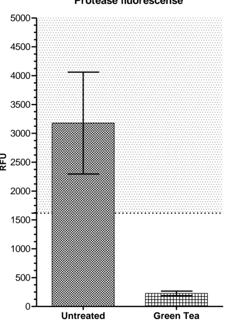
Protease fluorescence assay
The untreated samples had a greater caseinolytic activity than the samples treated with green tea (Fig. 1). This was statistically verified using Graphpad Statmate (v. 2.0) paired t-test (0.05). The mean relative fluorescence units (RFU) were 3178 for the untreated samples and 227 for the green tea samples. A dilution series of trypsin was made to confirm the reliability of the method. Values registered within the linear range of the trypsin dilution sequence are regarded as measurable with this method. None of the green tea samples showed enough activity to reach the linear range. Thus the samples treated with green tea were completely inhibited in regard to caseinolytic activity (p<0.001).
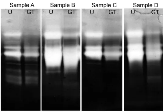
Zymogram assay
The zymogram assay (Fig. 2) most prominently displayed gelatolytic activity from enzymes in the GCF samples with molecular weights around 76kDa, 92 kDa and 255kDa. The untreated samples and green tea samples showed differences in band strength. The discrepancies were most pronounced in the bands of molecular weight around 255 kDA. In samples A, C and D the green tea samples showed a difference in visual density at 92 kDa. Activity, although vague, was displayed in the control samples around 130 kDa which was absent in the green tea samples. Both sample B and D showed a very high activity resulting in less distinguishable bands.
10
DISCUSSION
The purpose of this study was to investigate if green tea exerts change in the enzymatic activity of enzymes, in GCF, from patients with chronic periodontitis. The results from the fluorometric analysis showed that the caseinolytic activity from the enzymes in GCF were significantly lower in the samples treated with green tea. Casein substrate can be catalysed by stromelysins including MMP-1, MMP-7, MMP-12 and MMP-13. In high concentrations MMP-9 may also lyse the casein substrate (26). The trypsin dilution proved the method to be reliable for measuring enzyme concentration and activity. However the green tea samples showed fluorescence values below the linear range, meaning that the activity was too low to be accurately measured with this method. Inhibition of caseinolytic activity through green tea treatment was therefore significant with the concentration used.
The results from the zymogram assay showed a difference in band strength in all green tea samples most prominently in the bands corresponding to a molecular weight of 76 kDa, 92 kDa and ~255 kDa. Note that during SDS-Page pro forms of MMP become active as a result of denaturation (6). The following MMPs have substrate specificity for gelatin: MMP-2 and MMP-9. MMP-1, MMP-8 and MMP-13 are also able to lyse gelatin sufficiently for detection (26). The molecular weight of 255 kDa has proved in earlier studies to be analogous to complexes of MMP-9. It is most likely the homodimer of MMP-9 found at a molecular weight of ~220 kDa (27). A difference in visual density was also shown at a molecular weight around 92 kDa at which proMMP-9 and certain bacterial cysteine protease, namely gingipain R1, appear (28, 29). However gingipains do not exert proteolytic activity on collagen in its natural form, they have shown to do so when collagen is denaturated (30). PMN MMP-8 has a molecular weight range of 60 – 80 kDa and PMN membrane bound MMP-8 ranges from 30 – 110 kDa (31).
11 The results from the present study shows that green tea has a strong inhibitory effect on caseinolytic activity and a lesser, more specific, inhibitory effect on gelatinase activity. Metallomatrixproteinases and matrix-degrading serine proteinases are thought to be the main tissue degrading enzymes during chronic periodontitis. Ingman et al. (1996), have shown that higher levels of MMP-8 and MMP-9 than in the control samples could be measured from GCF during chronic periodontitis (3). Other studies have shown that MMP-8 and MMP-9 are the most prominent proteases during periodontitis. Even if periodontitis is induced by bacterial virulence factors the largest part of collagenolytic activity comes from endogenous enzymes (1). MMP-8 constitutes 80% of the total protein amount in GCF during chronic periodontitis (32). The result from this study points to the possibility that green tea exerts an inhibitory effect on both active and proMMP-9 activity and PMN MMP-8 activity. However, the methods used in this study does not allow for any single type of protease to be identified with any certainty exceeding substrate specificity and molecular weight correlation.
In a study by Chan et al. (2010), green tea and its effect on various organs in rats and mice was investigated. They concluded that the “no adverse effect”-level for the liver was 500 mg/kg in both species (33). In this study 2 g leafs of a Japanese green tea (Bancha) brewed in 100 ml distilled water in a temperature of 80⁰C for 20 minutes was used. This gives a calculated total catechin concentration of approximately 2500 µg/ml or 2.5 mg/ml (25). In 200 ml brewed green tea the catechin concentration is 500 mg. A typical cup of green tea is brewed with 2 g tea leafs and 200 ml water (70ºC for approximately 5 minutes) and has a concentration green tea catechins of 500 µg/ ml or 0.5 mg/ml (10, 25). In other studies, doses of 600 to 1800 mg/day, tea catechins have been used without adverse effects in humans. The concentration of green tea in this study has therefore been assumed as non-toxic, yet containing high concentrations of catechins. However, there are also reports of adverse effects with usage of green tea extracts as
12 supplements in dosages of 700 to 2100 mg/day causing clinical signs of hepatoxicity. It appears to be individual differences in metabolism and bioavailability of green tea polyphenols (19).
Some polyphenols have the ability to create noncovalent bonds to different proteins, such as casein and proline-rich proteins (PRP) in the saliva. This a potential source of error as casein was used as substrate for the fluorometric assay which may decrease the inhibitory potential of the polyphenols. PRPs constitute 70% of the protein content of the saliva (34). Thus contamination of saliva during the uptake of GCF from the gingival sulci is also a possible source of error. However the difference in enzyme activity from the assays is evident to such a degree that inhibition has undoubtedly occurred. The possible contamination of blood in the samples can cause an increased activation of MMP-2 through MMP-1 due to the heparin content of blood (35). This could give an increased enzyme activity in the samples.
Local oral application of tetracycline, such as doxycycline, has been used in periodontal treatment as a complement to scaling and root planing for some time with inconclusive evidence concerning its long term benefits. The recognized additive effects of tetracycline treatment have been credited due to its inhibitory properties on protease (36, 37). With the increasing problem of antibiotic resistance in bacteria, many health practitioners have taken a more conservative attitude towards the use of antibiotics in treatment. This study, along with current scientific results, supports the need for further analysis of green tea components in clinical research as a possible alternative to complementary antibiotic periodontal treatment.
In summary, the results of these tests indicates that green tea has general inhibitory properties on enzyme activity but not necessarily on all the enzymes found in the GCF. However these results cannot be generalized to the entirety of the tissues affected by periodontal disease. Many other
13 factors that are involved in the pathology of chronic periodontitis are unaccounted for with the in
vitro methods used.
ACKNOWLEDGEMENTS
For assistance in the laboratory, with statistical analysis and in general: Agnethe Henrikssen, Malmö University, Department of Oral Biology Bertil Kindby, Malmö University, Department of Oral Biology
14
REFERENCES
1. Sorsa T, Tjaderhane L, Salo T. Matrix metalloproteinases (MMPs) in oral diseases. Oral Dis. 2004; 10: 311-318.
2. Uitto VJ, Overall CM, McCulloch C. Proteolytic host cell enzymes in gingival crevice fluid. Periodontol 2000. 2003; 31: 77-104.
3. Ingman T, Tervahartiala T, Ding Y, Tschesche H, Haerian A, Kinane DF et al. Matrix metalloproteinases and their inhibitors in gingival crevicular fluid and saliva of periodontitis patients. J Clin Periodontol. 1996; 23: 1127-1132.
4. Lee W. Evidence of a direct relationship between neutrophil collagenase activity and
periodontal tissue destruction in vivo: role of active enzyme in human periodontitis. J Periodont Res. 1995; 30: 23.
5. Makela M. Matrix metalloproteinases (MMP-2 and MMP-9) of the oral cavity: cellular origin and relationship to periodontal status. J Dent Res. 1994; 73: 1397.
6. Nagase H. Activation mechanisms of matrix metalloproteinases. Biol Chem. 1997; 378: 151-160.
7. Pei D, Weiss SJ. Furin-dependent intracellular activation of the human stromelysin-3 zymogen. Nature. 1995; 375: 244-247.
8. Van den Steen PE, Dubois B, Nelissen I, Rudd PM, Dwek RA, Opdenakker G. Biochemistry and molecular biology of gelatinase B or matrix metalloproteinase-9 (MMP-9). Crit Rev Biochem Mol Biol. 2002; 37: 375-536.
15 9. Rai B, Kharb S, Jain R, Anand SC. Biomarkers of periodontitis in oral fluids. J Oral Sci. 2008; 50: 53-56.
10. Balentine DA, Wiseman SA, Bouwens LC. The chemistry of tea flavonoids. Crit Rev Food Sci Nutr. 1997; 37: 693-704.
11. Jung YD, Kim MS, Shin BA, Chay KO, Ahn BW, Liu W et al. EGCG, a major component of green tea, inhibits tumour growth by inhibiting VEGF induction in human colon carcinoma cells. Br J Cancer. 2001; 84: 844-850.
12. Yang F, de Villiers WJ, McClain CJ, Varilek GW. Green tea polyphenols block endotoxin-induced tumor necrosis factor-production and lethality in a murine model. J Nutr. 1998; 128: 2334-2340.
13. Demeule M, Brossard M, Page M, Gingras D, Beliveau R. Matrix metalloproteinase inhibition by green tea catechins. Biochim Biophys Acta. 2000; 1478: 51-60.
14. Garbisa S, Biggin S, Cavallarin N, Sartor L, Benelli R, Albini A. Tumor invasion: molecular shears blunted by green tea. Nat Med. 1999; 5: 1216.
15. Maeda-Yamamoto M, Kawahara H, Tahara N, Tsuji K, Hara Y, Isemura M. Effects of tea polyphenols on the invasion and matrix metalloproteinases activities of human fibrosarcoma HT1080 cells. J Agric Food Chem. 1999; 47: 2350-2354.
16. Garbisa S, Sartor L, Biggin S, Salvato B, Benelli R, Albini A. Tumor gelatinases and invasion inhibited by the green tea flavanol epigallocatechin-3-gallate. Cancer. 2001; 91: 822-832.
17. Dona M, Dell'Aica I, Calabrese F, Benelli R, Morini M, Albini A et al. Neutrophil restraint by green tea: inhibition of inflammation, associated angiogenesis, and pulmonary fibrosis. J
16 18. Jankun J, Selman SH, Swiercz R, Skrzypczak-Jankun E. Why drinking green tea could prevent cancer. Nature. 1997; 387: 561.
19. Lambert JD. The antioxidant and pro-oxidant activities of green tea polyphenols: a role in cancer prevention. Arch Biochem Biophys. 2010; 501: 65.
20. Sorsa T. Matrix metalloproteinases: contribution to pathogenesis, diagnosis and treatment of periodontal inflammation. Annals of medicine (Helsinki). 2006; 38: 306.
21. Okamoto M. Inhibitory effect of green tea catechins on cysteine proteinases in Porphyromonas gingivalis. Oral Microbiol Immunol. 2004; 19: 118.
22. Kushiyama M, Shimazaki Y, Murakami M, Yamashita Y. Relationship between intake of green tea and periodontal disease. J Periodontol. 2009; 80: 372-377.
23. Armitage GC. Development of a classification system for periodontal diseases and conditions. Ann Periodontol. 1999; 4: 1-6.
24. Parameter on periodontitis associated with systemic conditions. American Academy of Periodontology. J Periodontol. 2000; 71: 876.
25. Labbé D, Tremblay A, Bazinet L. Effect of brewing temperature and duration on green tea catechin solubilization: Basis for production of EGC and EGCG-enriched fractions. Separation and Purification Technology. 2006; 49: 1-9.
26. Snoek-van Beurden PA,S n o e k - B e u r d e n P A. Zymographic techniques for the analysis of matrix metalloproteinases and their inhibitors. BioTechniques. 2005; 38: 73.
27. Kjeldsen L. Isolation and primary structure of NGAL, a novel protein associated with human neutrophil gelatinase. J Biol Chem. 1993; 268: 10425.
17 28. Jie BaoG. Proteolytic Activities of Oral Bacteria on ProMMP-9 and the Effect of Synthetic Proteinase Inhibitors. Open Dentistry Journal. 2008; 2: 96.
29. Imamura T. The role of gingipains in the pathogenesis of periodontal disease. J Periodontol. 2003; 74: 111.
30. Potempa J. Comparative properties of two cysteine proteinases (gingipains R), the products of two related but individual genes of Porphyromonas gingivalis. J Biol Chem. 1998; 273: 21648.
31. Avellan NL. Capsaicin-induced local elevations in collagenase-2 (matrix metalloproteinase-8) levels in human gingival crevice fluid. J Periodont Res. 2006; 41: 33.
32. Marcaccini AM. Gingival crevicular fluid levels of MMP-8, MMP-9, TIMP-2, and MPO decrease after periodontal therapy. J Clin Periodontol. 2010; 37: 180.
33. Chan PC. Fourteen-week toxicity study of green tea extract in rats and mice. Toxicol Pathol. 2010; 38: 1070.
34. Jobstl E, Howse JR, Fairclough JP, Williamson MP. Noncovalent cross-linking of casein by epigallocatechin gallate characterized by single molecule force microscopy. J Agric Food Chem. 2006; 54: 4077-4081.
35. Crabbe T, O'Connell JP, Smith BJ, Docherty AJ. Reciprocated matrix metalloproteinase activation: a process performed by interstitial collagenase and progelatinase A. Biochemistry. 1994; 33: 14419-14425.
36. Sgolastra F. Long-term efficacy of subantimicrobial-dose doxycycline as an adjunctive
treatment to scaling and root planing: a systematic review and meta-analysis. J Periodontol. 2011; 82: 1570.
18 37. Preshaw PM, Hefti AF, Jepsen S, Etienne D, Walker C, Bradshaw MH. Subantimicrobial dose doxycycline as adjunctive treatment for periodontitis. J Clin Periodontol. 2004; 31: 697-707.
19
FIGURES
Protease fluorescense
RF
U
Untreated Green Tea
0 500 1000 1500 2000 2500 3000 3500 4000 4500 5000
Figure 1 – Protease fluorescense assay
Mean and standard deviation (vertical bars) of enzyme activity expressed in relative fluorescence units (RFU). The shadowed area represents the linear range derived from the trypsin dilution sequence.The results from the fluorometric analysis showed that the enzyme activity from the GCF was significantly lower in the samples treated with green tea (p<0.001).
20
Figure 2 – Zymogram assay
Zymogram stained gel using four GCF samples from separate individuals with chronic periodontitis indicated as untreated samples (U) and green tea treated samples (GT) with corresponding molecular weights.