Downloaded from https://journals.lww.com/prsgo by BhDMf5ePHKav1zEoum1tQfN4a+kJLhEZgbsIHo4XMi0hCywCX1AWnYQp/IlQrHD3hucOGBbZM6OE+/tsAoUl8pZ4xU1ZgAzwI7aIYKGfkzM= on 01/06/2019 Downloadedfrom https://journals.lww.com/prsgoby BhDMf5ePHKav1zEoum1tQfN4a+kJLhEZgbsIHo4XMi0hCywCX1AWnYQp/IlQrHD3hucOGBbZM6OE+/tsAoUl8pZ4xU1ZgAzwI7aIYKGfkzM=on 01/06/2019
Copyright © 2018 The Authors. Published by Wolters Kluwer Health, Inc. on behalf of The American Society of Plastic Surgeons. This is an open-access article distributed under the terms of the Creative Commons Attribution-Non Commercial-No Derivatives License 4.0 (CCBY-NC-ND), where it is permissible to download and share the work provided it is properly cited. The work cannot be changed in any way or used commercially without permission from the journal.
INTRODUCTION
Lower Limb Problem with CoverageSkin defects of the lower leg and a foot can be difficult to reconstruct due to terminal vascularization, thin skin, bony prominences, and small local muscle mass. Before the invention of microvascular surgery in the 70s, the only methods for coverage of lower leg defects were either lo-cal- or cross leg flaps. Since then, the trend to free flap reconstruction of traumatic lower limb defects has
signifi-cantly increased. Free-flap surgery does, however, require skilled personnel, sophisticated equipment, and more hospital resources. Local flaps, such as perforator and muscle flaps, therefore in many hospitals still have a role when used to cover lower limb defects.1
At our department, we have more than 20 years of ex-perience with the Extensor Digitorum Brevis (EDB) mus-cle flap,2 and we have used it to cover small defects in the distal leg, ankle, and foot. Relatively few reports have been published describing the technique, the use and extended use of the EDB muscle flap.3–11
The EDB-flap can be a good choice in particular when reconstructing small distal defects in the ankle or foot-region with acceptable contouring. Most methodological descriptions have restricted its use to defects of the foot and not proximal to the ankle. Reported shortcomings of From the *Plastic Surgery Unit, Surgery Department, Suez Canal
University, Ismailia, Egypt; and †Department of Hand Surgery, Plastic Surgery and Burns, and Department of Clinical and Experimental Medicine, Linköping University, Linköping, Sweden.
Background: Reconstruction of complex defects in the lower leg is a challenge. Although microvascular free tissue transfer is a popular technique, experience and available resources limit its use. Furthermore, free tissue transfer is not always required in the reconstruction of small lower leg defects, as many of them can be reconstructed with local alternatives such as an extensor digitorum brevis flap (EDB). Our aim was to describe our experience of the last 20 years with the EDB as a local muscle flap to cover small complex lower leg defects to establish its clinical feasibility and to document its associated complications.
Methods: All adult patients who were operated with EDB flap reconstruction of the lower limb during 1997–2017 at the Department of Hand and Plastic Surgery, Linköping University Hospital, were included in this retrospective study.
Results: Of 64 patients operated, only 7 had total flap failure, and the rate of com-plete success was 73% (47/64). Most of the skin defects were associated with frac-tures or complications thereof and were located in the ankle region, the dorsum of the foot, and the distal third of tibia or even the proximal tibia. Defects in the malleolar region and coexisting cardiovascular condition were factors associated with flap loss (either partial or total).
Conclusion: The pedicled EDB-flap has, in our hands, proved to be a versa-tile and safe reconstructive option in the reconstruction of small defects in the lower leg and foot. Long-time follow-up is, however, recommended. (Plast Recon-str Surg Glob Open 2018;6:e2071; doi: 10.1097/GOX.0000000000002071; Published online 17 December 2018.) Islam Abdelrahman, MRCS, FEBOPRAS, MD, PhD*† Moustafa Elmasry, MD, PhD*† Ingrid Steinvall, PhD† Pia Olofsson, MD, PhD† Hans Nettelblad, MD, PhD† Johann Zdolsek, MD, PhD†
Versatility of the Extensor Digitorum Brevis Muscle
Flap in Lower Limb Reconstruction
Disclosure: The authors have no financial interest to
declare. This work was supported by, and done, at the Department of Hand Surgery, Plastic Surgery and Burns, and the Linköping University, Linköping, Sweden. No other funding was received. The Article Processing Charge was paid for by the authors.
Reconstructive
Supplemental digital content is available for this article. Clickable URL citations appear in the text.
DOI: 10.1097/GOX.0000000000002071
Received for publication August 2, 2018; accepted October 19, 2018.
The study was approved by the Regional Ethics Review Board (No. 2018\18–31).
PRS Global Open
•
2018
the EDB-flap are its small size, healing problems of the donor site, and reduced mobility of the ankle joint. Anatomical Background
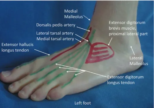
The EDB is a trapezoid-shaped muscle on the dorsum of the foot, lateral to the extensor hallucis longus tendon and deep to the tendons of the long toe extensors. It con-sists of 4 bellies that originate from ligamentous structures of the tarsal sinus and inserts on each of the long extensor tendons of the 4 medial toes. The size of the conjoined muscles in an adult is about 6 × 4.5 cm. The EDB muscle has a Mathes and Nahai type II vascular supply.12 The dominant pedicle is the lateral tarsal artery that runs in a plane deep to muscles. It branches from the Dorsalis Pedis artery at or immediately distal to the extensor retinaculum (Fig. 1). Veins and nerves usually follow the main arterial pedicle.
The aim was to describe our experience of the last 20 years with the use of the EDB as a local muscle flap for cov-erage of complex small lower limb defects and to delineate its clinical use as well as to analyze donor-site complications.
METHODS
All adult (≥ 18 years) patients who had intervention ICD-10 code ZZR30 (reconstruction with muscle flap) of the lower limb between 1997 and 2017 at the Depart-ment of Hand and Plastic Surgery at Linkoping University Hospital were screened. All patients who had been recon-structed with a pedicled EDB flap were included, and a retrospective analysis was done using data retrieved from the medical records. The following variables were used: age, sex, co-existing medical conditions, site, dimension of the defects, mechanism of injury, operating time (skin to skin), flap viability and complications, and donor-site
complications. The study was approved by the Regional Ethics Review Board (No. 2018\18–31).
Preoperative Procedure
All patients were examined with preoperative Doppler to identify anterior and posterior tibial vessels in the lower leg. Angiography or computed tomography (CT) angi-ography was performed in some patients when Doppler assessment was inconclusive or when the trauma to the lower leg was extensive.
Surgical Procedure
With the patient supine, a curvilinear skin incision is made on the dorsum of the foot starting from the lateral malleolus to the first web space, this incision follows the oblique course of the EDB muscle bellies. In the space be-tween the extensor digitorum longus (EDL) and extensor hallucis longus tendons, the lateral tarsal artery is identi-fied just distal to inferior border of extensor retinaculum along with the motor branch of the deep peroneal nerve. These course infero-laterally for approximately 2 cm be-fore passing underneath the proximal edge of EDB.
Dissection starts by creating a plane between EDB mus-cle and the superficial EDL tendons from medial to lateral taking care not to divide the branches of the superficial pe-roneal nerve. Elevation of the long extensors is simplified by early division of the extensor retinaculum. The tendons of the EDB are then divided distally where after the muscle is freed off the deep proximal attachment starting from the tarsal bones in the mid foot to its origin in the calcaneus. The neurovascular pedicle can then be dissected proxi-mally, which is facilitated by the ligation of a fairly constant branch to the sinus tarsi. The dorsalis pedis artery is then ligated distal to the origin of the lateral tarsal artery, which
will give extra arc of rotation, length, and better mobiliza-tion of the flap. The pedicle is dissected proximally, and if an even longer arc of rotation is needed, the extensor retinaculum is divided and the proximal dissection of the pedicle is followed even further. This latter procedure en-ables the flap to reach even the tibial tuberosity (Fig. 2). After elevation of the flap and pedicle, the extensor reti-naculum is reconstructed by single sutures of Polydioxa-none (PDS) 3-0, the donor site is sutured in layers. No drains are employed. Finally a skin graft is harvested from available donor site at the ipsilateral thigh with a Zimmer Pneumatic dermatome with thickness 0.01 inch, meshed 1:1.5, and fixed with staples on the surface of the EDB flap.
Postoperative Procedure
The ankle joint and foot are immobilized using a pos-terior below knee plaster slab for 6 weeks. Thereafter, gradual mobilization is initiated.
Analysis of Data
Descriptive data are presented as median (25th–75th centiles) unless otherwise stated. Probabilities of less than 0.05 were accepted as significant. Distribution was tested with the Lilliefors test for normality. Significance of dif-ference between the groups was assessed using the Mann Whitney U, and the chi-square, tests. Logistic regression was used to assess the effect of site (tibia or dorsal foot; Achilles or heel; malleolus), size of the defect, operating time, age, sex, and presence of co-existing medical condi-tions on flap outcome: viable flap (full success) coded as 0, complications and flap loss (dehiscence, fistulae, partial necrosis, and total necrosis) coded as 1.
RESULTS
A total of 64 patients were included. Figures 3–5 show 3 cases before and after operation, site of defects were dorsal foot, distal tibia, and medial malleolus. Four of the 64 patients had distally based flaps. The soft-tissue defects were associated with fractures in 40/64 (63%) pa-tients. The most frequent reason for flap-surgery (41%) was skin defects without exposed bone, followed by osteo-myelitis (27%), skin defects with exposed bone (17%), exposed Achilles tendon (14%), and 1 case with sarcoma (Table 1). The muscle flap was used to reconstruct de-fects in distal leg and foot in 94% (60/64) of the cases (Table 2) (see table, Supplemental Digital Content 1, which displays all the details of patients, http://links.lww. com/PRSGO/A938).
Fig. 2. the area possible to reach with the eDB-flap is substantially
enhanced by proximal dissection of the vascular pedicle. a low-grade osteitis in the upper part of the tibia was thus cured in the illustrated example.
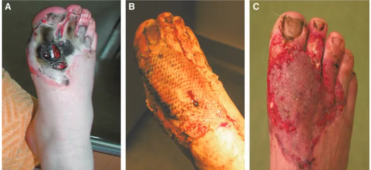
Fig. 3. a, Shows a friction injury in a 22-year-old male patient before operation. B, Shows 4 days after operation with a distally based eDB
muscle flap covering a 5 × 5 cm defect and additional skin graft on the adjacent wound. c, Shows the flap 3 weeks after operation. no co-existing medical conditions were recorded.
PRS Global Open
•
2018
The EDB flap reconstruction was successful in 47/64 patients (73%), 1 patient developed a fistula under the flap, 4 had dehiscence of the suture line, 5 had partial flap loss, and 7 had total flap loss. Additionally, 3 had a skin graft failure while the flap survived (Table 3). Thirteen of the patients developed donor-site complications of which delayed healing was the most common (n = 9), followed by dehiscence (n = 2), and fistulae (n = 2; Table 3). The differences between the full success group and those with complications are shown in Table 4. The proportion of patients with cardiovascular conditions was greater in the
group with complications. Defects of the malleolar region was the site with highest frequency of complications while tibia and dorsum of the foot had the lowest frequency.
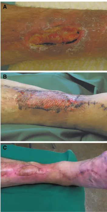
Fig. 4. a, Shows a chronic wound with a 7 × 4 cm defect in a
55-year-old male patient before operation. B, Shows 1 week after operation with a proximally based eDB muscle flap. c, Six months after op-eration. no complications were recorded and the only co-existing medical condition was epilepsy.
Fig. 5. a, Shows an exposed tibial plate in a 4 × 5 cm defect in a
65-year-old female patient before operation. B, Shows 2 weeks after operation with plate removal, bone debridement and external fixa-tion, and a proximally based eDB muscle flap. c, Shows the result 11 months after operation. no complications or co-existing conditions were recorded.
There was further a tendency for more complications with older age.
Simple logistic regression showed that malleolar site of injury [odds ratio (OR), 7.6; P = 0.005, compared with the reference group tibia and dorsal foot], and presence of cardiovascular condition (OR, 7.3; P = 0.003), were associ-ated with flap complications and flap loss.
Multivariable regression (adjusting for age and sex, and stepwise forward testing combinations of the other variables) showed that site of injury and cardiovascular condition were the differences between the 2 groups that were verified (significantly contributing to the model; Table 5).
DISCUSSION
Sixty-four patients were operated using the EDB flap to reconstruct defects of the lower leg and foot with me-dian size of 12 cm2. The 25th to 75th percentile of defect size was between 5 and 21 cm2. Total flap-failure was seen in 7 cases (11%) while 10 cases (16%) had partial loss of the flap. Malleolar defects and cardiovascular disease were factors associated with flap failure (either partial or total). We have used the EDB flap for different defects in differ-ent regions of the lower leg and even mid- and proximal parts of the tibia, which we think confirms the versatility Table 1. Indication for EDB-flap Surgery
Skin defect without exposed bone 26
Osteomyelitis 17
Skin defects with exposed bone 11
Exposed tendon Achilles 9
Other 1
Table 2. Site of Injury
Distal tibia 23 Medial malleolus 10 Achilles 10 Lateral malleolus 7 Heel 6 Dorsal foot 4 Proximal tibia 4
Table 3. Complications and Flap Loss
Complications Recipient Site Donor Site
Total necrosis 7 (11)
Partial necrosis 5 (8)
Wound dehiscence 4 (6) 2 (3)
Skin graft failure 3 (5)
Fistula 1 (2) 2 (3)
Delayed healing 9 (14)
n and (%) of 64
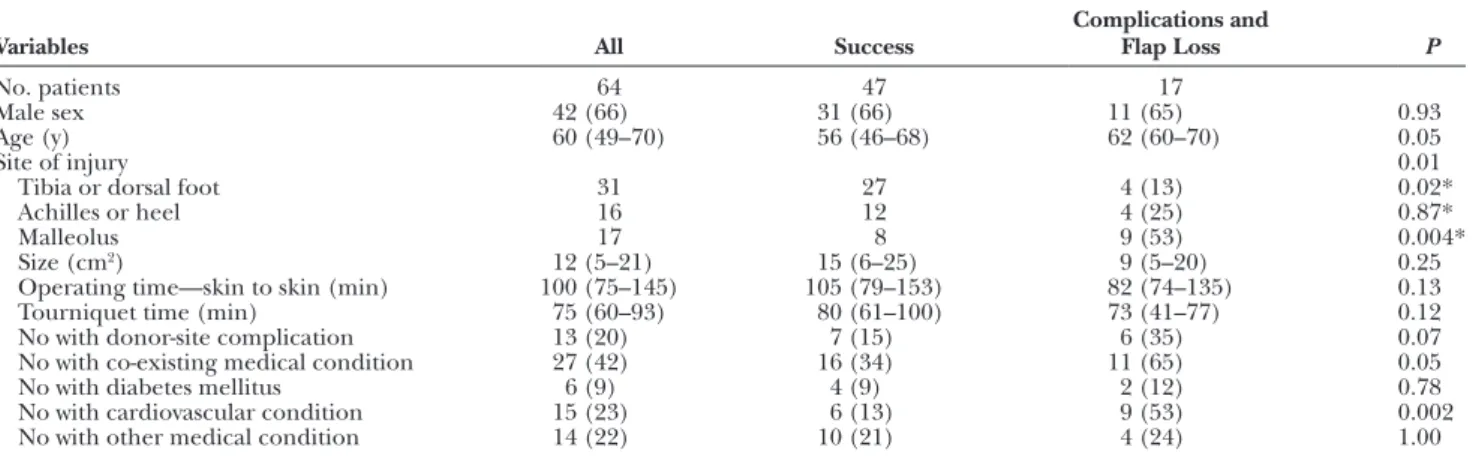
Table 4. Details of Patients Grouped by Flap Outcome
Variables All Success Complications and Flap Loss P
No. patients 64 47 17
Male sex 42 (66) 31 (66) 11 (65) 0.93
Age (y) 60 (49–70) 56 (46–68) 62 (60–70) 0.05
Site of injury 0.01
Tibia or dorsal foot 31 27 4 (13) 0.02*
Achilles or heel 16 12 4 (25) 0.87*
Malleolus 17 8 9 (53) 0.004*
Size (cm2) 12 (5–21) 15 (6–25) 9 (5–20) 0.25
Operating time—skin to skin (min) 100 (75–145) 105 (79–153) 82 (74–135) 0.13
Tourniquet time (min) 75 (60–93) 80 (61–100) 73 (41–77) 0.12
No with donor-site complication 13 (20) 7 (15) 6 (35) 0.07
No with co-existing medical condition 27 (42) 16 (34) 11 (65) 0.05
No with diabetes mellitus 6 (9) 4 (9) 2 (12) 0.78
No with cardiovascular condition 15 (23) 6 (13) 9 (53) 0.002
No with other medical condition 14 (22) 10 (21) 4 (24) 1.00
Data are median (25–75 centiles) or n (%) unless otherwise stated. Mann Whitney U test and chi-square for the difference between the outcome groups. Four patients had both a cardiovascular condition and either diabetes mellitus or another medical condition.
*Post hoc 2 × 2 chi-square for the difference between 1 site and the other 2 taken together.
Table 5. Multivariable Logistic Regression for Complications and Flap Loss
Variables Coefficient P OR 95% CI
Age 0.01 0.66 1.01 0.96–1.07
Sex 0.18 0.83 1.20 0.23–6.14
Cardiovascular condition 1.86 0.02 6.45 1.28–32.57
Site of injury
Tibia, dorsal foot 1.00
Achilles, heel 0.40 0.65 1.50 0.26–8.62
Malleolus 1.95 0.03 7.06 1.21–41.20
Constant ˗3.14 0.07
Viable flap (full success) coded as 0, complications and flap loss (dehiscence, fistulae, partial necrosis, and total necrosis) coded as 1. Model pseudo R2 0.24,
PRS Global Open
•
2018
of the pedicled EDB-flap in the reconstruction of lower leg defects.
Reconstruction of complex defects of the lower leg and the foot is a continuous challenge for reconstructive sur-geons due to poor blood supply, thin skin, and thick bony prominences and sparse muscle volume.13 The choice of reconstruction method depends on the nature of the de-fect, mechanism of trauma, and site of injury as well as surgeon preferences and capabilities. Free micro-vascular tissue transfer is in many parts of the world a popular, use-ful, and reliable method for the reconstruction of lower leg defects, in particular as experience with these tech-niques has increased globally.1 However, a high level of training, expertise, and experience is needed apart from higher costs than with local or pedicled flaps.14 Local solu-tions such as reversed flow island flaps, perforator flaps, keystone flaps, and local muscle or skin flaps are available options and in many hands easier to perform.1,15 A total assessment of the patient’s need of coverage in relation to local factors as well as age and comorbidities is crucial. If considering the use of an EDB-flap, the effects of the sac-rifice of a main artery, that is, anterior tibial/dorsalis pedis artery must always be evaluated in relation to future com-plications. Distally based sural flaps are also widely used in reconstruction of lower leg defects.16–18 A cross-leg flap could be considered as an old-fashioned and last resort method but certainly is still a reconstructive option. The morbidity and severe patient discomfort associated with this technique has reduced its popularity considerably.19 As shown previously, the EDB-flap can be considered a good reconstructive option for small defects in the lower leg and foot region,8,11,20 which also we have confirmed in this study. The small size and the thin contour of the flap in addition to the relative simplicity of harvesting are advantages that in some instances make the flap superior when compared with free tissue transfer or other local flap options. Also, in older age groups or in patients with significant comorbidities where free tissue transfer and perforator flaps could deemed more risky, the EDB can provide a good solution (Fig. 5). The ability to reconstruct defects in complicated areas such as the distal third of the tibia, the heel and Achilles tendon and malleoli is valu-able. We have, moreover, in this study also used the flap to reconstruct defects even of the proximal tibia, which confirms the extreme versatility of the EDB flap. In this group (proximal tibia defects), no flap loss was recorded. We further found the EDB-flap particularly useful in the treatment of osteomyelitis with fistulation where parts of, or some of, the muscle bellies also could be used to fill out smaller revised bone cavities.
Success, Failure Rates
In our study, the rate of complete reconstructive failure was about 11%, which is comparable with other studies using EDB11,13 and other flap techniques for reconstruc-tion of lower leg defects.21–26 However, our study group was considerably larger than in other reports. Our find-ing that cardiovascular co-existfind-ing medical condition was a risk factor for reconstructive failure might not come as a complete surprise.
The cumulative rate of partial and total necrosis after using perforator-pedicled propeller flaps for lower ex-tremity defects has in a recent review been reported to be 13.5%,26 which is lower than the corresponding number (18.8%) in our study, although the numbers change to 17.5% if case studies of less than 5 patients are excluded from the review26 to avoid positive reporting bias on ac-count of handpicked cases. The complete success in our study (73%) was similar to that of 70%, which was report-ed by Yasir et al.,21 although, none of the 8 patients in that study who were 50 years and older had flap necrosis,21 sug-gesting that perforator flaps can be one of the first choices for reconstruction in elderly patients.
The reversed sural flap with modifications (extended distally based) and medial sural artery perforator flap23,25 have reported necrosis rates of 10–15% of patients includ-ed in the study. However, the donor-site morbidity of this flap is well known, but its capacity to reconstruct bigger defects is an advantage.
According to the review by Reddy and Stevenson,1 the reconstruction of distal lower leg defects are mainly ac-complished using free tissue transfer. However, local fas-ciocutaneous flaps have been gaining some popularity nowadays. In a study by Kang et al.,27 the total failure rate for free tissue transfer was only 3.5%, however, including partial necrosis the percentage increase to 15%, which is similar to the other described studies and to this study as well. The main advantage of free tissue transfer is the lower local donor-site morbidity compared with local flaps and the capacity to reconstruct larger defects.
Malleolar defects had the worst outcome in our study. Chattar-Cora and Pederson9 reported 2 flap failures of to-tally 9 EDB done to reconstruct a malleolar defect, Koul et al.28 1 of 4, Pai et al.13 none of 5, which by crude num-bers is considerably better than our outcome in this site. However, in those studies, patient mean age was 49, 33, and 39 years in the malleolus subgroups, whereas it was 65 years in our study, which could explain the difference in outcome. The mean age in our study is notably higher than mean age in many studies,27 which is characteristic of our Swedish patient population. This makes the EDB tech-nique particularly useful in this part of the world due to the higher complication rate associated with both micro-surgical and propeller techniques in the same age group.
The EDB is technically not particularly challenging, and the median operating time in this study was 100 min-utes, which indicates that the operation in experienced hands is a short procedure with comparable complication rate to other techniques.
Anticipated Negative Effects after EDB-harvest
The reported complications after the use of EDB-flaps in other studies are few.11,13 Donor-site morbidity was 20% (13/64) in our study, which is within the range that of pre-vious publications of 10–45%.9,11,13,28 The donor-site mor-bidity on the dorsum of the foot of the EDB muscle flap is considered to be limited except for possible effects of loss of blood supply from the dorsalis pedis artery to the foot.13,29 According to the publication by Chattar-Cora and Pederson,9 none of the patients reported difficulty with
ambulation after EDB flap surgery. Koul et al.28 described that 2 of the patients had persistent sensory loss of the skin over the deep peroneal nerve distribution area. Gha-reeb29 found hypertrophic donor-site scars in 4 cases of their 14 EDB flaps, and that 1 patient had pain after walk-ing, whereas 4 had other sensation abnormalities at the donor site at long-term follow-up. It can be expected that loss of the synergistic effects of the EDB, and the Extensor hallucis longus and EDL could affect extension of the toes in conjunction with extensive scarring of the dorsum of the foot. Most of the studies have not studied long-term effects after these operations and the frequency of these complications in detail is not known.
Limitations of the Study
The retrospective design of the study resulted in dif-ficulty to assess longer term postoperative results in detail. We have relied on the patients’ medical records. There was an overrepresentation of negative records due to the increased focus generated as a complication results in more frequent visits and more notes. The long time-range of this study is a limitation due to changes over time in the instrumentation and increased surgeon experience with this technique. A small number of experienced recon-structive surgeons operated all these patients over a long period of time. There is now a need for further follow-up of this group of patients with regard to long-term results and donor-site morbidity.
CONCLUSIONS
The pedicled EDB-flap has proven to be a versatile and safe reconstructive option in the reconstruction of small defects in the lower leg and foot; however, long-time fol-low-up is recommended.
Ingrid Steinvall, PhD The Burn Centre Linköping University Hospital Linköping, SE-58185, Sweden E-mail: ingrid.steinvall@regionostergotland.se
REFERENCES
1. Reddy V, Stevenson TR. MOC-PS(SM) CME article: lower ex-tremity reconstruction. Plast Reconstr Surg. 2008;121:1–7. 2. Nettelblad H, Lidman D. Clinical experiences of the pedicled
extensor digitorum brevis flap. Scand J Plast Reconstr Surg Hand
Surg. 1997;31:339–343.
3. Landi A, Soragni O, Monteleone M. The extensor digitorum bre-vis muscle island flap for soft-tissue loss around the ankle. Plast
Reconstr Surg. 1985;75:892–897.
4. Leitner DW, Gordon L, Buncke HJ. The extensor digitorum bre-vis as a muscle island flap. Plast Reconstr Surg. 1985;76:777–780. 5. Ismail TI. The dorsalis pedis myofascial flap. Plast Reconstr Surg.
1990;86:573–576; discussion 577.
6. Mandrekas AD, Zambacos GJ, Grivas TM. The extensor digito-rum brevis muscle flap for the reconstruction of soft tissue de-fects in the lower limb. Eur J Plast Surg. 1995;18:82–84.
7. Bakhach J, Demiri E, Chahidi N, et al. Extensor digitorum brevis muscle flap: new refinements. Plast Reconstr Surg. 1998;102:103–110.
8. Martinet X, Forli A, Guinard D, et al. [Extensor digitorum mus-cle flap: its position in ankle and foot coverage. Report of 15 cases]. Ann Chir Plast Esthet. 2003;48:159–166.
9. Chattar-Cora D, Pederson WC. Experience with the extensor digitorum brevis muscle flap for foot and ankle reconstruction.
Ann Plast Surg. 2006;57:289–294.
10. Chateau F, Chabas JF, Niddam J, et al. [Use of extensor digitorum brevis flap in routine reconstructive surgery of lower limbs. Report of more than 50 cases]. Ann Chir Plast Esthet. 2012;57:600–605. 11. Torres LR, Paganelli PM, Dos Santos RP, et al. Extensor
digi-torum brevis flap on the treatment of lower limb injuries. Acta
Ortop Bras. 2014;22:86–89.
12. Hodges A. A-Z of Plastic Surgery: Oxford University Press. 13. Pai CH, Lin GT, Lin SY, et al. Extensor digitorum brevis
rota-tional muscle flap for lower leg and ankle coverage. J Trauma. 2000;49:1012–1016.
14. Abdelrahman I, Moghazy A, Abbas A, et al. A prospective random-ized cost billing comparison of local fasciocutaneous perforator versus free Gracilis flap reconstruction for lower limb in a develop-ing economy. J Plast Reconstr Aesthet Surg. 2016;69:1121–1127. 15. Dong JS, Peng YP, Zhang YX, et al. Reverse anterior tibial
ar-tery flap for reconstruction of foot donor site. Plast Reconstr Surg. 2003;112:1604–1612.
16. Rajacic N, Darweesh M, Jayakrishnan K, et al. The distally based superficial sural flap for reconstruction of the lower leg and foot.
Br J Plast Surg. 1996;49:383–389.
17. Hasegawa M, Torii S, Katoh H, et al. The distally based superfi-cial sural artery flap. Plast Reconstr Surg. 1994;93:1012–1020. 18. Jeng SF, Wei FC. Distally based sural island flap for foot and ankle
reconstruction. Plast Reconstr Surg. 1997;99:744–750.
19. Barclay TL, Sharpe DT, Chisholm EM. Cross-leg fasciocutaneous flaps. Plast Reconstr Surg. 1983;72:843–847.
20. Belmahi A, Fejjal N, Gharib NE, et al. [Extensor digitorum brevis muscle flap: its help in coverage of distal foot defects. Report of six cases]. Ann Chir Plast Esthet. 2005;50:282–287.
21. Yasir M, Wani AH, Zargar HR. Perforator flaps for reconstruc-tion of lower limb defects. World J Plast Surg. 2017;6:74–81. 22. Shen L, Liu Y, Zhang C, et al. Peroneal perforator pedicle
pro-peller flap for lower leg soft tissue defect reconstruction: clinical applications and treatment of venous congestion. J Int Med Res. 2017;45:1074–1089.
23. Liu L, Zou L, Li Z, et al. The extended distally based sural neuro-cutaneous flap for foot and ankle reconstruction: a retrospective review of 10 years of experience. Ann Plast Surg. 2014;72:689–694. 24. Tajsic N, Winkel R, Husum H. Distally based perforator flaps
for reconstruction of post-traumatic defects of the lower leg and foot. A review of the anatomy and clinical outcomes. Injury. 2014;45:469–477.
25. Wang X, Mei J, Pan J, et al. Reconstruction of distal limb defects with the free medial sural artery perforator flap. Plast Reconstr
Surg. 2013;131:95–105.
26. Bekara F, Herlin C, Mojallal A, et al. A systematic review and meta-analysis of perforator-pedicled propeller flaps in lower ex-tremity defects: identification of risk factors for complications.
Plast Reconstr Surg. 2016;137:314–331.
27. Kang MJ, Chung CH, Chang YJ, et al. Reconstruction of the low-er extremity using free flaps. Arch Plast Surg. 2013;40:575–583. 28. Koul AR, Patil RK, Philip V. Extensor digitorum brevis muscle
flap: modified approach preserving extensor retinaculum. J
Trauma. 2009;66:835–839.
29. Ghareeb FM. Using the extensor digitorum brevis muscle to improve donor-site morbidity of the dorsalis pedis flap. Plast