R E S E A R C H A R T I C L E
Open Access
Prevalence of temporomandibular disorder
in children and adolescents with juvenile
idiopathic arthritis
– a Norwegian
cross-sectional multicentre study
J. Fischer
1*, M. S. Skeie
1,2, K. Rosendahl
3,4, K. Tylleskär
5, S. Lie
1, X.-Q. Shi
1,6, E. Grut Gil
1, L. Cetrelli
2, J. Halbig
7,
L. von Wangenheim Marti
1, M. Rygg
8,9, P. Frid
7,10,11, P. Stoustrup
12and A. Rosèn
1,13Abstract
Background: Children and adolescents with juvenile idiopathic arthritis (JIA) may suffer pain from temporomandibular disorder (TMD). Still, routines for the assessment of temporomandibular joint (TMJ) pain in health and dental care are lacking. The aims of this study were to examine the prevalence of TMD in children and adolescents with JIA compared to their healthy peers and to investigate potential associations between JIA and TMD.
Methods: This comparative cross-sectional study is part of a longitudinal multicentre study performed during 2015– 2020, including 228 children and adolescents aged 4–16 years with a diagnosis of JIA according to the ILAR criteria. This particular substudy draws on a subset of data from the first study visit, including assessments of TMD as part of a broader oral health examination. Children and adolescents with JIA were matched with healthy controls according to gender, age, and centre site. Five calibrated examiners performed the clinical oral examinations according to a standardised protocol, including shortened versions of the diagnostic criteria for TMD (DC/TMD) and the TMJaw Recommendations for Clinical TMJ Assessment in Patients Diagnosed with JIA. Symptoms were recorded and followed by a clinical examination assessing the masticatory muscles and TMJs.
Results: In our cohort of 221 participants with JIA and 221 healthy controls, 88 (39.8%) participants with JIA and 25 (11.3%) healthy controls presented with TMD based on symptoms and clinical signs. Painful TMD during the last 30 days was reported in 59 (26.7%) participants with JIA vs. 10 (5.0%) of the healthy controls (p < 0.001). Vertical unassisted jaw movement was lower in participants with JIA than in controls, with means of 46.2 mm vs. 49.0 mm, respectively (p < 0.001). Among participants with JIA, a higher proportion of those using synthetic disease-modifying antirheumatic-drugs and biologic disease-modifying antirheumatic-drugs presented with painful masticatory muscles and TMJs at palpation.
(Continued on next page)
© The Author(s). 2020 Open Access This article is licensed under a Creative Commons Attribution 4.0 International License, which permits use, sharing, adaptation, distribution and reproduction in any medium or format, as long as you give appropriate credit to the original author(s) and the source, provide a link to the Creative Commons licence, and indicate if changes were made. The images or other third party material in this article are included in the article's Creative Commons licence, unless indicated otherwise in a credit line to the material. If material is not included in the article's Creative Commons licence and your intended use is not permitted by statutory regulation or exceeds the permitted use, you will need to obtain permission directly from the copyright holder. To view a copy of this licence, visithttp://creativecommons.org/licenses/by/4.0/. The Creative Commons Public Domain Dedication waiver (http://creativecommons.org/publicdomain/zero/1.0/) applies to the data made available in this article, unless otherwise stated in a credit line to the data.
* Correspondence:Johannes.Fischer@uib.no
1Department of Clinical Dentistry, The Faculty of Medicine, University of
Bergen, Årstadveien 19, N-5009 Bergen, Norway
(Continued from previous page)
Conclusion: Symptoms and clinical signs of TMD were seen in approximately half of the JIA patients compared to about one fourth of their healthy peers. Painful palpation to masticatory muscles and decreased vertical unassisted jaw movement were more frequent in participants with JIA than among healthy controls and should be part of both medical and dental routine examinations in patients with JIA.
Keywords: Juvenile idiopathic arthritis, Temporomandibular joint arthritis, Temporomandibular disorder, Temporomandibular joint disease, Children and adolescents
Background
Juvenile idiopathic arthritis (JIA) is currently the most common chronic rheumatic disease in children and ado-lescents [1, 2]. The International League of Associations of Rheumatology (ILAR) defines JIA as arthritis of un-known aetiology, starting before the age of 16 years with a duration of at least 6 weeks [3]. It encompasses seven categories, including systemic arthritis, oligoarthritis (persistent or extended), rheumatoid factor negative polyarthritis, rheumatoid factor positive polyarthritis, psoriatic arthritis, and enthesitis-related arthritis with different, though overlapping, characteristics. Cases that fit none or more than one of these categories are defined as undifferentiated arthritis. The burden of JIA is charac-terised by short and long-term functional disability and pain. Common features at presentation are morning stiffness, swelling of one or more joints, functional dis-turbances, and sometimes pain. The reported prevalence is around 1–2 cases per 1000 children, with girls more frequently affected than boys [1,2].
Temporomandibular disorder (TMD), known as an umbrella or collective term for muscle pain and jaw dys-function, covers a heterogeneous group of conditions [4]. TMD is associated with various clinical signs and symptoms involving the masticatory muscles, teeth, tongue, temporomandibular joint (TMJ), and/or their supportive tissues [5–7]. Changes in motor behaviour caused by musculoskeletal pain and pain-related move-ment disorders reflect sustained pain perception. In two recent studies from Western Norway [8, 9], the preva-lence of painful TMD among otherwise healthy adoles-cents was reported to be around 7% based on self-reported pain screening questionnaires adopted by Nils-son and colleagues [10]. In the study by Graue and col-leagues [9], the prevalence of TMD was 11.9% when using the Diagnostic Criteria for Temporomandibular Disorders (DC/TMD). In all three studies, females were more frequently affected than males.
In children and adolescents with JIA, the reported fig-ures are substantially higher. Previous studies of chil-dren and adolescents with JIA reported a broad spectrum of TMD prevalence ranging between 39 and 87% [11–13], depending on the study designs and the sample size. Therefore, it is a need to reinforce the
evidence with a relatively high number of samples and give insights to the effects of medication in JIA partici-pants when they are examined by palpation of mastica-tory muscles and TMJ.
Previous studies revealed that children and adolescents, irrespective of their JIA category, are prone to develop TMJ arthritis [11, 14]. Also, younger children with JIA might suffer pain from TMJs caused by inflammation and/or destructive changes, by muscular tensions from the surrounding muscles as a component of TMD, or by a combination of the two [12]. Symptoms indicating TMJ arthritis include decreased mouth opening and/or ear ache and pain during eating, chewing, or yawning [15–17]. At present, there are no precise clinical or imaging markers for active TMJ arthritis [18,19]. As for TMJ involvement, several studies have shown that even significant deform-ities may be undiagnosed due to a lack of symptoms or clinical findings [16,20–22]. In younger children, the clin-ical assessment of painful TMD symptoms might be biased by indirect input from their parents.
The aims of the present study were to examine the prevalence of TMD in children and adolescents with JIA compared to their healthy peers and to investigate po-tential associations between JIA and TMD.
Methods
Study design and participants
This cross-sectional study was part of a longitudinal multicentre study, the NorJIA study, performed during 2015–2020 and including 228 children and adolescents. Inclusion criteria were a diagnosis of JIA according to the ILAR [3] and age 4–16 years. In the exclusion
cri-teria, absent of written informed consent or major med-ical comorbidities such as congenital facial anomalies, skeletal dysplasia or malignancies were excluded.
This particular substudy (2015–2018), using a matched comparative cross-sectional design, drew on a subset of data from the first study visit, including assessments of TMD as part of a broader oral health examination. Chil-dren and adolescents were matched (1:1) with healthy controls according to gender, age, and centre site. The healthy controls were recruited from seven different Public Dental Service clinics representing both rural and urban areas in the western, middle, and northern parts
of Norway. The sample size estimate was based on a Swedish study reporting a TMD prevalence of 26% in children with JIA [23], and a sample size of 296 was re-quired for a precision of 5% with a 95% confidence interval.
Data collection
At the study visits, children and adolescents with JIA were examined by experienced paediatric rheumatolo-gists at Haukeland University Hospital in Bergen, Uni-versity Hospital of North Norway in Tromsø, and St. Olavs University Hospital in Trondheim. Registered data included background characteristics in terms of age at disease onset, disease category, disease status on the day of the examination, a thorough joint examination, blood tests, and validated measures for patient-reported dis-ability, general body pain, and health assessments. Fur-thermore, the applied dose was according to the international recommendations, while duration varied significantly, with or without combination with other medication. However, detailed drug history concerning duration and doses was not available in the study data-base. Both children and adolescents with JIA and con-trols underwent a thorough clinical oral examination performed by experienced dentists, including a TMD assessment.
TMD screening and assessment
The assessment procedures were standardised and were based on two shortened versions of the diagnostic tools “Axis I Clinical Examination for DC/TMD” [20] and the self-assessment questionnaire “TMJaw Recommenda-tions for Clinical TMJ Assessment in Patients Diagnosed with JIA” [21]. The latter was used to enhance the oper-ational specification of DC/TMD due to the fact that the DC/TMD tool alone is reported to show weak validity for TMJ assessment, e.g. disc displacement diagnosis (low sensitivity) and degenerative joint disease diagnosis (low sensitivity and specificity) [20].
Prior to and during the study period, calibration ses-sions for the five participating oral examiners were per-formed, including four calibration exercises according to procedures previously described by our research team [22]. Further details on the calibration results are pre-sented in Supplementary Tables S1and S2.
Variables and outcomes
The demographic variables were age, gender, JIA cat-egories, and medication status. The subjective symptom outcomes were TMD pain in the last 30 days (n, %) re-ported by the participants or the parents. The examining dentists also registered how many of the individuals expressed pain during jaw movement in the clinical examination (n, %). The clinical outcomes included
vertical and lateral unassisted jaw movements (mm), pain upon palpation of the masticatory muscles and the TMJ (n), and if the TMJ disc was clicking in a painful manner (n).
Statistical methods
Two-way mixed intraclass correlation coefficient (ICC) and percent agreement were used for calibration mea-surements. Differences between groups were tested using Chi-square statistics or a two-sample t-test as ap-propriate. All statistical tests were performed using SPSS version 25 (IBM, Chicago, IL). The level of statistical sig-nificance was set at 5% (p ≤ 0.05).
Ethical considerations
The study was approved by the regional ethics commit-tee (2012/542/REK vest). Written informed consents were obtained from all parents and/or participants as ap-propriate. The study was registered at ClinicalTrials.gov (No: NCT03904459).
Results
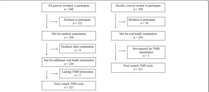
A total of 360 children and adolescents with JIA were eligible for the main study, of whom 228 accepted the invitation to participate, yielding a response rate of 63.3%. The acceptance rate for healthy controls was 224/ 294 (76.2%). The mean age for participants with JIA and healthy controls was 12.0 years (SD 3.17 and 3.21, re-spectively) (p = 0.98), and the mean age of the 228 par-ticipants with JIA was higher than for the 132 eligible patients that did not participate at 12.0 years vs. 10.5 years (SD 3.16 and 3.5, respectively) (p < 0.001). The proportion of girls with JIA was also higher than among the 132 patients not participating (59.2% vs. 58.3%, p = 0.027). Among the 228 participating children with JIA, 224 underwent an oral examination and 221 underwent the TMD assessment and were thus included in the present substudy (Fig.1).
Of the 221 children with JIA, 132 were girls (59.7%), the median age at disease onset was 6.1 years (IQR 8.1, 2.3– 10.4), the median age at the study visit by paediatric rheu-matologists at the hospital was 12.7 years (IQR (5.3, 9.4– 14.7), and the median disease duration was 4.6 years (IQR 5.7, 2.6–8.3) (Table 1). Oligoarticular JIA was the most common category and was seen in 98 of 221 patients (44.3%) with 77 having persistent oligoarticular disease and 21 having extended disease. In total, 146 of the 221 patients (66.1%) had on-going medication with synthetic disease-modifying antirheumatic-drugs (sDMARDs) and/or bio-logic disease-modifying antirheumatic-drugs (bDMARDs).
Clinical oral examination
Taking into consideration that self-reported pain is a combination of parent-reported and participant-reported
pain outcome, self-reported pain in the jaws during the last 30 days was reported in 59 (26.7%, 44 girls) partici-pants with JIA vs. 10 (5%, 8 girls) in healthy controls (p < 0.001). Pain during jaw movements at the clinical examination was reported in 112 (51%, 67 girls)
participants with JIA vs. 59 (26.8%, 34 girls) in healthy controls (p < 0.001) (Fig.2), ranging from 28.6 to 50% in the different JIA categories (Table1). No statistically sig-nificant differences in the presence of TMD according to JIA categories were found (p = 0.848) (results not shown).
Fig. 1 Flow chart of patients and healthy individuals included in the study
Table 1 Clinical characteristics of participants with Juvenile idiopathic arthritis (JIA) in relation to temporomandibular disorder (TMD)
Total cohort TMD No TMD
n = 221 n = 88 n = 133
Value Value Value
Girls, n (%) 132 (59.7) 61 (69.3) 71 (53.4)
Age at onset, median (IQR) 6.1 (8.1, 2.3–10.4) 6.8 (8.4, 0.7–14.2) 5.2 (7.2, 0.9–14.7)
Age at visit by paediatric rheumatologists at the hospital, median (IQR) 12.7 (5.3, 9.4–14.7) 13.1 (3.3, 5.2–16.1) 11.7 (6.5, 4.8–16.5)
Disease duration, median (IQR) 4.6 (5.7, 2.6–8.3) 4.6 (6.0, 0.2–14.2) 4.6 (5.5, 0.2–14.7)
JIA categories, n (%) Oligoarthritis persistent 77 (34.8) 27 (30.7) 50 (37.6) Oligoarthritis extended 21 (9.5) 11 (12.5) 10 (7.5) Systemic arthritis 7 (3.2) 2 (2.3) 5 (3.8) RF negative polyarthritis 49 (22.2) 17 (19.3) 32 (24.1) RF positive polyarthritis 4 (1.8) 2 (2.3) 2 (1.5) Psoriatic arthritis 9 (4.1) 6 (6.8) 3 (2.3) Enthesitis-related arthritis 23 (10.4) 9 (10.2) 14 (10.5) Undifferentiated JIA 31 (14.0) 14 (15.9) 17 (12.8) Ongoing medication, n (%) No DMARDs 75 (33.9) 26 (11.8) 49 (22.2) sDMARDs* 60 (27.1) 23 (10.4) 37 (16.7) bDMARDs** 86 (38.9) 39 (17.6) 47 (21.3)
JIA Juvenile idiopathic arthritis, TMD Temporomandibular disorder
*sDMARDs = Synthetic disease-modifying antirheumatic- drugs; methotrexate, mykofenolatmofetil, **bDMARDs = Biologic disease-modifying antirheumatic-drugs; etanercept, infliximab, adalimumab, tocilizumab, abatacept, certolizumab, golimumab
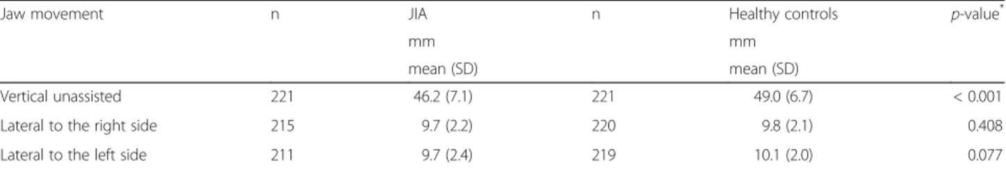
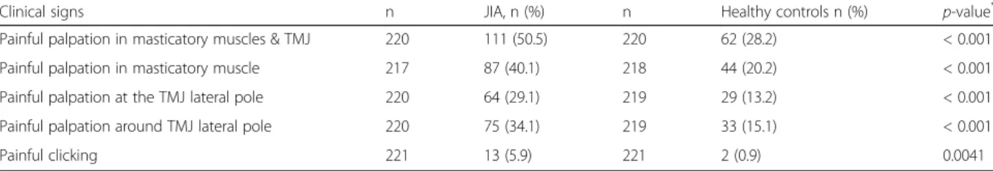
The clinical examination revealed that the mean vertical unassisted jaw movement was lower for participants with JIA than for controls, 46.2 mm vs. 49.0 mm, respectively (p < 0.001) (Table2). A total of 88 (39.8%, 61 girls) partic-ipants with JIA and 25 (11.3%, 17 girls) healthy controls had both symptoms and clinical signs of TMD (Fig. 2). When assessing the jaw muscles and TMJ, 111 (50.2%, 75 girls) participants with JIA had both painful masticatory muscles and TMJs on palpation vs. 62 (28.2%, 39 girls) of the healthy controls (p < 0.001) (Table3). A higher pro-portion of participants on current sDMARDs and/or bDMARDs treatment presented with painful masticatory
muscles and TMJ at palpation compared to participants with no biologic treatment (Table4).
Among participants with JIA, there were no significant differences in vertical unassisted jaw movement accord-ing to medication, with a mean of 46.4 mm (SD 7.1) in the JIA group and 45.8 mm (SD 7.1) among those not using DMARDs (p = 0.986) (results not shown). How-ever, in both groups, more than half of the participants had a vertical unassisted jaw movement of more than 40 mm. The proportion without this medication treatment was slightly higher (82.7%) compared to those on current sDMARDs and/or bDMARDs (77.4%).
Fig. 2 Prevalence of TMD in children and adolescents with JIA vs. healthy peers,≥ 10 years and < 10 years of age, 1) symptoms: pain the last 30 days and pain with jaw movements; 2) clinical signs: pain upon palpation of the masticatory muscles and TMJ, and 3) a combination of symptoms (1) and clinical signs (2)
Table 2 Jaw movement in 221 participants with JIA compared to controls
Jaw movement n JIA n Healthy controls p-value*
mm mm
mean (SD) mean (SD)
Vertical unassisted 221 46.2 (7.1) 221 49.0 (6.7) < 0.001
Lateral to the right side 215 9.7 (2.2) 220 9.8 (2.1) 0.408
Lateral to the left side 211 9.7 (2.4) 219 10.1 (2.0) 0.077
JIA Juvenile idiopathic arthritis, SD Standard deviation.*
Discussion
We have shown using a comparative cross-sectional multicentre design that around one third of the partici-pants with JIA in this cohort had TMD. Half of children and adolescents with JIA reported pain during jaw movements and pain on palpation of the masticatory muscles and TMJs as compared to one fourth of their healthy peers, palpatory pain was associated with sDMARDs and bDMARDs treatment, and children and adolescents with JIA had a significantly lower mean ver-tical unassisted jaw movement. Moreover, TMJ-related clinical signs and vertical unassisted jaw movement≤40 mm had the highest association in the JIA group.
The reported prevalence of TMD in children with JIA varies between 38 and 83% according to the definitions and methods of ascertainment used, to the cohort exam-ined, and to differences in populations [15,24–27]. Fer-raz and colleagues, in their study of 15 children with JIA ranging in age from 6 to 28 years (mean age 16.3 years), reported a high prevalence of 83%. Still, they did not de-scribe the method of ascertainment, i.e., whether the fig-ures were based on self-reporting or on clinical examination [28]. A previous study from Rongo and col-leagues based on 50 participants with JIA aged 9–16 years found a prevalence of TMJ damage from 100 joints to be 74% as assessed by MRI [25]. Others have reported a prevalence of 55% based on a questionnaire [29] and of 72% based on clinical signs [24]. However, none of those studies were based on the research diagnostic cri-teria RDC/TMD, and the children were older than those in our study. In contrast, a longitudinal study by Zwir et al., including 75 children (mean age 12.4 years),
revealed a prevalence of 38% based on symptoms and 47% based on clinical examination [30]. Their results are in line with ours.
In our study, the prevalence of TMD, either based on symptoms or clinical signs, in the healthy peers, were quite high at 28 and 29%, respectively. This was higher than in earlier studies among adolescents reported by Graue and colleagues (7 and 12%, respectively) and Østensjø and colleagues (7%) [8,9]. Studies from Finland and Brazil confirm our results with a high prevalence of TMD in the normal population. Vierola et al. [26] re-ported a TMD prevalence of 35% (mean age 7.9 years) and de Paiva Bertoli reported a TMD prevalence of 34% (mean age 11.0 years) [27]. The difference in TMD prevalence in the normal population of children and ad-olescents is probably due to the use of different diagnos-tic tools, different numbers of pardiagnos-ticipants, different ages of the studied populations, different countries, and dif-ferent study designs. In studies from Norway, Graue and colleagues [9] used two screening questions for pain re-lated to TMD [10] and DC/TMD [20] for symptoms and clinical signs in a population of 210 children and adoles-cents aged 12–19 years. Østensjø et al. [8] used the same two screening questions of TMD symptoms [10] for screening a population of 560 adolescents aged 13–19 years. Then a modified RDC/TMD examination [31] was used for those who answered yes to 1) having pain in the temples, face, TMJ, or jaws once a week or more and 2) having pain once a week or more when opening the mouth wide or chewing. The Finnish group [26] used the RDC/TMD [31] for clinical signs in 483 chil-dren aged 6–8 years, and the Brazilian group [27] used
Table 3 Pain on palpation and painful clicking in participants with JIA and controls
Clinical signs n JIA, n (%) n Healthy controls n (%) p-value*
Painful palpation in masticatory muscles & TMJ 220 111 (50.5) 220 62 (28.2) < 0.001
Painful palpation in masticatory muscle 217 87 (40.1) 218 44 (20.2) < 0.001
Painful palpation at the TMJ lateral pole 220 64 (29.1) 219 29 (13.2) < 0.001
Painful palpation around TMJ lateral pole 220 75 (34.1) 219 33 (15.1) < 0.001
Painful clicking 221 13 (5.9) 221 2 (0.9) 0.0041
JIA Juvenile idiopathic arthritis, TMJ Temporomandibular joints. *Chi-square test
Table 4 Clinical signs and pain at palpation according to DMARDs in 221 participants with JIA
Current sDMARDs and/or bDMARDs No current sDMARDs and/or bDMARDs p-value*
n n (%) n n (%)
Vertical unassisted jaw movement (> 40 mm) 146 113 (77.4) 75 62 (82.7) 0.361
Painful palpation to masticatory muscles & TMJ 145 80 (72.1) 75 31 (41.3) 0.052
Painful palpation to masticatory muscles 143 62 (43.4) 74 25 (33.8) 0.173
Painful palpation at the TMJ lateral pole 145 49 (33.8) 75 15 (20.0) 0.033
Painful palpation at the TMJ around lateral pole 145 56 (38.6) 75 19 (25.3) 0.049
sDMARDs Synthetic disease-modifying antirheumatic-drugs; methotrexate, mykofenolatmofetil, bDMARDs Biologic disease-modifying antirheumatic- drugs; etanercept, infliximab, adalimumab, tocilizumab, abatacept, certolizumab, golimumab,TMJ temporomandibular joint, * Chi-square test
the American Academy of Orofacial Pain [32] form for screening and the RDC/TMD [31] for clinical examin-ation in a populexamin-ation of 934 individuals aged 10–14 years. Thus it is clear that it can be challenging to get an exact figure on the prevalence of TMD in the normal population. A previous meta-analysis conducted by da Silva and colleagues showed the overall prevalence of intra-articular joint disorder to be 16% [33].
In our study, approximately half of the JIA subjects had clinical findings consistent with TMD, with no differences according to JIA category. Because the numbers for three of the categories – systemic arthritis, rheumatoid factor positive polyarthritis, and psoriatic arthritis – were rela-tively low, these results should be interpreted with caution.
The sensitivity and specificity of the clinical orofacial examination in relation to TMJ has been debated be-cause displacement of the disc, although eliciting a click-ing sound, might be asymptomatic [34–36]. Based on the DC/TMD criteria, asymptomatic TMJ clicking is still defined as TMD. However, several studies have shown that pain-free clicking represents a normal variant, typic-ally seen in girls during puberty [15]. Recently, a clinical examination protocol for JIA was developed by the Tem-poromandibular Joint Juvenile Arthritis Working Group (TMJaw). This examination protocol focuses on three general items, namely TMJ symptoms, TMJ dysfunction, and dentofacial deformity in JIA, and it shows acceptable reliability and validity [7].
We found, in accordance with other studies, that the TMJ area and the masseter muscle region were common locations for pain in JIA [29]. However, a recent study from Koos and colleagues reported a lower frequency of masticatory pain on palpation [15], and Kristensen and colleagues stated that masticatory pain complaints could develop over time [37]. In the present study, more than half of the participants with JIA showed clinical signs in the TMJ region and the masseter region, and more than one-fourth of the participants with JIA had TMD. A lon-gitudinal multicentre approach might elucidate the de-velopment of masticatory muscle pain, as Kristensen and colleagues have suggested [37].
The vertical unassisted jaw movement has been widely used as a valid marker for TMJ arthritis [38]. We showed that participants with JIA had lower vertical un-assisted movements compared to their healthy peers, but the differences were relatively small, thus questioning its clinical significance. Viewed differently, for children and adolescents aged < 11 years, the cut-off value of 40 mm was within the range of normal vertical jaw movement [39]. Further, our findings suggest that lateral movement did not differ significantly between the two groups, which is in line with the results of Twilt and colleagues [40] and Küseler and colleagues [19]. In the latter study of 15 children with JIA with a mean age of 12 years, the
recorded decreased lateral movements were≤ 5 mm with no significant relevance [19].
We found no statistically significant differences in the presence of TMD according to JIA categories. However, we found a significantly higher occurrence of clinical signs in participants with JIA currently on DMARDs medication (whether synthetic or biologic) compared to those not taking such medication. A high risk of devel-oping clinical signs of TMD was associated with a severe disease course, as indicated by the use of DMARDs.
The strengths of this study are the relatively large number of participants, in which the study groups were well matched, and the meticulous standardisation of the clinical TMJ assessment performed prior to and during the study period. However, the large number of partici-pants should not hide the fact that we are dealing with an underpowered sample size that was lacking 75 partic-ipants. An additional limitation is that the overall re-sponse rate of 63%, although considered acceptable, might have influenced the results because the group that did not participate was, on average, slightly younger and had a somewhat lower proportion of girls. Also, the shortened version of the DC/TMD used in this study is not directly comparable with studies having used the full DC/TMD score. In the present study, children and ado-lescents with JIA with TMD involvement were defined based on clinical examination, and both self-reported and parent-reported pain. Further studies will focus on the role of imaging on the diagnosis of TMJ arthritis in children and adolescents with JIA. Clinical orofacial examination may not be reliable for diagnosing disc dis-placement without reduction [5]. Imaging diagnosis is particularly important in JIA with non-symptomatic TMJ involvement because hard tissue loss in the condyle might hinder the growth of the mandible and subse-quently affect chewing function and cause aesthetic problems [15].
Conclusion
Symptoms or clinical signs of TMD were seen in ap-proximately half of the participants with JIA compared to about one fourth of their healthy peers. Painful palpa-tion of masticatory muscles and decreased vertical un-assisted jaw movement are more frequent in children with JIA than in healthy controls and should be part of both medical and dental routine examinations in the follow-up of JIA.
Supplementary information
Supplementary information accompanies this paper athttps://doi.org/10. 1186/s12903-020-01234-z.
Additional file 1 Table S1. Reliability tests (using intraclass correlation coefficients) between“a reference” and the examiners.
Additional file 2 Table S2. Percent agreement values between“a reference” and the examiners.
Abbreviations
DC/TMD:Diagnostic criteria for temporomandibular disorder; bDMARDs: Biological disease-modifying antirheumatic drugs; DMAR Ds: Disease-modifying antirheumatic drugs; sDMARDs: Synthetic disease-modifying antirheumatic drugs; ILAR: International League of Associations of Rheumatology; JIA: Juvenile idiopathic arthritis; RDC/TMD: Research diagnostic criteria for TMD; TMD: Temporomandibular disorder; TMJ: Temporomandibular joint
Acknowledgements
The authors thank Prof. Ellen Berit Nordal for her constructive comments on the manuscript. We thank the Center for Oral Health Services and Research of Middle-Norway (TkMidt), Trondheim, the Public Dental Service Compe-tence Centre of Northern-Norway (TkNN), Tromsø and the Oral Health Centre of Expertise in Western Norway (TkVest), Bergen, for contributing with time and personnel resources. Thanks and appreciation to Marie Sager and Gun-nar Lyngstad Center for Oral Health Services and Research of Middle-Norway (TkMidt). We thank the entire NorJIA research group (NorJIA.com) and Kasper Dahl Kristensen, Section of Orthodontics, Aarhus University, Denmark, for help with the questionnaires. Our gratitude also goes to all the participants accepting to be included in the study.
Authors’ contributions
JF: contributed to the conception and design of the study, agreed to be accountable for all aspects of the work and ensure questions related to the accuracy or integrity of any part of the work were appropriately investigated and resolved, was involved in writing the manuscript, and approved the final version to be published. MSS, KR, XQS, MR, PS, AR: contributed to the conception and design of the study and analysis/ interpretation of the data, were involved in drafting the manuscript and revising it critically for important intellectual content, and approved the final version to be published. SL: performed biostatistics. KT, EGG, LC, JH, LvWM, PF: contributed to data collection and provided valuable comments. All authors have read and approved the final manuscript.
Funding
The current study is supported in part by the Norsk Revmatikerforbund (Norway).
Availability of data and materials
The datasets used and analysed in the current study are available from the corresponding author on reasonable request.
Ethics approval and consent to participate
The study was approved by the regional ethics committee (2012/542/REK vest). Written informed consents were obtained from the parents/legal representatives and the adolescents. The study was registered at ClinicalTrials.gov (No: NCT03904459).
Consent for publication Not applicable. Competing interests
The authors declare that they have no competing interests. Author details
1Department of Clinical Dentistry, The Faculty of Medicine, University of
Bergen, Årstadveien 19, N-5009 Bergen, Norway.2Center for Oral Health
Services and Research of Middle-Norway (TkMidt), Trondheim, Norway.
3
Department of Radiology, University Hospital of North Norway, Tromsø, Norway.4UiT the Arctic University of North Norway, Tromsø, Norway. 5Department of Pediatrics, Haukeland University Hospital, Bergen, Norway. 6Department of Oral and Maxillofacial Radiology, Faculty of Odontology,
University of Malmö, Malmö, Sweden.7Public Dental Service Competence Centre of Northern-Norway (TkNN), Tromsø, Norway.8Department of Clinical
and Molecular Medicine, NTNU - Norwegian University of Science and Technology, Trondheim, Norway.9Department of Pediatrics, St. Olavs
Hospital, Trondheim, Norway.10Department of Otorhinolaryngology, Division
of Oral and Maxillofacial Surgery, University Hospital North Norway, Tromsø, Norway.11Department of Clinical Medicine, Faculty of Health Sciences, The
Arctic University of Norway, Tromsø, Norway.12Section of Orthodontics, Faculty of Health Sciences, Aarhus University, Aarhus, Denmark.
13Department of Oral and Maxillofacial Surgery, Haukeland University
Hospital, Bergen, Norway.
Received: 29 June 2020 Accepted: 25 August 2020 References
1. Riise OR, Handeland KS, Cvancarova M, Wathne K-O, Nakstad B, Abrahamsen TG, et al. Incidence and characteristics of arthritis in Norwegian children: a population-based study. Pediatrics. 2008.
2. Gabriel SE, Michaud K. Epidemiological studies in incidence, prevalence, mortality, and comorbidity of the rheumatic diseases. Arthritis Res Ther. 2009;11:229.https://doi.org/10.1186/ar2669.
3. Petty RE, Southwood TR, Manners P, Baum J, Glass DN, Goldenberg J, et al. International league of associations for rheumatology classification of juvenile idiopathic arthritis: second revision, Edmonton, 2001. J Rheumatol. 2004;31:390–2 PMID: 14760812.
4. LeResche L. Epidemiology of temporomandibular disorders: implications for the investigation of etiologic factors. Crit Rev Oral Biol Med. 1997. 5. Peck CC, Goulet JP, Lobbezoo F, Schiffman EL, Alstergren P, Anderson GC,
et al. Expanding the taxonomy of the diagnostic criteria for temporomandibular disorders. J Oral Rehabil. 2014;41(1):2.
6. Ohrbach R, Dworkin SF. The Evolution of TMD Diagnosis. J Dent Res. 2016: 0022034516653922.https://doi.org/10.1177/0022034516653922PMID: 27313164.
7. Stoustrup P, Herlin T, Spiegel L, Rahimi H, Koos B, Pedersen TK, et al. Standardizing the clinical orofacial examination in Juvenile idiopathic arthritis: An interdisciplinary, consensus-based, short screening protocol. J Rheumatol. 2019;190661 jrheum.
8. Ostensjo V, Moen K, Storesund T, Rosen A. Prevalence of painful Temporomandibular disorders and correlation to lifestyle factors among adolescents in Norway. Pain Res Manag. 2017;2017:2164825.https://doi.org/ 10.1155/2017/2164825.
9. Graue AM, Jokstad A, Assmus J, Skeie MS. Prevalence among adolescents in Bergen, Western Norway, of temporomandibular disorders according to the DC/TMD criteria and examination protocol. Acta Odontol Scand. 2016;74: 449–55.
10. Nilsson IM, List T, Drangsholt M. Prevalence of temporomandibular pain and subsequent dental treatment in swedish adolescents. J Orofac Pain. 2005.
15895837.
11. Cannizzaro E, Schroeder S, Müller LM, Kellenberger CJ, Saurenmann RK. Temporomandibular joint involvement in children with juvenile idiopathic arthritis. J Rheumatol. 2011;38:510–5.https://doi.org/10.3899/jrheum.100325. 12. Stoustrup P, Glerup M, Bilgrau AE, Küseler A, Verna C, Christensen AE, et al.
Cumulative incidence of Orofacial manifestations in early juvenile idiopathic arthritis: a regional, Three-Year Cohort Study. Arthritis Care Res (Hoboken). 2020;72(7):907.
13. Küseler A, Pedersen TK, Herlin T, Gelineck J. Contrast enhanced magnetic resonance imaging as a method to diagnose early inflammatory changes in the temporomandibular joint in children with juvenile chronic arthritis. J Rheumatol. 1998;25:1406–12.
14. Billiau AD, Hu Y, Verdonck A, Carels C, Wouters C. Temporomandibular joint arthritis in juvenile idiopathic arthritis: prevalence, clinical and radiological signs, and relation to Dentofacial morphology. J Rheumatol. 2007; PMID: 17696265.
15. Koos B, Twilt M, Kyank U, Fischer-Brandies H, Gassling V, Tzaribachev N. Reliability of clinical symptoms in diagnosing temporomandibular joint arthritis in juvenile idiopathic arthritis. J Rheumatol. 2014;41:1871–7.https:// doi.org/10.3899/jrheum.131337.
16. Stoll ML, Sharpe T, Beukelman T, Good J, Young D, Cron RQ. Risk factors for temporomandibular joint arthritis in children with juvenile idiopathic arthritis. J Rheumatol. 2012;39:1880–7.https://doi.org/10.3899/jrheum. 111441.
17. Keller H, Müller LM, Markic G, Schraner T, Kellenberger CJ, Saurenmann RK. Is early TMJ involvement in children with juvenile idiopathic arthritis clinically detectable? Clinical examination of the TMJ in comparison with
contrast enhanced MRI in patients with juvenile idiopathic arthritis. Pediatr Rheumatol. 2015;13:56.https://doi.org/10.1186/s12969-015-0056-2. 18. Weiss PF, Arabshahi B, Johnson A, Bilaniuk LT, Zarnow D, Cahill AM, et al.
High prevalence of temporomandibular joint arthritis at disease onset in children with juvenile idiopathic arthritis, as detected by magnetic resonance imaging but not by ultrasound. Arthritis Rheum. 2008;58:1189– 96.https://doi.org/10.1002/art.23401.
19. Küseler A, Pedersen TK, Gelineck J, Herlin T. A 2 year followup study of enhanced magnetic resonance imaging and clinical examination of the temporomandibular joint in children with juvenile idiopathic arthritis. J Rheumatol. 2005; PMID: 15630742.
20. Schiffman E, Ohrbach R, Truelove E, Look J, Anderson G, Goulet J-P, et al. Diagnostic Criteria for Temporomandibular Disorders (DC/TMD) for Clinical and Research Applications: Recommendations of the International RDC/ TMD Consortium Network* and Orofacial Pain Special Interest Group†. J Oral Facial Pain Headache. 2014;28:6–27.https://doi.org/10.11607/jop.1151. 21. Stoustrup P, Koos B. Clinical craniofacial examination of patients with
juvenile idiopathic arthritis. Semin Orthod. 2015;21:94–101.
22. Skeie MS, Frid P, Mustafa M, Aßmus J, Rosén A. DC/TMD examiner protocol: longitudinal evaluation on interexaminer reliability. Pain Res Manag. 2018; 2018.
23. Svensson B, Adell R, Kopp S. Temporomandibular disorders in juvenile chronic arthritis patients. A clinical study. Swed Dent J. 2000;24:83–92.11 061206.
24. Leksell E, Ernberg M, Magnusson B, Hedenberg-Magnusson B. Orofacial pain and dysfunction in children with juvenile idiopathic arthritis: a case-control study. Scand J Rheumatol. 2012.
25. Rongo R, Alstergren P, Ammendola L, Bucci R, Alessio M, D’Antò V, et al. Temporomandibular joint damage in juvenile idiopathic arthritis: diagnostic validity of diagnostic criteria for temporomandibular disorders. J Oral Rehabil. 2019;46(5):450.
26. Vierola A, Suominen AL, Ikavalko T, Lintu N, Lindi V, Lakka H-M, et al. Clinical signs of temporomandibular disorders and various pain conditions among children 6 to 8 years of age: the PANIC study. J Orofac Pain. 2012.22292136. 27. de Paiva Bertoli FM, Bruzamolin CD, Pizzatto E, Losso EM, Brancher JA, de
Souza JF. Prevalence of diagnosed temporomandibular disorders: a cross-sectional study in Brazilian adolescents. PLoS One. 2018;13.29420573. 28. Ferraz AML, Devito KL, Guimarães JP. Temporomandibular disorder in
patients with juvenile idiopathic arthritis: clinical evaluation and correlation with the findings of cone beam computed tomography. Oral Surg Oral Med Oral Pathol Oral Radiol. 2012;114:e51–7.https://doi.org/10.1016/j.oooo. 2012.02.010.
29. Rahimi H, Twilt M, Herlin T, Spiegel L, Pedersen TK, Küseler A, et al. Orofacial symptoms and oral health-related quality of life in juvenile idiopathic arthritis: a two-year prospective observational study. Pediatr Rheumatol. 2018;16.
30. Zwir LMLF, Terreri MTRA, Sousa SA, Fernandes ARC, Guimarães AS, Hilário MOE. Are temporomandibular joint signs and symptoms associated with magnetic resonance imaging findings in juvenile idiopathic arthritis patients? A longitudinal study. Clin Rheumatol. 2015;34:2057–63.
31. Dworkin SF, LeResche L. Research diagnostic criteria for temporomandibular disorders: review, criteria, examinations and specifications, critique. J Craniomandib Disord. 1992;6:301–55.1298767.
32. de Leeuw RRJ, Klasser G. American Academy of Orofacial Pain. General assessment of the orofacial pain patient. Orofacial pain - guidelines for assessment, diagnosis and management. 5th ed. Chicago: Quintessence; 2013. In: Orofacial pain; guidelines for assessment, diagnosis, and management, 5th ed. Quintessence Pub. Co.; 2013. p. 25–46. https://search-proquest-com.pva.uib.no/docview/1416243368?accountid=8579. 33. Da Silva CG, Pachêco-Pereira C, Porporatti AL, Savi MG, Peres MA, Flores-Mir
C, et al. Prevalence of clinical signs of intra-articular temporomandibular disorders in children and adolescents A systematic review and meta-analysis. J Am Dental Association. 2016;147(1):10.
34. Kircos LT, Ortendahl DA, Mark AS, Arakawa M. Magnetic resonance imaging of the TMJ disc in asymptomatic volunteers. J Oral Maxillofac Surg. 1987; 45(10):852.
35. Westesson PL, Eriksson L, Kurita K. Reliability of a negative clinical temporomandibular joint examination: prevalence of disk displacement in asymptomatic temporomandibular joints. Oral Surgery, Oral Med Oral Pathol. 1989.
36. Isberg A, Stenström B, Isacsson G. Frequency of bilateral
temporomandibular joint disc displacement in patients with unilateral symptoms: a 5-year follow-up of the asymptomatic joint. A clinical and arthrotomographic study. Dentomaxillofacial Radiol. 1991;20(2):73. 37. Kristensen KD, Stoustrup P, Kuseler A, Pedersen TK, Twilt M, Herlin T. Clinical
predictors of temporomandibular joint arthritis in juvenile idiopathic arthritis: a systematic literature review. Semin Arthritis Rheum. 2016;45:717– 32.https://doi.org/10.1016/j.semarthrit.2015.11.006.
38. Stoustrup P, Kristensen KD, Verna C, Küseler A, Herlin T, Pedersen TK. Orofacial symptoms related to temporomandibular joint arthritis in juvenile idiopathic arthritis: smallest detectable difference in self-reported pain intensity. J Rheumatol. 2012;39:2352–8.
39. Stoustrup P, Kristensen KD, Küseler A, Herlin T, Pedersen TK. Normative values for mandibular mobility in Scandinavian individuals 4–17 years of age. J Oral Rehabil. 2016;43:591–7.https://doi.org/10.1111/joor.12407. 40. Twilt M, Mobers SMLM, Arends LR, Ten Cate R, Van Suijlekom-Smit LWA.
Temporomandibular involvement in juvenile idiopathic arthritis. J Rheumatol. 2004;31:1418–22.15229966.
Publisher’s Note
Springer Nature remains neutral with regard to jurisdictional claims in published maps and institutional affiliations.