Department of Molecular Sciences
Evaluation of the antagonistic activity of
Rhodotorula babjevae and its extracellular
compounds towards postharvest pathogens
Caroline Sandberg
Evaluation of the antagonistic activity of Rhodotorula babjevae
and its extracellular compounds towards postharvest pathogens
Caroline Sandberg
Supervisor: Jonas Ohlsson, Swedish University of Agricultural Sciences, Department of Molecular Sciences
Examiner: Volkmar Passoth, Swedish University of Agricultural Sciences, Department of Molecular Sciences
Credits: 15 credits
Level: First cycle, G2E
Course title: Sjävständigt arbete i Livsmedelsvetenskap - Kandidatarbete
Course code: EX0876
Programme/education: Agricultural Programme – Food Science Course coordinating department: Department of Molecular Sciences
Place of publication: Year of publication: Title of Series: Part Number: Online publication: Keywords: Uppsala 2019 Molecular Sciences 2019:9 https://stud.epsilon.slu.se
Rhodotorula babjevae, PEFA, postharvest pathogens, yeast, mold
The growing population ultimately puts pressure on the food industry to become more effi-cient and to reduce postharvest food waste. The leading method of reducing postharvest decay of fruits has been through fungicide application. However, because of the public per-ception towards fungicide usage there is now a demand for new methods, that are environ-mentally friendly and unharmful for human consumption. The use of antimicrobial com-pounds and activities of microorganisms isolated from natural crop that possess antagonistic activity towards postharvest pathogens, has therefore become an emerging field of research. Earlier research has demonstrated that a yeast strain within the genus Rhodotorula marked antagonistic activity towards several postharvest pathogens, when the living culture was ap-plied onto fruit surface. The oleaginous yeast R. babjevae has been found to coproduce in-tracellular lipids and exin-tracellular polyol esters of fatty acids (PEFA) when exposed to ni-trogen limitation and an excess of carbon. The amphiphilic PEFA has been shown to possess several bioactivities, one of them being antifungal, thus making it a possible biocontrol agent.
Because of the aforementioned research on species in the Rhodotorula genus as potential biocontrol agents, one indicating the antagonistic activity of the living yeast cultures and one towards the potential antagonistic activity of the PEFA, the experiment was conducted to evaluate the difference. The aim of the experiment was therefore to evaluate the inhibitory effect of the living yeast culture of R. babjevae, and of its extracellular PEFA towards the postharvest pathogens Penicillium expansum, Botrytis cinerea and Aspergillus flavus. The yeast where cultured in a nitrogen limited medium with an access of glucose, to promote lipid production. During incubation of the yeast culture, the progress in biomass was meas-ured through optical density (OD) and samples were analyzed using HPLC to measure the glucose concentration. Intracellular lipid production was recorded by micrographs. A disk diffusion bioassay was prepared with two different cultures of living yeast culture (3 and 6 days old) and two concentrations of a PEFA pellet, on MEA plates inoculated with the molds. Continuous registration of the antagonistic activity followed during the incubation in the dark at 25 °C for 5 days. The results from the disk diffusion assay showed antagonistic activity from both the living yeast cultures towards B. cinerea, although neither of the two PEFA concentrations inhibited the mold. A. flavus and P. expansum had overgrown the plates after 2 days of incubation, probably because of too concentrated spore solution. The antagonistic activity towards B. cinerea could be explained by its slower growth rate, which provided the yeast time to acclimatize to the new environment. The antagonistic activity of
R. babjevae marked promising characteristics towards becoming a potential biocontrol agent
under commercial conditions.
Abstract
1 Introduction 4
2 Materials and methods 7
2.1 Culture media 7
2.2 Microorganisms 7
2.3 Disk diffusion bioassay 8
2.4 Measurements of optical density and glucose concentration 9
3 Results 10
3.1 Disk diffusion assay 10
3.2 Measurements on optical density and glucose concentration 13
4 Discussion 14
5 Acknowledgements 16
References 17
The world population is expected to reach 10.5 billion by 2050; it consequently means there will be 33% more people to feed (Aulakh and Regmi, 2013). According to a prediction made by the Food and Agriculture Organization of U.N, 1.3 billion tons of food are wasted or lost each year (Aulakh and Regmi, 2013).In the last 30 years it has been estimated that 95% of the research investments have gone to in-creasing the productivity of agricultural production and only 5% towards reducing food loss (Aulakh and Regmi, 2013). Although increasing the production is neces-sary to ensure global food security it may not be sufficient, considering the limited land and water resources, in addition to variable weather conditions due to climate change. Therefore, the reduction of postharvest losses in each line of the food chain is also crucial to sustainably reach the goals of global food security (Aulakh and Regmi, 2013) In developing countries, the postharvest loss of crops has, because of mishandling, pest infestation and spoilage, been estimated to approximately one quarter of the production. The additional losses of the less hardy and more perisha-ble products such as fruits, vegetaperisha-bles and root crops are hard to estimate but it is predicted to be a lot more (FAO, 1989).
Fruit decay, that comes as a result of postharvest infection by fungal pathogens, mainly occurs during transportation and storage (Zhang et al., 2011). The posthar-vest pathogen Botrytis spp. has many different host plants and because of its adapt-ability to a variety of microclimate conditions, rapid rates of germination and my-celium production, it poses a significant problem to the food industry (Elad et al., 2007). Botrytis cinerea (grey mold) and Penicillium expansum (blue mold) are among the most damaging postharvest pathogens towards apples (Zhou et al., 2001).
P. expansum produces the mycotoxin patulin (Baert et al., 2007). Aflatoxins are
carcinogenic mycotoxins produced by fungi in the genus Aspergillus, where A.
fla-vus is one of the major producers of aflatoxin in crop seeds in fields and storage
(Klich, 2007). Mycotoxins contaminate food and can cause serious health effects when ingested. The levels of the molecular toxins allowed in food is therefore reg-ulated, which consequently leads to additional food waste and economic loss in
several parts of the food chain (Paterson and Lima, 2010). Temperature and humid-ity are highly influential to the production of mycotoxins; the climate change is therefore prospected to affect mycotoxin production in food throughout the world. The increasing temperature is for instance predicted to promote production of afla-toxin in currently moderate temperature countries and patulin in colder countries (Paterson and Lima, 2010).
To control food decay, postharvest application of fungicides has long been the leading method (Zhang et al., 2011). However, because of reduced efficiency by development of pathogen resistance to key fungicides, lacking supply of new ones and restrictions towards fungicide application, the usage is facing obstacles (Jan-isiewicz and Korsten, 2002). In addition, there are also the public concerns about the possible health risks of pesticide residues in food and the negative effects on the environment (Zhang et al., 2011). There is therefore currently demand for efficient methods to control postharvest diseases which leave low levels of residues and have little or no toxic effect towards non-target organisms (Zhang et al., 2011). This has consequently encouraged research in the field of biocontrol of postharvest diseases to emerge (Janisiewicz and Korsten, 2002), which represents an alternative method based on the vulnerability of the pathogens to antimicrobial compounds derived from microorganisms of natural crop (Passoth and Schnürer, 2003). Microbial bio-control agents have a reduced risk of resistance development because of the gener-ally complex mechanisms used and they can also be assumed to be less demanding to the environment. Biocontrol can therefore be beneficial for the growing market of minimal pesticide and organic production systems (Elad and Stewart, 2007).
An experiment performed by Lima et al. (1998) evaluated the antagonistic activ-ity of yeasts towards postharvest pathogens through application on fruits. One of the yeast strains used belonged to the genus Rhodotorula which exhibited antagonistic activity against several postharvest pathogens and especially towards B. cinerea and
P. expansum. The culture was able to grow in the temperature range of 0 to 35
de-grees, beneficial qualities considering different postharvest pathogens, geographic variations as well as climate change. The ability to colonize unwounded fruit also showed potential for fruit surface application (Lima et al., 1998).Regarding public health concerns, the yeasts tested did not pose a risk to cause mycoses in human since it was unable to grow at 37 °C (Lima et al., 1998). The yeasts also had a low sensitivity towards fungicides and could therefore be used in addition to low doses of such compounds (Lima et al., 1998). Moreover, the antagonistic activities of bac-teria and yeast used in biocontrol have been prospected to, if not replace the fungi-cides, be integrated with the protection (Lima et al., 1998). Because the postharvest pathogens invade through fruit wounds, the antagonists used in biocontrol can be applied straight on the target area, through a single application use of delivery sys-tems, to reduce the decay (Janisiewicz and Korsten, 2002).
Among the range of isolates there is a large genetic diversity and different mech-anisms used for the antagonistic activity (Passoth and Schnürer, 2003). Some of the proposed mechanisms are parasitism of the pathogen, biofilm formation, secretion
of antifungal compounds (Liu et al., 2013), production of cell-wall lytic enzymes,
direct physical interaction by fungal hyphae and induction of host resistance (LIMA et al., 1998). Competing for essential organic and inorganic material in addition to space is also an important antifungal activity (Passoth and Schnürer, 2003). The usage of yeast during postharvest biocontrol has appeared to be preferable to bacte-ria, as several types of bacteria seem to extend their antagonistic behaviour through the production of the unwanted antibiotic substances (LIMA et al., 1998).
Oleaginous yeasts produce intracellular lipids, that mainly consist of triglycer-ides, when it is exposed to certain conditions such as nutrient limitation. The storage lipids accumulated by the yeast can reach 70% of dry cell weight (Guerfali et al., 2019). The oleaginous yeast Rhodotorula babjevae has been shown to coproduce intracellular storage lipids and extracellular polyol esters of fatty acids (PEFA) (Garay et al., 2017), when subjected to a nitrogen limitation with an excess amount of carbon (Guerfali et al., 2019). The amphiphilic PEFA has exhibited several bio-activities including antifungal, antibacterial, antiviral and anticarcinogenic activity (Guerfali et al., 2019). Thus, because of the antifungal activity, the PEFA have be-come interesting in the research for antimicrobial biocontrol agents.
As mentioned, earlier experiments have demonstrated antagonistic activity by living yeasts in the genus Rhodotorula and additional research has shown anti-fungal activity of the PEFA produced by R. babjevae. The experiment presented in this thesis was conducted to test the different antagonistic activities of the living culture and the PEFA. The aim was therefore to evaluate the antagonistic activity of
R. babjevae J195 strain, isolated from wild apples, and its extracellular products on P. expansum, B. cinerea and A. flavus, with the hypothesis that a stronger inhibitory
2.1 Culture media
Yeast Extract Peptone Dextrose agar (YEPD) 10 g/l yeast extract, 20 g/l peptone, 20 g/l glucose and 18 g/l agar.
Nitrogen limited cultivation medium was prepared in two Erlenmeyer flasks with 0.4 l and contained 2.5 g/l NaHPO4, 7.0 g/l KH2PO4, 0.23 g/l urea, 0.5 g/l yeast
extract, 1.5 g/l MgSO4 x H2O, 0.15 g/l CaCl2, 0.01 g/l ZnSO4 · 7H2O, 0.07 g/l
MnSO4 · H2O, 40 g/l glucose in deionized water, largely in accordance with Guerfali
et al. (2018) but substituting urea for (NH4)2SO4 to reduce H+ formation.
Saline was prepared for the disk diffusion bioassay, containing 0.9% NaCl and deionized H2O.
Malt Extract Agar (MEA) was prepared for the disk diffusion bioassay, contain-ing 20 g/l malt extract, 20 g/l dextrose, 6.0 g/l peptone, 15 g/l agar and 0.1 g/l chlo-ramphenicol to inhibit bacterial growth.
2.2 Microorganisms
Rhodotorula babjevae J195 strain from 2006, available in the SLU yeast strain
col-lection, stored in 50% glycerol stocks at -80°C. It was quickly thawed and inocu-lated on YEPD agar plates and then incubated for 2 days in 26°C.
100 ml of the nitrogen limited cultivation medium was distributed into 4 shake flasks with baffles. Each flask was then inoculated with a loop of yeast from the culture on the YEPD agar plate. The flasks were incubated on an orbital shaker (Ecotron, Infors HT, Bottmingen, Switzerland) at a temperature of 26°C with the agitation rate 180 rpm for 8 days.
To evaluate the inhibitory properties of R. babjevae J195 and PEFA extract on molds frequently associated with the strain in the wild, P. expansum, A. flavus and
B. cinerea were cultured on MEA plates one week before the experiment, and
incu-bated in the dark at 25 °C.
2.3 Disk diffusion bioassay
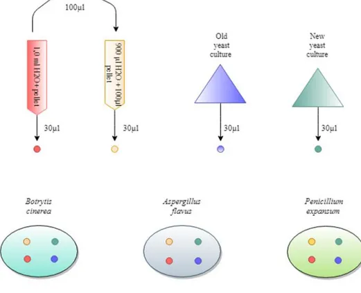
The disk diffusion bioassay was performed using four different yeast treatments to evaluate the efficiencies of the antagonistic activity on each of the molds. For a schematic overview of the procedure, see Figure 1. A previously centrifuged and frozen yeast culture with an apparent PEFA pellet was thawed in the refrigerator overnight and then diluted with 1.0 ml of sterile water before being centrifuged again. The pellet was then extracted with a spoon from the bottom of the 50 ml Falcon tube. 100 µl of the concentrated pellet solution was pipetted into a 15 ml Falcon tube with an additional 900 µl of H2O, resulting in a 10-fold dilution of the PEFA pellet solution. Broth from the 6 days old yeast culture (old yeast culture; OYC) and the 3 days old yeast culture (new yeast culture; NYC) were collected in 15 ml Falcon tubes. Neither of the yeast cultures were diluted before application.
3.5 ml of saline was transferred to each of the MEA plates with the mold cultures to solubilise the spores and to reduce the osmotic pressure in the medium. 400 µl of solution with spores was inoculated to the MEA plates and distributed with a dis-posable sterile bend rod. 30 µl of yeast solution was pipetted onto filter paper disks, which were dried and placed face down on the inoculated MEA medium.
The disks where slightly displaced when transported to the incubator, but quickly put back. However, to ensure the results another set of plates with disk diffusion assay were made following the same procedure but with only OYC and NYC. The disks were also placed on separate plates to minimise the risk of mix-up. 1 ml of saline was added to each mold to decrease the concentration of spores in the solu-tion. Instead of inoculating the MEA plates with 400 µl, 200 µl of the spore solutions were applied. However, the layer of spores was still significant in both A. flavus and
P. expansum. The plates were then incubated in the dark at 25°C for 5 days with
continuous registration of the results.
2.4 Measurements of optical density and glucose
concentration
Throughout cultivation of the yeast culture in the bioreactor, the amount of glucose and biomass were regularly measured. The optical density (OD) was measured in a spectrophotometer (CO8000 Cell density Meter, Nordic biolabs, Täby, Sweden). To measure the glucose content, samples of 1 ml for HPLC where taken in Eppendorf tubes and put in the freezer.
After thawing the samples overnight, 100 µl of the yeast solution was pipetted in Eppendorf tubes with 900 µl of 1% HCl and then centrifuged, for the PEFA to precipitate. 100 µl of the samples were then extracted to HPLC vials and put in the freezer. During the HPLC the compounds were analysed using Agilent 1100 (Ag-ilent Technologies) and run time 45 minutes, isocratic elution, flow rate 0.6 l/min, mobile phase 5 mM sulfuric acid on an ion exclusion column (Phenomex Rezex-ROA-Organic Acid H+), column temperature 60°C.
A graph was made to view the function of the amount of glucose (g/l) and bio-mass (logarithmic scale) through time (h) during the cultivation of yeast culture. To record the progress in the intracellular lipid production, light micrographs were taken using a microscope equipped with a digital camera of resolution 3.1 megapixel.
3.1 Disk diffusion assay
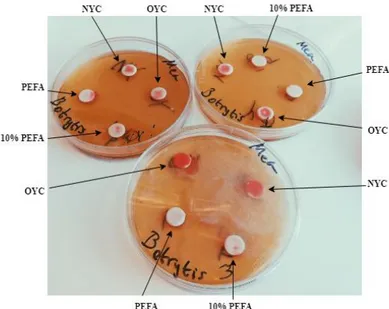
After 24 hours of incubation there was no visual growth from the plates inoculated with B. cinerea. By day 2, B. cinerea had started to grow and the yeast disks showed signs of proliferation. The disks inoculated with the yeast culture (OYC and NYC), showed clear growth on the disks and numerous marked antagonistic effects towards the mold (see Figure 2). No inhibition zones by the PEFA or 10% PEFA disk could be registered. However, in one of the three plates the PEFA disk showed some yeast growth outside of the disk although it had not proliferated to the same extent as NYC and OYC.
3 Results
Figure 2: Photograph of the disk diffusion bioassay after two days of incubation consisting of three MEA plates inoculated with B. cinerea with disks of each solution. Both OYC and NYC had grown on and outside the disks and one of the PEFA disks also showed signs of yeast growth outside the disk.
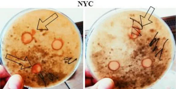
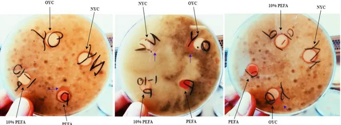
On plates inoculated with B. cinerea, disks with NYC and OYC displayed antago-nistic behaviour after 4 days of incubation (see Figure 3a and 3b). After 5 days of incubation yeast had colonized on and around disks with different treatments. Even though it was not significant enough to measure inhibition zones, antagonis-tic activity towards B. cinerea could be seen (see Figure 4),
Figure 3a: Photograph of two plates inoculated with B. cinerea after 4 days of incubation, containing discs with NYC (a) and OYC (b). Arrows displaying the growth that can be seen on and outside the disk, indicat-ing antagonistic activity towards the mold.
Figure 3b: Photograph of two plates inoculated with B. cinerea after 4 days of incubation, containing discs with NYC (a) and OYC (b). Arrows displaying the growth that can be seen on and outside the disk, indicating antagonistic activity towards the mold.
The plates inoculated with P. expansum and A. flavus were overgrown with mold after 2 days of incubation, with no signs of antagonistic activity by the yeast or PEFA (see Figure 5).
Figure 4: Photograph of three plates inoculated with B. cinerea, after 5 days of incubation. Black arrows marking the different yeast solutions and purple arrows the yeast growth outside the disks. Both NYC and the OYC indicates growth and antagonistic activity towards the mold. The yeast culture on the PEFA disk also exhibit antagonistic activity towards the mold.
Figure 5: Photographs of plates inoculated with A. flavus and P. expansum, after 2 days of incuba-tion. The plates are overgrown with mold.
3.2 Measurements on optical density and glucose
concentration
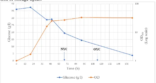
The graph in Figure 6 demonstrates the level of glucose (g/l) and OD (logarithmic scale) as functions of time (h), during the cultivation of the yeast culture. The de-creasing glucose concentration and inde-creasing OD illustrates exponential phase of the yeast culture. The graph shows that the exponential phase took place between approximately 20 and 43 hours of cultivation, after that the stationary phase was initiated. When the yeast reached the stationary phase, possibly because of nitrogen limitation, the glucose concentration continued to decrease, indicating that glucose was then instead used to produce storage lipids. Moreover, the glucose concentra-tion did not decrease between about 43 and 50 hours, which could possibly be ex-plained by the metabolic shift from formation of biomass to intracellular accumula-tion of storage lipids.
The progress of the intracellular lipid production between 20 h, 43 h, 92 h and 164 h of incubation is displayed in Figure 7. After 20 h of incubation there where no signs of started production whereas after 116 h the intracellular lipids where appar-ent. The progression of the production goes in accordance with the graph.
Figure 6: The graph shows the level of glucose (g/l) and OD (logarithmic scale) as functions of time (h) dur-ing cultivation of the yeast culture. Arrows markdur-ing for how long the yeast cultures were incubated.
Figure 7: Micrographs of the yeast culture after 20h, 43h, 92h and 164h of incubation. Indicating that after 116h there were significant signs of intracellular lipid production.
The results indicated a different outcome than predicted. Instead of the PEFA pellet showing the most antagonistic effect on the mold, the living yeast culture showed a stronger effect. Both the living yeast cultures, OYC and NYC, grew outside of the disks on several plates, restricting the mold culture’s growth and therefore revealed signs of antagonistic activity or outcompetition towards B. cinerea. The apparent difference in the antagonistic effect of the yeast cultures and the PEFA disks indi-cates that the yeast itself performs the antagonistic behaviour and not the PEFA. Since the PEFA pellet was extracted with a spoon a part of the yeast supernatant followed, thus disks inoculated with PEFA could vary in the proportion of yeast cells. Therefore, the diverging result in one of the disks could be because it possibly contained more of the living yeast culture. Additionally, there was no apparent dif-ference in antagonistic activity towards B. cinerea between the two yeast cultures OYC and NYC.
It should be considered that the molds were pre-cultured on MEA plates, the same medium as used in the disk diffusion assay, whereas the yeast was cultured in a N-limited medium. The yeast therefore had to go through a lag-phase whilst ac-climatizing to the new medium, which consequently might have provided the mold with an advantage. If the antagonistic activity was conducted through competing for nutrients, the slower growth rate of B. cinerea might have allowed the growth of the yeast culture by providing it with more time to acclimatize, thereby giving the yeast a chance to compete for nutrients before the mold had overgrown the plate. This would also be an explanation as to why it did not show any antagonistic activity towards P. expansum and A. flavus, since both molds had overgrown the plates al-ready after 2 days of incubation.
The overgrowth was also a result from a mistake made during the experiment, when 400 µl of the solubilised spores was pipetted instead of the planned 100 µl. On both
A. flavus and P. expansum there was a visible coat of the spores on the MEA plates
after the inoculation, in difference to the B. cinerea where the spores were not vis-ually as highly concentrated in the solution nor on the inoculated plates. The speed at which A. flavus and P. expansum spread on the plates in difference to B. cinerea could moreover indicate a difference in the concentration of spores between the molds.
The results indicate that the probable explanation of the bioactive mechanism of the yeast to inhibit the mold would be competition for nutrients. However, it can not be concluded without further research. The results from Lima et al. (1998) showed a difference in the inhibitory activity between the yeast strains used in the experi-ment. When observing the antagonistic activity, varying levels of the enzyme β-1,3-glucanase, participating in cell wall degradation, became apparent. The yeast with the highest amount of the enzyme, showed stronger inhibitory effect, suggesting that it benefited from the enzyme and presumably used it to utilize nutrients from the host cell walls. It would therefore be relevant in future experiments to investigate any beneficial effect of the intracellular lipids accumulated in the yeast when cul-tured in the N-limiting medium, as the storage lipids could provide the yeast with additional energy in an environment with a lack of nutrients. This is a property which could be beneficial during postharvest application on a crop with little nutri-ent supply. Culturing the yeast on differnutri-ent media would be a way to further inves-tigate any possible effects of the culture medium. Moreover, by applying the yeast cultures from the N-limiting medium at different phases during intracellular lipid production would also be a way to further investigate the role of the storage lipids.
Based on the results it can be assumed that the yeast culture needed some time to go through the lag phase before conducting any antagonistic activity on the mold. In future experiments, a possible change of method would therefore be to apply the yeast prior to inoculating the mold, which is also a relevant experiment considering the yeast’s potential to become a microbial bioagent that, during commercial appli-cation, likely would be applied on unwounded crops. Moreover, less concentrated spore solutions of P. expansum and A. flavus would be preferred, in order to evaluate if the antagonistic activity of the yeast culture would then increase, as it would have a better chance of competing for nutrients. Although the results strongly suggest that the antagonistic activity is due to the living yeast culture, the experiment did not conclusively investigate the effect of solely the PEFA. To conclude the lack of in-hibitory effect of the PEFA against the mold an extraction e.g., following the method of Guerfali et al. (2018), and chemical characterization of the extract would be nec-essary.
As a conclusion the results showed that the living yeast culture of R. babjevae J195 showed antagonistic activity against B. cinerea, however the PEFA did not. The antagonistic activity reveals a promising quality for the yeast as a potential bi-ocontrol agent. A way to continue the research towards a more commercialized ap-plication method would be to investigate the antagonistic behaviour through appli-cation of the yeast culture straight on the fruit surface, with the use of controls to estimate the development of decay.
I wish to acknowledge the help provided by Albina Bakeeva, who pre-cultivated the molds used in the experiment.
I am particularly grateful for the support and assistance given by my supervisor Jonas Ohlsson who has encouraged and given me continuous feedback throughout the entire process.
Aulakh, J., Regmi, A., 2013. Post-harvest food losses estimation-development of consistent methodology 34.
Baert, K., Devlieghere, F., Flyps, H., Oosterlinck, M., Ahmed, M.M., Rajković, A., Verlinden, B., Nicolaï, B., Debevere, J., De Meulenaer, B., 2007. In-fluence of storage conditions of apples on growth and patulin production by Penicillium expansum. International Journal of Food Microbiology 119, 170–181. https://doi.org/10.1016/j.ijfoodmicro.2007.07.061 Elad, Y., Stewart, A., 2007. Microbial Control of Botrytis spp, in: Elad, Y.,
Wil-liamson, B., Tudzynski, P., Delen, N. (Eds.), Botrytis: Biology, Pathology and Control. Springer Netherlands, Dordrecht, pp. 223–241.
https://doi.org/10.1007/978-1-4020-2626-3_13
Elad, Y., Williamson, B., Tudzynski, P., Delen, N., 2007. Botrytis spp. and Dis-eases They Cause in Agricultural Systems – An Introduction, in: Elad, Y., Williamson, B., Tudzynski, P., Delen, N. (Eds.), Botrytis: Biology, Pa-thology and Control. Springer Netherlands, Dordrecht, pp. 1–8. https://doi.org/10.1007/978-1-4020-2626-3_1
FAO, 1989. Prevention of post-harvest food losses. Fruits, vegetables and root crops. a training manual. Rome: FAO.
Garay, L.A., Sitepu, I.R., Cajka, T., Cathcart, E., Fiehn, O., German, J.B., Block, D.E., Boundy-Mills, K.L., 2017. Simultaneous production of intracellular triacylglycerols and extracellular polyol esters of fatty acids by
Rhodotorula babjevae and Rhodotorula aff. paludigena. J Ind Microbiol Biotechnol 44, 1397–1413. https://doi.org/10.1007/s10295-017-1964-6 Guerfali, M., Ayadi, I., Mohamed, N., Ayadi, W., Belghith, H., Bronze, M.R.,
Ri-beiro, M.H.L., Gargouri, A., 2019. Triacylglycerols accumulation and gly-colipids secretion by the oleaginous yeast Rhodotorula babjevae Y-SL7: Structural identification and biotechnological applications. Bioresource Technology 273, 326–334. https://doi.org/10.1016/j.biortech.2018.11.036 Janisiewicz, W.J., Korsten, L., 2002. Biological Control of Postharvest Diseases of
Fruits. Annual Review of Phytopathology 40, 411–441. https://doi.org/10.1146/annurev.phyto.40.120401.130158
Klich, M.A., 2007. Environmental and developmental factors influencing aflatoxin production by Aspergillus flavus and Aspergillus parasiticus. Mycosci-ence 48, 71–80. https://doi.org/10.1007/S10267-006-0336-2
Lima, G., DE CURTIS, F., Castoria, R., DE CICCO, V., 1998. Activity of the yeasts Cryptococcus laurentii and Rhodotorula glutinis against post-har-vest rots on different fruits. Biocontrol Science and Technology 8, 257– 267.
Liu, J., Sui, Y., Wisniewski, M., Droby, S., Liu, Y., 2013. Review: Utilization of antagonistic yeasts to manage postharvest fungal diseases of fruit. Interna-tional Journal of Food Microbiology 167, 153–160.
https://doi.org/10.1016/j.ijfoodmicro.2013.09.004
Passoth, V., Schnürer, J., 2003. Non-conventional yeasts in antifungal application, in: Johannes H. de Winde (Ed.), Functional Genetics of Industrial Yeast. Springer-Verlag, Berlin Heidelberg, pp. 305–329.
Paterson, R.R.M., Lima, N., 2010. How will climate change affect mycotoxins in food? Food Research International, Climate Change and Food Science 43, 1902–1914. https://doi.org/10.1016/j.foodres.2009.07.010
Zhang, H., Li, R., Liu, W., 2011. Effects of Chitin and Its Derivative Chitosan on Postharvest Decay of Fruits: A Review. International Journal of Molecular Sciences 12, 917–934. https://doi.org/10.3390/ijms12020917
Zhou, T., Chu, C.-L., Liu, W.T., Schaneider, K.E., 2001. Postharvest control of blue mold and gray mold on apples using isolates of Pseudomonas
syrin-gae. Canadian Journal of Plant Pathology 23, 246–252.
