Correlation between Retinoscopy and
Monocular and Binocular Subjective
Refraction
Li Stenberg
Degree project work in optometry
Level: C
Degree project works made at the University of Kalmar, School of Pure and Applied Natural Sciences, can be ordered from: www.hik.se/student
or
University of Kalmar
School of Pure and Applied Natural Sciences SE-391 82 KALMAR
SWEDEN
Phone + 46 480-44 62 00 Fax + 46 480-44 73 05 e-mail: info@nv.hik.se
Correlation between Retinoscopy and Monocular and Binocular Subjective Refraction
Li Stenberg
Optikerprogrammet 180 hp
Högskolan i Kalmar, Naturvetenskapliga Institutionen
Examensarbete 15 hp
Handledare:
Baskar Theagarayan, BSc Optom, Naturvetenskapliga Institutionen
Universitetsadjunkt Högskolan i Kalmar
391 82 Kalmar
Examinator:
Peter Gierow, Professor Naturvetenskapliga Institutionen Högskolan i Kalmar
391 82 Kalmar
Abstract
Purpose: The aim of the study was to investigate if static retinoscopy has a higher
correlation with monocular than binocular subjective refraction, and also to see if there is any difference between these results and Mohindra retinoscopy.
Methods: Retinoscopy and subjective refraction was performed on 32 adult subjects and the results from 29 of these were analysed.
Results: The statistical analyses showed that static retinoscopy has a higher correlation with monocular than binocular subjective refraction. Overall the correlation was high between all the refraction methods.
Conclusion: Static retinoscopy has a higher correlation with monocular than binocular subjective refraction in adults. Static and Mohindra retinoscopy are similar, and Mohindra retinoscopy is highly correlated with subjective refraction in adults.
SAMMANFATTNING
Retinoskopi är en klassisk objektiv refraktionsteknik. Tidigare studier har visat att statisk retinoskopi stämmer väl överens med subjektiv refraktion, och har många fördelar gentemot autorefraktion.
Syftet med den här studien var att utröna om retinoskopi stämmer bättre överens med
monokulär än binokulär refraktion. Tanken bakom detta är baserat på Grosvenors (2007 s.196) uttalande att retinoskopi är en monokulär refraktionsmetod och ska jämföras med monokulär snarare än binokulär subjektiv refraktion. Vidare jämfördes även Mohindra retinoskopi med statisk retinoskopi och subjektiv refraktion för att se om det fanns någon skillnad mellan resultaten.
Resultatet från 29 frivilliga försökspersoner visade ett genomgående högt samband mellan samtliga refraktionsmetoder. Dessutom visade resultatet att det inte fanns någon signifikant skillnad mellan statisk retinoskopi och monokulär refraktion, men att det fanns en signifikant skillnad mellan statisk retinoskopi och binokulär refraktion. Den statistiska analysen stöder studiens hypotes att skillnaden mellan statisk retinoskopi och monokulär refraktion är mindre än mellan statisk retinoskopi och binokulär refraktion. Anledningen till detta tros vara det faktum att de båda är monokulära metoder, och likheterna diskuteras.
Resultatet visade även att det inte fanns någon signifikant skillnad mellan Mohindra retinoskopi och vare sig monokulär eller binokulär refraktion. Anledningen till att resultatet skiljde sig åt mellan statisk och Mohindra retinoskopi kan vara att den genomsnittliga skillnaden mellan retinoskopi och binokulär refraktion var mindre för Mohindra än för statisk.
Slutsatsen är att statisk retinoskopi i genomsnitt ger ett mer negativt resultat än binokulär subjektiv refraktion hos vuxna, och att Mohindra retinoskopi är ett giltigt alternativ till statisk retinoskopi hos vuxna.
CONTENTS
INTRODUCTION………...1
Objective Refraction………..1 History………....1 Retinoscopy vs Autorefraction………...1 Retinoscopy………2 Optical Principles………...2The Illumination System………..3
The Focusing System………...3
The Reflex………5
The Focusing System in Practice……….7
Controlling Accommodation……….7
Static Retinoscopy……….7
Mohindra Near Retinoscopy………..8
Subjective Refraction……….9 Monocular Refraction………10 Astigmatism……….10 Spherical Endpoint………...11 Balancing Accommodation……….12 Binocular Refraction……….12 Previous Studies………....13 Aim………14
MATERIALS AND METHODS………15
Selection………15
Instruments Used………...15
Methods……….16
Mohindra Retinoscopy………..16
Static Retinoscopy……….16
Monocular Subjective Refraction………..17
Binocular Subjective Refraction………17
RESULTS ………..18
DISCUSSION……….23
CONCLUSION………...25
ACKNOWLEDGEMENTS………26
REFERENCES………....26
Bibliography………..27APPENDIX
Informerat samtycke Anamnes MätningarINTRODUCTION
Objective Refraction
Objective refraction is the determination of the refractive error of an eye without any input from the patient. This is essential in patients whose subjective response is absent, limited or unreliable, but it also gives a good starting point for the subjective refraction of every day patients. There are different ways to objectively obtain the refractive error of an eye. The most common are retinoscopy and autorefraction, where retinoscopy has the status of gold
standard. (Benjamin 2006 p.682, Elliott 2003 p.165, Eskridge et al 1991 p.155, Grosvenor 2007 p.183, Rabbetts 2004 p.345)
History
The principle of objective refraction was stumbled over in 1859, when Sir William Bowman noticed a peculiar light and shadow reflex in an astigmatic eye when moving an
ophthalmoscope beam across the pupil. After this discovery Bowman made use of the reflex in diagnosing astigmatism. However, it took until 1873 before the technique of retinoscopy, as we now know it, was introduced by the French ophthalmologist Cuignet and spread across the world. In 1878 Landolt made the first attempts to explain the underlying optical principles of retinoscopy. The theory was updated by Parent in 1880, and he also introduced the term
retinoscopie (“vision of the retina”) and later skiascopie (“vision of shadows”). (Benjamin
2006 p.686, Eskridge et al 1991 p.155, Grosvenor 2007 p.191, Rabbetts 2007 p.345)
Retinoscopy vs Autorefraction
The first autorefractors were created in the 1930’s, but it was not until the 1970’s that they started to become more reliable, the measurements more repeatable and the procedure more patient- and user friendly. In the 1980’s the autorefractor saw a widespread commercialisation and since that time several studies comparing it to retinoscopy have been carried out.
Although autorefraction provides a fast, accurate and reliable measurement in many cases it often yields a more myopic result and large differences occasionally occur. (Grosvenor 2007 p.202, Rabbetts 2007 p.368 and 376)
Jorge et al (2005) showed that non-cycloplegic autorefraction on average yielded a more myopic result than static retinoscopy in 199 adults, and concluded that the autorefractor’s auto-fogging system is unable to adequately neutralize the patient’s accommodation. Based on their study Jorge et al claim that, when performed by an experienced clinician, retinoscopy is a more reliable method to obtain the objective starting point for refraction under non-cycloplegic conditions.
This is supported by Grosvenor (2007 p.206) who stated that the only way of ensuring that the patient’s accommodation is fully relaxed is the performance of retinoscopy by an experienced clinician. Apart from a more accurate result the advantages of retinoscopy over autorefraction are many. While the autorefractor gives no other information than the refractive state of the eye retinoscopy provides a wide range of information about the ocular health and function. As well as detecting ocular media opacities and corneal dystrophies it gives information about, and enables control of, the accommodative state of the eye throughout the procedure. This allows the examiner to identify abnormalities such as latent hyperopia or pseudomyopia. And while autorefraction will give inaccurate or no readings at all in a substantial number of patients due to factors like small pupils, media opacities, intraocular lenses, irregular corneas or nystagmus retinoscopy will often obtain a better result. (Benjamin 2006 p.712 and 752, Elliott 2003 p.165, Rabbetts p.377).
In countries like Sweden, where the use of cycloplegia by Optometrists is prohibited, it is of greatest importance to use an objective refraction technique in which the patient’s
accommodation is relaxed and can be monitored for changes throughout the procedure. As shown in the above discussion this technique should be retinoscopy.
Retinoscopy
Optical Principles
The modern day retinoscope is a modified form of the ophthalmoscope. It consists of an illumination system directing light into the patient’s eye, and an observation system allowing the examiner to observe the light reflected by the ocular fundus of the eye. By placing trial lenses of different powers in front of the eye the speed and motion of the reflex in the
patient’s pupil is altered until the point of neutrality is reached. When the reflex is at
neutrality the far point of the eye is at the retinoscope aperture and the eye thus made myopic by a known amount, namely the dioptric distance between the retinoscope aperture and the spectacle plane of the observed eye. To obtain the true refractive error of the eye, where the far point is at infinity, the dioptric working distance is subtracted from the lens power resulting in neutrality. (Eskridge et al 1991 p.158, Grosvenor 2007 p.192)
Contrary to the situation in ophthalmoscopy the refractive state of the examiner’s eye is not involved in the process of retinoscopy. Calculations have been made showing that the
refractive error of the examiner would have to be at least 35 D of myopia in order to affect the findings by 0.12 D (Grosvenor 2007 p.192).
The Illumination System
A light source in the retinoscope handle emits a divergent ray bundle which is refracted by a convex condensing lens, and reflected by a perforated or semi-silvered plane mirror (the observation system). The mirror directs the light into the patient’s eye, and the ocular fundus of the eye directs it back through the aperture of the retinoscope, into the pupil of the
observer. The retinoscope light source was originally a spot, but in the early 1920’s Copeland developed the streak retinoscope which uses a line light source instead of a spot. The
advantage of the streak retinoscope is in detecting astigmatism and determining the cylinder axis. Both spot and streak retinoscopes are manufactured today, but the streak is more widely used. Other than the light source there is no difference between the modern focusing spot and streak retinoscopes. (Benjamin 2006 p.686, Elliott 2003 p.166, Eskridge et al 1991 p.156, Grosvenor 2007 p.192, Rabbetts 2007 p.358)
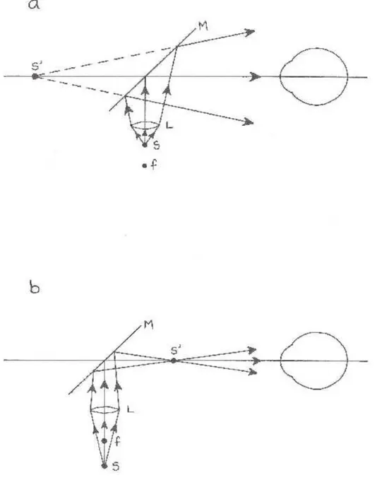
The Focusing System
The focusing system incorporated in all streak retinoscopes and most modern spot
retinoscopes consists of a sleeve or collar by which the light source is displaced with respect to the condensing lens, changing the vergence of the emitted light. In the plane mirror position the light source is situated inside the focal point of the condensing lens, creating a divergent beam of light (see Figure 1a). When swept across the patient’s pupil the beam moves in the same direction as the retinoscope.
When the sleeve or collar is changed the light source moves outside the focal point of the condensing lens, resulting in a convergent beam of light that moves opposite the direction of the retinoscope, mimicking the effect of a concave mirror (see Figure 1b).
(Benjamin 2006 p.689, Eskridge et al 1991 p.159, Grosvenor 2007 p.193). The practical use of the focusing system will be discussed below.
Figure 1. The plane and concave mirror positions. S is the light source, L the condensing lens, f the focal point
of the condensing lens, S´ the immediate source (the image of the light source) and M the plane mirror. Based on the original from Borish’s Clinical Refraction (2006 p.687).
The Reflex
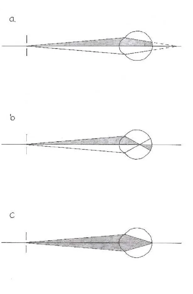
With the focusing system set in the plane mirror position and the light beam swept across the patient’s pupil the streak reflex in the pupil will move either in the same or opposite direction (“with” or “against” motion) or not at all (“neutrality”). A “with” motion indicates that the far point of the tested eye is either at infinity (in emmetropes), behind the retinoscope aperture (in low myopes) or behind the tested eye (in hyperopes). As the retinoscope beam is moved in one direction the image on the fundus of the tested eye will do the same, see Figure 2a. In order to create a known amount of myopia and place the far point of the eye at the retinoscope aperture positive lenses are added until neutrality. If the reflex shows an “against” motion the far point of the tested eye is located somewhere between the eye and the retinoscope aperture, meaning the eye is myopic. When the retinoscope beam is moved in one direction the
reflected light will move in the opposite direction, as shown in Figure 2b. Neutrality is
reached by adding negative lenses. If the reflex shows neither “with” nor “against” motion but instead fully illuminates the pupil it means that all the emitted light rays are reflected back to the retinoscope and neutrality is reached (see Figure 2c). Although this might sound simple in theory, a lot of the time neutrality is not an easily detected point but rather a range of
uncertainty between “with” and “against” motions. The retinoscopic endpoint will then be the dioptric power midway between just noticeable “with” and “against” motions. (Benjamin 2006 p.688 and 692, Eskridge et al 1991 p.156, Grosvenor 2007 p.192, Rabbetts 2007 p.347)
A dull, slow and wide reflex indicates a large refractive error while a bright, fast and slim reflex is close to neutrality. At neutrality the reflex actually moves infinitely fast, but as the ocular system is incapable of perceiving motion at this magnitude the reflex appears not to move at all (Benjamin 2006 p.696).
The Focusing System in Practice
If the retinoscope setting is changed to the concave mirror position the beam of light, as explained above, will move opposite the direction of the retinoscope. This means that the reflex in the patient’s pupil will move in the opposite direction compared to when the plane mirror position is used. A myopic eye will display “with” instead of “against” motion, and a hyperopic “against” instead of “with” motion. The possibility of changing the vergence of the light is useful in confirming the direction of motion of the reflex in case of uncertainty, as a “with” motion in one position will always be an “against” motion in the other. Also many practitioners find “with” motions easier to work with, and can always do so if using a focusing retinoscope. (Eskridge et al 1991 p.159, Grosvenor 2007 p.193)
Controlling Accommodation
To obtain an accurate result of refractive error, no matter the method used, it is necessary that accommodation is relaxed. This is most commonly achieved by fogging or cycloplegic drugs. Fogging causes the quality of the visual stimuli to deteriorate, thereby allowing
accommodation to relax. However, when pseudomyopia, latent hyperopia or other
accommodative abnormalities are present fogging works to a lesser extent and cycloplegia may be necessary in order to paralyse accommodation. (Benjamin 2006 p.704, Jorge et al 2005, Grosvenor 2007 p.195, Owens et al 1980, Rabbetts 2007 p.356)
Static Retinoscopy
While the tested eye is dazzled by the retinoscope light the non-tested eye fixates a distant target. Grosvenor (2007 p.689) advocates the use of a large optotype together with the
bichrome filter, which reduces the illumination on the screen and thus the bothersome reflexes on the trial lenses. The room illumination is lowered to obtain a high-contrast view of the reflex, and to allow the pupil to dilate and the reflex become larger and brighter (Benjamin 2006 p.689, Rabbetts 2007 p.356).
Before starting retinoscopy the non-tested eye is fogged in order to keep accommodation relaxed and stable. Fogging is done by adding trial lenses of appropriate power until the reflex shows a clear “against” motion in all meridians. The working distance should be 50 or 67 cm,
depending on the arm length of the examiner. A long working distance is preferred, since the longer the working distance the smaller the error of being slightly “off distance”. (Eskridge et
al 1991 p.166, Grosvenor 2007 p.194)
It is important that the examiner is seated as close as possible to the line of sight of the tested eye as too large an angle will cause oblique astigmatism and an incorrect result. Jackson et al (2005) showed that retinoscopy performed 5 degrees off the visual axis caused a significant myopic shift. But if the examiner is seated in a way that his or her head blocks the view of the tested eye and only allows the non-tested eye to focus the distant target the obliquity of observation is less than 3 degrees. However it is important not to block the view of the non-tested eye. If this happens accommodation is stimulated and an incorrect result is obtained. (Eskridge et al 1991p.156, Benjamin 2006 p.708)
Since the majority of people have at least a small amount of astigmatism the examiner must start retinoscopy by sweeping the retinoscope beam across the vertical, horizontal and oblique meridians of the patient’s pupil. While doing so the brightness, speed, width and direction of motion of the reflex are observed. If the reflex looks the same in all meridians no astigmatism is present and the reflex is neutralized with appropriate lens power. If astigmatism is present, the two primary meridians are identified and the retinoscope beam is aligned with the
meridian showing the fastest, brightest and slimmest reflex, meaning the most hyperopic/least myopic meridian. After this is neutralized the beam is aligned with the meridian perpendicular to it. The cylinder axis is aligned with the streak and minus cylinder power is added until neutrality. The dioptric working distance is subtracted from the sphere power to obtain the refractive state of the eye. (Elliott 2003 p.167, Grosvenor 2007 p.194)
Mohindra Near Retinoscopy
In order to avoid the use of cycloplegia in infants and young children and to overcome the difficulty of directing their attention to a distant target during static retinoscopy Mohindra (1977) came up with a method of performing retinoscopy at near. The technique uses the non-accommodative stimuli of the retinoscope light (Owens et al 1980) as fixation target at a 50 cm distance. Retinoscopy is performed in a totally dark room with the non-tested eye
working distance with a tonus adjustment of +0.75 D for dark focus, Owens et al 1980) is subtracted from the result. Although Mohindra obtained a good correlation between near and cycloplegic retinoscopy in infants and children, and between near retinoscopy and subjective refraction in adults, it should be noted that the tonus adjustment is not ideal for all individuals and subjective confirmation should be obtained whenever possible (Owens et al 1980).
However, a number of studies comparing near retinoscopy and cycloplegic retinoscopy in infants and children reveals a significant discrepancy between the two. Saunders and Westall (1992) compared near retinoscopy to cycloplegic refraction in 31 infants and 43 children and found near retinoscopy to be on average 0.39 D more myopic. In order to obtain a closer correlation between the two methods Saunders and Westall suggested the correction factor be changed from 1.25 D to 0.75 D for infants and 1.0 D for children. Twelker and Mutti (2001) compared near and cycloplegic retinoscopy in 29 infants, using the correction factor of 0.75 D proposed by Saunders and Westall. In spite of this the mean near retinoscopic finding was 0.97 D more myopic than cycloplegic retinoscopy.
Subjective Refraction
In subjective refraction the refractive error of the eye is determined on the basis of the
patient’s subjective response. The aim is to determine the spherical and cylindrical lenses that provides the best visual acuity with the patient’s accommodation relaxed, meaning the lenses needed to place the far point of the eye at infinity (Grosvenor 2007 p.183 and 209). Even though retinoscopic findings are highly correlated with subjective refraction a subjective check should always be made or attempted. The patient’s vision in every day life is a subjective response and may be influenced by other factors than optical ametropia. For example a previously uncorrected astigmatic might not accept the full cylindrical correction due to the visual distortions created. (Rabbetts 2007 p.362, Benjamin 2006 p.710)
Subjective refraction should be performed under conditions that resemble the patient’s normal viewing situation. For example too dim room illumination or occlusion of one eye will cause the pupils to dilate and can cause refractive changes due to spherical aberration. This is the reason why subjective refraction, whenever possible, should be carried out under binocular viewing conditions. Also binocular refraction has a variety of advantages over monocular
refraction. It allows greater relaxation of and control over accommodation, and thus gives a more accurate result in conditions such as latent hyperopia and pseudomyopia. The fusion present eliminates cyclophorias and prevents manifestation of latent nystagmus, both of which would otherwise influence the refractive result. Another advantage of binocular refraction is that balancing of accommodation is not required, and the procedure is thus slightly faster than monocular refraction. (Benjamin 2006 p.791, Elliott 2003 p.172, Eskridge et al 1991 p.190 and Rabbetts 2007 p.104 and 119)
Monocular Refraction
In monocular refraction the non-tested eye is occluded and accommodation controlled by fogging of the tested eye. The recommendations to how much fog should be used vary between authors. Grosvenor (2007 p.212) means that the visual acuity should be reduced to 0.5 while Benjamin (2006 p.798) suggests 0.2. In short an adequate amount of fog is used in order to relax accommodation. The fog is then slowly reduced in 0.25 D steps until further decrease in power does not improve visual acuity. The circle of least confusion is now on the retina, and it is time to test the eye for astigmatism.
Astigmatism
The most common technique to determine the astigmatic component is the Jackson Crossed Cylinder, JCC. The crossed cylinder is an astigmatic lens in which the principal meridians are equal in numbers but opposite in sign, the mean power thus being zero. The JCC is available in three different powers, ±0.25 ±0.37 and ±0.5 D, where the first is the most commonly used. The negative cylinder axis is usually marked with red dots and the positive with white. The JCC is mounted 45 degrees between the principal meridians, in the zero power position. (Grosvenor 2007 p.211, Rabbetts 2007 p.109)
Procedure, as described by Elliott (2003 p.175):
With the best spherical correction in place the JCC is placed before the patient’s eye. The zero power position is set at 180 degrees and the crossed cylinder flipped from one side to the other. The patient is to report which of the two images is the clearest, and the orientation of the minus cylinder axis is noted. The zero power position is then turned to 45 degrees and the
crossed cylinder flipped again. The orientation of the minus cylinder axis for the clearest image is again noted, and the zero power position is set between the two preferred
orientations. If both images are equally clear in the two positions above, no astigmatism is present and the procedure ends here.
A -0.25 D cylinder is added, its axis parallel with the zero power position. The crossed
cylinder is flipped and the zero power position adjusted toward the direction of the red dots of the clearest image until both images appear equally clear – the zero power position is now aligned with the astigmatic axis of the eye. The JCC is then rotated so the red or white dots are aligned with the axis. To determine the amount of cylinder needed the JCC is again flipped and more cylinder added if the image is clearest for the red dots, and cylinder power removed if it is clearest for the white dots. For every -0.5 D of added cylinder the sphere has to be adjusted by +0.25 D in order to keep the circle of least confusion on the retina. When the two images are equally clear the cylinder power is obtained. The cylinder axis should be rechecked and the spherical power refined.
Spherical Endpoint
The endpoint of subjective refraction should be the maximum amount of positive power providing the best visual acuity with accommodation relaxed. When this occurs the circle of least confusion is on the retina and the far point of the eye at infinity. (Grosvenor 2007 p.209) When unfogging a patient it is important to ensure that the letters actually become clearer and not just blacker. If the patient reports that the letters become blacker or smaller it means that accommodation is brought into play and more positive power should be added. (Benjamin 2006 p.831, Eskridge et al 1991 p.179, Rabbetts 2007 p.106)
Sometimes the spherical endpoint can be somewhat uncertain, and it might be necessary to use a different method to confirm it. One of the more common ones is the Red-Green or Bichrome test, which uses chromatic aberration to determine the spherical endpoint. As green light is refracted stronger than red the green image on the retina will be slightly in front of the red. The effect of chromatic aberration is enhanced when the pupils dilate and the difference between the images thereby more distinct, why it is advisable to keep the room illumination switched off during the test. If the patient reports that the symbols on the red half of the board are clearest it means that the red image is closest to the retina and more negative power should
be added. If the green image is the clearest positive power is added, and when both images are equally clear the endpoint is obtained. If equality can not be obtained a young person should be left on the green side, as a small amount of accommodation will move the circle of least confusion to the retina, and a presbyope should be left on the red side in order to preserve accommodation. Since patients with a colour defect will find the green background brighter and patients with yellowing lenses the red it is important to emphasize that it is the clarity of the black symbols in the image and not the background that should be judged. (Benjamin 2006 p.832, Elliott 2003 p.175, Eskridge et al 1991 p.179, Grosvenor 2007 p.209, Rabbetts 2007 p.106)
Balancing Accommodation
During monocular refraction, even when using the fogging technique properly, it is possible that accommodation is not completely relaxed during the endpoint determination. And as the eyes are refracted separately the amount of accommodation present may differ between the eyes, resulting in an imbalance when used together. Therefore, in patients with normal binocular vision, a monocular refraction must always be followed by a binocular balance which equalizes and relaxes accommodation. Binocular balance can be achieved using a variety of methods involving polarization, prism dissociation and fogging techniques. None of them will be explained here as they are of no value to this study. (Benjamin 2006 p.836, Eskridge et al 1991 p.185, Rabbetts 2007 p.117)
Binocular Refraction
The advantages of binocular over monocular refraction, as previously explained, are that accommodation is stable and more relaxed due to the fact that both eyes are open and fusion thus present. Although both eyes are fogged by about +1.0 D fusion is maintained through the peripheral vision. The non-tested eye remains fogged throughout the procedure, while the tested eye is refracted following the same principles as described for monocular refraction. (Benjamin 2006 p.848, Elliott 2003 p.185, Eskridge et al 1991 p.191, Rabbetts 2007 p.118). It is a well established fact that binocular refraction often yields a more hyperopic result than monocular refraction, shown by among others Norman (1953). In his study on 350 eyes
Norman showed that binocular refraction was 0.25 D more hyperopic than monocular refraction for 35% of the subjects, and 0.5 D more hyperopic for 12% of the subjects.
As binocular refraction allows greater relaxation of accommodation than monocular refraction a greater amount of hyperopia can often be corrected. Another reason why binocular
refraction often yields a more hyperopic result has to do with the depth of focus of the eye. During monocular refraction the pupils dilate due to the occlusion of one eye. This causes the depth of focus to decrease and results in a more myopic endpoint. During binocular refraction the pupils are of normal size and the depth of focus is larger, allowing more plus power to be accepted before blur becomes perceptible. (Eskridge et al 1991 p.193, Rabbetts 2007 p.117)
Previous Studies
Millodot and O’Leary (1978) carried out a study on 1078 eyes of all ages and concluded that there was a hyperopic shift of 0.3-0.4 D between static retinoscopy and subjective refraction in subjects below the age of 15. The hyperopic shift diminished with age and became negative beyond the age of 50.
The results found by Millodot and O’Leary are contradicted by the results shown by
Grosvenor et al (1985). In their study on 100 children aged 6-10 the mean difference between static retinoscopy and subjective refraction was an insignificant 0.01 D.
Jorge et al (2004) compared static retinoscopy with subjective refraction in 192 adults aged 18 to 34 and found an insignificant average difference of 0.02 D.
Considering the previous studies the effect of age should be of no importance to the sample in this study and the correlation between static retinoscopy and subjective refraction should be high.
Mohindra (1977) showed a high correlation between static retinoscopy and subjective refraction in adults. It seems that there has been no interest in comparing near and static retinoscopy in adults, since no reports on the matter have been found.
Grosvenor (2007 p.196) stated that retinoscopy is a monocular finding and should be compared to monocular rather than binocular subjective refraction.
Aim
The aim of this study is to investigate if static retinoscopy has a higher correlation with monocular than binocular subjective refraction, and also to see if there is any difference between these results and Mohindra near retinoscopy.
MATERIALS AND METHODS
Selection
32 subjects were recruited and 3 were excluded because of pseudomyopia or because a reliable endpoint could not be obtained during one or more of the measurements. The final sample size was 29 subjects, 7 males and 22 females. They were all volunteers collected among the population in Kalmar and fulfilled the following inclusion criteria:
Healthy eyes
Binocular vision (fusion)
• No tropia
• No anisometropia of more than 2.0 D
• No amblyopia of more than two lines difference between the eyes No pseudomyopia
No presbyopia No high astigmatism
To avoid media opacities and small pupils the age span was set to 18-40 years. The average age was 27. Contact lens wearers were told not wear their lenses one hour before the session.
Instruments Used
A Keeler Professional streak retinoscope
A manual Topcon Visiontester phoropter for static retinoscopy and subjective refraction A trial frame and spherical and cylindrical trial lenses for Mohindra retinoscopy
Methods
All the measurements were done on the right eye alone. They were performed in one session following the same order for all the subjects. The aim of the study and the tests included were explained to the patient and a consent form was signed. Patient health history was taken and preliminary tests (visual acuity, cover test, pupillary reflex, motility and near point of convergence) were carried out to ensure that the patient fulfilled the inclusion criteria. The eyes were checked for abnormalities using a slit-lamp biomicroscope.
Mohindra Retinoscopy
Mohindra retinoscopy was performed at a 50 cm working distance. The patient looked into the retinoscope light, ensuring retinoscopy was performed in the line of sight of the tested eye. The fellow eye was patched and the room completely darkened to avoid any stimuli of
accommodation. A trial frame and loose spherical and cylindrical lenses were used to reach neutrality. In case of uncertain neutrality the endpoint was decided to be midway between just noticeable with and against motion, down to 0.25 D. In case of only 0.25 D between the two positions the endpoint was decided to be the most positive/least negative position.
A correction factor of 1.25 D was subtracted from the neutralizing lens to obtain the refractive error of the eye.
Static Retinoscopy
Static retinoscopy was also performed at a 50 cm working distance, with the non-tested eye focusing the distant target of a large optotype together with the bichrome filter. The non-tested eye was fogged/unfogged until a clear against motion was seen. The room illumination was switched off to obtain a high contrast view of the reflex. The examiner was seated in the line of sight of the tested eye, making sure to block the tested but not the fellow eye. The patient was instructed to report if the view of the non-tested eye was obstructed, and to focus the distant target at all times. A manual phoropter with spherical and cylindrical lenses was used to reach neutrality, and the endpoint criterion was the same as for Mohindra retinoscopy. The dioptric working distance of 2.0 D was subtracted from the neutralizing lens to obtain the refractive error of the eye.
Monocular Subjective Refraction
Monocular refraction was performed using a manual phoropter, the non-tested eye occluded and the room illumination slightly dimmed. The lens power obtained during retinoscopy was used as starting point, leaving the patient fogged by +2.0 D. The patient was unfogged and any astigmatism corrected. The spherical endpoint was the maximum plus power providing the best visual acuity, using the tumbling-E test chart to minimize the risk of the patient remembering than actually seeing the optotypes.
Binocular Subjective Refraction
A manual phoropter was used and the room illumination slightly dimmed. The monocular refraction fogged by +1.0 D was used as starting point, and the non-tested eye was
fogged/unfogged until visual acuity reached 0.5. The Worth Four Dot test was used to ensure binocular vision. The tested eye was then unfogged and in case of astigmatism the cylinder power and axis were rechecked. The endpoint criterion was the same as for monocular refraction, and the tumbling-E optotypes were again used. The left eye was refracted
following the same procedure while the right eye was fogged to a visual acuity of 0.5. Finally both eyes were fogged by +0.5 D and simultaneously unfogged to achieve the maximum plus power for the best visual acuity.
Statistical Methods
The result from all the refraction methods was converted into spherical equivalent. The collected data was analysed in Excel using correlation analyses, two-sided paired t-test and regression analyses.
RESULTS
Static retinoscopy. The spherical refractive error of the sample ranged from -8.0 to +1.25 D
(mean -1.80 D) and the maximum amount of cylinder was -1.5 D. Mean spherical equivalent was -2.09 D and the standard deviation 2.75. A total of 19 subjects (65.5%) had myopia, 4 (13.8%) emmetropia and 6 (20.7%) hyperopia. All but one (96.6%) were astigmatic.
Mohindra retinoscopy. The spherical refractive error ranged from -8.0 to +1.5 D (mean
-1.74 D) and the maximum amount of cylinder was -1.0 D. Mean spherical equivalent was -2.01 D and the standard deviation 2.78. A total of 21 subjects (72.4%) had myopia, 1 (3.4%) emmetropia and 7 (24.1%) hyperopia. 26 subjects (89.7%) were astigmatic.
Monocular refraction. The spherical refractive error ranged from -8.0 to +1.0 D (mean
-1.81 D) and the maximum amount of astigmatism was -1.25 D. Mean spherical equivalent was -2.04 D and the standard deviation 2.78. A total of 19 subjects (65.5%) had myopia, 2 (6.9%) emmetropia and 8 (27.6%) hyperopia . 27 subjects (93.1%) were astigmatic.
Binocular refraction. The spherical refractive error ranged from -7.5 to +1.0 D (mean
-1.64 D) and the maximum amount of astigmatism was -1.25 D. Mean spherical equivalent was -1.87 D and the standard deviation 2.75. A total of 18 subjects (62.1%) had myopia, 2 (6.9%) emmetropia and 9 (31.0%) hyperopia. 27 subjects (93.1%) were astigmatic.
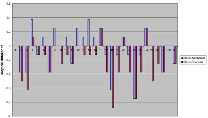
The monocular refraction was on average 0.17 D more myopic than the binocular refraction. For 16 subjects the binocular spherical endpoint was 0.25 D more hyperopic than the
monocular, representing 55.2% of the sample. For 2 subjects, or 6.9%, the binocular spherical endpoint was 0.5 D more hyperopic than the monocular. For 11 subjects (37.9%) there was no difference in spherical endpoint between monocular and binocular refraction.
Static retinoscopy was on average 0.22 D more myopic than binocular refraction, but only 0.06 D more myopic than monocular refraction. The mean difference between static and Mohindra retinoscopy was 0.08 D. Mohindra retinoscopy was on average 0.14 D more myopic than binocular refraction, and 0.03 D more hyperopic than monocular.
The summary of the results are shown in Table 1, the mean refractive findings in Figure 1 and the difference between retinoscopy and subjective refraction in Figure 2 and 3.
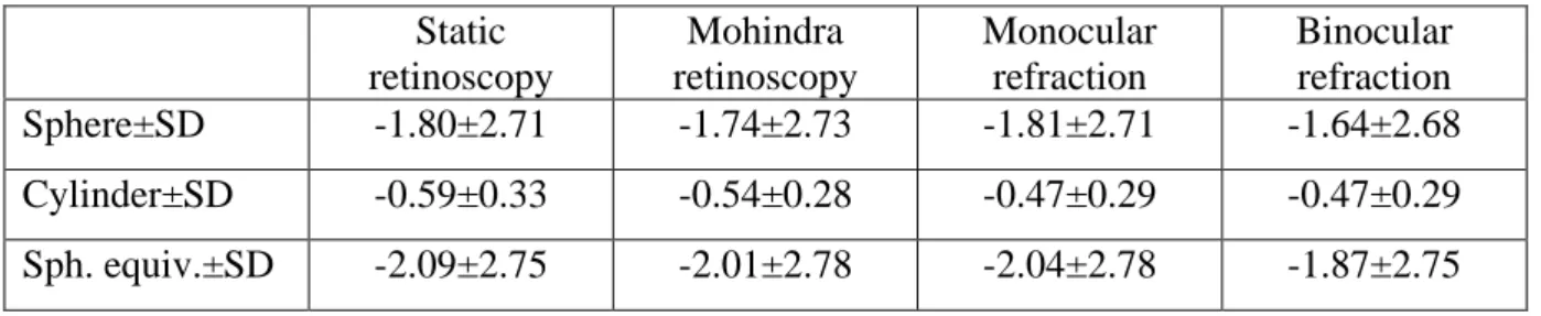
Table 1. Sphere, cylinder and spherical equivalent mean±SD in dioptres.
Static retinoscopy Mohindra retinoscopy Monocular refraction Binocular refraction Sphere±SD -1.80±2.71 -1.74±2.73 -1.81±2.71 -1.64±2.68 Cylinder±SD -0.59±0.33 -0.54±0.28 -0.47±0.29 -0.47±0.29 Sph. equiv.±SD -2.09±2.75 -2.01±2.78 -2.04±2.78 -1.87±2.75
Figure 1. Mean refractive findings±SD in dioptres.
-1.87 -2.04 -2.09 -2.01 -5 -4 -3 -2 -1 0 1
Figure 2. Dioptric difference between static retinoscopy and subjective refraction for each subject. -1 -0,8 -0,6 -0,4 -0,2 0 0,2 0,4 0,6 1 2 3 4 5 6 7 8 9 10 11 12 13 14 15 16 17 18 19 20 21 22 23 24 25 26 27 28 29 Number of subjects D io p tr ic d if fe re n c e Static-monocular Static-binocular
Figure 3. Dioptric difference between Mohindra retinoscopy and subjective refraction for each subject.
-1,5 -1 -0,5 0 0,5 1 1 2 3 4 5 6 7 8 9 10 11 12 13 14 15 16 17 18 19 20 21 22 23 24 25 26 27 28 29 Number of subjects D io p tr ic d if fe re n c e Mohindra-monocular Mohindra-binocular
The correlation between all the measurements was high, ranging from 98.70 to 99.86% (see Table 2). However, the two-sided paired t-test revealed a significant statistical difference between monocular and binocular refraction (p<0.001), and also between static retinoscopy and binocular refraction (p<0.001).
Furthermore the t-test showed that there was no significant difference between static retinoscopy and monocular refraction (p=0.295) or Mohindra retinoscopy and monocular refraction (p=0.719). There was no significant difference between static and Mohindra retinoscopy (p=0.292) or Mohindra retinoscopy and binocular refraction (p=0.102). The correlation and p-values are shown in table 2.
Table 2. Correlation and P-values for the different measurements.
Monocular/ binocular Static/ Mohindra Static/ monocular Static/ binocular Mohindra/ monocular Mohindra/ binocular Correlation 99.86% 98.79% 99.48% 99.50% 98.70% 98.74% P-value <0.001 0.292 0.295 <0.001 0.719 0.102
To further establish the relationship between retinoscopy and monocular and binocular subjective refraction regression analyses was done. The slope for monocular refraction was higher than for binocular for both static and Mohindra retinoscopy. The difference between the slopes was small but statistically significant, 0.02 for static and 0.01 for Mohindra retinoscopy. The difference was the same for all refractive errors. The coefficient of
determination (R2) was high for all the measurements, ranging from 0.97 to 0.99. Figure 4 and 5 shows the results. The top equation in each figure shows the results for monocular
Figure 4. Regression analyses for static retinoscopy and subjective refraction. y = 1,0063x + 0,0692 R2 = 0,9897 y = 0,9946x + 0,2128 R2 = 0,9901 -10 -8 -6 -4 -2 0 2 -10 -8 -6 -4 -2 0 2 Static retinoscopy S u b je c ti v e r e fr a c ti o n Mono Bino Mono Bino
Figure 5. Regression analyses for Mohindra retinoscopy and subjective refraction.
y = 0,9864x - 0,0575 R2 = 0,9743 y = 0,9751x + 0,088 R2 = 0,975 -10 -8 -6 -4 -2 0 2 -10 -8 -6 -4 -2 0 2 Mohindra retinoscopy S u b je c ti v e r e fr a c ti o n Mono Bino Mono Bino
DISCUSSION
Static retinoscopy vs subjective refraction. The results in this study show that there is no
significant difference between static retinosopy and monocular subjective refraction, but that there is a significant difference between static retinoscopy and binocular subjective refraction. This supports the hypothesis of the study that static retinoscopy has a higher correlation with monocular than binocular subjective refraction. The reason for this could be that they are both monocular methods. Also retinoscopy is performed in reduced room illumination, which in many cases affects the eyes in the same way as monocular viewing. The relaxation of and control over accommodation is not as good in monocular as binocular viewing, which results in a more myopic endpoint. Both monocular viewing and reduced room illumination results in more instrument myopia than binocular viewing (Wesner and Miller 1986). Reduced room illumination or occlusion of one eye causes the pupils to dilate and spherical and chromatic aberrations to increase (Elliott p.172, Rosenfield et al 1993), further establishing the
relationship between static retinoscopy and monocular refraction. The overall high correlation between retinoscopy and subjective refraction is a well established fact, shown by among others Grosvenor et al (1985) and Jorge et al (2005).
Mohindra retinoscopy vs subjective refraction. The results in this study show that there is no
significant difference between near retinoscopy and subjective refraction, which supports the results found by Mohindra in 1977. Contrary to the results for static retinoscopy there is no significant difference between Mohindra retinoscopy and binocular subjective refraction. This might be explained by the mean shift in refraction between Mohindra retinoscopy and
subjective refraction being less than between static retinoscopy and subjective refraction.
Static vs Mohindra retinoscopy. When comparing near and static retinoscopy one must
consider the differences between the methods. Since they are both monocular methods, carried out in dark surroundings and using a non-accommodative stimuli for fixation, the major difference is the correction factor used in Mohindra retinoscopy. Since there was no difference between static and Mohindra retinoscopy in this study it shows that the amount of tonic accommodation was indeed 0.75 D (Owens et al 1980) for all the subjects.
Monocular vs binocular subjective refraction. The discrepancy between monocular and
binocular refraction found in this study is a well established fact in literature, shown by among others Norman (1953).
Clinical factors that could possibly have influenced the outcome of the study involve the patient not keeping steady focus at the target during retinoscopy, retinoscopy off-the-axis and the patient remembering rather than actually seeing the optotypes during subjective refraction. Any time the patient looses fixation of the distant target and focuses on the examiner instead accommodation will come into play and the refractive error obtained will be incorrect. But as accommodation is seen as a change in size of the pupil, and as the patient was constantly reminded to fixate the distant target fluctuating accommodation should not have any effect on the outcome of this study. As the distance from the visual axis has to be 5 degrees or more (Jackson et al 2004) in order to cause a significant myopic shift it is highly unlikely that the position of the examiner had any effect in this study. If the patient remembers rather than actually sees the optotypes the refractive endpoint might not be correct. As the projector used in this study was incapable of randomizing letters the tumbling-E test chart was used to reduce the learning effect. This seemed to work accordingly as none of the patients read the lines as if they had seen them before.
It should also be noted that this study was performed unmasked and by one examiner only, which might have influenced the outcome. If the study was to be repeated it is suggested that it be blinded, either by an assistant changing the lens power or one examiner performing static retinoscopy and monocular refraction and the other Mohindra retinoscopy and binocular refraction.
Due to the fact that 7 subjects (24% of the sample) were previously uncorrected hyperopes and that it is easier to loose control of accommodation during monocular refraction of
hyperopes this might have had an effect on the outcome. Since this study did not include any high hyperopes, and as the range of myopes was somewhat limited any future study should involve a larger sample with a wider spread of refractive errors.
CONCLUSION
The results of this study have shown that static retinoscopy has a higher correlation with monocular than binocular subjective refraction. It has also shown that static and Mohindra retinoscopy are similar, and that Mohindra retinoscopy is highly correlated with subjective refraction.
ACKNOWLEDGEMENTS
I would like to thank my supervisor Baskar Theagarayan for all his time, support and invaluable help.
I would also like to thank all the subjects participating in the study. And to Kitten, my rock in a stormy sea, thank you for keeping me afloat!
REFERENCES
Grosvenor T, Perrigin DM and Perrigin J. 1985. Three-Way Comparison of Retinoscopy, Subjective and Dioptron Nova Refractive Findings. Am J Optom Physiol Opt. 62(1):63-65.
Jackson DW, Paysse EA, Wilhelmus KR, Hussein MAW, Rosby G and Coats DK. 2004. The Effect of Off-the-visual-axis Retinoscopy on Objective Refractive Measurement. Am J
Ophthalmol. 137(6):1101-1104.
Jorge J, Quieros A, Almeida JB and Parafita MA. 2004. Retinoscopy/Autorefraction: Which is the Best Starting Point for a Noncycloplegic Refraction? Optom Vis Sci 83(1):64-68.
Jorge J, Quieros A, González-Méijome J, Fernandes P, Almeida JB and Parafita MA. 2005. The Influence of Cycloplegia in Objective Refraction. Ophthal Physiol Opt. 25(4):340-345.
Millodot M and O’Leary D. 1978. The Discrepancy between Retinoscopic and Subjective Measurements: Effect of Age. Am J Optom Physiol Opt. 55(5):309-316.
Mohindra I. 1977. A Non-Cycloplegic Refraction Technique for Infants and Young Children.
J Am Optom Assoc. 48(4):518-523.
Norman SL. 1953. Plus Acceptance in Binocular Refraction. Optom Weekly 44:45-46.
Owens AD, Mohindra I and Held R. 1980. The Effectiveness of a Retinoscope Beam as an Accommodative Stimulus. Invest Ophthalmol Vis Sci 19(8):942-949.
Rosenfield M, Ciuffreda KJ, Hung GK and Gilmartin B. 1993. Tonic Accommodation: a Review 1. Basic Aspects. Ophthal Physiol Opt. 13:266-284.
Saunders KJ and Westall C. 1992. Comparison between Near Retinoscopy and Cycloplegic Retinoscopy in the Refraction of Infants and Children. Optom Vis Sci. 69(8):615-622.
Twelker JD and Mutti DO. 2001. Retinoscopy in Infants Using a Near Noncycloplegic Technique, Cycloplegia with Tropicamide 1%, and Cycloplegia with Cyclopentolate 1%.
Optom Vis Sci. 78(4):215-222.
Wesner MF and Miller RJ. 1986. Instrument Myopia Conceptions, Misconceptions, and Influencing Factors. Documenta Ophthalmologica 62, 281-308.
Bibliography
Benjamin WJ. 2006. Borish’s Clinical Refraction, 2nd ed. Butterworth-Heinemann Elsevier. Elliott DB. 2003. Clinical Procedures in Primary Eye Care, 2nd ed. Butterworth-Heinemann Elsevier.
Eskridge JB, Amos FJ, Bartlett JD. 1991. Clinical Procedures in Optometry. Lippincott Williams & Wilkins.
Grosvenor T. 2007. Primary Care Optometry, 5th ed. Butterworth-Heinemann Elsevier. Rabbetts RB. 2007. Bennett and Rabbetts’ Clinical Visual Optics, 4th ed. Butterworth-Heinemann Elsevier.
Appendix 1
Informerat samtycke
Tack för att du ställer upp som försöksperson i mitt examensarbete!
Arbetet går ut på att jämföra ögats objektiva och subjektiva synfel med hjälp av fyra olika mätmetoder.
Till att börja med får du svara på några frågor gällande din allmänna hälsa och din ögonhälsa, då endast friska ögon får delta i studien. Därefter gör jag några korta tester för att försäkra mig om att du uppfyller övriga urvalskriterier. Om allting ser bra ut går jag vidare till de faktiska mätningarna.
Ögats objektiva synfel mäts med ett retinoskop, ett instrument med vilket jag riktar en ljusspalt mot ditt öga och utifrån reflexen bedömmer ögats synfel. Detta är helt ofarligt, men du kan bli lite bländad för en kort stund efteråt.
Ögats subjektiva synfel mäts genom en vanlig synundersöknig, där jag sätter glas med olika styrkor framför ditt öga och du får avgöra vilket som är bäst.
Undersökningen tar ca en timme, och du får självklart säga till om du behöver ta en paus och sträcka på benen!
De enda personuppgifter som samlas in är namn, kön och födelseår, och det är endast kön, födelseår och resultatet av mätningarna som redovisas i examensarbetet.
Ditt deltagande i studien är frivilligt och du kan när som helst utan närmare förklaring avbryta undersökningen.
Jag har läst informationen och godkänner att mina resultat används i studien:
_______________________ Ort och datum
_______________________ _______________________
Namn Underskrift
Optikerstudent: Handledare: Li Stenberg Baskar Theagarayan xxxx-xxxxxx
Appendix 2
Anamnes
Födelseår_______ Man Kvinna Använder du glasögon eller kontaktlinser? Ja Nej Upplever du någon gång dubbelseende? Ja Nej
Om Ja, hur ofta och vid vilket tillfälle? ________________________________ ________________________________ Har du några ögonrelaterade besvär? Ja Nej
Om Ja, vad, hur ofta och vid vilket tillfälle? ________________________________ ________________________________ Blev du lappbehandlad som barn? Ja Nej
Har du någon ögonsjukdom? Ja Nej
Om Ja, vad? ________________________________ Har du varit utsatt för någon ögonskada? Ja Nej
Om Ja, vad och för hur länge sedan? ________________________________ ________________________________ Har du några andra (ej ögonrelaterade) sjukdomar? Ja Nej
Om Ja, vad? ________________________________ Tar du några mediciner (gäller även receptfria)? Ja Nej
Om Ja, vad/vad tar du den mot? ________________________________ Hur ofta? ________________________________ Har du tagit någon medicin idag? Ja Nej
Appendix 3
Mätningar
Fri visus H_______ V_______ B_______ Covertest avstånd _____________________________________________________ Covertest nära _____________________________________________________ Pupillreflex direkt_________ indirekt__________ swinging_________ PERLA__________ Motilitet ________________________________________________________________ KNP _______ cm PD H_______ mm V_______ mm B_______ mm Spaltlampa H_____________________ V___________________