Drug Eluting Hydrogels
Design, Synthesis and Evaluation
Lage Ahrenstedt
Doctoral Thesis
Department of Surgery
University of Cape Town
Cape Town
Aug 2012
The results in this thesis were produced in collaboration between the University of Cape Town (UCT; South Africa) and the Royal Institute of Technology (KTH; Stockholm, Sweden).
Supervisor: Dr. Deon Bezuidenhout (UCT) Co-supervisor: Prof. Anders Hult (KTH)
© N. Lage Ahrenstedt
Department of Surgery University of Cape Town Chris Barnard Building ZA-7925 OBSERVATORY South Africa
PROLOGUE
n 1897 Josef Conrad wrote that an author must be like an artist in capturing the movement of a dart into stone or the magic intonation into a piece of music. In the same manner a scientist must use artistic abilities to grasp the complexity of his work and to interpret its content. But in contrast to the artist who works with ideas the scientist follows the path of facts.
Within this context the author, artist and scientist behind this work will take the reader on a journey through the development of drug eluting hydrogels and put those in the context of ameliorating the healing of cardiovascular diseases.
ABSTRACT
ydrogels have successfully proved themselves useful for drug delivery applications and several delivery routes have been developed over the years. The particular interest in this work was to design, synthesise and evaluate in situ forming drug eluting hydrogels, which have the potential to ameliorate the healing of cardiovascular diseases.
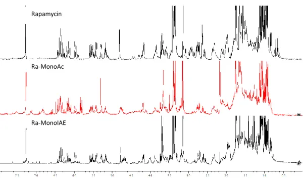
With this aim the anti-inflammatory and immunosuppressant drugs rapamycin (Ra) and dexamethasone (Dex) were made water soluble by conjugation with polyethylene glycol (PEG). Ra was attached pendant from the terminal of PEGs while Dex was incorporated into dendritic structures grown from PEGs. These conjugates were further crosslinked into hydrogels by either conjugate or thiol-ene addition. The gel degradation was tuned to take between 5 and 27 days by using gel building block combinations that induced either 2 or 4 hydrolytically labile bonds per crosslink or by varying the number of crosslinking sites of the building blocks. The use of thiol-ene addition prolonged the degradation time nearly seven folded compared to conjugate addition as a more stable crosslink was formed.
Two different formulations for gelling via conjugate addition were used (acrylate-thiol or vinyl sulphone-thiol) to deliver Ra, which was carried by either a 4- or 2-armed PEG. The elution kinetic for the respective gel formulation was of zero order during 15 and 19 days of gel degradation. In addition, Ra was PEGylated via esters, with a distance of either one or two carbons to a nearby thio-ether functionality. The difference in ester conjugation resulted in a slight but significant change in drug-PEG conjugate stability, which was mirrored by the increased time to reach the half amount of total drug elution; from 9.3 to 10.2 days and from 5.1 to 9.7 days for the two gel formulations, respectively. Dexamethasone was incorporated via an ester into dendrons of first and second generation pending from 2- and 4-armed PEGs at loadings of 2, 4 or 6 Dex molecules per carrier molecule. The resulting elution kinetic was of zero order during degradation periods of 5-27 days. Released Dex still possessed biological activity as determined by an in vitro cell assay.
The novelties in this thesis are: (A) slow release of rapamycin obtained by covalent incorporation into hydrogels, (B) the use of unique PEG-based dendrimers to incorporate dexamethasone into a hydrogel and (C) zero order sustained release of dexamethasone at physiological pH.
SAMMANFATTNING
ydrogeler har framgångsrikt visat sig användbara för att leverera läkemedel och ett flertal metoder har utvecklats de senaste 20 åren. Fokuset i den här avhandlingen ligger på att designa, framställa och utvärdera läkemedelsutsöndrande hydrogeler som spontanhärdar in situ, vilka har potential att förbättra läkningen efter kardiovaskulär sjukdom.
Med det syftet gjordes de anti-inflammatoriska och immunsänkande läkemedlen rapamycin (Ra) och dexametason (Dex) vattenlösliga genom att konjugeras med polyetylenglygol (PEG). Ra fästes kovalent längst ut på PEGar medans Dex inkluderades i dendritiska strukturer vilka byggdes från ändpunkten av PEGar. De här konjugaten tvärbands till hydrogeler via antingen konjugerad addition eller radikal polymerisation. Nedbrytningen av gelerna trimmades till att ta mellan 5 och 27 dagar genom att använda kombinationer av gelbyggstenar som bildar antingen 2 eller 4 hydrolyserbara estrar per tvärbindning eller genom att variera antalet tvärbindningspunkter hos byggstenarna. Användandet av radikal polymerisation i sig ledde till att nedbrytningen av geler tog nära sju gånger längre tid jämfört med geler gjorda via konjugerad addition eftersom stabilare tvärbindningar då formas.
Två olika kombinationer för härdning via konjugerad addition (akryl-tiol eller vinylsulfon-tiol) användes för att leverera Ra som bars av antingen en 4- eller 2-armad PEG. Utsöndringskinetiken av Ra för de två kombinationerna var av nollte ordningen under de 15 och 19 dagar som gelerna degraderade. Dessutom, Ra PEGylerades via estrar med ett avstånd på antingen ett eller två kol till en närliggande tioeter. Skillnaden i avstånd ledde till en liten men signifikant skillnad i stabiliteten hos Ra-PEG konjugaten, vilket speglades i den förlängda tiden att nå halva mängden av den totala läkemedelsutsöndringen; från 9.3 till 10.2 dagar och från 5.1 till 9.7 dagar för de två respektive gelkombinationerna. Dex kopplades in via en esterbindning till dendroner av första och andra generationen byggda från PEGar med 2 eller 4 armar, vilket resulterade i att 2, 4 eller 6 Dex levererades per bärarmolekyl. Dex eluerade med nollte ordningens kinetik under degraderingsperioder på mellan 5 och 27 dagar. Vidbehålllen biologisk aktivitet av eluerad Dex bekräftades genom cellexperiment in vitro.
Nyheterna i den här avhandlingen består av: (A) kontrollerad utsöndring av rapamycin uppnådd genom kovalent inbindning till hydrogeler, (B) användandet av unika PEGbaserade dendrimerer för kovalent inbindning av dexametason till hydrogeler och (C) nollte ordningens utsöndring av dexametason vid fysiologiskt pH.
LIST of PUBLICATIONS
The work in this thesis is presented or will be presented in the following publications.
Covalent Incorporation and Controlled Release of Active Dexamethasone from Injectable Polyethylene Glycol Hydrogels
Bezuidenhout D, Oosthuysen A, Davies N, Ahrenstedt L, Dobner S and Zilla P 2012, Submitted to J Biomed Mater Res A
Tailored Zero Order Rapamycin Release from Degradable PEG Hydrogels
Ahrenstedt L and Bezuidenhout D
2012, Manuscript in preparation
Hydrogels Made of PEG Didendrons with Pending Dexamethasone Result in Zero Order Sustained Release
Ahrenstedt L, Hed Y, Hult A, Zilla P, Malkoch M, Bezuidenhout D
2012, Manuscript in preparation
Dendritic Hydrogels for Controlled Drug Delivery
Ahrenstedt L, Hed Y, Malkoch M, Hult A, Zilla P, Bezuidenhout D
11th International Conference on Frontiers of Polymers and Advanced Materials 2011, Poster Presentation
ABBREVIATIONS
The abbreviations are listed in alphabetical order, regarding the left hand side column.
3D Three-dimensional
Å Ångström
Ac Acrylate
AcCl Acryloyl chloride Acet Acetonide protection
Act Acetylene At Atorvastatin CVDs Cardiovascular deceases d Doublet DCC N,N'-dicyclohexylcarbodiimide DCM Dichloromethane dd Doublet of doublets Dex Dexamethasone DMAP 4-Dimethylaminopyridine DMF Dimethylformamide
DMPA 2,2-dimethoxy-2-phenyl acetophenone
Dox Doxorubicin
eq Equivalent
ESI-MS Electrospray ionisation mass spectroscopy EtOAc Ethyl acetate
EtOH Ethanol
FI Fold induction
g Gram
HDC Huisgen 1,3-dipolar cycloaddition
Hep n-Heptane
Hex n-Hexane
HPLC High pressure liquid chromatography I Photo initiator
IAE Iodoacetic ester kDa kilo Dalton
KTH Royal Institute of Technology
MALDI Matrix-assisted laser desorption ionisation time-of-flight mass spectroscopy MeCN Acetonitrile MeOH Methanol mg Milligram MHz Megahertz MI Myocardial infarct ml Millilitre mm Millimetre mmol Millimole MMP Matrix metalloproteases
mTOR Mammalian target of rapamycin
nm Nanometre
NMR Nuclear magnetic resonance
NVP N-vinyl-2-pyrrolidone
PBS Phosphate buffered saline PEG Polyethylene glycol PGA poly(glycolic acid) pH -log[H+]
pKa -log[Ka], Ka =acid dissociation constant PLA poly(lactic acid)
PLGA poly(lactic-co-glycolic acid) ppm Parts per million
pTSA Para toluene sulphonic acid
Ra Rapamycin
s Singlet
TEA Triethylamine
THF Tetrahydrofuran
TLC Thin layer chromatography
Trizma 2-Amino-2-hydroxymethyl-propane-1,3-diol
UK United Kingdom
UV Ultraviolet
TABLE of CONTENTS
1 INTRODUCTION ...1 1.1 Hydrogels ...2 1.1.1 Gelling Methods ...6 1.1.1.1 Physical Gels ...6 1.1.1.2 Chemical Gels...7 1.1.1.2.1 Photo-Polymerisation ...7 1.1.1.2.2 Thiol-ene Addition ...8 1.1.1.2.3 Conjugate Addition ...91.1.2 Crosslink Density and Swelling ... 10
1.1.3 Degradation ... 11
1.1.3.1 Hydrolytic Degradation ... 12
1.2 Approaches towards Controlled Drug Delivery ... 14
1.2.1 Encapsulation ... 14
1.2.2 Covalent Incorporation ... 15
1.2.2.1 Hydrolytically Labile Linkers ... 16
1.2.2.2 Enzymatically Labile Linkers ... 16
1.2.2.3 Redox Labile Linkers ... 17
1.3 Drug Release ... 17
1.3.1 Mechanisms and Kinetics ... 17
1.3.2 Controlled by Diffusion, Swelling or Degradation ... 18
1.3.3 Controlled by Hydrolysis ... 21
1.4 Cardiovascular Diseases ... 23
1.4.1 Rapamycin ... 25
1.4.1.1 The Effect of Rapamycin on Cardiovascular Diseases ... 25
1.4.1.2 Controlled Release of Rapamycin ... 26
1.4.2 Dexamethasone ... 28
1.4.2.1 Controlled Release of Dexamethasone ... 29
1.5 Hypothesis and Aims ... 31
1.6 Strategy ... 33
2 MATERIALS and METHODS ... 37
2.1 Nomenclature ... 37
2.2 Syntheses of Gel Building Blocks ... 37
2.2.1 Preparation of the Multi-arms ... 37
2.2.2 Preparation of the Thiol Crosslinkers... 39
2.2.3 Mono-functional Approach ... 40
2.2.3.1 Preparations of Rapamycin Derivatives ... 40
2.2.3.2 Preparations of Ra-PEG conjugates ... 42
2.2.4 Dendritic Approach ... 44
2.2.4.1 Preparations of Acetylene monomer anhydride ... 44
2.2.4.2 Preparation of Dex-azide conjugate... 47
2.2.4.3 Preparations of First Generation Linear PEG-Dex Conjugate ... 48
2.2.4.4 Preparations of First Generation 4-armed PEG-Dex Conjugate ... 51
2.2.4.5 Preparations of Second Generations Linear PEG-Dex Conjugate ... 54
2.3 Atorvastatin... 56
2.4 General Procedures ... 57
2.4.1 Dendritic Synthesis ... 57
2.4.2 DOWEX Purification ... 57
2.4.4 mHPLC Analysis... 57
2.4.5 MS Analysis... 58
2.4.6 MALDI Analysis ... 58
2.4.7 Phosphate Buffered Saline ... 58
2.4.8 Gel Formation ... 59
2.4.8.1 Photo-Polymerisation... 59
2.4.8.2 Mono-functional Approach, RaMonoX Method 1 ... 60
2.4.8.3 Mono-functional Approach, RaMonoX Method 2 ... 60
2.4.8.4 Mono-functional Approach, RaDiIAE ... 60
2.4.8.5 Dendritic Approach, Conjugate Addition ... 60
2.4.8.6 Dendritic Approach, Thiol-ene Addition... 61
2.4.9 Drug Elution ... 61
2.4.10 Determination of pH dependence of gel formation ... 62
2.4.11 Curve Fitting ... 62
2.4.12 Swelling ... 64
2.4.13 Volume Determination ... 64
2.4.13.1 Calculation of Crosslink Density ... 65
2.4.14 Reporter Assays ... 66
2.4.14.1 Rapamycin ... 66
2.4.14.2 Dexamethasone ... 67
3 RESULTS and DISCUSSION ... 69
3.1 Synthesis of Gel Building Blocks ... 69
3.1.1 Multi-arms ... 69
3.1.2 Crosslinkers ... 70
3.1.3 Mono-functional Approach ... 72
3.1.4 Dendritic Approach ... 78
3.2 Gel Formation Chemistries ... 85
3.2.1 Conjugate Addition ... 85
3.2.2 Thiol-ene Addition ... 86
3.2.2.1 Conjugate versus Thiol-ene Addition ... 86
3.3 Drug Release and Hydrogel Characterisation ... 88
3.3.1 Mono-functional Approaches... 88
3.3.1.1 Ra-Ac/IAE, 4-armed carrier ... 88
3.3.1.2 Ra-Ac/IAE, 2-armed carrier ... 92
3.3.1.3 RaDiIAE, no carrier ... 94
3.3.2 Dendritic Approaches ... 97
3.3.2.1 First Generation, 2-armed backbone ... 97
3.3.2.1.1 Gelling Through Conjugate Addition ... 97
3.3.2.1.2 Gelling Through Thiol-ene Addition ... 99
3.3.2.2 First Generation, 4-armed backbone ... 102
3.3.2.3 Second Generation, 2-armed Backbone ... 104
3.3.3 Summary ... 106 3.4 Reporter Assays ... 107 3.4.1 Rapamycin ... 107 3.4.2 Dexamethasone ... 109 4 CONCLUSIONS ... 113 4.1 Complementary Work ... 114 5 ACKNOWLEDGEMENTS... 115 6 APPENDIX I; ATORVASTATIN ... 117 REFERENCES ... 119
-1-
1 INTRODUCTION
ver the last century the concept of hydrogels has been developed into an interesting branch of polymer science with enormous potential. Particularly fruitful is the use of hydrogels in medical research where gels are commonly used in a wide spectrum of applications. There are multiple parameters to consider in the design of a hydrogel, which must all individually be adapted to obtain desired gel properties. This broad register of choices is the origin of the success for hydrogel applications, since it allows for the preparation of gels with very divert but specific properties. Hence, with proper design and methodology it is possible to create gels that fulfil the strict demands of a biomaterial (Hoffman 2002) which can be used as wound covers, implant coatings, slow-release devices or materials that mimic in vivo functionality (Hubbell 1998; Lin et al. 2009). Commonly, drugs or growth factors are delivered with the hydrogels to obtain these properties in a biological system.
Many drugs however, are hydrophobic and therefore insoluble in the human aqueous media, which is a problem for drug delivery and efficiency. The non-solubility is a particular problem in cases where the drug is delivered from a solution, as only aqueous solutions should be introduced into living tissue. Therefore various approaches towards in situ delivery of hydrophobic drugs have been developed and a common method is to conjugate the drug to a water soluble polymer, such as PEG (polyethylene glycol). This conjugation does not only bring the drug into solution but also increases its circulation time and protects it, primarily in case of peptides and proteins, against proteolytic activity (Woghiren et al. 1993; Gombotz et al. 1995; Elbert et al. 2001; Roberts et al. 2002; Li et al. 2003; Frokjaer et al. 2005; Veronese et al. 2005).
Rapamycin (McMurray et al.), a macrolide compound originally developed as an antifungal agent, and Dexamethasone (Dex), a synthetic member of the glucocorticoid class of steroid drugs, both have immunosuppressant and anti-inflammatory properties. These drugs have high potential for ameliorating cardiovascular healing, which is of interest to our group, and have previously been used to prevent restenosis (Morice et al. 2002; Koenig et al. 2009), quench pathological cardiac hypertrophy (McMullen et al. 2004) and to reduce inflammation from implanted devices (Baeyens et
al. 1998).
One approach to ameliorate cardiovascular recovery is by means of drugs and a crucial step in such approach is to develop suitable drug delivery devices. This thesis describes the development of methods to obtain controlled and sustained drug delivery of Ra and Dex via hydrogels and is part of a larger overall project (which describes healing post myocardial infarction using animal models and
O
-2-
1
1
1 b a f fother gel formulations than described here). The methods are based on drug PEGylation and the work shows the importance of proper design of the hydrogel formulation and drug incorporation methods to achieve desired properties.
1.1 Hydrogels
A hydrogel is formed by creating a sufficient number of crosslinks between polymeric building blocks so that a continuous three-dimensional (3D) network is created. The most representative feature of hydrogels is the ability to absorb large amounts of water.
In theory a multi-arm with only two functional groups (fa =2) could be crosslinked by a 2-armed crosslinker (fb =2), but then the hydrogel would not consist of an inter-crosslinked polymer network but a random entanglement of copolymers, which demands a conversion of 100% to form according to Equation 1. Practically however, a functionality of three is the minimum requirement for the multi-arm to form an infinitely connected network when crosslinked by a 2-armed crosslinker and according to Equation 1 a conversion of 71% is needed for this combination to reach the gel point.
Critical conversion (1)
The final degree of conversion is individual for each hydrogel system and increased by high polymer concentration and long gelling time (DuBose et al. 2005).
Figure 1 illustrates the resulting continuous network after crosslinking an 8-armed star shaped
multi-arm with a 2-multi-armed linear crosslinker. A stoichiometric ratio (1:1) for the functional groups of the two pregel components is fundamental for the gel to form. Only a few percent deviations may prevent the gelling, or at least drastically slow down the process. In the case where too many of the arms are present mainly dimers are formed while an excess of crosslinker saturates the multi-arm and hence, no continuous network is formed. Nonetheless, even for a stoichiometric relation the crosslink efficiency is not 100% since the progressing network formation itself makes the forming gel more rigid, which lowers the possibility for remaining functional groups to come in close enough proximity to react, hence there will be unreacted groups in a gel.
-3-
+ 4n n
Figure 1: A continuous polymer network formed by crosslinking an 8-armed star shaped multi-arm with a 2-armed linear crosslinker.
One approach to categorise hydrogels is by their building blocks and a major distinction is usually made between natural and synthetic polymers, some of which are listed in Table 1 (Gombotz et al. 1995; Hoffman 2002). Many of the naturally occurring polymers contain charged groups, signalling peptides or other functionalities, which make them useful for many applications but also unpredictable since different polymer sources may vary in the substitution pattern. Synthetic polymers on the other hand have greater potential to be tailored into desired properties with good repeatability.
Table 1: Common naturally occurring and synthetic building blocks used for hydrogel assembling.
Natural Polymers Synthetic Polymers
Alginate Chitosan Collagen Dextran Fibrin Gelatine Heparin Hyaluronic acid
poly(acrylic acid), PAA poly(ethylene glycol), PEG poly(glycolic acid), PGA poly(lactic acid), PLA
poly(lactic-co-glycolic acid), PLGA poly(N-isopropyl acrylamide), PNIPAAm poly(N-vinyl-2-pyrrolidone), PVP poly(vinyl alcohol), PVA
When hydrogels are required for in vivo use, biocompatibility is crucial. A biocompatible material should be inert to proteins and other substances in biological fluids as well as to receptors and other parts of cell membranes (Lee et al. 2000). Materials made of PLGA are hydrophobic, and in contact with blood the surface of such materials non-specifically adsorb a protein layer from the body fluids
-4-
(Miller et al. 2005). This protein layer possesses an intrinsic cell adhesive property, which might be the reason why hydrophobic polymers are more prone to initiate an inflammatory response than hydrophilic polymers. A general method to decrease the hydrophobicity and simultaneously increase backbone flexibility of PLA, PGA and PLGA polyesters is to incorporate them in block copolymers with PEG. These block copolymers and devices made thereof are degradable since the polyesters degrade by hydrolysis, and in addition enzymatically in vivo (Reeve et al. 1994). Even though these polyesters are hydrophobic, there is a widespread use of the PEG copolymers for the preparation of hydrogels (Qiao et al. 2005), micro/nano particles (Gombotz et al. 1995) and micelles (Song et al. 2011) in applications where degradability is essential.
In contrast, polymer coatings of the hydrophilic polymer PEG is known to increase platelet and bacterial repellence on implants and is thus a suitable material for biomedical applications (Lee et al. 2000). PEG hydrogels are also extremely non-adhesive towards blood proteins and cells and are approved by the Food and Drug Administration in the United States for various clinical uses such as biosensors, valves and surface mono layers in microarray devices (Peppas et al. 2006). In vivo, a degrading PEG material is slowly dissolved into the circulatory system and further cleared via both renal and hepatic pathways (Veronese et al. 2005). The “bio-inert” property of PEG-based gels can be explained by its extensive interaction with water (a typical hydrogel absorbs water until it contains only ~1-10% dry material), which acts like a camouflage for the immune system. A wide range of hydrophilic synthetic polymers have been thus used to create hydrogels for medical applications (Hoffman 2002).
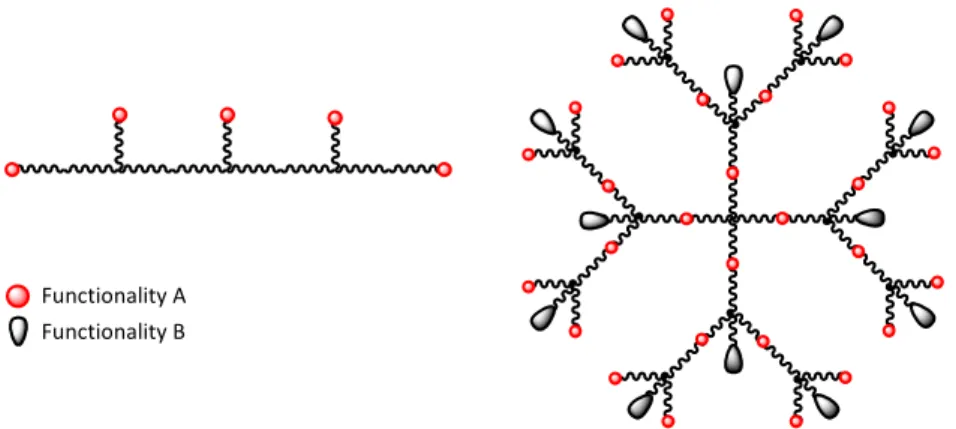
Up to this point, the origin and some of the intrinsic properties of the polymeric building blocks used to assemble hydrogels have been presented. However, to obtain desired gel properties the geometry of the building blocks is as important as the polymer properties itself. The geometry determines the general gel properties, such as swelling (DuBose et al. 2005), mechanical strength (Anseth et al. 1996) and drug diffusion properties (van de Wetering et al. 2005). Polymer geometries which are often used include linear, star shaped and dendritic structures. In Figure 2 a linear polymer is shown with the functional groups pending along its backbone. It is noticeable that this structure carries only one kind of functionality, consequently drug incorporation and hydrogel network formation must occur via the same chemistry, often simultaneously in a one-pot reaction. Also shown is a 4-armed star shaped dendritic structure of second generation. The dendrimer carries two different functionalities of orthogonal character (i.e. one can be reacted specifically without affecting the other). Therefore drug attachment is carried out in a separate reaction step prior gel formation, which is more controlled and repeatable.
-5-
Functionality A Functionality B
Figure 2: Examples of two PEG architectures; to the left a linear backbone with pending functional groups and to the right a 4-armed PEG dendrimer of second generation.
Dendritic materials have excellent intrinsic properties for drug delivery purposes and have successfully proved their usefulness in a number of delivery routes (Cheng et al. 2008). The specific advantages of dendritic structures compared to multi-arm polymers are the increased multivalency and the possibility of making them bi-functional. Recent developments of dendritic structures allow for a higher number of functional groups in fewer synthetic steps (Antoni et al. 2009), which for drug delivery purposes can be used to obtain high drug loading densities. An example of such material is the 2-Amino-2-hydroxymethyl-propane-1,3-diol (trizma) based dendrimer of third generation, synthesised by Antoni et al. in 6 steps, with 24 functional A groups and 21 B groups, where A and B are orthogonal. For dendritic materials in drug delivery applications functionality A is used to incorporate the drug while B is conjugated to tissue targeting molecules, polymers to increase water solubility or used for crosslinking to form a hydrogel (Ihre et al. 2002; Cheng et al. 2008). Even though dendritic materials have shown low cytotoxicity (Padilla De Jesus et al. 2002; Antoni et al. 2009) these carriers tend to accumulate in various organs within a few hours after intravenous administration. However, both the biodistribution and the retention time are improved by dendrimers PEGylation (Cheng et al. 2008).
The hydrogel scaffolds used in this thesis were constructed from PEG building blocks of several geometries. One approach used mono-functional 2- or 4-armed PEGs with terminal functional groups used for both drug incorporating and crosslinking. Alternatively, dendritic 2- or 4-armed bi-functional structures of first and/or second generation were used (Figure 3).
-6-
OH functionality Act functionality
Figure 3: The geometries used for the work in this thesis. Top structures; 2- and 4-armed mono-functional PEGs. Lower structures; 2- and 4-armed dendritic PEGs of first generation followed by a 2-armed dendritic PEG of second generation.
1.1.1 Gelling Methods
Gelling occurs by establishing several crosslinks between the participating polymeric building blocks. The connections could be of either non-covalent character (electrostatic or solvent induced) as for “physical hydrogels” or covalent bonds, making “chemical hydrogels”.
1.1.1.1 Physical Gels
For physical gels the polymer is either functionalised by charged groups, which are crosslinked by species of the opposite charge, or made of blocks that associate to minimise the contact with the surrounding media. Physical gels could also form from hydrogen bonding of functional groups along the backbone of the polymer. For example, a solution of alginate (a brown algae polysaccharide highly substituted with anionic carboxylic acid groups) gels upon addition of cationic polymers like chitosan or divalent cations, such as Mg2+ or Ca2+ (Figure 4).
-7- O O OH OH O O OH OH O O -O O O -m n
Cationic polymer Divalent cations
Ca2+ Ca2+
Figure 4: Formation of a physical hydrogel from alginate by crosslinking with either charged polymers or ions.
A potential niche of physical hydrogels is polymer-clay composite gels where clay particles are used as crosslinkers in free radical polymerisation of monomers. Those composites have shown extraordinary mechanical properties such as a 3 300 times higher fracture energy than conventional chemically crosslinked hydrogels (Haraguchi 2007).
1.1.1.2 Chemical Gels
In contrast to physical gels, polymers used to form a chemical gel carry functional groups which react by free radical reactions, or with a crosslinker carrying complementary functionality, to form covalent bonds (Hennink et al. 2002). Chemical gels have the advantage over conventional physical gels of being both stronger and more easily tailored to obtain desired properties, while physical gels benefit from their reversibility.
The work in this thesis is based on chemical gels formed by free radical polymerisation reactions or conjugate addition between PEG multi-arms carrying acrylate or thiol functionalities.
1.1.1.2.1 Photo-Polymerisation
Gelling through photo-polymerisation occurs when polymers containing olefinic groups are exposed to radicals, most commonly initiated by heat or ultraviolet (Sen et al.) irradiation. The resulting crosslinks are of carbon-carbon character (Srinivasan et al. 2010), which are considered non-degradable (the importance of this is explained later).
-8- Thiol-ene chemistry I + R'-SH R'-S R R O O R R O O SR' SR' R'-SH R R'-SH R O O R O O SR' R O O R O O SR' + R'-S R SR' R SR' R + R'-S Allyl Acrylate 100:0-25 100:150 Initiation: Propagation Chain transfer VS Homopolymerisation 1.1.1.2.2 Thiol-ene Addition
To introduce covalent crosslinks other than carbon-carbon bonds during free radical polymerisation the concept of thiol-ene chemistry may be used. In this concept the propagating radical is formed on a thiol species which then reacts with the “ene” (double bond). Scheme 1 pictures the general mechanism of the thiol-ene addition reaction. In the initiation step a photo initiator (I) is activated to produce radicals, which immediately abstracts a thiol hydrogen to form a thiyl radical.
Scheme 1: General thiol-ene reaction mechanisms for allyl-thiol and acrylate-thiol systems, respectively. Red colour indicates bonds formed during thiol-ene addition.
Most often the olefin in thiol-ene reactions is an allyl, but in this thesis acrylates were used instead. Therefore the propagation and chain transfer steps of both groups are shown. In the propagation step the initially formed thiyl radical combines with the double bond to form a new bond, marked in red, while transferring the radical into the former double bond. The following reaction step is crucial for the gel properties (explained later) since the radical could react either via chain transfer by abstracting the hydrogen from a thiol or via homo-polymerisation to form a dimer. For a stoichiometric thiol-acrylate system the chain transfer step is slow compared to the propagation step (Cramer et al. 2003), hence the acrylate radical intermediate is rather stable and has the opportunity to undergo both reactions. Cramer and Bowman measured the rate constants for chain transfer (KCT) and homo-polymerisation (KH) respectively for a stoichiometric thiol-acrylate system and calculated the ratio (KH /KCT) to be 1.5 while for allyls the amount of homo-polymerisation was only a few percent up to 25% depending on the allyl character (Cramer et al. 2001). The consequence of
homo--9- R O Nu RCN, R2NH, ROH, RSH, RBr + R O Nu R O R' R''
polymerisation is that carbon-carbon bonds are formed on the expense of thio-ether bonds. Thiol-ene reactions ends by radical termination, which occur when two radicals of any origin recombines.
A general advantage of thiol-ene crosslinking methods is that many solvents can be used, also aqueous, and that no leaving groups are formed. A disadvantage is the large degree of side-reactions, which form non-degradable carbon-carbon bonds, and the need of a potentially toxic initiator.
However, thiol-ene addition is a versatile method to form hydrogels and many different “thiol” and “ene” combinations are reported (Rydholm et al. 2007; Aimetti et al. 2009; Benton et al. 2009; Diaz
et al. 2010; Lundberg et al. 2010). The use of those systems for drug delivery purposes is described
later.
1.1.1.2.3 Conjugate Addition
A gelling method, which does not rely on radical chemistry, is the conjugate addition reaction (in some texts referred to as “Michael-type addition”). In this reaction, an α,β-unsaturated carbonyl reacts with a nucleophile (Scheme 2). The conjugating feature of the double bond, making the terminal carbon electrophilic and liable for a nucleophilic attack, renders this reaction possible. The selectivity of the nucleophile to react via conjugation, instead of an attack of the carbonyl carbon, can be explained by the “hard/soft” concept, which is outside the scope of this thesis (LoPachin et al. 2008).
Scheme 2: General conjugate addition mechanism where an α,β-unsaturated carbonyl reacts with a nucleophile. Red colour indicates the formed bond.
From here on only conjugate addition of acrylates (R’ and R’’ = H) and thiols (RSH) will be considered. Acrylates are the least sterically hindered species among all α,β-unsaturated carbonyls, a feature which makes them easily accessible towards conjugate addition (McCarthy et al. 1994). Reactivity of thiols is determined by their individual pKa, which is a result of the surrounding structure. Adjacent electron withdrawing atoms or functional groups lowers the pKa, thereby making the proton easier to abstract and the thiol a better nucleophile, while an electron donating atmosphere has the opposite influence on the thiol reactivity (Lutolf et al. 2001). Since thiol reactivity is closely related to
-10-
the pKa it is crucial to control the pH of the reaction mixture during conjugate addition in order to control the reaction rate.
Conjugate addition holds many potential advantages over other gel formation chemistries, such as photo-polymerisation and thiol-ene addition, as it can be performed without the addition of initiator or radiation and does not produce a leaving group. In addition, a solution of mixed pregel components spontaneously turns into a solid gel within minutes at physiological conditions (pH 7.4 and 37° C). This feature has been used to design injectable hydrogels from PEG (Elbert et al. 2001) and PVA (Ossipov et al. 2008) to mention some synthetic hydrophilic polymers.
The concept of using conjugate addition to form degradable hydrogels for drug delivery was essentially developed by Hubbell and co-workers during the last ten years (Elbert et al. 2001; Lutolf
et al. 2001; Schoenmakers et al. 2004; Fittkau et al. 2005; Metters et al. 2005; van de Wetering et al.
2005; Jo et al. 2009). Since then, others have adopted and developed the methodology to obtain properties desired for their applications.
In this thesis conjugate addition was primarily used to form hydrogels from mono-functional or dendritic PEG based compounds bearing 4 or 8 acrylate groups crosslinked by 2- or 4-armed PEG thiols. In cases where the pregel compounds were not water soluble the related thiol-ene addition was employed instead.
1.1.2 Crosslink Density and Swelling
Following the gelling phase of a hydrogel, the gel is normally immersed in an aqueous phase, for in
vitro or in vivo studies, where absorption of water swells the gel. The degree of swelling depends on
factors such as molecular weight of the building blocks, concentration of building blocks during gelling and the number of crosslinking sites (DuBose et al. 2005; van de Wetering et al. 2005). Those parameters influence the level of crosslink density, which is a measure of the amount of covalent/ionic bridges between gel building blocks formed during gelling. Both the average molecular mass between crosslinks (Mc; g/mol) and the average mesh size (; Å) are used to represent crosslink density and these are calculated from experimental data (Andreopoulos et al. 1998).
The mesh size represents the average distance between two crosslinks, in Ångström, and a short distance between crosslinks is equivalent to a high crosslink density. A normalised mesh size (Swollen/ Gelled) larger than 1 indicates that the mesh size for a swollen gel has increased compared to its initial value, not necessarily due to bond breaking. Instead, the main reason for an
-11-
initial increase in mesh size is a result of swelling where polymers in the gel network are stretched. Any further increase, however, indicates a lowered crosslink density resulting from bond breaking. Flory and Rehner developed the first equation to predict swelling of hydrogels (Flory et al. 1943). The equation is based on an entropy concept were two opposing forces either favours or disfavours swelling.
The driving force for swelling is the entropy increase when water diffuses into the polymer network. This thermodynamic mixing of polymer and water is described by the Flory-Huggins interaction parameter (Horta et al. 2005). The interaction parameter is an individual constant for each polymer-solvent system and indicates to which extent the polymer chains are hydrated (Horta et al. 2005). A gel made of hydrophobic polymers have a non-favoured interaction with water and does not swell much while gels made from hydrophilic polymers absorb large amounts of water and consequently swells to a great extent. A typical hydrogel swells until it contains up to 90-99% water by mass.
The opposing force, which discourage further swelling, is a pure consequence from the swelling itself, since swelling brings tension into the gel network by stretching it and hence, lowers the entropy. Rubber-elasticity theories are used to describe this term in the equation. Swelling continues until equilibrium is reached, that is when the driving force of mixing polymer and water equals the contracting force of the elastic polymer network (Richter et al. 2008).
Since the crosslink density is very important for the gel characteristics (DuBose et al. 2005), the equation is rather rearranged to instead calculate crosslink density from experimental swelling data.
A disadvantage of the Flory-Rehner theory is that crosslinks are assumed to form in the absence of solvent. However, the original equation has since been developed to account for the presence of water during gelling (Peppas et al. 2006), different degrees of swelling, both ideal and non-ideal hydrogel network formation (Metters et al. 2005) and for ionic gels (Sen et al. 2000).
Importantly, these calculated numbers only gives an average of the cavity size within the gel but not the range; and there is a range of mesh sizes since drugs larger than the calculated network dimensions still find paths through the gel and diffuses through it.
1.1.3 Degradation
Depending on the structural feature of the gel building blocks the resulting crosslinks may be non-degradable or non-degradable. One method to obtain non-non-degradable gels is conjugate addition of vinyl sulphone (VS) and thiol moieties (Scheme 3).
-12- O O R S R O O S R R H2O OH R OH R HO O S R HO O S R + + S S O O O PEG PEG S HS O O O PEG PEG +
Scheme 3: Formation of a hydrolytically stable crosslink by conjugate addition.
Our group applied such hydrogel in a rat infarction model in order to improve pathological remodelling in the early healing phase (Dobner et al. 2009). The applied non-degradable gel acts as a mechanical support and resulted in initial benefits by increasing the wall thickness and reducing the wall dilatation, but there was also a substantial inflammatory response to the material.
For many in vivo applications, however, it is desired to remove the applied hydrogel by degradation of the gel network into its respective water soluble building blocks. The advantage of using degradable hydrogels is a lowered exposure to the gel and its potential risk to cause an inflammatory response. Degradation can be achieved by proper design of either the polymer backbone or the crosslinks between individual polymers. Since swelling results in an increased surface area of the polymer network exposed to water the introduction of hydrolytically labile bonds increases the degradation rate of the network. Such degradation can be designed to occur over days to months, depending on desired application.
1.1.3.1 Hydrolytic Degradation
The hydrogel crosslinks used for the work in this thesis were formed by connecting the gel building blocks via ester bonds, consequently the gels were degraded by ester hydrolysis (Scheme 4). Esters however, are relatively stable at near-neutral pH (Zacchigna et al. 2008), but can be weakened. For example, introduction of an adjacent thio-ether moiety decreases the ester stability and increases its degradation rate (Hiemstra et al. 2007; Rydholm et al. 2007). The electron withdrawing effect of the thio-ether is used to explain the weakening effect of thio-ethers on esters (Schoenmakers et al. 2004; Metters et al. 2005).
Scheme 4: Tailored degradation rate of hydrogel crosslinks, where red bonds are cleaved by hydrolysis.
-13-
The weakening effect is even greater if there are more electron withdrawing groups in the structure beyond the thio-ether (Jo et al. 2009). In addition to crosslink stability, the gel degradation rate is also related to crosslink density, where a higher density results in a slower degradation (DuBose et al. 2005).
Previously (chapter 1.1.1.2.2), during the introduction of free radical polymerisation as a crosslinking method, the formation of carbon-carbon bonds was mentioned as a disadvantage.
Scheme 5 shows the resulting bonds when acrylate-containing compounds are crosslinked via
photo-polymerisation, thiol-ene addition and conjugate addition, respectively. The ester crosslinks produced by photo-polymerisation are not weakened by an adjacent electron-withdrawing group (Scheme 5, top reaction) and are consequently expected to hydrolyse very slowly.
Scheme 5: Three alternative routes to form ester containing gel crosslinks, red bonds are formed during crosslinking.
Thiol-ene addition on the other hand introduces a thio-ether moiety, in this case at a distance of two carbons from the ester, which significantly increases the degradation rate of formed gels. However, the thiol-ene reaction of acrylates and thiols has a rather high degree of homo-polymerisation, which forms the more stable ester crosslinks (Scheme 5, middle right reaction product). The presence of those stable ester crosslinks makes the gel less degradable. Conjugate addition of acrylates and thiols (Scheme 5, lower reaction) on the other hand, results specifically in thio-ether weakened ester crosslinks and hydrogels formed by this method degrades readily.
In case of gels formed by thiol-ene or conjugate addition the degradability originates from hydrolysable crosslinks. Alternative routes for the introduction of degradability include the design of the crosslinker itself, which may consist of hydrolytically degradable polyesters or enzyme sensitive oligopeptides (Zisch et al. 2003; Ehrbar et al. 2005).
SH R O O Gel O O S Gel O O Gel O O S 10:15 Gel O O Gel O O Gel R'
Free Radical Polymerisation
Thiol-ene Addition Conjugate Addition + SH R' O O S Gel Gel Gel H
-14-
1.2 Approaches towards Controlled Drug Delivery
Introduction of drugs into the blood circulation can take place in multiple ways, either directly via intravenous injection or secondary via transdermal, suppository, intramuscular or oral routes from where the drug is absorbed into the circulation. One of many challenges of drug delivery is to control the drug concentration in the blood, since a too low concentration is inactive while a too high concentration is toxic. Therefore much effort has been dedicated to master the kinetics of drug release where a constant dosing over time is the goal.
When a drug enters the circulation it has the potential to come in contact with every single cell accessible to the blood system. Consequently, a large number of healthy cells in different organs are exposed to unnecessary treatment. This is a major cause for unwanted side effects of drugs. As an alternative, if the drug was delivered locally to the very spot in need of treatment, many side effects could be avoided. A highly potential delivery concept meeting the demands of both controlled and local release is drug delivery through hydrogels.
In such in vivo applications of hydrogels, the void of pathogens is of greatest importance. Depending on the hydrogel system, different sterilisation techniques are applicable. For UV cured implantable gels, UV sterilisation of the formed gel is suitable (Guvendiren et al. 2010), while for injectable gels it is possible to filter sterilise the gel precursor solutions prior mixing and injection (Bahney et al. 2011).
The following text describes the main hydrogel based drug delivery systems.
1.2.1 Encapsulation
The simplest concept for drug delivery through hydrogels is the encapsulation method where the drug is embedded, or trapped, within a hydrogel. There are numerous methods of drug encapsulation of which the most common will be described here. In general, a homogeneous distribution of drug molecules throughout the gel network is achieved upon crosslinking a solution of pregel polymers in which the drug is also dissolved (Peppas et al. 2006). The drug molecules are then captured within the cavities of the gel during its formation. In contrast to these matrix systems there are reservoir systems where the drug is stored in the interior of the gel surrounded by a second gel barrier.
Drug delivery using micro- or nano-particles combine the attractive features of both systems. The gel feature contributes with a high water content (biocompatibility) and flexibility while the particle feature adds availability and the possibility to control the drug release rate by choosing the appropriate particle size (Pan et al. 2002). Micro- and nano-spheres can be fabricated in multiple
-15-
ways, such as self-assembly of chitosan-PEG copolymers into micelle structures, precipitation of chitosan into nanoparticles or emulsion polymerisation methods to mention some. The delivered drug is either trapped in a reservoir system, homogeneously dispersed throughout the matrix or attached to the particle matrix (Hamidi et al. 2008).
Stimuli-responsive gels are characterised by their ability to react on changes in their physical environment, where the most studied systems respond to changes in pH (Sen et al. 2000) or temperature (Martellini et al. 1999). Other systems may respond to light, electric or ionic potentials or magnetic fields (Ahn et al. 2008). Some stimuli-responsive systems can be applied in-situ via injection, followed by gelling (He et al. 2008). Responsive behaviour could be designed for both chemical and physical gels, by the composition of the polymer backbone or the presence of pendant charged groups that bring the responsiveness. Gels made of poly(acrylic acid) are pH sensitive since a rise in pH deprotonates the acidic groups after which water diffuses into the gel to hydrate the charged groups and the gel starts to swell (Richter et al. 2008). For poly(acrylamide) a pH decrease protonates the amides, resulting in water covering the charged anions and the gel starting to swell (Richter et al. 2008). Poly(N-isopropylacrylamide) is the most commonly used polymer for thermo-sensitive gels and when the temperature rise above a critical level for such gels, the polymer backbone loses its bound water, which forces the hydrophobic polymers together to minimise the unfavourable polymer-water contact area upon the gel network deswells (Lin et al. 2006). There are also systems where swelling is trigged by an increase in temperature (Ganji et al. 2009).
Encapsulated drug molecules in those stimuli-responsive gels are released upon swelling since the growing network allows the drug to diffuse out of the gel. The swelling behaviour of those systems is reversible. Therefore, if the external stimulus ends the gel shrinks and the drug release diminish (He et al. 2008).
1.2.2 Covalent Incorporation
The drug delivery described hitherto constitutes systems where the drug is stored or dispersed within the gel by different methods and the release is controlled by designing the physical parameters of the gel, such as crosslink density, degree of swelling and degradation rate.
An alternative approach to controlled drug release is to covalently attach the drug to the hydrogel via a linker, thus, drug release is dependent on the breaking of a covalent bond. One of the main goals with drug release research is to deliver drugs into a biological system, hence when designing the in vivo breaking of such linker there is only a narrow range of chemistry available. The main approaches for releasing covalently bound drugs in vivo are hydrolysis, enzymatic digestion and reduction of disulphide bonds (West et al. 2005; Khandare et al. 2006).
-16- Dox OH N N H O PEG Dox OH O H2O pH 5.5
1.2.2.1 Hydrolytically Labile Linkers
By connecting already existing functional groups on the drug, such as hydroxyl, carbonyl, amine or imine groups, with certain linkers release by hydrolysis is obtained, which can be tailored to occur at either acidic or physiological pH (West et al. 2005). Drug-gel linkers containing the acyl hydrazone moiety are acid-labile and used when drug delivery is directed to inflamed tissues, which are associated with acidic conditions (Farr et al. 1985), or other sites with lowered pH. Ulbricht et al. PEGylated the anticancer agent doxorubicin (Dox) via an acid labile acyl hydrazone containing linker (Scheme 6) and showed that release occurred exclusively at a lower pH (Ulbrich et al. 2004).
Scheme 6: Acid catalysed hydrolysis of an acyl hydrazone drug-PEG linker, which recreates the original Dox structure.
If instead the drug is attached via an ester, or ester in the vicinity of charged groups, the release can be tuned to occur at different rates at a physiological pH. In the work by Jo et al. release of covalently bound bovine serum albumin from a PEG hydrogel was obtained by hydrolysing the ester containing linker at a physiological pH (Jo et al. 2009). The release rate was furthered tailored by incorporating charged amino acids in line with the ester group.
1.2.2.2 Enzymatically Labile Linkers
Hydrogels in medical devices, such as dressings for improved wound healing, are specifically applied to the desired target, therefore the presence of certain cell types, such as epidermal, endothelial or inflammatory cells, is predictable (Singer et al. 1999). These cells secret enzymes, such as matrix metalloproteases (MMPs) and plasmin, which cleave specific peptide sequences within the surrounding fibrin network to provide passage for the migrating cells. By designing the drug-gel linker to contain particular peptide sequences corresponding to the substrate of these enzymes drug release mediated by enzymatic activity is obtained (Gombotz et al. 1995; Zisch et al. 2003; Benton et
al. 2009).
Cell-demanded release of vascular endothelial growth factor (VEGF) was obtained by Zisch et al. by covalently attaching the factor via a MMP degradable linker (Zisch et al. 2003). In a similar approach Ehrbar et al. showed that the resulting proliferation of human umbilical vein
-17-
endothelial cells increased in a dose-dependent manner upon plasmin mediated VEGF release from fibrin matrices with different VEGF loads (Ehrbar et al. 2005).
1.2.2.3 Redox Labile Linkers
The difference in redox environment between the inside and outside of a cell is used to design redox-sensitive disulphide linkers. These are ideally stable in extra-cellular environments, but reduced as a result of cellular uptake resulting in intracellular drug release. The main agent considered for in vivo reduction of disulphides is the tripeptide glutathione, which is the most abundant thiol compound in the cytoplasm where the concentration is ~1-10 mM, compared to extracellular levels, ~2 µM (Hong
et al. 2006). Importantly, the glutathione levels in some cancer tissues are significantly increased
compared to healthy tissue (Yeh et al. 2006). Consequently, drug release depending on disulphide reduction can be used for targeted drug release.
In an in vitro study by Navath et al. glutathione mediated release of a model drug resulted in a 70% intracellular release of the drug load, while no release was detected extracellularly, from highly loaded (16 or 18 per carrier) dendritic drug conjugates (Navath et al. 2008).
Alternatively, the thiol containing model compound can be bound to a gold particle by self-assembly and exchanged by glutathione in vivo (Hong et al. 2006).
1.3 Drug Release
1.3.1 Mechanisms and Kinetics
The elution of encapsulated or covalently attached drug may be controlled by different mechanisms and released with different kinetics, thus giving rise to different elution profiles.
The most commonly considered mechanisms for drug release are swelling, degradation, diffusion or the breaking of a covalent bond. To significantly ascribe experimentally obtained release data to any of these mechanisms mathematical models are used. To mathematically describe an elution process there are multiple parameters to consider, such as the change of matrix dimensions by diffusion of water into the gel matrix and its degradation and concentration dependant diffusion of the drug out of the gel (Siepmann et al. 2000). Each model makes certain assumptions and is therefore only suitable for systems that fit those criteria.
A commonly used model to describe diffusion controlled elution is the Higuchi equation, but is only valid for non-swelling and non-degrading thin films and is therefore not applicable to describe the elution from the gels in this work (Siepmann et al. 2011).
-18-
However, there are models that do consider swelling and degradation as a release mechanism, such as the Hixson-Crowell and Korsmeyer-Peppas equations (Shoaib et al. 2006). If experimentally obtained release data fits to the latter equation it may also be possible to distinguish whether the drug elution is primary controlled by the diffusion or swelling mechanisms or a combination thereof (Siepmann et al. 2001).
However, these equations were developed to describe the elution of entrapped drugs. The release of covalently incorporated drugs is generally determined by the degradation rate of the covalent linker. Most of these links are designed to degrade by hydrolysis and gives rise to unique zero order elution profiles, unattainable by diffusion controlled release mechanisms (Siepmann et al. 2001; DuBose et
al. 2005; Lin et al. 2006). To distinguish between burst, first order or zero order release kinetics
obtained release data is compared to representative equations. A significant curve fitting to those equations indicates whether the release rate is dependent (first order) or independent (zero order) on the drug concentration (Shoaib et al. 2006).
1.3.2 Controlled by Diffusion, Swelling or Degradation
Drug release from a hydrogel scaffold is controlled by a number of factors, such as the drug incorporation method, the drug diffusion coefficient and the gel architecture. No matter which method or what type of gel is used the drug must travel out of the gel boundaries by diffusion to accomplish any effect on the surroundings, unless release is controlled by surface degradation of the gel. This logic explains why the diffusion-controlled mechanism is the most applicable mechanism to describe drug release from hydrogels. Smart designs and methodologies thus aim to control diffusion barriers in order to render the drug delivery sustained over a longer time period than a purely diffusion-controlled system provides (Peppas et al. 2006). There are two main hydrogel types where drug release is diffusion-controlled, reservoir devices and matrix devices (Figure 5).
-19- t = 0 t > 0 Hydrogel membrane Drug reservoir Reservoir system t = 0 t > 0 Hydrogel matrix Dispersed drug Matrix system
Figure 5: Diffusion controlled drug delivery from reservoir and matrix systems, t = time.
In the reservoir model a second hydrogel covers the drug-containing core as an effective diffusion barrier. In matrix systems the drug is homogeneously dispersed throughout the gel. In matrix systems affinity gels are smart expansions where the drug interacts with the gel network either through columbic or polar affinity, which lower the apparent diffusion coefficient and slows the drug elution.
To add complexity the gels made of hydrophilic polymers start to swell at t >0. For gels with high crosslink density the drug diffusion through the gel may be restricted making the drug release limited to the rate of swelling (Figure 6). Many stimuli-responsive gels belong to this category (He et al. 2008). The distinction between diffusion and swelling controlled drug delivery can be made using the Deborah number (De = λ/t) where the time scale of swelling, represented by the polymer relaxation time (λ), is compared to the time scale of diffusion (t). For a system controlled by swelling (De >1) the swelling is slow (big λ) and restricts the drug diffusion. For diffusion controlled systems the relation is the opposite (De <1), the diffusion is slow (large t) and limits the rate of drug elution (Lin et al. 2006). By varying the molecular weight (2-10 kDa) of 8-armed PEG acrylates van de Wetering et al. could control the gel swelling and regulate the release of a model compound from a burst release to sustained delivery over 25 days (van de Wetering et al. 2005).
-20- t = 0 t > 0 Hydrogel matrix Dispersed drug Swelling gels Stimuli-Responsive gels t = 0 t > 0 pH, Temperature t = 0 t > 0 Hydrogel matrix Dispersed drug Degradable gels
Figure 6: Swelling controlled drug delivery from a matrix system, t = time.
For degradable hydrogels, the degradation rate of the gel network effects the drug release and is in some cases the rate-limiting factor (Figure 7). Degradation occurs when labile bonds within the gel network are cleaved, either by hydrolysis or enzymatic activity. There are two modes of hydrogel degradation, surface erosion and bulk degradation. Surface erosion would occur in a hydrophobic gel where surrounding water degrades the gel surface, but this is a rather theoretical situation since hydrogels contain huge amounts of water per definition. Instead surface erosion takes place in a hydrogel crosslinked by peptide sequences or enzyme degradable polymers when the enzyme activity is faster than the transport of enzymes into the gel interior. Most degradable hydrogel systems, though, can be ascribed to bulk degradation, where the degradation is equally likely to occur throughout the entire gel. A general example of bulk degradation is for a hydrogel where either the monomer building blocks are hydrolytically labile or the non-degradable monomers are crosslinked by a degradable crosslinker, or a combination of both. Hydrogel degradation facilitates drug diffusion and is rate limiting if the drug diffusion is significantly slower than the gel degradation.
Figure 7: Degradation controlled drug delivery from a matrix system, t =time.
The reservoir and matrix hydrogel systems mentioned above may release the drug with an initial rapid burst followed by a prolonged release phase controlled by diffusion, swelling or degradation. Such burst may be decreased by increasing crosslink density or coating the drug containing gel with additional drug-free layers (Lin et al. 2006). Although no comprehensive theories have been put forth
-21- O O + O O S
Drug R SH Drug Gel
O O Drug Drug O O N3 I O O Gel Drug O O N N N O O R + R SH O O S Drug Gel + Conjugate Addition Nucleophilic Substitution Cycloaddition
to fully describe the phenomenon many possible causes have been identified including material/drug interactions, fabrication conditions and sample geometry (Huang et al. 2001).
1.3.3 Controlled by Hydrolysis
The complexity, and also the controllability of the drug eluting system, is taken to the next level by incorporating the drug covalently into the gel structure. Copolymerisation of drug and gel monomers incorporate the drug into the gel backbone, alternatively and more commonly used is the method to covalently attach the drug pendant from the polymer backbone. The character of the drug-polymer link determines whether the link degrades under acidic, alkaline or other conditions. Hydrazone bonds are acid-labile and drugs conjugated via these links are released in acid environment, such as certain inflammatory sites (Liu et al. 2010). Esters on the other hand degrade under slightly alkaline healthy in vivo-conditions (DuBose et al. 2005).
The drug release described in this thesis is controlled by ester hydrolysis, probably in combination with diffusion, swelling and gel degradation. The drugs used (described later) were incorporated into the pre-gel polymers by conjugate addition, nucleophilic substitution or the 1,3-dipolar cycloaddition (HDC) of azides and terminal acetylenes (Act), also known as one Huisgen of the click reactions. In all three cases ester links are formed between the drug and the polymer network of the gel (Scheme 7).
Scheme 7: Conjugate addition, nucleophilic substitution and cycloaddition were used for drug incorporation in this thesis. Red bonds are formed during drug incorporation.
As already stated in the case of gel degradability, esters in the vicinity of electron withdrawing groups are readily hydrolysed at a physiological pH. This is also the case for the drug-gel conjugates formed by conjugate addition and nucleophilic substitution depicted above. Schoenmakers et al. synthesised the drug-PEG conjugates shown in Scheme 8 which differ in the number of methylene groups spacing the carbonyl and the thio-ether moieties. The measured hydrolysis rate of the two conjugates turned out to be approximately 3.3 times slower for the longer linker (Schoenmakers et al. 2004).
-22- Cu L L Cu PEG H 1.Deprotonation 2.R'-N3 Cu L L Cu PEG N N N Drug PEG N Drug N N Cu L L Cu +H+ PEG N Drug N N Cu L L Cu H O O Drug S PEG O O S Drug PEG H2O HO O S PEG HO O S PEG OH Drug OH Drug + +
Scheme 8: Two drug-PEG conjugates with different release rates.
To explain this feature molecular modelling experiments were conducted in order to calculate the atomic charge on the carbon of the carbonyl group. Calculations were performed for the above structures (Scheme 8) and also for the case where only one methylene group separates the ester and the thio-ether. The results revealed that the positive charge of this carbon decreases with an increased number of methylene spacers (Schoenmakers et al. 2004), which is a clear indication why the hydrolysis rate is also decreased. Later research has shown that even the structure beyond the thio-ether moiety affects the ester degradation rate. In experiments where different amino acids were incorporated in line with the thio-ether it was obvious that the presence of electron withdrawing groups act to decrease the ester stability in the same fashion as the thio-ether itself (Jo
et al. 2009).
Using the HDC reaction instead results in a triazole moiety connecting the drug and the polymer network (Scheme 7, lower reaction). The ester closest to the drug is formed in a separate reaction prior to incorporation and is not weakened by electron withdrawing species.
Normally cycloadditions proceed through a concerted mechanism. However, molecular modelling and experimental data favours a stepwise catalytic cycle for the HDC reaction. Roughly, the cycle can be described by four major steps (Scheme 9); an initial formation of a π-complex between the alkyne and the Cu(I) catalyst dimer whereupon the alkyne is deprotonated, the introduction of the azide into the complex followed by intra-complex rearrangements to form the triazole and a terminating protonation to release the product and reform the catalyst (Hein et al. 2008).
-23-
The HDC reaction is unusually robust as it tolerates a wide range of pH values (5-12), can be performed over a wide range of temperatures (0-160° C) and tolerates the presence of water and oxygen. In addition the HDC always give 1,4-substituted products and proceeds approximately 107 times faster than the uncatalysed counterpart (Hein et al. 2008).
A disadvantage with this particular click reaction is the requirement of a copper catalyst. Although copper is an essential element to maintain normal body functions and has also been used with success to treat tumours, excessive amounts lead to the development neurological disorders such as Parkinson’s disease and Alzheimer’s disease (Wang, T. et al. 2006). However, the copper catalyst can be reduced to undetectable levels from click formed hydrogels by washing with ethylenediaminetetraacetic acid (EDTA) solution (Malkoch et al. 2006).
Two alternative mechanisms for release of tethered drugs by hydrolysis are proposed by Dubose et
al. The dominant mechanism is primary release where the ester connecting gel and drug is
hydrolysed and the drug released in its unmodified form. Alternatively, by cleaving esters further away along the PEG chain, or a combination of esters within the gel network, a drug-PEG conjugate is released, making it secondary or higher order release (DuBose et al. 2005). For the PEG gels studied by DuBose et al. (PEG-acrylate crosslinked by dithiothreitol with simultaneous incorporation of a thiolated dye) the amount of primary release was higher than 90%.
1.4 Cardiovascular Diseases
The specific interest of our research group (Cardiovascular Research Unit) concerns the treatment of cardiovascular diseases (CVDs). Therefore drugs with potential to ameliorate the healing of different aspects of CVDs were chosen to benchmark the hydrogel concepts developed in this work. Additionally, the use of drugs with combined anti-inflammatory and cardioprotective properties is beneficial in case the implanted gels would produce an inflammatory response.
CVDs is a collection term for a number of pathologies concerning the heart and the arteries supplying it with oxygen rich blood, but in a wider interpretation CVDs also include the general vascular system throughout the body. Statistics for the United Kingdom (Stojkovic et al.) revealed that 0.2% of the population suffered from a heart attack in 2010 and one third of all deaths in the UK was a result of CVDs (Scarborough et al. 2010). In fact, CVDs are the major cause of death worldwide accounting for 29% of all deaths (Mathers et al. 2004).
Several risk factors for developing CVDs have been identified giving a sad picture of the general awareness since most are self-inflicted. The risk factors are obesity, smoking, low