An evaluation of gingival
recession and orthodontic
treatment with fixed
appliances.
- A pilot study, on the prevalence of recession
and diagnostic validity of intra oral photos.
Dan Håkansson
Supervisor:
Mikael Sonesson,
Lecturer
Dep. of Orthodontics
Malmö University
Master´s degree (30 hp)
Malmö University
Programme in Dentistry
Faculty of Odontology
ABSTRACT
English
Introduction: Around one fourth of all children and adolescents born during the same year go
through some sort of orthodontic treatment. In some literature gingival recession has been mentioned inconclusive as being a possible risk from orthodontic treatment.
Aim: The primary aim of this study is to investigate if a correlation can be seen between
gingival recession and the use of fixed appliances in patients treated at Department of Orthodontics, Malmö University.
Method: Study casts, intra oral photographs and clinical examination (a recall 2014) were
used to identify gingival recession with a yes or no as regarding presence. A measurement was done if recession was present. Subjects were selected from patients that finished treatment in 2008/2009.
Result: Study casts, for 2008 it was a negative correlation (fewer gingival recessions after
treatment), p-value for 2008 showed p=0,0034 which is regarded as statistical significant. There were too little data material to use for analyse regarding clinical examination and oral photographs.
Conclusion: Gingival recessions seem to have no connection with orthodontic treatment or
possibly a negative connection at Department of Orthodontics, Malmö University. It is based on limited data though and not conclusive. Study with larger group of patients would be needed.
Swedish
Introduktion: Ungefär en fjärdedel av alla barn och ungdomar födda under samma år
genomgår någon form av behandling som innefattar förflyttning av tänder.
Syfte: Primär fokus med denna studie är att undersöka om ett samband finns mellan gingival
retraktion och användning av fast apparatur på avdelningen för Ortodonti, Malmö Högskola.
Metod: Studiemodeller, intraorala bilder och klinisk undersökning (återbesök 2014) användes
för att identifiera gingival retraktion med ja eller nej. Om det fanns gjordes en mätning. Försöksobjekt valdes från patienter som blivit färdigbehandlade 2008/2009.
Resultat: Studiemodeller för 2008 visade färre gingivala retraktioner efter behandling p-värde,
(p=0,0034 som är statistiskt signifikant). För lite data fanns från klinisk undersökning och intraorala bilder för att göra analays.
Slutsats: Gingivala retraktioner verkar inte ha någon koppling med ortodontisk behandling på
avdelningen för Ortodonti, Malmö Högskola. Kliniska intraorala foton är ett grovt mått diagnostiskt verktyg för att bedöma gingivala retraktioner.
Det är baserat på begränsad data och inte slutgiltigt. Studie med större grupp av patienter behövs.
INDEX
1. INTRODUCTION ... 5-11 1.1 Definitions and aetiology... 5-7 1.2 Epidemiology... 7-8 1.3. Ethical aspects and costs for society... 8-9 1.4. Available methods... 9-10 1.5 Aims and hypotheses... 10 2. MATERIAL AND METHODS... 11-16 2.1 Material and exclusion/inclusion... 11-12 2.2 Tools and measurements... 12-14
2.3 Assessment of gingival recession via the use of study casts, intra oral photographs and clinical examination... 14-15
2.4 Correlation between gingival recession and treatment... 15-16 2.5 Ethical committee and sponsorship... 16 2.6 Statistical analysis... 16 3 RESULT... 16-18 3.1 Subjects... 16-17 3.2 Gingival recession... 17 3.3 Reliability analysis... 18 3.4 Prevalence of gingival recession and correlation with orthodontic treatment... 18 4. DISCUSSION... 18 4.1 Gingival recession and hypotheses... 18 4.2 Subjects and used methods... 20-21 4.3 Clinical relevance... 21-22 4.4 Aspects for society and the individual ... 22-23 4.5 Future study... 23-25 5. CONCLUSION... 25 6. REFERENCES... 26-28 7. TABLES... 29-30
1. INTRODUCTION
Around one fourth of all children and adolescents born during the same year go through some sort of orthodontic treatment. Aesthetics is the most common reason why the patient seeks an orthodontic treatment. (1) The patient is often not aware, or lack the knowledge of other indications that justify the treatment. It has also been shown that aesthetics influences the individual's self conceived life quality. (1) Complications can sometimes arise in connection with treatment of different malocclusions. One complication that has been discussed the last decade is gingival recessions and how often it occurs after an orthodontic treatment. Beyond the negative impact on aesthetics an exposed root surface can cause discomforts like tooth sensibility, dental caries, plaque retention and with more severe scenarios attachment loss of the tooth. To evaluate this type of complication several methods have been used like plastic models, photographs and clinical measurements (2-6).
1. 1 Definitions and aetiology
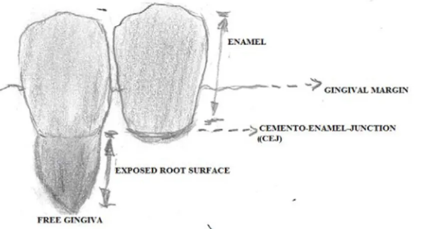
Gingival recession (soft tissue recession) means that the gingiva surrounding the teeth retracts so the root surface of the tooth becomes exposed to the oral cavity.
Figure 1. Tooth on the left has got a gingival recession. Copyright: Dan Håkansson
Definition for gingival recession is according to American Academy of Periodontology, "Location of the gingival margin apical to the cemento-enamel junction."(s.47) (7). Meaning
that the soft tissue (the gingiva) moved back apical from its original position by the cement-enamel-junction (CEJ). Gingival recession is believed to have several aetiological factors. One study (8) describes that there may be endogenous factors or exogenous factors to why it happens.
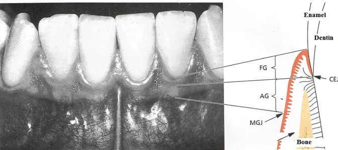
Figure 2. Description of anatomical structures seen in periodontium. FG= Free gingiva, AG=, Attached ginigiva, MGJ=Muco-Gingivial-Junction, CEJ=Cemento-Enamel-Junction. After (9) Lindhe
Exogenous factors may be a mechanical trauma (tooth brushing), toxins (bacterial composition), fillings, a periodontal treatment (scaling of dental calculus) or a non-appropriate or bad orthodontic treatment (tooth movement) (8).
Endogenous factors may be a traumatic overbite, a lesion in the alveolar bone or anatomical features (like frenulum labii inferioris).
It has in literature also been mentioned that thinner dimensions of the tissue that lies on top of the root surface may be a risk factor for developing gingival recession (8). One explanation is that during a possible inflammation, a larger part of the tissue with thinner dimension gets more affected compared to the tissue with a thicker dimension. If a thicker dimension is present it might enclose the area of inflammation with a healthy tissue (8). Tugnait et al. 2001
(10) describes in a review how orthodontic treatment may cause damage on the gingiva if a tooth is moved outside the cortical bone. Such a trauma to the bone may then cause a gingival recession. Tugnait et al. 2001 (10) also describes that malocclusion from example a deep bite might lead to a trauma on the gingiva and a gingival recession.
Some symptoms in the oral cavity are connected with an exposed root surface, for example hypersensitivity, when the teeth in the mouth are exposed to cold, heat or chemical stimulation. (10) If the gingiva has withdrawn very far from the CEJ towards the apex there is also an increased risk that the tooth has suffered attachment loss. (10)
Toxins from micro organisms then induce gingivitis in the soft tissue. Finally, the exposed root surface develops easier dental caries compared to covered root surface. (10)
1.2 Epidemiology
Mohlin (11) describes how gingival recession may arise as a complication of bad or inappropiate orthodontic treatment which may cause difficulties for the patient in a short or long term.
The Swedish Council on Health Technology Assessment (SBU) also concluded that there is no scientific evidence to support the statement that orthodontic treatment increases the risk for gingival recession of lower jaw incisors. (1)
Overall SBU showed insufficient evidence to support or discard a connection between gingival recession and orthodontic treatment. (1)
Finally in the thesis of Andlin-Sobocki et al. 1993 (12) it is described that gingival recession is often located on mandibular incisors and vary in prevalence from 1-19 %.
The SBU have in a systematic review showed a certain degree of support in literature for a lowering of the marginal bone level on upper jaw molars from some type of orthodontic
appliance. However, the SBU-report stated that this change in the tissue had no clinical relevance. (1)
1.3 Ethical aspects and costs for society
A study cast is a tool that makes it easier for the practitioner to find and diagnose malocclusions but also easier with therapy planning. At the same time in order to have a study cast an impression of the patient's mouth is required. For a child, it can be difficult or unpleasant to have these impressions taken. In addition the impressions are often sent to a dental technician, which costs (impression material, fee from technician) and time (waiting for study cast). Photographs on the other hand requires less cooperation from the patient since they need to merely open their mouth, is more comfortable and takes less time to get the result.
A report shows that it is common that treatments are made on aesthetic reasons and that the caregiver initiates the individual awareness of a malocclusion, thus the patients demand for treatment of the malocclusion is elevated. (1)
Like all health care, the orthodontic treatments are connected with costs, approximately four hundred million Swedish crones per year. (1) Since money is limited that means resources for providing and giving treatments are limited. That's why it is important that the caregiver offers and does treatments that do not harm and are as cost-effective as possible. Gingival recession might be a complication and if it happens on the incisors it may cause need of treatment in the future. Since it was concluded in the same systematic review that the appearance of teeth was the most common reason for seeking orthodontic treatment (1), it is not unlikely they will seek additional treatment in case a gingival recession occurs.
This will then demand attention and resources from the caregiver and society that could have been spent on other patients. It might also induce a lowered satisfaction from the individual
on his/her oral health. That's why it is important that both the caregiver and patient are aware of all side-effects to treatment, where gingival recession has been mentioned of being a possible risk.
In Odontology a knowledge gap exists concerning gingival recession according to SBU and reviews touching the subject is inconclusive. (1) Meanwhile another review stated that movement of incisors in the lower jag through appliance had no correlation with occurence of gingival recession. (13) A systematic review concerning how inclincation of incisors during orthodontic treatment can cause gingival recession concluded there was conflicting evidence regarding correlation between movement of incisors and gingival recession. (14) The same systematic review also concluded that the studies included were of low evidence in quality. Another review made the conclusion that a weak correlation existed between gingival recession and orthodontic treatment but the recession was not of clinical importance (0,03 mm). (13)
In 2013 the Angle Society concluded in a report, that there is weak connection between gingival recession and orthodontic treatment and further clinical studies are needed (15).
1.4 Available methods
In a candidate report made in 2013 at Malmö University, it was concluded that a majority of studies looking at gingival recession used clinical examination and/or study casts and/or intraoral photographs when examining gingival recession or other malocclusions. (16)
Measurement of pocket depth is shown by the SBU (17) to be lacking when it comes to assessment of attachment loss and that the error of measuring lies at +/- 1 mm between observers and for the same observer. Dental radiographs with correct projection (bitewing projection) can show a decrease of marginal bone level with a measurement error of 0.2 mm
to 0.6 mm. (17) On the other hand possible bone loss in the bucco-lingual direction is much harder to detect on the pictures. And those surfaces are the most interesting to look at for this study. Therefore these methods are not suitable for evaluation of gingival recession.
There are systematic reviews that tried to determine the validity of new methods like digital scanning compared to traditional study casts. Study casts are in those reviews referred to be golden standard. (18, 19)
Therefore intra-oral photographs taken with a camera, study casts from alginate impressions and clinical examinations were the chosen methods. Digital scanning as mentioned is also a method to depicture structures in the mouth. Today there are hardware and software within the field of digital scanning that can offer high resolution of both soft tissue and hard tissue. However digital scanning is still not so commonly used. Since this is a pilot study for future studies, it was decided to use methods that are already available at clinics today treating malocclusions.
1.5 Aims and hypotheses
The primary aim of this study is to investigate if there is correlation or not between gingival recession and the use of fixed appliances at Department of Orthodontics, Malmö University. The hypothesis is, a correlation exists between gingival recession and orthodontic treatment with fixed appliance.
Secondary aim is to investigate if intra oral photographs are as good as study casts and clinical examinations, to detect gingival recessions. The hypothesis is that gingival recession will be easiest to detect during a clinical examination.
2. MATERIAL AND METHODS
2.1 Material and exclusion/inclusionThe patients were identified via paper journals in an archive consisting of patients with finished treatment. The patient were asked through a letter or phone call to take part in the study. If patients accepted they were sent an information letter. If no response or contact were made with selected patients, an additional attempt was made through phone call.
A check up was done on all available study casts to see if there were any incomplete sets. Incomplete set was defined as a study cast being not accountable from before or/and after treatment. Attempts were made to contact all patients for a recall with letter and/or phone call. A total of 10 patients accepted to participate in the study.
The patients must have been treated at the Department of Orthodontics, Malmö University and finished in 2008 or 2009. A summary of inclusion criteria is shown in Table 1. The selected patients were called for a new clinical examination. During the examination a clinical assessment of gingival recession was made.
At the clinical examination, impressions were taken for study casts and both intra oral and extra oral photographs were taken. Also periodontal conditions were observed and any possible visual recessions were measured according to the definition for gingival recession according to American Academy of Periodontology.
The patients received information by letter with a short story about the aim of the study. The visit was free of charge for the patient. There was no sponsorship for this study.
Measurement of all available study casts before and after treatment were performed on all qualified seventy-two patients. This was also done for all available intraoral photographs.
First all study casts from before treatment were observered, then two months later all study casts from after treatment were observed. Observation for presence of gingival recession was done on all buccal sites of the incisors. A measurement was performed if any visible gingival recession was detected on study cast or clinical examination.
2.2 Tools and measurements
All measurements were done with one digital calliper (Biltema, Art nr 16-105, Sweden) to minimize any differences caused because of different measurement tool. The digital calliper displayed measured distance with an error of +/- 0.03 mm according to the manufacturer. Intraoral photographs were displayed on a computer screen (HP Compaq LA2205wg 22-inch Widescreen LCD Monitor, Native Resolution 1680 x 1050 @ 60 Hz (WSXGA+), 1.76 Million pixels). Photographs were used that was in the digital journal system, called T4 made by CareStream Dental Stockholm, for the patient. Measuring on study casts and clinically were in all cases done by approaching the tooth at its buccal site (Figure 3). Measuring on intraoral photographs was as well done from the buccal site of the tooth (Figure 4). All measurements were done by one observer (DH). The intraoral photographs on all patients were taken by the same photographer with camera of type Digital Camera Fujifilm FinePix S2 Pro with an objective of the type Nikon 55 mm. This was also true for the study casts that were all made by the same dental technician at the department of Orthodontics, Malmö University, Malmö, Sweden. Study casts were made in plaster, type III white dental stone.
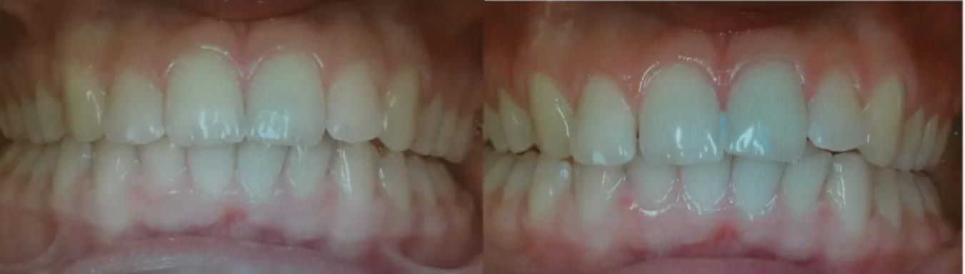
Figure 3. Digital calliper inserted from the CEJ to MGJ.. This was done on each incisor for study casts and clinical examinations. Image on the left is an upper jaw before treatment, and right image the same upper jaw after treatment.
Figure 4. Detection of gingival recession on intraoral photographs were performed on a computer screen.. Observer looked at photographs from before treatment, after treatment and at the follow up. Image on the left is after treatment, right recall visit in 2014.
All impressions were made by alginate. Based on earlier studies (20, 21) alginate shows to have a good reproducibility, it keep a consistency with old study casts, Dentsply Blueprint xCreme was used in present study. An impression tray made of metal with holes was chosen for all impressions. (Figure 5).
Figure 5. Type of impression material and tray that was chosen for the clinical examinations to make new study casts during the recall.
2.3 Assessment of gingival recession via the use of study casts, intra oral photographs and clinical examination
Recessions were categorized as YES, when an identified recession was observed or NO, when no recession was identified. If it was registered YES at the clinical examination or on the study cast a measurement was done. Recessions on the photos were registered only as YES or NO, because the pictures were not taken with any preset conditions like distance between camera and object, fixation of the object ( head of the patient) or ruler in the picture for scale.
Concerning study casts, measurements were done on both central and lateral incisors in upper jaw and lower jaw, before and after finished orthodontic treatment and at recall.
During the recall some general questions were asked concerning oral health and condition. This was mostly done to get information in case something special had happened since the last visit at the department and to provide assistance in discovering any possible surfaces of gingival recession.
Study casts were analysed regarding gingival recessions one month after the clinical examinations were performed. Analysis were done on study casts made before treatment, after finished treatment and newly taken study casts in association with the clinical examination. After another month the photographs were analysed. Photographs taken before treatment, after finished treatment and from the new appointments were evaluated. To test the reliability of the observer's measurements, two months later after both measurement of all study casts and photographs were finished, an additional new measurements were performed on random selected casts.
Recall five years after completed treatment 2008-2009 was chosen because this is the standard the SBU has stated as a minimum for any study to be used in a systematic review for an evaluation of treatment in a long term perspective. (1) The timeframe was therefore set to five years back in time from the year 2014.
2.4 Correlation between gingival recession and treatment
The objective is to compare a) and b) (Figure 6). If the difference between these is completely random, then the prevalence of observed gingival recessions should be roughly evenly divided between a) and b). This means if there are 100 observed gingival recessions when combining a) and b) together, if observations are randomly occurring then there would be roughly 50
observations of a) and 50 observations of b). The connection can be positive (less observed instances of recession after treatment), negative (more observed instances of recession after finished treatment) or neither (random occurrence of recession). In this way an evaluation and comparison is made concerning gingival recession before and after treatment.
2.5 Ethical committee and sponsorship
An application was handed to the Ethical Committee at the Faculty of Odontology, Malmö University. This was done with the purpose of having an ethical trial before the study could be started. The decision, Dnr: OD 2013/389-59, that came on the 19th of December 2013 from the committee, was that the study was a case of control of quality within health care and did not need ethical approval. No sponsorship existed for this study.
2.6 Statistical analysis
Results from study casts before treatment/after treatment/recall, photographs before treatment/after treatment/recall and clinical examination (recall) would be tabulated. p <0.05 for a result to be considered significant.
Paired samples t-test was performed for analysing statistical significance regarding gingival recession. A significant result was set at p<0,05. In addition, the intra individual coherence was tested by a paired t-test.
3. RESULT
3.1 SubjectsAll patients who finished their treatment in 2008 and 2009 at the department of Orthodontics, Malmö University were included. A total of forty-two patients had been treated 2008, twenty
being completed with treatment in 2009. Fifty-two patients fulfilled the criteria, for full disclosure Table 2.
Combining the included patients from 2008 and 2009 gave a total of seventy-two patients. After finished review it was concluded that two sets of plaster models from 2008 and nine sets from 2009 were incomplete. Age and gender of the patients are shown in Table 3.
3.2 Gingival recession
Gingival recession were recorded for all study casts from 2008, 2009 and from the additional visit in 2014 were included for the final study. Surfaces with undetermined recession are referred to as surfaces excluded, Table 4 (gingival recession). Data concerning gingival recession on study casts are presented in two tables, 5 and 6. Table 5 shows the gingival recession in mm divided with the number surfaces found having gingival recession. Table 6 shows how many percentage of the buccal sites for each tooth displayed gingival recession on the observed surfaces.
Gingival recession on intraoral photographs were registered if visible on incisors in upper and/or lower jaw.
Gingival recessions on intra-oral photographs were so few that no comparison could be made before and after treatment in the patient group who visited the clinic in 2014 for the additional clinical examination.
Since only ten patients showed up for the clinical examination the data collected from the clinical examination could not be used properly for comparison. It was noted that 9 out of 10 patients were pleased with their treatment. The 10th patient was displeased because a relapse in the lower jaw.
3.3 Reliability analysis
The ambition to compare intraoral photographs taken from the clinic recall in 2014 was not fulfilled because of lack of photos due to illness of the photographer during the period of clinical examinations and patients affected could not come for a second appointment.
Overall the reliability for the measured distances were good for the observer.
3.4 Prevalence of gingival recession and correlation with orthodontic treatment.
Prevalence of gingival recession on observed surfaces before and after treatment in total (2008 and 2009) were 6,5 %. A total of 960 surfaces were observed both and after treatment where 8 % were excluded because of obstacles on the surface of the study casts. A total prevalence of gingival recession (2008 and 2009) before treatment were 3,8 % and after treatment 2,1 %.
An analysis concerning gingival recession and its eventual connection with appearing after orthodontic treatment was made on study casts. For 2008 it was a negative correlation, p-value for 2008 showed p=0,0034 which is regarded as statistical significant (Table 8). Meaning there were fewer observations of gingival recession after finished treatment then before received treatment. The group of patients who finished their treatment in 2009 showed no statistical significant differences in the observation of recession before and after treatment. (Table 7).
4. DISCUSSION
4.1 Gingival recession and hypothesis
The hypothesis for this study could be falsified. Also not expected, the trend seemed to indicate that at least for the year 2008 there was no correlation between gingival recession and treatment with fixed appliances at Department of Orthodontics, Malmö University. Meaning after finished treatment, there were less gingival recessions than before started treatment. However it must be remembered that only a total of thirteen surfaces on study casts were detected among subjects from 2008. This pilot study seem to indicate that occurrence of gingival recession is as stated from previous studies very low. For a future study a much larger population would be needed as the prevalence of recession is low.
Results from this study seem to confirm what some previous studies concluded that orthodontic treatment can reduce the severity of attachment loss around teeth by moving the root package of incisors further in the alveolar process away from the compact bone. However the data is very limited and therefore any conclusions from this pilot study can not be said with full certainty.
The secondary aim to assess if photos, casts and clinical investigations could be used to locate gingival recessions were not possible to do. However, one can argue that study cast is the best way currently to register gingival recessions. In this study casts were naturally available from before and after treatment. They had also kept up to date over the years. Compared with intra oral photographs where the camera type and picture type had changed. And in dental practice the orthodontist do not do typically do measuring regarding gingival recessions as was seen in the journals.
4.2 Subjects and used methods
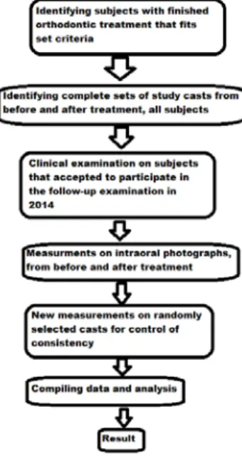
Figure 8 sums up how the study was executed.
Figure 8. Flowchart for how the study was done.
This study can not say anything about the reliability of measuring vertical distance of the crown on photos which might be important for measuring the length of gingival recessions. It can be said though that since frontal intral-oral photographs are normally taken with the jaws in occlusion. It can also be concluded that any type of vertical measurement on the crown for a method validation would make it hard to validate photographs overall for incisors in the lower jaw. It is recommended by SBU to have at least two observers when observing an occurrence (22). This could however not be done for this study because of time and that the author had no additional observer available that had the time to calibrate. This makes data less reliable. It is shown though that the one observer for this study has been consistent in measuring.
One of the larger set backs with the study is the low number of individuals who participated in the clinical examinations at the recall 2014. For which only ten participated at the recall of seventy-two possible subjects.
Also the intra-oral photographs have not been taken with the same distance from the patients nor have the patient or camera been stabilised while taking the photos making measurements impossible to do. When observing photographs it was noticed that that depending on the angle between camera and object, shadows from the soft tissue could be cast on the tooth surface. These shadows made it harder to detect or rule out gingival recession. It was also noticed that saliva covering the tooth and marginal gingiva could reflect the light from the flash on the camera making it harder to distinguish a possible transition from enamel and exposed root surface. Photographs where the teeth had been dried with air before photos were taken, eliminated any issue with saliva. However every photograph have been done by the same photographer for all subjects.
Photographs can only display 2D-images of a 3D-reality which means any curved object will be distorted in a 2D-image. This distortion can affect the data from measurements and that's why teeth are chosen that are fairly straight in the arch, which the lateral and central incisors are. How much the distortion increases the more posterior you move in the arch is outside the scope for this study. It was noticed during the collection of data that the photographs were divided between digital system and analog slides. In this study the set criteria was to look at only digital available photographs.
4.3 Clinical relevance
The fact that so few, if any recessions, could be seen on the photographs raise the question how large clinical problem a gingival recession is. From the data collected on study casts, the value of the gingival recessions is often at 0.5 mm or less. This is a level of lesser severity of attachment loss if compared with the periodontal classification system for attachment loss on teeth where it falls in the category of periodontitis levis (23). And almost all intra oral
photographs showed no gingival recessions. Therefore it seems that any possible gingival recession created from an orthodontic treatment has a little clinical relevance. It might however be of aesthetic relavence for the patient.
A further method validation comparing study casts and what is seen in the mouth regarding vertical distances could be appropriate before making too long going conclusions. However since all study casts are made by one experienced dental technician, impressions taken by experienced dentists and observer showed reliability in measuring, it can be stated with fairly certainty that possibilities of error were few regarding observations on study casts.
This study does not look into what type of malocclusion a patient has had before an orthodontic treatment. A patient with severe form of malocclusion is helped by orthodontic treatment but after treatment might have persisting issues concerning gingival recession. In that case, to claim an orthodontic treatment for being responsible for defect in soft tissue, is not appropriate.
4.4 Aspects for society and the individual
As mentioned before does the presence of gingival recession play a role for the individual's perspective of self estimated need for treatment. It can also make the patient inclined to seek additional health care which then will take time from the caregiver, time that could have been spent on helping other patients. However one must also notice and raise the question if this is a problem when contemplating which treatment to do. This study shows that the measured vertical distances of gingival recession did not go above 0,5 mm in almost all cases. A review made in 2011 concluded the gingival recession were approximately 0,03 mm (18). This in clinical practice is barely visible and in this study even falls in the range for error of
measurement in the digital caliper that was used. This means for clinical practitioner the implication from a possible correlation is probably very low from a biological perspective on the tissue.
However again for the patient that notice a gingival recession and cares about appearances it might be of large subjective importance and cause problems for the patient's life and the clinic in how to help the patient. How self perceived value of health and appearance, factors into the patient's view on gingival recession goes beyond this study. It must however not be discarded from an ethical stand point.
Implications on a society level is hard to conclude from this study. It can however be speculated that so far no established correlation exists and therefore any possible cost for additional treatment can be regarded as low. The report from SBU where it presented calculations for cost of orthodontic treatment did not mention any additional cost caused from dealing with gingival recession (1).
Therefore the problem of gingival recession seems to be as described previously (27,28) ,of huge importance on an individual level but low to very low importance for dentists and society.
4.5 Future study
In a future perspective, it would be interesting to incorporate digital scanning with study casts. Some studies have been done in this field. But it would be interesting to compare how the patients, the children, would receive digital scanning instead of impressions or for that matter intra-oral photographs. Since dental anxiety can cause problems for patients in adulthood it is of importance to study new methods that might minimize the risk for this to happen.
In the future maybe digital scanning, could replace taking impressions and intra-oral photographs, which can make it more time effective for the patient, the caregiver and
cost-effective for society (more patients in shorter time) but also make the visit at the dentist a more pleasant experience.
For future study, there are some aspects that would be changed. The study would include children from a whole region with several clinics, not only one orthodontic clinic. This is to more accurate observe occurrences. Treatments observed in this study were all done by orthodontists at Malmö University. It would be a prospective study where patients would be followed in the same way as in this study with the difference of adding clinical examination before and after finished treatment. Study would end with a five year follow-up. The study would for more conclusive results have a randomised design. The groups would consist of one with patients declining treatment, one group with patients accepting treatment and a third group of patients with no need of treatment. Criteria from this study could be kept and maybe narrowed down by only looking on one type of malocclusion, recommended most common type of malocclusion requiring fixed appliances as treatment.
All photographs would be digital, taken with a fixed distance between the patient and camera or optional having a ruler of some kind in the picture for scale.
A study like that would have the possibility to give a higher chance for a more accurate evaluation and higher chance of getting a better understanding of gingival recessions. Also the study would first, if possible, have a part of method validation where the methods intraoral photographs, study casts, clinical examination and digital scanning were compared regarding ability to recreate surfaces and usage for vertical measurement. That type of method validation goes beyond what current study could do in its set time.
It must be remembered that this study had to adjust to its limited time and resources when the layout of the study was constructed.
5. CONCLUSION
Gingival recessions seem to have no connection with orthodontic treatment or possibly a negative connection at Department of Orthodontics, Malmö University, Malmö, Sweden. It is based on limited data though and not conclusive. Study with larger group of patients would be needed. The study was aiming on investigating gingival recession with methods of plaster models, intra oral photographs and clinical examination. But lack of participation from patients made data from the clinical examination inconclusive and parts of the photographical archive was incomplete since some of the pictures were stored analogy using devices that are not used in practice today.
Future study should include looking at the method digital scanning compared with intra-oral photographs and study casts through impressions with a method validation concerning accuracy of measuring vertical distances on teeth. Also future study in this field would benefit from being prospective in its structure where a randomised control group is used. Where one group consists of patients declining treatment, one group with patients accepting treatment and a third group with patients of no need of treatment.
6. REFERENCES
1 . Statens beredning för medicinsk utvärdering(SBU), Bettavvikelser och tandreglering i ett hälsoperspektiv- En systematisk litteraturöversikt. Stockholm: Statens beredning för medicins utvärdering(SBU), Oktober 2005.
2. Closs LQ, Branco P, Rizzatto SD, Raveli DB, Rosing CK. Gingival margin alterations and the pre-orthodontic treatment amount of keratinized gingiva. Braz Oral Res. 2007;21:58-63. 3. Vasconcelos G, Kjellsen K, Preus H, Vandevska-Radunovic V, Hansen BF. Prevalence and severity of vestibular recession in mandibular incisors after orthodontic treatment. Angle Orthod. 2012;82:42-47.
4. Zimmer B, Seifi-Shirvandeh N. Changes in gingival recession related to orthodontic treatment of traumatic deep bites in adults. J Orofac Orthop. 2007;68:232-244.
5. Allais D, Melsen B. Does labial movement of lower incisors influence the level of the gingival margin? A case-control study of adult orthodontic patients. Eur J Orthod. 2003;25:343-352.
6. Djeu G, Hayes C, Zawaideh S. Correlation between mandibular central incisor proclination and gingival recession during fixed appliance therapy. Angle Orthod. 2002;72:238-245. 7. The American Academy of Periodontology, Glossary of Periodontal Terms 4 th edition. Chicago: The American Academy of Periodontology, 2001.
8. Baker P, Spedding C. The aetiology of gingival recession. Dent Update. 2002;29:59-62. 9. Lindhe J., Lang P. N. och Karring T., Clinical Periodontology and Implant dentistry Volume 1: Basic concept. Singapore; Blackwell Publishing Ltd, 2008.
10. Tugnait A, Clerehugh V. Gingival recession-its significance and management. J Dent. 2001;29:381-394.
12. Andlin- Sobocki A. Gingival Recession, Keratinized and Attached Gingiva in Anterior Teeth of Children. Volume 46 of Umeå Universitet 1993: Umeå University odontological dissertations abstracts.
13. Aziz T, Flores-Mir C. A systematic review of the association between appliance-induced labial movement of mandibular incisors and gingival recession. Aust Orthod J. 2011;27:33-9. [Abstract only available]
14. Joss-Vassalli I, Grebenstein C, Topouzelis N, Sculean A, Katsaros C. Orthodontic therapy and gingival recession: a systematic review. Orthod Craniofac Res. 2010;13:127-41.
15. Johal A, Katsaros C, Kiliaridis S, Leitao P, Rosa M, Sculean A, Weiland F, Zachrisson B State of the science on controversial topics: orthodontic therapy and gingival recession (a report of the Angle Society of Europe 2013 meeting). Prog Orthod. 2013;11:14-16.
16. Håkansson D. Gingival retraktion i samband med ortodontisk behandling- En systematisk litteraturöversikt, 2013 Malmö University. [Not published]
17. Statens beredning för medicinsk utvärdering(SBU), Kronisk parodontit- Prevention, diagnostik och behandling. Stockholm: Statens beredning för medicins utvärdering(SBU), 2004.
18. Fleming, P., Marinho, V. and Johal, A., Orthodontic measurements on digital study models compared with plaster models: a systematic review. Orthodontics & Craniofacial Research 2011;14:1–16.
19. Matthew G. Wiranto, W. Petrie Engelbrecht, Heleen E. Tutein Nolthenius, W. Validity, reliability, and reproducibility of linear measurements on digital models obtained from intraoral and cone-beam computed tomography scans of alginate impressions, American Journal of Orthodontics and Dentofacial Orthopedics, 2013;140:140-147.
20. Rådebrg S, Selléus. Två algniatmaterials dimensionsstabilitet beroende av tillsatt vätmängd vid förvaring och retention till underlag över tid, 2012. [Not published]
21. Appelros C, Olsson J. Effekt av förvaringstid och förvaringstemperatur på linjär dimensionsstabilitet hos tre dentala avtrycksmaterial. En jämförande in vitro studie, 2007. [Not Published]
22. Statens beredning för medicinsk utvärdering(SBU), Utvärdering av metoder i hälso- och sjukvården- En handbok. Stockholm: Statens beredning för medicins utvärdering(SBU), 2014. http://www.sbu.se/upload/ebm/metodbok/sbushandbok.pdf [2015-04-18]
23. The Periodontal Disease Classification System of the American Academy of Periodontology — An Update. Putnins E. E, Wiebe B. C.D, J Can Dent Assoc;2000;66:594-597.
7. TABLES
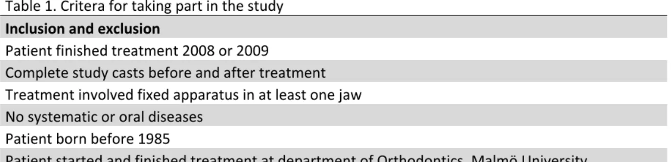
Table 1. Critera for taking part in the study
Inclusion and exclusion
Patient finished treatment 2008 or 2009
Complete study casts before and after treatment Treatment involved fixed apparatus in at least one jaw No systematic or oral diseases
Patient born before 1985
Patient started and finished treatment at department of Orthodontics, Malmö University
Table 2. Patients treated at the Department of Orthodontics Malmö University
Year 2008 Year 2009 Year 2008 and 2009
Patients did not qualify (n=20) Patients did not qualify (n=27) Patients that qualified (n= 72)
Number. Reason Number. Reason Year Total
1. Born before 1985 1. Born before 1985 2008 20
2. Born before 1985 2. Born before 1985 2009 52
3. Born before 1985 3. Born before 1985 2008+2009 74
4. Adminastrive reason 4. Patient transferred 5. Adminastrive reason 5. Patient transferred 6. No treatment recieved 6. Removable appliance 7. No interest from patient 7. Declined treatmend
8. Declined treatment 8. Cancelled treatment
9. Cancer treatment 9. No received treatment
10. Removable appliance 10. Cancelled treatment 11. Removable appliance 11. Declined treatment 12. Removable appliance 12. Fractured mandible
13. Poor compliance 13. Moved away
14. No response 14. No treatment
15. Cancelled treatment 15. Declined treatment 16. No interest from patient 16. No treatment 17. Cancelled treatment 17. Declined treatment
18. Treatment done 2006 18. Moved away
19. Born before 1985 19. No treatment
20. Cancelled treatment 20. Other medical treatment
Loss: 20 21. Belonged to different archives
22. Belonged to different archives 23. Economy
24. Moved away
25. Unclear about treament 26. Unclear about treament 27. Debt
Table 3. Description of age and gender of subjects selected for measurement before/after treatment
Year 2009 Before After Year 2008 Before After
Average age 13,5 16,7 Average age 13,9 16,6
Max 18 21 Max 19 23
Min 9 12 Min 9 11
Male 18 18 Male 8 8
Female 26 26 Female 12 12
Total 44 Total 20 20
Table 4. Gingival recession
Year 2008 2009
Surfaces IndividualsSurfaces Individuals
Total number of surfaces 288 17 672 43
Surfaces with ging. recession 14 9 44 22
Surfaces excluded 27 13 48 28
Surfaces left 253 17 624 43
No ging recession 239 10 580 27
Only before treatment 12 7 22 11
Only after treatment 1 1 18 12
Before and after 1 1 4 2
Table 5.Summarize of gingival recession 2008/2009 measured in mm
Tooth 32 31 41 42
Before treatment (average mm) 0,7 0,48 0,48 0,26
After treatment (average mm) 0,34 0,42 0,23 0
Tooth 12 11 21 22
Before treatment (average mm) 0,61 0,39 0,42 0,39
After treatment (average mm) 0,79 0,56 0,57 0,45
Table 6.Summarize of gingival recession 2008/2009 in percentage
Tooth 32 31 41 42 Before treatment (%) 3,4 16 16 1,7 After treatment (%) 7,4 6,8 8,9 3,6 Tooth 12 11 21 22 Before treatment (%) 3,6 11 11 3,6 After treatment (%) 5,7 3,8 2 1,8
Table 7. Gingival recession before and after treatment from 2008 and 2009 Year Before treatment After treatment p-value, two-tailed p-test
2008 12 1 0,0034